fabrication and optical properties of ordered sea urchin-like zno
Bạn đang xem bản rút gọn của tài liệu. Xem và tải ngay bản đầy đủ của tài liệu tại đây (822.18 KB, 3 trang )
Fabrication and optical properties of ordered sea urchin-like ZnO
nanostructures by a simple hydrothermal process
Yi Zhou
a,
n
, Ce Liu
a
, Mengyao Li
a
, Hongyan Wu
a
, Xian Zhong
a
, Dang Li
a
, Difa Xu
b
a
Department of Chemistry and Biological Engineering, Changsha University of Science and Technology, Changsha 410114, China
b
Department of Science and Technology, Changsha University, Changsha 410003, China
article info
Article history:
Received 28 March 2013
Accepted 29 April 2013
Available online 8 May 2013
Keywords:
Nanocrystalline materials
Sea urchin-like ZnO
Crystal growth
Solar energy materials
Photoelectric properties
abstract
Ordered sea urchin-like zinc oxide (ZnO) nanostructures were fabricated via a simple hydrothermal
process at relatively mild conditions by one step. The microstructure, morphology and the photoelectric
properties of the as-prepared products were investigated by x-ray diffraction, transmission electron
microscopy, field emission environment scanning electron microscopy, UV–visible optical absorption and
photocurrent–voltage measurements. The results demonstrated that the ZnO crystals had hexagonal
wurtzite structures, and were assembled by a central nucleus and many needle-like ZnO which grew
radially from the nucleus. Compared with ZnO nanorod arrays, the sea urchin-like ZnO nanostructures
showed preferable photoelectric properties as the anode of dye-sensitized solar cells. This can be
attributed to special geometric morphologies.
& 2013 Elsevier B.V. All rights reserved.
1. Introduction
Zinc oxide has attracted a great deal of attention as a photo-
anode in dye-sensitized solar cells (DSSCs) due to its large band
gap (3.37 eV) and large exciton binding energy (60 meV) [1].1D
(one-dimensional) ZnO nanostructures with controlled dimen-
sions and morphologies such as nanowires [2,3], nano/microtubes
[4,5] and nanorods [5,6] have been successfully synthesized by
hydrothermal processes. The particle size of the prepared oxides
can be controlled by changing preparation condition, such as the
concentrations of the zinc precursor, the deposition time, and
temperature [7]. Previously, 1D ZnO nanostructures were com-
pared with ZnO particulate films exhibiting a better electron
transport due to relatively low junction densities which decrease
the ohmic loss within the photoanode [3,8]. However, the 1D
nanostructures exhibit low surface area, which results in low
efficiency of power conversion of DSSCs and limits their industry
application. Recently, many efforts have focused on the prepara-
tion of 3D ZnO nanostructures to improve the surface area. Many
3D ZnO nanostructures have been prepared, such as jack-like ZnO
[9,10], flower-like ZnO [11,12], and hedgehog-like ZnO [13]. These
structures have 1D nanoscale with 3D architectures combining
properties of 3D and 1D materials, which may be used as an
interesting alternative with higher specific surface and porosity
than those of simple arrays of ZnO nanowires [14], especially for
application in DSSCs [15].
In this paper, new type 3D ordered urchin-like single-crystal
ZnO nanostructures were fabricated and their photoelectric prop-
erties in the field of DSSCs were studied and compared with those
of ZnO nanorod arrays. 3D ordered urchin-like single-crystal ZnO
nanostructures are assembled by a central nucleus and many
needle-like ZnO which grew radially from the nucleus. The photo-
electric properties of such an ordered architecture are better than
those of ZnO nanorod arrays.
2. Experimental
Ordered sea urchin-like zinc oxide (ZnO) nanostructures were
prepared through a simple hydrothermal process in a Teflon-
sealed autoclave. Zinc nitrate hexahydrate (Zn(NO
3
)
2
Á 6H
2
O) and
hexamethylenetetramine (C
6
H
12
N
4
) were added in sequence to
40 mL double distilled water to make [Zn
2+
]¼ [(CH
2
)
6
N
4
]¼
0.1 mol/L. The above solution was maintained at room tempera-
ture with strong magnetic stirring for 0.5 h to obtain a transparent
and homogeneous solution. 60 drops/min of NH
3
(25%) were then
added into the solution to adjust the pH value to 9. Afterward, the
above solution was transferred to a Teflon-sealed autoclave. The
reaction was run at 90 1C for 9 h to synthesize the ordered sea
urchin-like ZnO nanostructures. Finally, the samples were har-
vested and thoroughly washed with distilled water, followed by
drying in air at 45 1C.
The configuration of DSSC can be found in the literature [16].
The surface morphology of product was observed by field-
emission scanning electron microscopy (FESEM, JEOL JSM-6700F)
and a high-resolution transmission electron microscope (HRTEM,
Contents lists available at SciVerse ScienceDirect
journal homepage: www.elsevier.com/locate/matlet
Materials Letters
0167-577X/$ - see front matter & 2013 Elsevier B.V. All rights reserved.
/>n
Corresponding author. Tel./fax: +86 731 85258328.
E-mail addresses: , (Y. Zhou).
Materials Letters 106 (2013) 94–96
JEOL JEM-2100F) equipped with selected area electron diffraction
(SAED). The photocurrent–voltage (I–V) characteristics were mea-
sured with a computer-controlled digital source meter (Keithley,
model 2400) under illumination with a Newport solar simulator
(AM1.5, 100 mW cm
2
). The X-ray diffraction patterns were
recorded on an X-ray diffraction system (SIEMENS D5000).
UV–visible optical absorption was measured by UV–visible spec-
trophotometer (Beijing,TU-1901).
3. Results and discussion
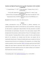
Fig. 1a shows the XRD pattern of the sea urchin-like ZnO by a
simple hydrothermal process at relatively mild conditions. All the
diffraction peaks are labeled and can be readily indexed to hexago-
nal phase of ZnO (JCPDS card no. 361451). No other diffraction peaks
are presented, which demonstrates that high-purity ZnO products
have been synthesized. Besides, the diffraction peaks are sharp,
which confirm the well-crystallized particles of prepared ZnO.
Fig. 1bshowstheUV–visible optical absorption characteristics of
the sea urchin-like ZnO. Excitonic absorption peak of the ZnO
sample is at about 360 nm in the ultraviolet region, which is blue-
shifted due to the nanometric size effect when compared with the
absorption edge of bulk ZnO which appears at 400 nm [17].From
Fig. 1b, it is also found that the average excitonic absorption in the
ultraviolet region is up to 75%.
SEM images, TEM image, HRTEM image and SAED pattern of
the sea urchin-like ZnO nanostructures are presented in Fig. 2.
Fig. 2a and b displays the low magnification and high magnifica-
tion SEM images of the sea urchin-like ZnO nanostructures,
respectively. The intact morphology of an individual sea urchin-
like ZnO nanostructure is observed in Fig. 2b. It is clear that the
ZnO crystals are assembled by a central nucleus and many needle-
like ZnO which grow radially from the nucleus. The TEM image in
Fig. 1. X-ray diffraction pattern (a) and UV–visible optical absorption characteristics (b) of the sea urchin-like ZnO nanostructures.
Fig. 2. SEM images (a,b), TEM image (c), HRTEM image (d) and SAED pattern (e) of the sea urchin-like ZnO nanostructures.
Y. Zhou et al. / Materials Letters 106 (2013) 94–96 95
Fig. 2c reveals that the surface of the needle-like ZnO nanowires
with a width of approximately 25 nm is smooth. The HRTEM
image in Fig. 2d shows that the (0001) lattice spacing is about
0.26 nm, which corresponds to the distance between two adjacent
(002) planes of ZnO, suggesting that the sample is single-
crystalline structure with the preferential (0001) growth direction.
SAED pattern in Fig. 2e further depicts that the sample is single
crystalline, which is in agreement with analysis results of Fig. 2d.
For analyzing the photoelectric properties of the sea urchin-like
ZnO nanostructures, simple battery devices were fabricated. Fig. 3
shows the I–V curve of the sea urchin-like ZnO nanostructures
based DSSCs. As shown in Fig. 3, the current density at short circuit
(J
sc
), voltage at open circuit (V
oc
), fills factor (FF) and efficiency of
power conversion (η) are better than those of our previous work
ZnO nanorod arrays, among which η was increased by 77.6% ([16]).
This suggests that the photoanode with a highly branched net-
work possesses higher photoconversion efficiency when com-
pared with the 1D nanostructures. The higher photoconversion
sea urchin-like ZnO nanostructures efficiency is ascribable to the
contributions from electron through the enrichment of dye load-
ing without sacrificing electron-transport properties [18,19]. In
addition, the geometry of ordered sea urchin-like nanostructures
possesses larger pores, which can provide an effective path for
electrolyte diffusion; thus light harvesting and overall efficiency
are improved.
The conversion efficiency of the sea urchin-like ZnO nanos-
tructures is lower than the highest value reported in the literature
[20]. This may relate to the difference of cell configurations and
the quality of the dye. However, in the same condition, the
conversion efficiency of sea urchin-like ZnO nanostructures is
superior to that of ZnO nanorod arrays. This shows that the sea
urchin-like ZnO nanostructures fabricated via a simple hydrother-
mal process at relatively mild conditions have potential as a
photoelectrode for the DSSCs.
4. Conclusions
Ordered sea urchin-like zinc oxide (ZnO) nanostructures were
fabricated via a simple hydrothermal process at relatively mild
conditions. XRD shows the sea urchin-like ZnO crystals as hex-
agonal wurtzite monocrystals. TEM shows that the ZnO crystals
were assembled by a central nucleus and many needle-like ZnO
which grow radially from the nucleus. When using the sea urchin-
like ZnO nanostructures as the anode of dye-sensitized solar cells
(DSSC), the efficiency of power conversion (η) was increased 77.6%
over those of ZnO nanorod arrays.
Acknowledgments
This work was supported by the National Natural Science
Foundation of China (Grant no. 21171027). The authors are also
grateful to the aid provided by the Science and Technology
Innovative Research Team in Higher Educational Institutions of
Hunan Province.
References
[1] Anderson J, Chris GVW. Rep Prog Phys 2009;72:126501–30.
[2] Lupan O, Guerin VM, Tiginyanu IM, Ursaki VV, Chow L, Heinrich H, et al. J
Photochem Photobio A: Chem 2010;211:65–73.
[3] Walter W, Fang TH, Ji LW, Ching CL. Mater Sci Eng 2009;158:75–8.
[4] Liu ZF, Liu CC, Ya J, Lei E. Renew Energy 2011;36:1177–81.
[5] Li QC, Vageesh K, Li Y, Zhang HT, Tobin JM, Robert PHC. Chem Mater
2005;17:1001–6.
[6] Liu CS, Yoshitake M, Wu YY, Osamu T. Thin Solid Films 2006;503:110–4.
[7] Polsongkram D, Chamninok P, Pukird S, Chow L, Lupan O, Chai G, et al. Physica
B 2008;403:3713–7.
[8] Elena G, Jonathan R, Chen HH, Gaurav S, Lu YC, Anders H, et al. J Phys Chem B
2006;110:16159–61.
[9] Chen ZG, Ni AZ, Li F, Cong HT, Cheng HM, Lu GQ. Chem Phys Lett
2007;434:301–5.
[10] Sudhagar P, Kumar RS, June HJ, Woohyung C, Sathyamoorthy R, Jongok W,
et al. Mater Res Bull 2011;46:1473–9.
[11] Li H, Liu ET, Chan FYF, Lu Z, Chen R. Mater Lett 2011;65:3440–3.
[12] Edit P, Viktória H, Albert O. Colloid Surf A 2009;340:1– 9.
[13] Neda N, Antonín B, Zdenek R, Karel H, Marie K, Alexander K, et al. Appl Surf
Sci 2012;258:3485–9.
[14] Lou XW, Lynden AA, Yang ZC. Adv Mater 2008;20:3987–4019.
[15] Irene GV, Monica LC. Energy Environ Sci 2009;2:19–34.
[16] Zhou Y, Li D, Huang Y, He WH, Xiao B, Li H. Trans Nonferrous Met Soc China
2012;22:2736–41.
[17] Duan XF, Lieber CM. Adv Mater 2000;12:298–302.
[18] Matt L, Lori EG, Justin CJ, Richard S, Yang PD. Nat Mater 2005;4:455–9.
[19] Chen HN, Li WP, Hou Q, Liu HC, Zhu LQ. Electrochim Acta 2011;56:8358–
64.
[20] Chen LY, Yin YT. Nanoscale 2013;5:1777–80.
Fig. 3. I–V characteristics of the sea urchin-like ZnO nanostructures based DSSCs.
Y. Zhou et al. / Materials Letters 106 (2013) 94–9696