point of care biosensor systems
Bạn đang xem bản rút gọn của tài liệu. Xem và tải ngay bản đầy đủ của tài liệu tại đây (1.05 MB, 39 trang )
Point-of-Care Biosensor Systems
Arvind Sai Sarathi Vasan
1
, Ravi Doraiswami
1
, Dinesh Michael Mahadeo
1
,
Yunhan Huang
1
and Michael Pecht
1,2
1
Center for Advanced Life Cycle Engineering, University of Maryland, College Park, MD, USA
2
Center for Prognostics and System Health Management, City University of Hong Kong, Kowloon, Hong Kong
ABSTRACT
Point-of-care biosensor systems can potentially improve patient care through real-time and remote health
monitoring. Over the past few decades, research has been conducted in the field of biosensors to detect
patterns of biomarkers and provide information on their concentration in biological samples for robust
diagnosis. In future point-of-care applications, requirements such as rapid label-free detection,
miniaturized sensor size, and portability will limit the types of biosensors that can be used. This paper
reviews label-free detection techniques using Biological MicroElectroMechanical Systems as a potential
candidate for point-of-care biosensing applications. Furthermore, detailed surveys have been carried out
on both the wireless networking schemes applicable for a point-of-care environment and prognostic
techniques that will enable decision-support services. This paper concludes by providing a list of
challenges that must be resolved before realizing biosensor systems for next-generation point-of-care
applications.
1. INTRODUCTION
A study conducted by the Milken Institute reported that the total number of individuals in the U.S.
affected by chronic diseases during 2003 was around 162 million. The treatment expenditures and loss in
productivity projected for 2023 are as high as $790 billion and $3.363 trillion, respectively. It was also
estimated that costs totaling $1.333 trillion could be avoided if improvements were made in the
prevention and treatment of diseases [1]. Figures 1(a) and 1(b) present details on the number of people
affected by the seven most common chronic diseases in the U.S. and the associated costs in 2003,
respectively. Figure 1(c) presents the estimated costs incurred due to the chronic diseases.
Genomics and proteomics research has elucidated many new biomarkers that have the potential to
improve healthcare [2–3]. The availability of multiple biomarkers is believed to be critical in the
diagnosis of complex diseases [4]. Detection of biomarkers associated with different stages of disease
pathogenesis could further facilitate early detection of diseases and their infection rate [5]. Widespread
use of these biomarkers will depend upon the development of point-of-care (POC) biosensor devices that
will allow real-time, rapid, label-free, and multiplexed detection with high selectivity and sensitivity.
In spite of the rapid explosion of genomics and proteomics research, very few of these biomarkers
have transitioned into clinical settings. This is due to the lack of rapid diagnostic techniques that can
successfully detect them. For example, tuberculosis (TB), a widespread public health problem [20], has
proven to be curable [21]. However, WHO estimates for 2009 suggest a global detection rate of 63%,
with only 50% of TB cases in Africa being detected [22]. Delayed diagnosis has serious consequences,
because one untreated pulmonary case can increase the chances of an epidemic [23–24]. To obviate such
problems, a diagnostic tool is needed that does not require laboratory facilities or demand specialist
training. It has been estimated that a single diagnostic test offering 100% accuracy could save 625,000
lives per year if widely implemented, and a test with only 85% sensitivity and 97% specificity might save
392,000 lives, or 22.4% of the current annual worldwide deaths [25]. These numbers signify that there is
a pressing need to develop new POC diagnostic tools to reduce the number of fatalities and the costs
incurred due to growing health concerns.
As health care costs are increasing and with an ageing world population [26], there is an increasing
need to remotely monitor the health condition of patients. This includes situations where patients are not
confined to hospitals. To address this issue, a variety of POC system prototypes have been produced.
Such systems provide real-time feedback information about the health condition of patients, either to the
patients themselves or to a medical database accessible to health care providers. Thus, POC systems
constitute a new approach to address the issue of managing and monitoring the health of patients suffering
from chronic diseases, elderly people, and postoperative rehabilitation patients by performing tasks that
are traditionally performed using laboratory testing [27–28]. In this paper, information is presented on
potentially new technologies in the form of POC biosensors and their integration with appropriate
technology to provide clinically relevant information, thereby assisting physicians and clinicians in
disease diagnosis. The major technology platform that will be the focus of this discussion is biosensors
and their integration into POC systems for the analysis of clinically significant biomarkers.
Figure 1. (a) Number of people reported as having common chronic diseases in U.S. in 2003; (b) economic impact
of chronic diseases in U.S. in 2003; and (c) projected annual avoidable costs for the year 2023.
POC systems for health monitoring may comprise different types of biosensors that are wearable or
implantable and capable of measuring physiological parameters such as heart rate, oxygen saturation,
changes in plasma protein profile, and patterns of multiple biomarkers and their concentration. The sensed
information is then communicated through a wired or wireless link to a central data acquisition node such
as a Personal Digital Assistant (PDA), which can in turn transmit the sensed signals to a medical center
[29]. These systems would not only reduce response time but would also make testing available in
environments where laboratory testing is not feasible [6]. These systems not only have applications to
medical diagnostics and biological warfare agent detection [7], but the sensor configuration can be
modified to suit other applications, such as food quality assurance [8], environmental monitoring [9], and
industrial process control [10–11]. Other possible applications are also emerging, such as the
identification of animal and plant pathogens, field tissue and gene analysis, and diagnostic testing such as
for malarial strains and water purity analysis [6].
Furthermore, early diagnosis of critical health changes could enable the prevention of fatal events
[30]. Such early diagnosis requires continuous monitoring, yet current physiological sensing systems are
unsuitable for unobtrusive, recurrent, long-term, low-cost health monitoring. The next generation of POC
biosensor systems could enable better early detection of critical changes in a patient’s health condition.
(a)
(b)
(c)
Hence, POC biosensor systems are expected to act not only as data collection systems, but they must also
learn the health baselines of individual patients and discover problems autonomously by detecting
alarming health trends using advanced information processing algorithms. Also, next-generation POC
systems should satisfy certain medical criteria while operating under several ergonomic constraints and
significant hardware resource limitations. Designing such systems is a very challenging task, since a lot of
highly constraining and often conflicting requirements have to be considered. Specifically, the design
needs to take into account size and weight factors; also, its presence should not hinder a patient’s actions.
Furthermore, radiation concerns and other issues need to be accounted for. Also, the security and privacy
of the collected clinical data must be guaranteed, while system power consumption must be minimal in
order to increase the operational life of the system [29–32].
In this paper we identify the important elements of an integrated POC system that will satisfy the
requirements of future health care services. We also discuss in detail the state of the art in each of these
elements and the issues related to their implementation in a complete POC system. The aim of this review
is not to criticize but rather to serve as a reference for researchers and developers to provide direction for
future research requirements. Section 2 presents the important elements that have to be integrated in a
POC system. In Section 3 biosensors as the main tool for diagnostics are discussed and their classification
based on sensing method, transduction mechanism, and the type of receptor is presented. Section 4
reviews the requirements of wireless biosensors to enable remote health monitoring. This is followed by
network-level architecture for a POC system to route information from the POC system to clinicians
(Section 5). Section 6 introduces the concept of prognostics in health care using the information from
POC systems. Research issues for implementing a POC wireless system are discussed in Section 7, and
conclusions are presented in Section 8.
2. ELEMENTS OF FUTURE POC SYSTEMS
POC systems are viewed as integrated systems that can process clinical samples for a number of
different types of biomarkers in a variety of settings, such as clinical laboratories, doctors’ offices, and,
more. Basically, POC systems make state-of-the-art technology platforms accessible to a large population
pool. From a diagnostic or prognostic perspective, POC systems must provide the clinician with the
ability to have access to a wealth of molecular information for providing profiles of a variety of
biomarkers using novel biosensing technology that in the past have been accessible only at major
healthcare centers. The development of new biosensing technologies will provide opportunities for better
screening of at-risk patients, tighter surveillance of disease recurrence, and better monitoring of treatment.
In addition, POC technologies are by their very nature low-cost in their implementation, making large-
scale screening for disease prevention more attractive to health care insurers [4].
The small size and simple construction of Biological MicroElectroMechanical Systems (BioMEMS)
used for the detection of biological and chemical agents are ideally suited for POC lab-on-a-chip systems
that are capable of detecting multiple biomarkers in the environment and on humans as well as
performing sampling and analysis for critical data evaluation [33]. For this reason, BioMEMS has been
receiving widespread attention in the biosensor research community in recent years. Applications dealing
with diagnosis, sensing, and detection are the areas targeted most by BioMEMS engineers. The design,
fabrication, and process steps for BioMEMS for diagnostics are different depending on the application
and the materials used. BioMEMS have been used to handle biological samples for the detection of
microorganisms, cells, proteins, viruses, and DNA [34]. Moreover, the areas of low-cost POC testing,
battlefield diagnostics, and homeland security require inexpensive, low-power, reliable devices [35].
Current challenges in developing BioMEMS for diagnosis, sensing, and detection are 1)
characterization and development of biosensors with low detection limits (as low as few femtamolar
concentration or pg/mL), high sensitivity, and specificity [36]; 2) methods to identify and mitigate noise
effects that are both intrinsic and extrinsic; 3) accurate identification of analytes; 4) use of materials that
are biocompatible and have mechanical properties that enable improved sensitivity with low electrical
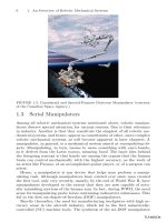
losses; and 5) creating low-noise readout methods. Procedures for separation and manipulation of
pathogens have also been one of the major problems in developing such sensors [33] [37]. Also, since
biosensors for POC applications are sometimes used as either implanted sensors or wearable sensors, it is
important to ensure that the sensor materials are biocompatible so as to ensure that there are no harmful
effects on patients; also, sensor materials should not degrade rapidly over time [38–39]. Biocompatibility
in turn can cause sensor node failure [39] due to biofouling, hermiticity of encapsulation, electrode
passivation, and limited lifetime of the immobilized enzymes [40–41].
Recently, there has been an increase in interest to integrate POC biosensors with microfluidics, which
has permitted the miniaturization of conventional techniques to enable high-throughput and low-cost
measurements through lab-on-a-chip systems [12]. Lab-on-a-chip systems take advantage of several
intrinsic characteristics of microfluidics, including laminar flow, low consumption of costly reagents,
minimal handling of hazardous materials, short reaction time required for analysis, multiple sample
detection in parallel, portability, and versatility in design [13]. Systems for biomolecular assays [14–15]
and bio-separations [16–17], including the separation of circulating tumor cells or plasma from whole
blood, have been reported in the past [18–19].
In order to realize remote monitoring and provide real-time feedback information, biosensors with
wireless link capability are desirable. Transmission of sensed data in the overall context of a POC
biosensor system needs to be performed for communicating the collected clinical information to the
healthcare provider or to a remote medical station. Typically, a biosensor network is deployed to aid data
transfer from the POC to the medical database. A biosensor network is defined as a collection of sensor
nodes that comprises a variety of biosensors, memory, data processing capabilities, and communication
block [42]. A combination of wireless MEMS and biosensors meets the demands of future medical
wireless biosensor systems. For reliable data transfer, a POC wireless biosensor network uses two-tiered
network architecture [43-44]. The lower tier involves the distribution of biosensors in the POC, and the
higher tier includes data routing to a central server. In a biosensor network, each biosensor senses its
environment and sends the raw data collected to a base station [45]. Direct transmission by a biosensor to
the base station is not a very efficient way of routing data. Hence, a network topology is employed to
efficiently transfer the data. The most common network topologies used to route data are 1) the cluster-
based approach and 2) the tree-based approach.
A cluster is basically a group of co-located sensor nodes that have formed a group or cluster to
minimize their communication energy costs. A typical cluster is characterized by a leader mode called the
cluster head. The cluster head is the sensor which is responsible for collecting data from its cluster
members, aggregating it and forwarding it to the base-station. Therefore, in this topology the cluster heads
are the nodes that perform the long distance communication to the base-station and hence consume more
energy than the cluster member that performs only short-distance communication. In order to prevent the
cluster head node from dying due to a lack of energy, the cluster node is periodically rotated. In the long
run, therefore, the energy consumption due to communication is averaged out or spread over the entire
network [45].
Another network topology that is commonly preferred is the tree-based approach. Here the nodes
form a tree-based structure for routing information. The final data collection node is called the sink node.
A set of nodes transmit data to the sink node and form the first level of the tree. These nodes in turn
receive information from another set of nodes forming the second level of tree structure. In this level, the
former set of nodes act as parent nodes to the latter. In the next level, the child nodes in the second level
in turn act like parent nodes and receive information from another set of nodes. This hierarchy continues
till all the nodes form a part of the network. Wireless Autonomous Spanning Tree Protocol (WASP) is a
medium access protocol that is used in a tree-based approach. Here, each node will tell its children in
which slot they can send their data by using a special message: a WASP scheme. This WASP scheme is
unique for every node and constructed in the node. A node uses the schemes to control the traffic of its
children and simultaneously to request more resources from its parents for these children. This minimizes
the coordination overhead because each scheme is used by the parent and the children of the sending
node. Everything the node has to know to generate this scheme can be obtained by listening to the
WASP-schemes. Coming from its parent node (i.e., one level up in the tree) and from its children (i.e.,
one level down in the tree). Consequently, the division of the time slots is done in a distributed manner
[46]. Figure 2 shows a typical cluster and tree topology.
Figure 2. (a) Cluster topology and (b) tree topology.
In addition to the above described requirements, an important and possibly required feature in a POC
system is the ability to provide embedded decision support, i.e., to extract higher level information from
raw bio-signals obtained from the biosensors. In a scenario where numerous POC biosensor systems are
deployed to continuously monitor several patients, a large quantity of multidimensional data will be
accumulated for each patient. It will be tedious for the professionals to examine the entire data set in order
to detect anomalies in health trends. Hence, the other requirement is to perform early identification
(prognosis) and thus prevention (health management) of diseases and health episodes. This requires
advanced inference logic and embedded signal processing capabilities to identify anomalies in the health
condition of the patient. In related work [47–48], researchers have embedded machine learning algorithms
in mobile phones to detect heart arrhythmias in the monitored ECG signals. Using a network of
biosensors, additional physiological information can be obtained and can be fused using machine learning
techniques to make an estimation of the patient’s health state [49].
3. MEMS FOR BIOSENSING
Biosensors are devices used to selectively detect the presence of specific biomolecules or compounds
in a given environment by carrying out physical or chemical transduction. Biosensors produce an
electrical equivalent of the change reflected in the biologically sensitive element acting as the sensor head
[50–51]. BioMEMS are defined as biosensors devices that are constructed using techniques based on
micro/nano-scale fabrication. BioMEMS are used for identifying, immobilizing, growing, purifying,
processing or manipulating, analyzing, and identifying biological and chemical analytes [37][52-53]. The
increased surge in BioMEMS for biosensing applications is primarily due to the fact that microfabrication
(a)
(b)
technology has resulted in miniaturization of sensing devices. This has led to increased sensitivity
resulting from sensors reduced in size to the scale of the analyte, leading to better performance, increased
reliability of extracted data, and reduction in detection time. Also, effective reagent volumes are reduced,
making the system cost-effective. Furthermore, miniaturization allows for portability, which is one of the
main requirements for POC applications [50][54]. For example, Feltis et al. [6] constructed a fully self-
contained, hand-held biosensor based on the Surface Plasmon Resonance (SPR) technique, with the size
of the complete sensor unit being 15 x 8 cm. They were able to measure changes as small as 3.3×10
−6
refractive index units and were able to detect the biological toxin ricin at 200 ng/mL in 10 min.
The range of potential applications of BioMEMS technology is broad. BioMEMS is the largest and
most studied area is diagnostics for POC testing applications. Some of the recent research works in the
field of POC diagnostics using BioMEMS include: Hwang et al. [55], who proposed and experimentally
proved a novel detection method for detecting bacterial cells in blood; Abbas et al [56], who developed a
millimeter wave BioMEMS for the detection of nitric oxide synthase (NOS) for ex-vivo applications; and
Tremerler and Sarikaya [57], who combined genetic tools with synthetic nanoscale constructs to create a
research methodology called Molecular Biomimetics.
3.1 Biosensors for POC technologies
A biosensor is traditionally defined as a bioanalytical device incorporating a biological material or a
biomimic (e.g., antibodies or nucleic acids) that is intimately associated with or integrated within a
physicochemical transducer or transducing microsystem. The transducer may be optical, electrochemical,
thermometric, piezoelectric, or magnetic [4][58]. The purpose of the transducer is to aid in producing an
electronic signal that is proportional to a single analyte or a related group of analytes. The essential
elements of a biosensor (Figure 2) are (1) the bio-recognition element (bio-receptor), (2) the transducing
element, (3) the excitation element, and (4) the readout modality. However, some biosensors might not
contain any excitation element.
Figure 3. Schematic representation of a biosensor containing the biorecognition element, transducer, and physical
output that is related to the concentration of the analyte of interest.
One of the critical functionality requirements of biosensors is bio-recognition element selectivity for a
specific target analyte and the ability to maintain this selectivity in the presence of interfering species or
compounds [59]. Selectivity depends on the ability of the bio-receptor to bind to the analyte. Highly
selective biological recognition systems have been developed using bio-receptors that are developed from
biological origins (such as antibodies and ligands) or that have been patterned after biological systems
(synthetic-based recognition element such as aptamers, peptides, surface-imprinted polymers, or
molecularly imprinted polymers) [57]. Another functionality requirement of a biosensor is sensitivity.
Sensitivity depends on multiple factors, including the geometry of the sensing surface [60], sensor
material properties [61], resolution of the measurement system, and the surface chemistry used in
immobilizing the bio-recognition element on the sensing surface [62]. The two commonly used forms of
surface chemistry are (1) silane chemistry and (2) thiol chemistry. Silane chemistry is preferred in
situations where the sensing surface material is silica, silicon, or plastic. On the contrary, thiol chemistry
is utilized when the sensing surface is potentially made of metals (mostly gold). In 2006, Bailey et al. [63]
introduced the DNA-encoded antibody library (DEAL) technique. Both for pathological examination or
for fundamental biology studies, different classes of biomolecules are measured typically in
heterogeneous samples, thus introducing unavoidable sources of noise that are hard to quantitate. The
DEAL technique was proposed for spatially multiplexed detection of single-stranded DNAs (ssDNAs)
and proteins on the same diagnostic platform. DEAL is based on the coupling of ssDNA oligomers onto
antibodies that are then combined with the biological sample of interest. Spotted DNA arrays are then
utilized to spatially stratify the biomolecules. Bailey et al demonstrated the DEAL technique for (1) the
detection of multiple proteins, (2) the co-detection of ssDNAs, proteins, and cells, and (3) the sorting of
cell lines and primary immune cells.
Widespread use of biosensors in POC applications will ultimately depend upon the development of
biosensing techniques that allow rapid, label-free detection of multiple biomarkers with high selectivity
and sensitivity. There are two major strategies for the detection of biomarkers: label-based and label-free
detection. Existing bio-detection systems mainly rely on fluorescence transduction methods (also called
labeled detection, as a fluorescent dye/tag is used to identify the biomarker concentration by either
labeling the bounded biomarkers or the unbounded bio-recognition sites) to detect the binding of
biomarkers to a biorecognition element due to its high sensitivity and selectivity and sufficient temporal
and spatial resolution [65]. In other words, label-based techniques require the labeling of query molecules
with labels such as fluorescent dyes, radioisotopes, or epitope tags [66]. This includes the enzyme-linked
immunoabsorbent assay (ELISA), a common clinical approach for protein marker detection that has a
detection limit as low as a few femtamolar concentrations, and the Frequency Resonant Energy Transfer
(FRET) transduction method. However, these labeling strategies often alter the surface characteristics and
natural activities of the query molecules. Moreover, labeling procedures are laborious and lengthy and
they limit the number and types of query molecules that can be studied. This makes labeling impractical
for use in POC applications [64][67]. In contrast to label-based techniques, label-free detection methods
depend on the measurement of an inherent property of the query itself, such as mass or dielectric
property, and it also avoids interference due to tagging molecules, thereby aiding in the rapid
determination of reaction kinetics of biomolecular interactions in real time [68-70]. This is the reason that
label-free detection holds potential for rapid diagnosis in a POC setting.
3.2 Biosensor classification
Apart from classification based on sensing strategy, i.e., label-based or label-free sensing (see Figure
4), biosensors are also classified based on the type of bio-receptor used (Table 1) and the transduction
mechanism (Table 2).
The ability to recognize the “target” or biomarker in a mixed population is viewed as a critical step in
any diagnostic assay. The biomarker can be present intracellularly or extracellularly. Whatever the
sampling matrix might be, the biomarker must be recognized and collected from a heterogeneous
population. In addition, the marker can be as complex as a whole cell or as simple as a single molecule
(such as a prostate-specific antigen (PSA)) [4]. Hence, the specificity of the biosensor is determined by
the receptor or the recognition element, and this directly affects the sensitivity of the biosensor.
Biosensors can be classified into two types based on the bio-receptor used. The bio-receptors can be
classified as (1) the bioaffinity type and (2) the biocatalytic type receptors [71]. In bioaffinity-based
reception, the receptor does not affect or change the target material, while in a biocatalytic receptor; the
receptor catalyzes a biochemical reaction. Most enzymes correspond to biocatalytic receptor types. In
some cases where enzymes are not available to detect certain biomolecules, antibodies are used for highly
selective reception.
Figure 4. Classification of biosensors based on sensing method.
Table 1. Biosensor classification based on transduction mechanism
Transduction Mechanism Method
Mechanical Stress sensing
Mass sensing
Optical Fluorescence
Chemiluminescence
Bioluminescence
Surface Plasmon
Scattering
Evanescent Waves Interferometry
Electrical Conductometric
Capacitive
Piezoelectric Quartz Crystal Microbalance (QCM)
Surface Acoustic Wave (SAW)
Electrochemical Potentiometric
Amperometric
Ion sensitive FET
1
(ISFET)
Chemical FET (ChemFET)
Thermal Calorimetric
1
Field Effect Transistor
Table 2. Types of biosensors based on receptors
Receptor Type
Enzyme Bioaffinity/Biocatalysis
Antibody/Antigen Bioaffinity
(immunosensor)
Nucleic Acids/DNA Biocatalysis
Biomimetic materials Bioaffinity
Cellular Structures/Cells Biocatalysis
Ionophore Bioaffinity
The most commonly used bio-receptors are 1) antibodies, 2) enzymes, 3) nucleic acids, and 4)
synthetic bio-recognition elements. Antibodies are biological molecules that exhibit very specific binding
capabilities for specific structures. An antibody is a complex molecule made up of hundreds of individual
amino acids arranged in a highly ordered sequence. Biosensors that depend on antigen-antibody binding
are called immunosensors. One of the most widely used immuno assays is the enzyme-linked
immunoabsorbent assay (ELISA) technique [73].
Enzymes are often chosen as bio-receptors based on their specific binding capabilities as well as their
catalytic activities. In biocatalytic recognition mechanisms, the detection is amplified by a reaction
catalyzed by macromolecules called biocatalysts. The catalytic activity provided by enzymes allows for
much lower limits of detection than would be obtained with common binding techniques. Presnova et al
[72] demonstrated electrochemical biosensors based on horseradish peroxidase where the peroxidase is
the enzyme that catalyzes the oxidation of a variety of organic molecules in the presence of hydrogen
peroxide.
Another biorecognition mechanism that has been receiving a lot interest in the last decade
involves the hybridization of deoxyribonucleic acid (DNA) or ribonucleic acid (RNA). Here, if the
sequence of bases composing a certain part of the DNA is known, then the complementary sequence
(which is called the probe) can be synthesized. By unwinding the double-stranded DNA into single
strands and adding the probe, and then annealing the strands, the probe will hybridize to its
complementary sequence on the target DNA. Ueberfeld et al. [74] designed a reversible fluorescent DNA
probe that can be used to determine the concentration of single-stranded DNA in solution by using a
ratiometric fluorescence measurement.
Recently, synthetic ligands have been studied for a variety of targets, such as aptamers and
ligands. The advantage of using synthetic bio-receptors over other bio-recognition elements is that they
are robust structures that can be placed in different environments without losing their specificity, can be
made by using wet chemistry techniques, and can be easily structurally modified to support the addition
of other sensing elements. Yao et al. [75] developed the “one-bead one-compound” (OBOC)
combinatorial library method (where each resin bead displays a unique peptide) and whole cell binding
assay to synthesize and identify D-amino acid-containing cancer cells.
As stated earlier, label-free detection techniques hold potential for inexpensive, noninvasive, and
informative clinical diagnoses, particularly in point-of-care settings. Recently, there has been increasing
interest among industry and the scientific community to use BioMEMS sensors for carrying out label-free
detection. The miniaturized size of MEMS sensors aids in reducing measurement time, increasing
sensitivity, and minimizing invasiveness. A couple of research efforts to realize label-free detection using
RF MEMS have been reported recently. Kim et al. [76] demonstrated a detection method based on RF
electric signals and MEMS to detect glucose oxidase (GOx). Dalmay et al. [77] developed a detection
method using microwave frequencies to study cell electrical parameters. Arvind et al. [78] demonstrated
two parallel modes of sensing using a single RF MEMS shunt capacitor to detect Staphylococcus Aureus
using mouse monoclonal IgG
3
proteins. Surface stress change and RF signal losses due to specific
biomolecular binding are two parallel sensing modes realized.
3.3 Label-free detection techniques using BioMEMS sensors
Many label-free techniques (Figure 4) have been successfully demonstrated in the past using micron-
size biosensors. However, these techniques also have issues regarding sensitivity and specificity. Further,
expensive fabrication techniques, morphological anomalies, and insufficient knowledge of biosensors
often restrict their use. Here we present the most commonly employed label-free detection techniques,
namely 1) surface plasmon resonance (SPR), 2) microcantilevers, 3) quartz crystal microbalance (QCM),
and 4) biological field effect transistors (BioFETs). All these sensors are constructed using techniques
based on micro-/nano-scale fabrication and thus can be considered as BioMEMS sensors.
Surface Plasmon Resonance (SPR)
SPR is a surface-sensitive spectroscopic method that measures change in the refractive index of bio-
sensing material at the interface between metal surfaces, usually a thin gold film (50–100 nm) coated on a
glass slide, and a dielectric medium [70]. Owing to high loss in the metal, the associated charge density
wave, also called the surface plasma wave (SPW), propagates with high attenuation in the visible and
near-infrared spectral regions. Changes in the refractive index of the sensing surface may be determined
by optically interrogating the SPR. During interrogation, optical radiation is used to excite the SPW,
which results in the resonant transfer of energy into the SPW. SPR manifests itself by resonant absorption
of the energy of the optical wave [79]. In order to detect biomarkers, the gold surface in SPR is
immobilized with the bio-recognition element. Unlabeled query molecules (target) are added, and any
changes in the angle of reflection of light caused by binding of these molecules to the immobilized bio-
receptors are measured to characterize biomolecular interactions in real time. The angle at which the
minimum intensity (corresponding to the maximum resonant energy transfer) of the reflected light is
obtained is known as the “SPR angle” (see Figure 5), which is directly related to the amount of
biomolecules bound to the gold surface. The advantages of SPR include real-time [85], multiplexed,
qualitative, as well as quantitative detection. Also, SPR is sensitive to conformational changes on the gold
surface. However, SPR is limited to the use of gold or silver surface alone.
Figure 5. Detection principle of an SPR device. Biomolecular interactions at the sensing surface layer are monitored
as a shift in the resonance wavelength.
SPR has been employed in a wide variety of bio-assays such as the analysis of association or
dissociation kinetics of biomolecules [80], drug discovery [81], rapid detection of cancer biomarkers [82],
and antigen–antibody interactions in protein microarrays [83]. Yu et al. [84] exploited the highly sensitive
nature of SPR to detect very low concentrations (0.1ng/mL) of domoic acid analyte having very low
molecular weight (310g/mol). Recently, Feltis et al. [6] constructed a fully self-contained, handheld SPR
device that is capable of duplicating many laboratory-based tests with a sensitivity range similar to many
commercial ELISA-based immuno-assays.
BioMEMS microcantilever
Electro-mechanical detection of biochemical entities and reactions has been demonstrated at the
nano- and micro-scale using microcantilever structures [60]. Past research works have reported the
observation that when specific biomolecular interactions occur on one surface of a microcantilever beam,
the cantilever bends [86–89] (see Figure 6(b)). The recent discovery of the origin of nanomechanical
motion generated by DNA hybridization and protein–ligand binding [88] provided some insight into the
specificity of the technique. In addition, its use for DNA–DNA hybridization detection, including
accurate positive/negative detection of one-base pair mismatches, has also been reported [88–89]. Besides
being label-free, this technology readily lends itself to the formation of microarrays using well-known
microfabrication techniques [90], thereby offering the promising prospect of high throughput protein
analysis. However, it remained unclear whether the cantilever sensing technique had sufficient sensitivity
and specificity to detect disease-related proteins at clinically relevant conditions and concentrations. Gu et
al. [91] in 2001 demonstrated the feasibility of using microcantilevers for detecting disease-related
proteins by detecting PSA as an example of both protein–protein binding in general and tumor marker
detection in particular.
In microcantilever biosensors there are two modes of sensing used for detection. They are (1) stress
sensing and (2) mass sensing. Stress sensing is carried out by coating one side of the cantilever beam
using a bio-receptor that adsorbs target biomolecules. The adsorption results in the expansion or
compression of the bio-receptor layer, which then induces surface stress on the cantilever beam, and thus
the cantilever bends due to stress. In the latter type of sensing, the cantilever is actuated to vibrate in its
resonant frequency. The binding of the biomolecule with a bio-receptor changes the frequency of
vibration. The shift in resonant frequency is analyzed to detect the concentration of the biomolecule [92].
Surface stress–based micro-cantilevers have been proposed and utilized because of their ease of
operation, higher sensitivity, and the ease to study surface stress during adsorption through optical
detection (as in atomic force microscopy (AFM)) and piezoresistive detection [92–94]. The
microcantilever having precise deflection measurement techniques allows for the detection of surface
stresses as small as 10
-4
Nm
-1
[95].
DNA hybridizations were the first biological sensing methods demonstrated using microcantilever-
type sensors. McKendry et al. [96] used an eight-cantilever array to detect unlabeled DNA hybridizations
at nanomolar concentrations within minutes. Zhang et al. [97] used microcantilevers in rapid and label-
free detection of biomarker transcripts in human RNA in the picomolar range. Protein-based cantilever
structures started evolving after the discovery of appropriate enzymes and antibodies. The late evolution
of protein-based cantilevers is due to the fact that the sensitivity of microcantilevers for protein detection
is much less compared to the detection of DNA. Biomolecule detection and analysis using proteins have
also been reported by Wu et al. [91]. Arntz et al. [98] used a biosensor similar to the McKendry array
[96] for real-time detection of cardiac biomarker proteins—myoglobin and kinase—whose level indicates
heart disease. Further, Arntz et al. [98] were the first to report that the sensitivity of protein detection by a
cantilever array technique is several orders of magnitude less than that reported in DNA hybridization.
The general concept of a BioMEMS cantilever device is that it detects changes in surface stress due to
adsorption-induced force and resonant frequency variation due to mass loading [99]. Fritz et al. [86]
reported that the bending motion is caused by changes in the surface stress of a cantilever beam, but the
origin of the surface-stress change was first elucidated by Wu et al. [88], who also reported that deflection
may be upward or downward depending on the type of molecules involved. Wu et al. [88] also suggested
that cantilever deflection is created because of the interplay between changes in configurational entropy
and intermolecular energy induced by specific biomolecular reactions between biomolecules such as
antibodies and analytes. Figure 6(a) lists the operation modes, signal transductions, and read-out
mechanisms with respect to microcantilever-based biosensors. The most commonly used molecular
detection methods for measuring the bending of a microcantilever (Figure 6(b)) are the optical deflection
method or piezoresistive techniques. In the optical deflection method a low-intensity laser is reflected off
of the cantilever and is focused onto a position-sensitive detector (PSD) (Figure 6(c)). Low intensity laser
beams are used so that the biomolecules coated on the surface of the cantilever are not affected [88][99].
The major advantage of the optical detection method is that it is capable of detecting deflections on
the order of 10
-10
m [100]. This detection scheme demands high precision and alignment, which makes
the optical diagnosing system expensive. Also, the liqueous environment causes thermal management
issues, leading to erroneous readings. Due to these disadvantages, a piezoresistive method of detection is
preferred for portable sensors. In a piezoresistive microcantilever system (Figure 6(d)), a loop of
piezoresistive material, such as polysilicon, is embedded onto the cantilever surface. When the cantilever
bends, it causes the bending of piezo material, which results in a change in its resistance (i.e., deflection
implies that the piezoresistive material undergoes a stress that causes a strain, which leads to a change in
resistance). This change in resistance can be determined by using a Wheatstone circuit. Though less
sensitive than optical detection methods, piezoresistive methods can detect deflections on the order of
nanometers. The readout system can be integrated on the chip (which is not the case with optical
detection) and is thus more appropriate for wireless sensing.
Quartz Crystal Microbalace
Quartz crystal microbalance (QCM) based biosensors are a viable alternative to microcantilevers due
to their sensitive mass detection capabilities and their ability to monitor in real time. The general
operating principle of a QCM biosensor is a decrease in the crystal’s resonant frequency following the
binding of the target biomolecules. When a rigid mass binds to the oscillating crystal surface, the shift in
the resonance frequency (∆f) is proportional to the mass bound. This was first proved by Sauerbrey [101]
in 1959. Henceforth, QCM has been used to quantify the number of target cells in a sample. Targets
detected by QCM include Chlamydia [102], Salmonella [103], Bacillus [104], Pseudomonas [105], and
Escherichia coli [106]. Recently, Poitras et al. [107] developed a biosensor using QCM with dissipation
monitoring (QCM-D) and validated it by detecting Escherichia coli (E. coli) O157:H7. The advantage of
using QCM-D over traditional QCM is that it gives insight into the viscoelastic properties of the adsorbed
mass. The biosensor response in QCM-D is the initial slope of the dissipation shift as a function of
elapsed time (D
slope
). The D
slope
provides an important advantage of rapid online bacteria detection.
Figure 6. (a) Operation modes and transduction mechanism used in BioMEMS microcantilever. (b) Cantilever beam
response: (i) initial state and (ii) sensing state. (c) Schematic of optical detection system for measuring
microcantilever deflection. (d) Schematic of a piezoresistive BioMEMS microcantilever.
Electrochemical Biosensors
Recently, an impressive number of inventive designs for biosensors based on electrochemical sensing
have appeared. These biosensors are also considered to be chemically modified electrodes (CMEs), since
electronic conducting, semiconducting, and ionic conducting materials are coated with a biochemical
films [108–109]. These types of sensors combine the bio-receptors with electrochemical transducers to
produce a biosensor to provide a simple, accurate, and inexpensive platform for patient diagnosis.
(a)
(b)
(c)
(d)
There are different types of electrochemical biosensors that have been commonly reported in the past:
1) amperometric, 2) potentiometric, 3) field-effect transistor (FET), and 4) conductometric biosensors
[110]. Amperometry is based on the measurement of the current resulting from the electrochemical
oxidation or reduction of an electroactive species. It is usually performed by maintaining a constant
potential at a working electrode (usually gold or carbon) or on an array of electrodes with respect to a
reference electrode. The resulting current is directly correlated to the bulk concentration of the
electroactive species [111–112]. Similarly, potentiometric measurements involve the determination of the
potential differences between an indicator and reference electrode when there is no significant current
flowing between them. The most common potentiometric devices are pH electrodes or gas selective (CO
2
and NH
3
) electrodes [110]. The potential differences between these indicator and reference electrodes are
proportional to the logarithm of the ion activity or gas fugacity (or concentration), as described by the
Nernst-Donnan equation [113–114]. Also, many enzyme reactions, such as that of urease, and other
biological membrane receptors may be monitored by ion conductometric or impedimetric devices using
interdigitated microelectrodes [115]. Drummond et al. [116] carried out an in-depth survey of the
approaches to electrochemical detection of selected DNA sequences, such as direct electrochemistry of
DNA, electrochemistry at polymer-modified electrodes, electrochemistry of DNA-specific redox
reporters, electrochemical amplification with nano-particles, and electrochemical devices based on DNA-
modified charge transport chemistry.
Another important class of electrochemical devices is the biologically active field effect transistors
(BioFETs). These are biosensor types that use a semiconductor transducer. The basic idea of BioFETs
stems from ion-selective field-effect transistors (ISFETs). In ISFETS, the gate of a MOSFET (metal-
oxide-semiconductor field-effect transistor) is replaced by an ion-selective layer, an aqueous solution, and
a reference electrode [118–120]. Such devices measure the concentrations of certain ion species in the
liquid and therefore act as pH sensors. More recently, biosensors such as EnFETs (enzyme FETs),
ImmunoFETs, and DNAFETs were devised building upon the ISFET concept by replacing the ion-
selective layer of ISFETs with layers of immobilized enzymes, antibodies, or DNA strands [121–123].
Gupta et al. [124] used a different kind of FET called the heterojunction (HFET) for in-vivo
electrochemical detection of physiological salt concentration. They reported the detection of streptavidin
and clinically relevant levels of biotinylated monokine induced by interferon γ (MIG) at physiological salt
concentrations with AlGaN heterojunction field effect transistors (HFETs). High charge sensitivity and
electrical stability in buffers makes HFETs an attractive platform for in-vivo detection.
3.4 Lab-on-a-chip for point-of-care testing
Lab-on-a-chip is a system or device that can be used to perform a combination of analyses on a single
miniaturized device for biological and clinical assays [125]. These systems are designed to either detect a
single or a class of biomolecules, or they have system-level analytical capabilities for a broad range of
biomolecules, known as a micro total analysis system (µTAS) [126]. These devices offer advantages such
as integrating sample handling and preparation, mixing, separation, lysing of cells, and detection [37].
These devices are also capable of purifying, isolating, and characterizing samples, and such a platform
integrates sample treatment modules together with separation and detection modules, all brought together
using the functionality of MEMS techniques [127]. Currently, the focus on bioparticle manipulation
methods to separate and identify biomolecules is based on electrostatic techniques such as
electrophoresis, electroosmosis, electrofusion, electrowetting, and dielectrophoresis (DEP) [125]. Most of
the bioparticle separation is done using DEP techniques. Pohl et al. [128] introduced the term DEP; they
described the translational motion of neutral matter caused by the polarization effects in non-uniform
electric fields.
Recently, there has been an increase in interest regarding the use of microfluidic paper-based
electrochemical devices, also called µPEDs, for quantifying the concentration of various analytes in
aqueous solutions, including biological fluids such as urine, serum, and blood [117]. The surge in interest
is due to the advantages of µPEDs: (i) it is light-weight, portable, single-use, and disposable; (ii) it is
flexible and foldable; (iii) it has excellent reproducibility with high sensitivity and accuracy; (iv) it does
not require professional medical personnel or complicated instruments; and (v) it allows for the
integration of high-density detection systems into a small device [129-132].
Also, there have been research efforts in the past to realize rapid multiplexed detections using lab-on-
a-chip devices. Fan et al. [133] reported an integrated microfluidic system to address the issue of
multiplexed detection of protein in microliter quantities of blood using fluorescent labels. Fan et al.
developed the integrated blood barcode chip (IBBC) to address the need for microchips that integrate on-
chip plasma separations from microliter quantities of whole blood with rapid in-situ measurements of
multiple plasma proteins. The immunoassay region of the chip is a microscopic barcode, integrated into a
microfluidics channel and customized for the detection of many proteins and/or for the quantification of a
single or few proteins over a broad concentration range. Zheng et al. [134] reported label-free, real-time
multiplexed detection of protein cancer markers with high selectivity and femtamolar sensitivity using
antibody-functionalized silicon-nanowire field-effect sensors.
4 WIRELESS BIOSENSORS
Wireless networks allow secure and instant access to patient reports and medical and administration
records. This helps in reducing errors, making decisions more quickly, and increasing quality of care
[135]. Wireless technology is credited with improving the flow of information unfettered by the
constraints of location or time. Secure wireless healthcare enables healthcare organizations to confidently
access medical applications and services, such as electronic health records, decision support, e-
prescribing, and e-research [136]. Also, secure wireless healthcare offers the wireless foundation to
deploy mobile care applications and location-aware services. This allows easy access to information for
POC services regardless of the location. Access to patient information by healthcare professionals at the
POC is critical for improving patient safety and medical care [137].
Biosensors with wireless link capabilities are desirable because wireless sensing systems can monitor
health conditions in real time and also bring forward new health care services, such as real-time drug
delivery. Although bio-assays for various pathogenic bacteria, proteins, and DNA based on BioMEMS
systems have been reported, their potential holds only if they can be reused for further sensing by
dissociating the detected pathogens from the corresponding antibodies. Traditionally, biosensors in the
labs are reused by dissociating biomarkers such as detected proteins using strong chemical formulations.
This method handicaps the use of both wearable and implantable sensors. Chen et al. [138] demonstrated
a wireless BioMEMS sensor for the detection of C-Reactive Proteins (CRP). This BioMEMS sensor dealt
with the problem of reusability through electrically controlled protein-antibody binding. The advantage of
the sensor system was dissociation, which was realized by applying a low-frequency 0.2 Hz 1-V AC
electric field to the sensor. Previously, Brod et al. [139] were the first to report and demonstrate electrical
control on antigen-antibody binding for the modulation of molecular recognition next to an electrode in a
pH biosensor. They demonstrated that the binding of an antibody to an antigen can be controlled
electrically in a reversible manner. The antigen-antibody binding was monitored using electrochemical
surface plasmon resonance (SPR). Application of a bias that is -0.5 V or more negative with respect to an
Ag/AgCl reference electrode caused the antigens to detach from the antibodies. When the negative bias
was removed, the antigens and antibodies had their binding ability restored. Similar efforts to control of
molecular interactions using electrical means have been reported. This includes modulation of an enzyme
immobilized on a membrane [140], redox reactions [141–143], dissociation of molecules by reducing
covalent bonds [144], and electrophoretic and electrostatic control over DNA hybridization [145–146].
However, in some applications the antibody can potentially be desorbed from the sensing surface. In such
applications, the DEAL approach circumvents the denaturation of antibodies. This is because DNA
patterns are robust to dehydration and can survive elevated temperatures (80–100 °C) [133].
4.1 Wireless biosensor architecture
A sensor provides the interface between the physical world and electronic systems, and a sensor node
incorporates the sensor along with the signal conditioning unit, processing unit, and communication
module. It is the function of the sensor nodes to acquire the sensed data and transmit them to the personal
server or data acquisition hardware. With advancements in MEMS technology, micro-size inexpensive
sensor nodes are becoming more realizable [147]. A wide range of commercially available components-
off-the-shelf (COTS) sensors have been deployed to monitor vital signals, the behavior of humans, and
the surrounding environment. These include accelerometers and gyroscopes, electrocardiography (ECG),
electromyography (EMG), electroencephalography (EEG) electrodes, pulse oximetry, respiration, carbon
dioxide (CO
2
), blood pressure, blood sugar, humidity, and temperature sensors [148].
Figure 7. Functional block diagram of wireless sensor module.
Figure 7 shows the functional blocks of a wireless sensor module. The major four blocks of a sensor
module are the 1) power supply, 2) radio system, 3) processing unit, and 4) sensing module. The
processing unit is composed of volatile and nonvolatile memory and a microcontroller. The sensor
module in turn consists of a filter, analog to digital converter (ADC), and a sensor head. The type of
sensor being used depends on the application [149]. The sensor senses the physiological data and is
preconditioned (by amplification and filtering) to levels suitable for digitization. The digitized data is
transmitted to the data acquisition device. A processor is programmed to control and coordinate the
activities of the sensor node for internal data acquisition and establishing communication with the
personal server. The processor also controls switching the individual components of the sensor node
between sleep and wakeup modes to save battery power when not in use.
Power consumption is constrained in wireless sensor nodes for medical applications. Lowering the
power consumption can prolong the lifetime of sensors. However, this adds an additional challenge to the
digital signal processor (DSP) for supporting radio-frequency (RF) transmissions between the severely
power-limited nodes. Self-powered wireless sensors are preferred in applications where sensors are
implanted, because, once implemented, it is tough to access the sensor to recharge the battery.
Because of advances in MEMS along with improvements in nano-engineering techniques, the
fabrication and manufacturing of micro-size sensors has become feasible [150]. This increased the
potential for producing smaller implantable and wearable sensors and also changes the traditional way of
measuring human physiological parameters. Recently, MEMS electrodes for ECG acquisition have been
developed by the fabrication of micro-size needles or electrodes on silicon polymers [147]. Brown et al.
[151] developed a fluorescent hydrogel-alginated microspherical optical glucose sensor, which serves as
an example for miniaturized nano-engineered sensors. Najafi et al. [152] carried out a study on the use of
battery-less MEMS implants for cardiovascular applications. This is a physiological sensor that includes
an array of micro-needles for use in an implanted blood pressure sensor. A more detailed review of
BioMEMS for implanted biosensors was provided by Grayson et al. [153].
4.2 POC wireless system model and architecture
Prognostics in health care refers to methods of survival analysis and estimating the rate of health
deterioration. In order to carry out prognostics, physiological parameters and health deterioration
parameters must be monitored. To understand and evaluate the prognostics of human health, biosensor
networks must be deployed both in the environment and on patients. Works related to wireless sensor
networks for environment sensing, event detection, and specific target sensing have been studied in the
past. Buratti et al. [154] reviewed wireless sensor technologies, their applications, standards, features in
design, and evolution. Mahfuz and Ahmed [155] studied the suitability of wireless sensor networks
(WSN) from the available communication system standards (ZigBee and Bluetooth) and CMOS-level
architecture for environmental monitoring, protection, and control. Cao et al. [147] presented a
comparison of IEEE 802 wireless standards for communication with an emphasis on the physical layer of
the ISO/OSI layered architecture model.
In the context of POC systems, transmission of measured data needs to be performed 1) for
communicating the data from the biosensors to the central node and 2) for sending the aggregated data
from the central node to a remote medical database or the healthcare provider [29]. In the first scenario,
the user’s mobility and comfortableness are hindered by the use of wires. Hence, in this case, the sensor
nodes can form a body area network (BAN) for transmitting the data to a central node, which can be a
personal digital assistant (PDA) or a custom-designed microcontroller-based device. Wireless BAN
(WBAN) is a radio-frequency-based networking technology that interconnects small nodes around the
human body, each of which has sensing capabilities [156–157]. A WBAN network is slightly different
from a Wireless Personal Area Network (WPAN) in the sense that WPAN interconnect devices are
centered on an individual person’s workspace and are intended to provide coverage distances of 10 m.
Also, WBANs differ from general WSN, which imposes new technical challenges [157]. First, there are
two types of nodes existing in WBANs: 1) sensors that are placed on the body or implanted inside the
human body, and 2) router nodes whose main function is to relay data to the medical database [158-159].
This is different from WSNs wherein all nodes act both as sensor and relay nodes [160]. Second, the
number of nodes deployed in a WBAN is comparatively less than in WSNs, as nodes are either placed on
or inside the body. This places a limitation on the reliability of data due to the reduction in redundant
information provided by multiple nodes sensing the same physiological parameter [156]. Third, WBANs
exhibit relatively stable data rates compared to WSNs (which perform event-based monitoring) due to the
need for periodic physiological monitoring, which demands continuous operation and thus requires power
management. Finally, WBANs are used for critical parameter monitoring; latency must be largely
avoided. In WSNs the nodes can be stationary or mobile, and node mobility does not occur in a group.
WBAN nodes are associated with the wearer’s mobility, and thus mobility occurs in a group [160–161].
Figure 8. Two-level network architecture for point-of-care wireless systems.
A WBAN is a health monitoring network of sensor nodes that is implanted or worn by a person called
the host [162]. The biosensor network consists of a group of biosensors, an external device called the
control node that is placed on or closer to the human body, and an external base station to relay data to the
global network (Figure 8) [167]. The sensor nodes in the WBAN are heterogeneous, possessing the ability
to measure multiple stimuli from the host’s body [163–164] and each consist of a processor, memory,
transceiver, sensors/actuators, and a power unit [167]. They are assumed to be built to survive extreme
conditions, such as variation in temperature and humidity [165], and are powered using batteries or
energy harvesting mechanisms that harvest energy from body movements, vibration, body heat
production, and bio-fuels [166]. These biosensors perform the tasks of sensing physiological information,
processing it, transmitting it to the control node, receiving external signals, and triggering actions such as
drug delivery. The control node acts as both the data aggregation and routing point. The control node also
sends the data collected periodically to an external base station, where it is stored for further processing.
The control node and the base station have significantly higher transmission and processing capabilities
than the biosensors [167].
The Vanderbilt Advanced NETwork Systems Lab in conjunction with the University of California,
Berkeley and Cornell University developed an integrated wireless sensor network for remote health care
called the CareNet (Figure 9). It was developed with to the goal of reliably and securely collecting
physiological data and transmitting them to a medical database where healthcare providers can access
patient health information [168–169]. Laerhoven and Gellersen [163] suggested that a large number of
low quality sensors can perform the task of monitoring as effectively as a few high quality sensors. Such a
network will have multiple sensors measuring the same stimuli, providing redundancy and increasing
fault tolerance, while at the same time being less intrusive and more favorable for regular wearing due to
their light-weight and small form factor [162]. In line with such a claim, networks up to 192 and 255
nodes have been proposed by Choi et al. [170].
Figure 9. CareNet network model.
Research projects such as CodeBlue, MobiHealth, and iSIM have also led to the concrete
establishment of a WBAN system [171-173]. IEEE 1073 has been focusing on standardizing a layered
architecture solution for wireless communication in wireless body area networks (WBANs) [158][174].
Figure 10 shows the IEEE 1073 layered architecture model for WBAN. The entire system architecture
(presented by Jovanov et al. [175]) involves a two-tiered wireless heterogeneous networking
infrastructure. Lo and Yang [176] developed a WBAN hardware platform based on this architecture using
COTS (components off-the-shelf).
Sensor devices are lightweight, wearable and mobile, which also means they have low computation
and communication power and a small amount of memory. A tele-healthcare system would employ a
network comprising individual health monitoring systems that connects to the healthcare providers
(centered on a medical server) through the Internet [177–178]. The medical server resides at the top of
this hierarchy and is optimized to provide service for a large number of users and encompasses a complex
network that also connects medical personnel and healthcare professionals. This network is a multi-hop
IEEE 802.11-based wireless network or mobile phone network (e.g., GPRS, 3G) [179]. The lower tier
consists of a number of sensor nodes that are strategically placed on an individual’s body. The sensor
nodes sample vital signals and transfer the relevant data to a personal server (PS; technically the control
node), which is generally a PDA for mobile patients or a home-based PC for elderly patients for in-home
monitoring [180–181] through a wireless link. The PS and the sensor nodes collectively form a network
that is generally called a personal network [182] and has a computerless architecture to access sensor
data. The personal server interfaces with the WBAN nodes through a network coordinator that
implements ZigBee (IEEE 802.15.4) or Bluetooth connectivity [182]. Practically, this interconnectivity
can be realized using radio waves, lasers, sound waves, light waves, or infrared radiation. Radio
connectivity (IEEE 802.15.4) is preferred because for shorter range the connectivity can be established
with lesser power and smaller antenna size than other schemes [183]. The personal server sets up and
controls the WBAN and provides a graphical or audio interface to the user and transfers information
about the individual’s health status to the health care provider. Table 3 compares commonly used short
distance communication schemes.
Figure 10. ISO and IEEE 1073 layered architecture
Table 3. Comparison of short distance communication schemes
ZigBee
(IEEE 802.15.4)
Bluetooth
(Open Standard)
WLAN
1
(IEEE 802.11)
Frequency Band
2.4 GHz 2.4 GHz 2.4 GHz
Bandwidth
5 MHz 1 MHz 20 MHz
Data rate
250 kbps
2
(2.4 GHz) 1 Mbps
3
>11Mbps
Multiple Access Scheme
CSMA/CA
4
FHSS
5
OFDMA
6
,CSMA/CA
Transmitter Power
0 dBm 4 dBm - 10 dBm 24 dBm
Range
0-10m 0-10m 0-100m
1
Wireless Local Area Network
2
kilo bits per second
3
mega bits per second
4
Carrier Sense Multiple Access/Collision Avoidance
5
Frequency Hop Spread Spectrum
6
Orthogonal Frequency Division Multiple Access
Yang et al. [183] fabricated and demonstrated a wearable RFID-enabled sensor node for continuous
biomedical monitoring and discussed the dielectric characterization of fabric substrates, inkjet-printing of
conductive nano-particle silver ink, design of RFID antennas, and integration of sensor active and passive
devices for a wireless biomedical application. A simple and cost-effective health care system was
developed and named UbiMon [184]. NASA, in collaboration with Stanford University, developed a
wearable physiological monitoring system for astronauts for continual monitoring of physiological
parameters during space missions [185]. The MIT Media Lab developed MIThril, a wearable computing
platform compatible with both custom-made and COTS sensors that interfaces humans and computers
[186–187]. Harvard University further extended and demonstrated the formation of ad-hoc networks in
their project CodeBlue [188–189] to facilitate automatic vital signal collection and real-time analysis of
human health when placed on patients in hospitals or disaster environments.
WBANs target diverse applications, including healthcare, real-time monitoring of athletes, workplace
safety, consumer electronics (short range wireless technologies for human-computer interaction (HCI))
[190-191], secure authentication and safeguarding of uniformed personnel, and health monitoring of
astronauts during space mission. Also, handicapped people can be assisted using intra-body
communications (IBCs), as proposed by Riaz and Shimamoto in [190]. Categorization of WBANs is done
based on factors such as the type of sensor for a particular application, radio system requirements (e.g.,
data rate), network topologies, and type of application [156].
WBANs extend conventional bedside monitoring to ambulatory monitoring, providing a POC to
patients, the elderly [192], and others for both hospital and home-based scenarios [168]. The objective of
POC wireless systems is to provide monitoring, diagnostic, autonomous diagnostic, alarm and emergency
services along with management of electronic medical record (EMR) of patients [193]. The CodeBlue
project at Harvard University was developed with an intention of providing a solution for pre-hospital and
in-hospital emergency care, stroke patient rehabilitation, and disaster response. Some of the sensors
developed for CodeBlue include a wireless two-lead EKG [194], accelerometer, gyroscope, and an EMG
sensor for monitoring patients with strokes. It uses the ZigBee protocol to communicate among the nodes.
It also uses a mesh topology and without any centralized server or database for storage and control of
information. Due to multi-hop transmission the system experiences packet loss, and thus the bandwidth
per receiver is limited to 40 kbps [163][184][195].
The Advanced Health and Disaster Aid Network (AID-N) developed at Johns Hopkins University has
an architecture similar to that of CodeBlue [196]. It provides monitoring service for mass casualties
during the time of disaster by using electronic tags similar to RFID tags. The system uses a global
positioning system (GPS) for outdoor localization and a Mote Track system for indoor tracking. Further
Wi-Fi and cellular networks are employed to facilitate the communication between personal servers and
the central server.
Wearable health monitoring systems (WHMS) developed at the University of Alabama aim at
providing telemedicine system for ambulatory health status monitoring. WHMS has a star-topology
network. The personal server is implemented on a PDA or a PC, which co-ordinates data collection from
sensor nodes using a TDMA scheme [195]. The MIThril project [186-187] includes an ECG, skin
temperature, and galvanic skin response sensor that is wearable and used for sensing and context-aware
interaction [197]. Further extensions were made to a multi-user wireless distributed wearable computing
platform that utilizes Wi-Fi connectivity to relay data from a personal server (implemented on a PDA) to
the central server [184].
Similar to WHMS, another research project involves the efforts of eight European countries and
fifteen other partners to create an ambient intelligent microsystems platform for mobile services and
applications (MIMOSA). It employs a mobile phone as a user-carried interface device. To enable local
connectivity between the sensor nodes, WiBree (Bluetooth low-energy technology) and RFID tags were
used. Also to provide an application programming interface for local connectivity and to facilitate sensor
readings, NanoIP and simple sensor interface (SSI) protocols have been integrated into MIMOSA [198].
Human gesture recognition has been proven to be very useful for the handicapped. Schwiebert et al.
[199] came up with a smart sensor with a novel approach to provide a complete system for restoring
vision to visually-impaired persons from the signals generated by an external camera to an array of
sensors that electrically stimulate the retina via a wireless interface. The Wireless Sensor Node for Motion
Capture System with Accelerometers (WiMoCA) [200] is concerned with the design and implementation
of a distributed gesture recognition system. The network system uses a star topology, and data
connectivity is realized using a time division multiple access (TDMA) approach. Sensing modules are
made of tri-axial accelerometers operating at 868 MHz with a peak data rate of 100 kbps [201].
Lo and Yang [200][202] studied the need for gesture recognition and came up with a way to identify
hand movement recognition. Other related work with respect to wireless medical system can be found in
[203–210]. A more detailed discussion on WBAN application in cardiovascular disease, cancer detection,
asthma, battlefield, and telemedicine systems can be found in [158].
In the context of WBAN, the IEEE 802.15.6 Task Group (TG) 6 is developing a communication
standard for low-power wireless sensor nodes operating in and around a patient’s body. As pointed out by
Pantelopoulos and Bourbakis [29], the standard under development should support scalable data rates
ranging from 1 kbps to hundreds of Mbps over a distance of 2–5m with a network size of 100 nodes and
guarantee very low latency and power consumption (around 0.1–1 mW) [157].
4.3 WBAN infrastructure
Table 4 provides a reference to the most important frequency bands for POC wireless communication
[157]. In order to design and develop a reliable system suitable for WBAN, knowledge of radio
propagation channels is needed [211]. WMTS is the licensed WBAN for medical telemetry systems, but it
has a restricted bandwidth (around 14 MHz). The other bands, such as ISM (2.4 GHz), include bands to
protect adjacent channel interference. The MICS band operating in the range of 402–405 Hz is dedicated
for implanted sensor communication. UWB operates at a higher frequency allocated between 3.1 to 10.6
GHz and will be looked upon more in future research. Currently, most of the WBAN services are
confined to the 2.36–2.4 GHz band so as to leverage COTS integration and avoid interference from
neighboring wireless channels.
Table 4. List of frequency bands for WBAN
1
Description Frequency Band
Implant MICS
2
402–405 MHz
On-body - 13.5 MHz
On-body - 5–50 MHz
(HBC
3
)
On-body WMTS
4
600 MHz
On-body ISM
5
900 MHz
On-body ISM(ZigBee) 2.4 GHz
On-body UWB
6
3.1–10.6 GHz
1
Wireless Body Area Network
2
Medical Implant Communications Service
3
Human Body Communication
4
Wireless Medical Telemetry Service
5
Industrial, Scientific, and Medical
6
Ultra-wideband
5 PROGNOSTICS IN POC APPLICATIONS
Reliability is a prime factor of concern for industrial and medical devices since the functional
degradation of products during performance in the field can be catastrophic [212]. Such a situation
occurred during World War II, when the reliability of military equipment became a serious matter of
concern [213]. Since then there has been an increase in interest in industry to study the durability or
lifetime of devices by monitoring the ongoing health of products and systems in order to predict failures
and provide warnings in advance of catastrophic failure [214]. Vichare and Pecht [215] have defined
prognostics as a method of prediction or estimation of the future state of health based on current and
historical health conditions. Further, this lifetime prediction study was extended and applied to study the
survival time of cancer patients. The term “remaining useful life (RUL)” used by industrial reliability
engineers is called “survival analysis” by cancer researchers [213]. In recent times, survival analysis
refers to a method for analyzing data in disciplines including medicine, epidemiology, environmental
health, marketing, and astronomy. Analogous to system-level failure prediction, prognosis in health care
involves techniques that combine sensing, recording, and interpretation of environmental, operational,
and performance parameters to carry out survival analysis for public health.
Health monitoring is one of the main application areas for pervasive computing [216]. The evolution
of biosensors for POC diagnostics and prognostics has been motivated by the advancement in information
and communication technologies, microfluidics, and microelectronics. Future POC diagnostic systems are
expected to support artificial intelligent algorithms through the use of decision support process, in order to
provide accurate and efficient diagnostic results in real time [213]. Decision support processing is a
critical component of a POC system since they can assist novice practitioners and nurses in their
diagnostic and clinical judgment [217]. POC wireless systems require the integration of current lab-on-a-
chip technologies with decision support services operated by standalone devices.
In the context of POC systems, prognostics must be performed for two different purposes. First,
prognostics is used to estimate the RUL of the POC system at each time instant so as to avoid system
failure and reduce system downtime. This can be implemented through the use of traditional techniques of
prognostics and health management (PHM) by interpreting the parameter indicative of (i) performance
degradation, i.e., deviation of operating parameters from their expected values; (ii) physical degradation
of critical subcomponents; or (iii) changes in operation environment [214]. The second type of health
monitoring at POC refers to the diagnosis of diseases and estimating the survival time. Survival time no
longer means the time to death [213]. It refers to the time period from a starting point to the occurrence of
a certain event.
In POC systems, prognosis of human health conditions is a theoretical model, as in the traditional
PHM approach. This theoretical model is built on the assumption that the health symptoms that are used
to identify the presence of health risks are embedded in the physiological signals from the patient. Hence,
the objective of these theoretical models is to represent the collected physiological information in an
efficient manner, identify patterns from the extracted information that corresponds to pathological
symptoms, and finally use this information to detect the health condition. Systems that provide this type
of functionality are called clinical decision support systems (DSS). Some efforts in the past pertaining to
DSS can be found in [218–221].
5.1 Prognostics approaches for devices and systems
In this section, the different types of prognostic approaches as applied to devices and systems are
discussed. Since POC systems have just started receiving attention in the clinical arena, no reports on
previous work relating to prognostics and POC systems is available in the public domain. However, with
the increasing demand for availability of medical systems, the need for prognostics will also soon be
realized.
Prognostic approaches
Early studies in prognostics focused on component-level reliability evaluation based on an
understanding of the component degradation and their loading conditions [222–223]. Based on these
approaches, Pecht et al. [224] proposed methods to use life cycle models to improve design and
manufacturing capability for electronic components. These studies led to the new theory of predicting
failures based on physics-of-failure (PoF) models [225–226]. Methods to select parameters and modeling
approaches for failure prediction have also been investigated [227–228]. In order to address situations
where PoF methods are not suitable, data-driven approaches were developed that use statistical techniques
to predict component failures based on their performance information [229]. These approaches rely on the
assumption that the statistical characteristics of performance data are relatively unchanged unless there is
a fault or malfunctioning of the system or component. Thus the main objective in data-driven approaches
involves precisely detecting anomalies in performance data. Commonly used data-driven approaches
include the sequential probability ratio test (SPRT) [230], support vector machines (SVMs) [231],
Bayesian SVM [232], and Mahalanobis distance [233-238]. Cheng and Pecht [239] also investigated the
use of a multivariate state estimation technique as an input for the prediction of RUL.
Fusion prognostics
Data-driven approaches fail to distinguish between failure mechanisms when the physical
understanding of component failure is not incorporated. Recognition of this fact has led to the
development of a fusion model that integrates a physical understanding of failure with data-driven
techniques thereby exploiting the benefits of both PoF and data-driven methods [240–241]. Recent
publications from the Center for Advanced Life Cycle Engineering (CALCE) at the University of
Maryland have focused mainly on identifying a pre-cursor parameter related to the physics of component
failure and employing machine learning techniques to forecast anomalies in the performance trend. Gu et
al. [242] developed a method to assess failures in electronic components under vibrational loading. Kwon
et al. [243] presented a prognostic technique to detect interconnect failure precursors using RF impedance
monitoring and the sequential probability ratio test (SPRT) under thermo-mechanical loading
conditions. Patil et al. [244] focused on identifying precursor parameters for enabling prognostics of
insulated gate bipolar transistors.
System prognostics
In relation to system-level prognostics, Pecht [245] presented a road map for how prognostic
approaches at the component level should be carried out to predict and avoid system failures. Sun et al.
[246] analyzed the benefits of applying prognostics to systems. However, due to the presence of multiple
critical components in a system, there are multiple failure modes and mechanisms that need to be tracked
to predict system failure. Wang et al. [247] developed a model to address this situation where each
component and failure mode is modeled individually and then pooled together to form the system
inspection model.
5.2 Prognosis of health condition
Prognostics in health care refers to the method incorporated in tracking the progression of health
symptoms from which an estimate can be obtained about the patient’s health condition. According to
Pantelopoulos and Bourbakis [49], “Prognostics uses a theoretical model to follow the progression of
health symptoms so as to be able to derive, at any given time, an estimation of the user’s health condition
in order to detect health risks by identifying dangerous health trends.”
In POC systems, these models utilize the information extracted using biosensors as input for
estimating future health conditions. However, such signals are fuzzy in nature. Hence, Pantelopoulos and
Bourbakis [49] proposed a prognostics methodology based on fuzzy language for estimating the health
conditions of patients whereby the current state of the corresponding fuzzy finite-state machine (FSM)
signifies the current estimated health state. Also a stochastic petri net model (SPN) has been employed for
the interaction scheme between the user and the biosensor system. This approach classifies the bio-signals
used in the identification of health symptoms into three categories to define the states of the FSM. The
bio-signals that are used as input are classified as 1) value-specific signals, whose instantaneous values
carry the diagnostic content; 2) morphology-specific signals, which contain diagnostic information in the
structure of the signal; and 3) patient voice recording, which is used as feedback.
The most important component of the POC diagnostic/prognostic system concerns the modeling of
knowledge from a patient’s clinical data and real-time monitored data. This component serves two
purposes: (1) to extract new and useful knowledge and (2) to provide prediction methods for diagnosis as
well as detection of susceptibility for a particular disease [248]. In order to extract new and potentially
useful knowledge, cluster analysis [249] and association rule mining [250] are applied. Classification
techniques [251] are applied to create predictive models for the diagnosis and detection of disease.
5.3 Survival analysis
Traditionally, prognostics in the medical arena have been performed using survival analysis
techniques. Various types of statistical approaches have been used for analyzing survival data. As stated
earlier, the primary variable in survival analysis is the survival time, which is modeled as a non-negative
random variable [252]. Standard statistical methods are not suitable for analyzing survival time because
(1) the survival time may be longer than the study time (one type of censoring of survival data) and (2)
the distribution of survival times is more skewed than symmetrical [213]. According to McGarty [253], in
order to adopt a statistical technique for analyzing survival data, the evaluation of some natural process is
embedded in the statistical model with the assumption that the model is a useful approximation of the real
process [252]. The major applications of survival analysis in public health include estimation of the
distribution of the survival time, testing hypotheses of equal survival distributions, and identification of
prognostic factors [213].
Survival time can be defined by three survival functions [254]: (1) the survivorship function; (2) the
probability density function, which is also known as unconditional failure rate; and (3) the hazard
function which is also known as instantaneous failure rate, age-specific failure rate, or conditional
mortality rate. The hazard function is a measure of the proneness to failure as a function of the age of the
individual [215][255].
The objective in carrying out survival analysis is to estimate the survival function and cumulative
hazard function, which helps in predicting the survival time. Various parametric and non-parametric
approaches have been proposed in the past for estimation of the survival and hazard function [254].
Let T denote survival time, a non-negative random variable. As stated earlier, T is the time period
from the starting point of the study to the occurrence of a certain event. The event may be death or
hospitalization [213]. Mathematically, let
0
T
be a time-to-event variable [256], and let
C
be the fixed
right censoring time [252], which may occur when a patient leaves during the study or survives past the
end of the study period. Then the survival variable
T
and the censoring indicator
δ
are defined as
follows:
},min{
0
CTT
=
(1)
=
0
1
δ
if
if
CT
CT
≤
>
0
0
(2)
The survivorship or survival function is denoted by S(t), which is defined as
)(1)()( tFtTPtS −=>=
(3)
where
)(tF
is the distribution function of
T
. The probability density function is defined as
t
ttTtP
tf
t
∆
∆
+
<
<
=
→∆
)(
lim)(
0
(4)
On the other hand the hazard function is defined as
t
tTttTtP
th
t
∆
>
∆
+
<
<
=
→∆
)|(
lim)(
0
(5)
The three functions are mathematically equivalent, and even if one of them is known, the other two
functions can be derived. According to the proportional hazard (PH) model the hazard function is
proportional to the baseline hazard function, i.e.,
)exp()()(
0
Xthth
β
=
(6)
where
)(
0
th
denotes the baseline hazard function,
β
represents the distribution parameter, and
X
denotes
an observed predictor of T [253]. The baseline hazard function is estimated using parametric or non-
parametric models [257]. Thus, once a baseline hazard function is estimated, the proportional hazard,
survival function, and the probability density function of survival time can be predicted.
In certain cases the effects of left truncation and right censoring data are required. Joint survival
analysis approach is required to tackle such problems. Shemayakin and Youn [258] presented a brief
overview of using Copula models for joint survival analysis. Copula models are popular for modeling
dependencies between random variables, and thus their application includes failure of paired organs or
body functions or physiological parameters in health science and predicting human mortality.
6 EXISTING CHALLENGES IN POC SYSTEMS
In this section we discuss the challenges for WBAN that need to be addressed before WBAN
technologies are widely deployed for POC solutions.
6.1 Biocompatibility and sensor packaging
Since WBAN sensors are deployed on the body or even implanted inside, the size and physical
compatibility with human tissues are critical. Sensors must be made from biocompatible materials so that
the sensor material does not negatively affect patients or degrade over time [259]. Common materials for
biomedical devices include synthetic or biological polymers, metals, and ceramics. Aluminium nitride is
inert with respect to the body and thus makes it a feasible choice for biomedical sensors [260].
Biocompatibility itself can cause failures due to biofouling, hermeticity of encapsulation, electrode
passivation, and the limited lifetime of the immobilized enzymes [261–263]. To confront the failure due
to bio-fouling is to use micro-sensor arrays. This helps in improving the reliability, as it is very unlikely
that all the sensors will fail at the same time [263]. Apart from biocompatibility, the material chosen for
biosensors at the POC must also have certain electrical and mechanical properties to reduce electrical
losses and improve sensitivity.
6.2 Wireless operation
Lowering the power consumption of the radio link can greatly reduce the size and prolong the
lifetime of a sensor. Otis et al. proposed a 400µW receive and 1.6mW transmit transceiver for wireless
sensor networks [264]. Yoo et al. [265] proposed an approach that uses the human body as the medium of
data transmission for low energy on-body data transmission. As mentioned earlier, UWB has proven to be
the dominant emerging technology for wearable sensors [266]. UWB can theoretically produce a data rate
up to 1Gbps.
Antenna design is one of the determining factors for a reliable and energy-efficient wireless
communication link. The antenna has to be optimized in order to maximize the throughput of the radio.
Alomainy et al. [267] proposed a planar compact antenna structure for miniaturized on-body sensors.
Higgins suggested different types of antenna design for implantable sensors [268–269]. Ullah et al. [270]
discusses the design of an implantable antenna for operation in the MICS band. In the same paper, the
physical layer and medium access control (MAC) layer performance of body sensor networks are
discussed. Zhen et al. [271] analyzed the performance of clear channel assessment (CCA) by in-body and
on-body nodes and demonstrated that on-body sensor nodes cannot see the activity of in-body nodes
when they are 3m away from the surface of the body.
Apart from low-power wireless transceivers, communication protocol is also an essential component
for the development of energy efficient wireless sensors [263]. Dave et al. [272] studied the energy
efficiency and quality of service (QoS) performance of IEEE 802.15.4 and IEEE 802.11e. Timmonsa and
Scalon [273] analyzed the performance of IEEE 802.15.4 for use in biomedical sensors in BANs. They
showed that when properly configured with low data rate asymmetric traffic, the sensors can operate for
at least 10 years without battery replacement. Li and Tan [274] proposed a novel time division multiple
access (TDMA) protocol for an on-body sensor network that exploits bio-signal features to perform
TDMA synchronization and improve energy efficiency. Other protocols, such as wide area system secure
(WASP) and CICADA, have also been proposed [275–277]. A number of low power communication
protocols have been proposed for wireless sensor networks, such as low-energy adaptive clustering
hierarchy (LEACH) [278], power aware multi-access with signaling (PAMAS) [279], sensor-MAC (or)
S-MAC [280] and Berkeley-MAC (or) B-MAC [281]. Very recently Marinkovic et al. [282] presented an
energy-efficient medium access control protocol suitable for communication in a wireless body area
network for remote monitoring of physiological signals using a low duty cycle TDMA strategy. Buddhika
de Silva et al. [283] investigated the impact of inter-user interference and its behavior with respect to the
number of networks and the rates at which these networks communicate. They observed that inter-user
interference can cause up to 35 % loss in packet delivery ration.
Though energy-efficient MAC protocols have been devised, there is still a need for accurate models
that take into account factors such as reliability, latency, mutual interference, energy consumption, and
mobility effect. Sheryl Howard [284] studied the energy efficiency of error-correcting coding in low-
power links that connect the implant and the external device. However, there is a need for more in-depth
evaluation in all other scenarios.
6.3 Security
Privacy requires effective and efficient authentication techniques in WBANs. It was suggested that
the ideal way to provide user authentication is to use complex but distinguishable human body
characteristics. Sriram Cherukuri et al. [167] used the same method and proposed an approach wherein
biometrics derived from the body are used for securing the keying material. A similar approach was
presented by Venkatasubramanian et al. [285] with two schemes that rely on the novel technique of using
physiological values from the wearer’s body for securing a cluster topology formation.
Venkatasubramanian et al. [286] presented a novel key agreement scheme that allows two sensors in a
WBAN to agree to a common key generated using electrocardiogram (EKG) signals for providing
security on real EKG data. Still, a mechanism needs to be devised to differentiate between different levels
of security to identify and distinguish between life-threatening events from other events with various
security priorities.
6.4 Power restrictions
Sensors for WBAN application have power restrictions. This is mainly due to their small size and the
absence of wires. Using an integrated power supply such as a battery will drain out of power. ence, there
is a need to switch to passive power sources such as solar power, vibration, motion, air flow, temperature
difference, and infrared radiation. Raghunathan et al. [287] discuss the key issues and tradeoffs that arise
in the design of solar energy harvesting in wireless embedded systems and present a design,
implementation, and performance evaluation of Heliomote sensors. Mitcheson et al. [288] developed a
vibration-based generator designed for wearable/implantable devices that is capable of delivering up to
2µJ/cycle. Similarly, Reilly et al. [289] proposed a system to harvest vibration energy from a
piezoelectric thin film. IMEC developed a 3 x 3 x 1.5 cm
3
thermal micro-power generator that is capable
of converting thermal energy to 4µW power at 5ºC temperature difference on the thermopile. Although
techniques for generating power from passive sources have been proposed, the power generated is
insufficient for continuous operation. Thus, to deal with this issue, there is a need for an external wireless
power source to generate and wirelessly transfer power to the sensors. Recently, MIT researchers have
reported transferring energy through a wireless medium to power electronic devices over a short range
using evanescent waves [290]. Huang et al. [291] demonstrated a technique to harvest power from solar
energy.
6.5 Standards and rules
Efforts have been made to develop standards for WBAN for POC [292]. However, pervasive health
monitoring and treatments (POC) employing WBAN require standardized rules for operation in
ambulatory environments and providing POC services without limitations on the wearer’s location or
mobility while also taking into account individual privacy. Also, sensor fabrication, minimum
biocompatible property requirements, power consumed, and minimum detectable levels of a particular
biomarker must be standardized.
7 DISCUSSION
The growing number of fatalities resulting from chronic diseases has led to increased efforts to
develop POC systems for remote health monitoring and patient care. To understand the improvements