Inactivation of bacteria by non thermal plasma
Bạn đang xem bản rút gọn của tài liệu. Xem và tải ngay bản đầy đủ của tài liệu tại đây (2.55 MB, 31 trang )
2
Inactivation of Bacteria by
Non-Thermal Plasmas
R. Morent and N. De Geyter
Research Unit Plasma Technology – Department of Applied Physics
Faculty of Engineering and Architecture – Ghent University
Belgium
1. Introduction
In physical sciences, “plasma” refers to the forth state of matter, while in medicine and
biology, plasma is known as the non-cellular component of blood (Fridman et al., 2008).
Interestingly, the term “plasma” has been coined by Irving Langmuir to emphasize that the
characteristics of ionic liquids ubiquitous in biology and medicine are analogous to plasma
in the physical sciences (Fridman et al., 2008, Langmuir, 1928). Despite this historical
connection, plasmas are mainly associated with the solid-state processing technology
(Stoffels et al., 2003), while being rarely used in biomedical applications directly. This
situation is however rapidly changing and multiple plasma applications in life sciences are
recently emerging (Daeschlein et al., 2010, Vandamme et al., 2010, Kalghatgi et al., 2010,
Kong et al., 2009, Nie et al., 2009, Kalghatgi et al., 2007).
The plasma state can be considered to be a gaseous mixture of oppositely-charged particles
with a roughly zero net electrical charge (Denes&Manolache, 2004). Besides charged
particles, plasmas also contain neutral atoms and molecules, excited atoms and molecules,
radicals and UV photons. Generally, plasmas can be subdivided into 2 categories: thermal
plasmas and non-thermal (or cold) plasmas (Denes&Manolache, 2004, Fridman et al., 2008,
Bogaerts et al., 2002). Thermal plasmas are characterized by very high temperatures of
electrons and heavy particles, both charged and neutral. In contrast, non-thermal plasmas
are composed of low temperature particles (charged and neutral molecular and atomic
species) and relatively high temperature electrons (Bogaerts et al., 2002, Denes&Manolache,
2004). Because the ions and the neutrals remain relatively cold, a non-thermal plasma does
not cause any thermal damage to articles it comes in contact with. This characteristic opened
up the possibility to use these non-thermal plasmas for the treatment of heat-sensitive
materials including biological matter such as cells and tissues (Laroussi, 2009). Non-thermal
plasmas are already routinely used in material processing applications, such as etching,
activation and deposition (Borcia et al., 2006, Bruce et al., 2010, De Geyter et al., 2008, De
Geyter et al., 2009, Morent et al., 2009a, Morent et al., 2009b, Maruyama et al., 2010). More
recently, the biological and medical applications of these plasmas have witnessed a great
interest from both plasma as well as medical research communities.
This review paper focuses on one specific fascinating application of non-thermal plasmas in
biomedical science, namely the inactivation of bacteria, also called plasma sterilization
www.intechopen.com
Biomedical Engineering – Frontiers and Challenges
26
(Stoffels et al., 2008). We need to stress that the term sterilization is somewhat ambiguous
since this term is only used when all initial micro-organisms are killed, which is however
not always the case when applying non-thermal plasmas to contaminated surfaces (Boudam
et al., 2006). Conventional sterilization methods include the use of dry heat (oven), moist
heat (autoclave) or chemicals like gaseous ethylene oxide, liquid formaldehyde and
glutaraldehyde (Kelly-Wintenberg et al., 1998, Moisan et al., 2001, Moisan et al., 2002, Park
et al., 2003). Some major drawbacks of these conventional techniques are the high processing
temperatures (ovens and autoclaves) which makes it impossible to sterilize heat-sensitive
materials like polymers, the use of toxic chemicals and the long sterilization times needed
(approximately 12 hours in the case of ethylene oxide exposure) (Park et al., 2003, Moisan et
al., 2001, Montie et al., 2000). Another interesting sterilization method is the use of gamma
irradiation, but this is an expensive technique and may cause the material to undergo
undesirable changes during sterilization (Moisan et al., 2001, Henn et al., 1996,
Ishigaki&Yoshii, 1992). The limitations of these conventional methods have encouraged the
search for new approaches and an alternative method of sterilization is treatment with a
non-thermal plasma (plasma sterilization). These plasmas operate under moderate
temperatures and use non-toxic gases, therefore, thermal and chemical damage to the
substrate is limited (Philip et al., 2002, Sladek&Stoffels, 2005). Moreover, plasmas are not
only capable of killing bacteria and viruses, they can also remove these dead micro-
organisms from the surfaces of the objects being sterilized (Chau et al., 1996). This chapter
on plasma sterilization is organized as follows: a first part will focus on the inactivation of
bacteria on non-living surfaces, which has reached a state of maturity. In this first section,
the kinetics of bacterial inactivation processes will be described, followed by the effects of
various plasma-generated agents on bacterial cells. Afterwards, a brief review on the
inactivation of bacteria on non-living surfaces by vacuum and atmospheric pressure
plasmas will be presented. A second part of the chapter will deal with state-of-the-art
applications of non-thermal plasmas in bacterial inactivation, namely the sterilization of
teeth and human/animal tissue, which are both relatively new research topics.
2. Plasma sterilization on non-living surfaces
2.1 Survival curves to determine the inactivation efficiency
Plasma effects on micro-organisms can be evaluated using various methods, however, a
commonly used approach is the determination of survival curves (Stoffels et al., 2008,
Boudam et al., 2006, Moisan et al., 2002). These curves are plots of the logarithm of the
number of surviving micro-organisms as a function of exposure time to the sterilizing agent.
Although the precise procedures to obtain these curves may vary, usually a suspension
containing a well-defined concentration of micro-organisms is placed on a substrate and let
to dry. After plasma exposure, the remaining micro-organisms are let to inoculate for
several hours before counting. Considering that counting large numbers of cells is
troublesome, the number of colony forming units (CFU) is determined instead of counting
individual cells after inoculation (Stoffels et al., 2008). For conventional sterilization
methods, the survival curve is usually a unique straight line: the inactivation process is an
exponential function of time (Moisan et al., 2002, Cariou-Travers&Darbord, 2001). In
contrast, plasma sterilization can provide survival curves with different shapes depending
on the type of micro-organism, the type of medium supporting the micro-organisms and the
method of plasma exposure (direct or remote) (Laroussi et al., 2000, Laroussi, 2002). In some
www.intechopen.com
Inactivation of Bacteria by Non-Thermal Plasmas
27
cases, the survival curves after plasma exposure are straight lines (similar to conventional
sterilization methods) (Laroussi et al., 2000, Herrmann et al., 1999, Yamamoto et al., 2001),
however, in most cases, two or even three different linear segments occur, each segment
being a different inactivation phase (Kelly-Wintenberg et al., 1998, Moisan et al., 2002,
Laroussi et al., 2000). This implies that the number of surviving micro-organisms is also an
exponential function of time, but with different time constants. To characterize the slope of
each segment, an interesting parameter has been extensively used by several researchers
studying plasma sterilization: the so-called “D-value” (decimal value) (Moisan et al., 2002,
Laroussi, 2002, Fridman, 2008). This parameter is the time required to reduce an original
concentration of micro-organisms by 90 % (one log
10
reduction) and is expressed in the unit
of time.
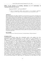
Single-slope survival curves have been observed in atmospheric pressure plasma
sterilization by Herrmann et al. (Herrmann et al., 1999), Laroussi et al. (Laroussi et al., 2000)
and Yamamoto et al. (Yamamoto et al., 2001) and an example of such a single-slope curve is
presented in Figure 1.
Fig. 1. Example of a single-slope survival curve: E.coli exposed to an atmospheric pressure
glow discharge in a helium/air mixture [Reprinted with permission from (Laroussi, 2005)].
Herrmann et al. (Herrmann et al., 1999) employed a remote atmospheric pressure plasma jet in
a helium/oxygen mixture to treat Bacillus globigii spores on glass coupons and found a D-value
of 4.5 seconds. Laroussi et al. (Laroussi et al., 2000) and Yamamoto et al. (Yamamoto et al.,
2001) utilized an atmospheric pressure glow discharge (DBD) in a helium/air mixture and an
argon/H
2
O
2
corona discharge respectively to treat Escherichia coliphage (E. coli). In these
studies, single-slope survival curves were reported with D-values ranging from 15 seconds for
the corona discharge to 5 minutes for the DBD-discharge (Laroussi et al., 2000, Laroussi, 2002,
Yamamoto et al., 2001). More recently, Stoffels et al. (Sladek&Stoffels, 2005, Stoffels et al., 2008)
and Choi et al. (Choi et al., 2006) presented results on plasma-induced deactivation of E. coli
using a plasma needle operating in helium/air mixtures and a dielectric barrier discharge
(DBD) in air respectively and also found a straight line as survival curve.
Two-slope survival curves can occur in both vacuum and atmospheric pressure plasma
sterilization and were observed for the first time in 1998 by Hury et al. (Hury et al., 1998).
These authors reported on the inactivation of different Bacillus spores in an oxygen plasma
www.intechopen.com
Biomedical Engineering – Frontiers and Challenges
28
operating at low pressure (0.5 Pa) and did not observe a linear survival curve, but two
successive lines with different slopes. According to their findings, the first slope has the
smallest D-value (D
1
), while the D-value of the second slope (D
2
) is larger. As a result, the
authors concluded that the inactivation of spores in their low pressure oxygen plasma is a
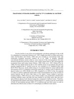
two-step process: a fast process followed by a much slower one. Similar two-slope curves, as
illustrated in Figure 2, were found in 2000 and 2002 by Moreau et al. (Moreau et al., 2000)
and Philip et al. (Philip et al., 2002), who employed low pressure (133-933 Pa) microwave
discharges in pure argon and N
2
/O
2
mixtures (7 % oxygen) respectively for the inactivation
of Bacillus subtilis spores. According to both Hury (Hury et al., 1998) and Moreau (Moreau et
al., 2000), the first phase of their survival curve corresponds to the action of UV irradiation
on isolated spores or on the first layers of stacked spores. The second phase, which is
characterized by slower kinetics, represents spores that are shielded by others and thus
require longer irradiation times to accumulate a lethal UV dose.
Fig. 2. Evolution as a function of time of the population of spores submitted to a pure argon
afterglow at low pressure [Reprinted with permission from (Moreau et al., 2000)].
As previously mentioned, two-slope survival curves have also been observed in
atmospheric pressure plasmas. Kelly-Wintenberg et al. (Kelly-Wintenberg et al., 1998) and
Laroussi et al. (Laroussi et al., 2000) employed an atmospheric pressure glow discharge
(DBD) for the inactivation of E. coli, Staphylococcus aureus and Pseudomonas aeruginosa. In
contrast to the vacuum plasmas, the D-value of the observed second slope (D
2
) was smaller
than the D-value of the first slope (D
1
) in these plasma systems. A general example of the
observed survival curves is shown in Figure 3.
Montie et al. (Montie et al., 2000) found similar survival curves for the inactivation of E. coli
and B. subtilis on glass, polypropylene and agar and claimed that the D
1
-value depends on
the species being treated, while the D
2
-value depends on the type of surface supporting the
micro-organisms (Laroussi, 2002, Fridman, 2008). A hypothesis for the two-slope survival
curve was given by Kelly-Wintenberg et al. (Kelly-Wintenberg et al., 1998): during the first
killing stage, active plasma species react with the outer membrane of the cells leading to
damaging alterations. After this process has sufficiently advanced, the reactive species can
quickly cause cell death, resulting in a very rapid second phase.
www.intechopen.com
Inactivation of Bacteria by Non-Thermal Plasmas
29
Fig. 3. Survival curve of Pseudomonas aeruginosa exposed to an atmospheric pressure glow
discharge in a helium/air mixture [Reprinted with permission from (Laroussi et al., 2000)].
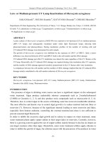
Multi-slope survival curves with three (or more) linear segments have also been observed in
both vacuum as well as atmospheric pressure plasma sterilization. Moreau et al. (Moreau et
al., 2000), Moisan et al. (Moisan et al., 2002) and Philip et al. (Philip et al., 2002) found three
inactivation phases when employing a low pressure (133-933 Pa) microwave discharge in
argon/oxygen and N
2
/O
2
mixtures (0.7-2 % oxygen) respectively for the inactivation of B.
subtilis. These authors claim that the first phase, which has the shortest D-value, is mainly
due to the action of UV photons on isolated spores or on the first layers of stacked spores.
The second phase (with the slowest kinetics) can be attributed to the erosion of the spores by
active species, such as atomic oxygen. The third phase starts when the spores that were not
inactivated during phases 1 and 2 have been sufficiently eroded, hence allowing UV
photons to hit the genetic material of the still-living spores and finally kill them. A schematic
illustration of the three-phase survival curve can be found in Figure 4.
Fig. 4. Example of a three-phase survival curve: inactivation of Pseudomonas aeruginosa with
an atmospheric pressure glow discharge in a helium/air mixture [Reprinted with
permission from (Laroussi, 2005)].
www.intechopen.com
Biomedical Engineering – Frontiers and Challenges
30
These multi-slope survival curves were also recorded by Laroussi et al. (Laroussi et al., 2000)
and Roth et al. (Roth et al., 2000) who employed atmospheric pressure plasma sources for
sterilization, however, a clear elucidation for this type of survival curve is lacking for the
moment. The explanation given earlier for low pressure plasma sterilization is most likely
not applicable to atmospheric pressure plasmas, for which it has been shown that UV
photons only play a secondary role in the killing process (Herrmann et al., 1999, Yamamoto
et al., 2001, Kostov et al., 2010, Laroussi, 1996). Based on the above-mentioned bacterial
inactivation kinetics, one can conclude that the mechanisms of sterilization by both vacuum
and atmospheric pressure plasmas are very complex and far from understood at this
moment.
2.2 Plasma species responsible for sterilization
As mentioned at the end of the previous section, plasma sterilization is a complex process
since multiple plasma species, such as UV photons, excited species, reactive neutrals,… can
interact with the bacterial cells being treated. This section tries to give an overview of the
contributions of various plasma agents (heat, UV radiation, charged particles and reactive
species) to the sterilization process.
2.2.1 Effect of heat
The first factor that usually comes to mind when discussing sterilization is the effect of heat.
As previously mentioned, conventional sterilization methods are based on the use of either
dry or moist heat. Moist heat (or autoclaving) is widely used for sterilization by subjecting
objects to steam under pressure at temperatures of at least 120 °C for 15 minutes or longer
(Gopal, 1978, Ratner et al., 1990). Dry heat sterilization requires even higher temperatures
(close to 170 °C) with treatment times of about one hour (Laroussi, 2005). Most non-thermal
plasmas operate at low temperatures (room temperature to approximately 70°C), therefore,
no substantial thermal effects on bacterial cells are expected in plasma sterilization processes
(Laroussi&Leipold, 2004). However, it should be noted that some non-thermal plasmas (like
gliding arc discharges and dielectric barrier discharges) can be characterized by elevated
temperatures in localized intervals in space or in time and for these discharges thermal
effects should be sometimes also taken into account (Fridman, 2008). However, in general,
heat is not a major contributor to the sterilization effect in non-thermal plasmas.
2.2.2 Effect of UV radiation
Non-thermal plasmas are a source of UV with different wavelengths, however, only UV
radiation in the 200-300 nm wavelength range with doses of several mJ/cm
2
are known to
cause lethal damage to cells (Fridman, 2008, Laroussi, 2005). This damage is mainly the
dimerization of thymine bases in bacterial DNA strands, which inhibits the ability of
bacteria to replicate properly (Fridman, 2008, Laroussi, 2005).
The presence of UV radiation in the plasma strongly depends on the operating pressure. It is
well-known that vacuum plasma discharges are able to provide significant UV radiation in
the range of wavelengths effective in sterilization. This can explain the important
contribution of UV radiation in plasma sterilization at low pressures, as discussed in the
previous section. In contrast, the great majority of papers published on the inactivation of
micro-organisms by atmospheric pressure plasmas conclude that the contribution of UV
radiation to the inactivation process is not important, when not at all relevant. By comparing
www.intechopen.com
Inactivation of Bacteria by Non-Thermal Plasmas
31
the killing kinetics of UV radiation from a low pressure mercury-vapour lamp with that of a
glow discharge plasma at atmospheric pressure, Laroussi et al. (Laroussi, 1996) concluded in
1996 that UV was not the main killing agent. In 1998, this claim was supported by Kelly-
Wintenberg et al. (Kelly-Wintenberg et al., 1998) who exposed different types of micro-
organisms to an atmospheric pressure glow DBD in air. These authors observed that there is
no significant difference in the time needed to inactivate micro-organisms on polypropylene
samples, whether exposed directly or sealed in bags. They concluded from this observation
that UV is not a significant contributor to lethality since the opaque bags would have
blocked much of the UV light (Kelly-Wintenberg et al., 1998). In the following years, several
researchers (Laroussi, 2002, Laroussi, 2005, Choi et al., 2006, Laroussi&Leipold, 2004,
Birmingham, 2004) have carried out various experiments that also supported the claim that
UV plays a minor role in plasma sterilization. In 1999, Herrmann et al. (Herrmann et al.,
1999) exposed Bacillus globigii to a remote atmospheric pressure plasma jet operating in
helium/oxygen mixtures. When the reactive plasma effluent was blocked by a quartz
window, allowing only UV radiation and heat to reach the spores, no substantial reduction
in the initial concentration of bacteria was observed. In 2004, Laroussi and Leipold
(Laroussi&Leipold, 2004) used the flowing afterglow of a DBD in air at atmospheric
pressure to inactivate spores of the Bacillus genus. They quantified the UV emission from the
discharge and observed no significant UV emission in the wavelength range effective in
sterilization. Also in 2004, Birmingham (Birmingham, 2004) tested what he calls a “plasma
blanket”, which is an aluminium foil layer covered by a micro-machined dielectric
polyimide layer. This structure provides small corona discharge spots in air and can be
wrapped around a contaminated object. The author states that the “plasma blanket” does
not generate sufficient photons of the appropriate wavelength and thus concludes that the
inactivation process only results from the interaction of the ionized gas with the biological
material. Choi et al. (Choi et al., 2006) subjected different samples to a DBD operated in air
at atmospheric pressure and found no UV radiation below 290 nm. As a result, they
concluded that UV plays no significant role in plasma sterilization. In an interesting review
paper, Laroussi et al. (Laroussi, 2005) specifically concluded that there is no UV radiation in
the 200-285 nm wavelength for dry air non-thermal plasmas at atmospheric pressure.
Therefore, UV photons are not likely to take part in the inactivation of micro-organisms
when air plasmas are used.
In contrast to the previous literature overview, some authors (Park et al., 2003, Heise et al.,
2004, Lee et al., 2005, Trompeter et al., 2002) do mention the possible role of UV photons in
plasma sterilization at atmospheric pressure, however, the arguments brought forward are
quite often not fully convincing or incomplete (Boudam et al., 2006). For example,
Trompeter et al. (Trompeter et al., 2002) employed an atmospheric pressure filamentary
DBD in different gases and found that argon was the most efficient gas in terms of spore
inactivation. As a result, they concluded that the inactivation process is purely due to UV
radiation since argon is chemically inactive. Heise et al. (Heise et al., 2004) employed a
similar discharge in argon and claimed that the inactivation of spores is partly due to UV-
induced effects. Both Trompeter et al. (Trompeter et al., 2002) and Heise et al. (Heise et al.,
2004) present their results as being performed in pure argon, nonetheless this is not the case
since impurities are always present in the discharge gas (Boudam et al., 2006). This
contamination can strongly reduce the UV emission (Boudam et al., 2006), therefore, the role
of UV in the inactivation process might not be as large as mentioned by these authors.
Recently, Boudam et al. (Boudam et al., 2006) subjected micro-organisms to an atmospheric
www.intechopen.com
Biomedical Engineering – Frontiers and Challenges
32
pressure DBD operated in an N
2
/N
2
O
2
mixture. These authors claim that they have proven
that UV photons, under specific operating conditions, can be the dominant inactivation
species. The role of UV in atmospheric pressure plasma sterilization is thus still open for
debate and further research should be definitely conducted before the controversy can be
resolved.
2.2.3 Effect of reactive species
According to several authors (Laroussi, 2002, Laroussi, 2005, Fridman, 2008), reactive
neutral species (such as O, O
2
*, O
3
, OH
•
, NO and NO
2
) can make a significant contribution
to the plasma sterilization process, especially at high pressures. To show the effect of these
reactive species on the destruction of bacteria, Laroussi and Leipold (Laroussi&Leipold,
2004) employed an atmospheric pressure DBD in three different gases: pure helium, 97%
helium-3% oxygen mixture and air to inactivate Bacillus spores. When helium is used as
discharge gas, only very small concentrations of radicals originating from impurities are
expected. When helium is mixed with oxygen, oxygen-based species such as O and O
3
will
be present in the discharge. When air is used as discharge gas, both oxygen- and nitrogen-
based species will be generated. Figure 5 shows a comparison between the inactivation
kinetics in the case of pure helium and the helium/oxygen mixture.
Fig. 5. Percent of surviving Bacillus spores as a function of plasma treatment time for pure
helium (black) and a helium/oxygen mixture (97% He-3% O
2
) (gray) [Reprinted with
permission from (Laroussi&Leipold, 2004)].
When pure helium was used as discharge gas, the surviving spore population percentage
was still much greater than 10% after 10 minutes of plasma treatment. In fact, in this case,
the D-value was greater than 20 minutes. When a helium/oxygen mixture was employed, as
shown in Figure 5, a D-value of 10 minutes was achieved. Laroussi and Leipold
(Laroussi&Leipold, 2004) also showed that in the case of an air plasmas, the D-value could
be even further decreased to approximately 20 seconds. According to these authors, this
dramatic decrease in inactivation efficacy can thus be attributed to the presence of
chemically reactive species such as NO, NO
2
, O, O
3
,… Besides Laroussi and Leipold, other
authors (Herrmann et al., 1999, Moreau et al., 2000, Richardson et al., 2000) have also
experimentally shown that discharges containing oxygen provide a strong germicidal effect.
In particular, Herrmann et al. (Herrmann et al., 1999) compared results obtained by an
atmospheric pressure plasma jet with and without oxygen. These authors found that the D-
www.intechopen.com
Inactivation of Bacteria by Non-Thermal Plasmas
33
value in the case of absence of oxygen is higher than in the case when oxygen is added.
Richardson et al. (Richardson et al., 2000) also showed that their resistive barrier discharge
became more effective in killing B. subtilis when oxygen was added to the discharge gas
helium. It is believed that the oxygen species attribute to the sterilization process due to
their strong oxidative effects on the outer structures of cells (Fridman, 2008). Moreover,
discharges containing oxygen also generate ozone (O
3
) which interferes with the cellular
respiration system and is known to have a strong bactericidal effect (Laroussi, 2002). After
experimenting with different gas mixtures, Kuzmichev et al. (Kuzmichev et al., 2000)
concluded that the best bactericidal effects were achieved in moistened oxygen and air. In
the presence of moisture, hydroxyl (OH) radicals are generated in the plasma, which play a
significant role by chemically attacking the outer structures of bacterial cells. In the case of
air, the production of NO and NO
2
adds to the lethality of the process.
2.2.4 Effect of charged particles
In the discussion of the mechanisms of inactivation, a lot of attention is given to the effect of
UV radiation and reactive species and most researchers have neglected to investigate the
influence (if any) of charged particles. As a result, the exact role of electrons and ions is not
yet fully resolved, but there are several indications of their importance. Fridman et al.
(Fridman et al., 2007, Fridman, 2008) have stated that charged particles play an essential role
in sterilization, especially in the case of direct plasma treatment, i.e. when the plasma is in
direct contact with the micro-organisms. These authors employed an atmospheric pressure
air DBD in direct mode and afterglow mode (so-called “plasma jet”) at similar discharge
powers and with the same amount of UV radiation and the results comparing the direct and
indirect effects of plasma sterilization are illustrated in Figure 6. From this figure, it can be
clearly seen that direct application of plasma yields a better sterilization efficiency than the
treatment by plasma afterglow. Although this high efficiency in the direct mode cannot be
solely described to the role of charged particles (the concentration of reactive species is also
lower in case of indirect exposure), it could be possible that charged-induced mechanisms
contribute to the sterilization process in direct plasma exposure.
Fig. 6. Effect of direct application of plasma (a: 5 seconds, b: 15 seconds) and effect of plasma
afterglow (c: 30 seconds, d: 2 minutes) [Reprinted with permission from (Fridman et al.,
2007)].
www.intechopen.com
Biomedical Engineering – Frontiers and Challenges
34
Mendis et al. (Mendis et al., 2000) also suggested that charged particles might play a
significant role in the rupture of outer cell membranes. According to these authors, charge
accumulation on the outer surface of the membrane induces an electrostatic force, which can
overcome the tensile strength of the membrane causing its rupture. They also state that this
mechanism is only effective for Gram-negative bacteria, which posses thin outer membranes
and a thin murein layer. The scenario will probably not occur for Gram-positive bacteria,
which lack an outer membrane but have a much thicker murein layer thereby providing
these bacteria higher strength and rigidity. Their conclusions can be supported by several
experimental observations. Kelly-Wintenberg et al. (Kelly-Wintenberg et al., 1999) employed
an atmospheric pressure glow discharge for the inactivation of the Gram-negative E. coli and
used transmission electron microscopy (TEM) to visualize the plasma-induced physical
damage to the micro-organisms. Figures 7 (a) and (b) show the cells before plasma exposure
and after 30 seconds of plasma treatment respectively. As observed in these figures, plasma
exposure rapidly disrupts the cell wall and leads to a release of cellular contents in the
surrounding medium. Laroussi et al. (Laroussi, 2002) employed a similar discharge for the
inactivation of the Gram-positive B. Subtilis and found intact cell walls after plasma
sterilization, which clearly supports the claim of Mendis et al Based on the above-
mentioned discussion, one can conclude that there may be a contribution of charged
particles in bacterial inactivation, but most likely only in direct plasma treatment, however,
this interesting possibility remains to be investigated in detail.
Fig. 7. TEM images of E. coli (A) before and (B) after 30 seconds of plasma exposure
[Reprinted with permission from (Kelly-Wintenberg et al., 1999)].
2.3 Vacuum plasmas
Glow discharge plasmas operated in vacuum have been successfully employed to sterilize
surfaces for quite some time. Patents written by Ashman and Menashi (Ashman&Menashi,
1972) as well as by Gut Boucher (Gut Boucher, 1980) showed that an electrical discharge in
an appropriate gas can lead to sterilization. In these patents, samples are inserted within a
chamber which is subsequently evacuated with a pump to 1-5 Pa. Afterwards, this chamber
is filled with gas set at the required pressure, typically 5-300 Pa. The gas discharge is then
achieved by applying an RF field to the gas by means of a coil located on the outside of the
chamber. Ashman and Menashi (Ashman&Menashi, 1972) used different discharge gases,
such as chlorine, bromine, iodine, water vapour, oxygen and nitrogen, but only observed an
www.intechopen.com
Inactivation of Bacteria by Non-Thermal Plasmas
35
effective sterilizing effect when halogens were used as discharge gas. Gut Boucher (Gut
Boucher, 1980) added aldehyde vapours to different carrier gases (oxygen, argon or
nitrogen) and observed the best results with oxygen as carrier gas. In 1987, Jacobs and Lin
(Jacobs&Lin, 1987) employed a similar RF discharge in a two-step process: (1) injection and
contact of hydrogen peroxide (H
2
O
2
) with the item being sterilized; (2) application of an RF
discharge to ensure that no toxic residues remain on the sterilized item. It is however
important to note that the previously mentioned systems are not really plasma-based
sterilization systems due to the use of gas mixtures that contain components with germicidal
properties (such as H
2
O
2
and aldehydes) before the plasma is ignited (Laroussi, 2005).
Nevertheless, some of these sterilization systems have been commercially available since the
1990s (Montie et al., 2000, Rutala et al., 1998, Vassal et al., 1998).
One of the first reports on plasma sterilization where gases were used with no germicidal
properties on their own was published in 1976 by Fraser et al. (Fraser et al., 1976). These
authors employed a low pressure RF discharge in different gases (argon, nitrogen, oxygen,
helium,…) and obtained reductions in spore populations of 99% after 2 minutes of plasma
exposure. However, it was not specified for which discharge gas this result was obtained. A
few years later, in 1982, Bithell (Bithell, 1982) observed that articles placed in a porous
envelope could be sterilized in a low pressure (40 Pa) oxygen RF plasma after a plasma
exposure time of 60 minutes. In 1989, Nelson and Berger (Nelson&Berger, 1989) also claimed
that an oxygen RF plasma is very effective as biocidal medium.
Different authors have examined which discharge gas would be the most efficient to
inactivate bacterial spores. Hury et al. (Hury et al., 1998) compared the mortality of Bacillus
subtilis spores after exposure to low pressure argon, oxygen and CO
2
plasmas and
concluded that oxygen and CO
2
plasmas are much more efficient in destroying B. subtilis
spores than a pure argon plasma, most likely due to the presence of reactive oxygen species.
A similar study was performed by Purevdorj et al. (Purevdorj et al., 2003) who used inert
gas plasmas, oxygen-based plasmas and various moisturized air plasmas at low pressure for
the inactivation of B. pumilus spores. These authors found that spore survival widely varied
depending on the composition of the feed gas. In contrast to the results of Hury et al. (Hury
et al., 1998), a pure oxygen plasma caused a lower spore mortality that an argon plasma,
however, no clear explanation for this observation was given. Experiments also showed that
a 50:50% oxygen-argon mixture leads to a much higher spore mortality, while the highest
mortality could be achieved by employing moisturized air. Based on this latter result, the
authors claim that the presence of water vapour enhances the inactivation process, most
likely because of the generation of OH radicals.
Taking into account the high efficiency of oxygen-based plasmas, literature on low pressure
sterilization has mainly focused on these kind of plasmas, nevertheless, inert gas plasmas
are in some cases still employed for bacterial inactivation (Liu&Chen, 2008, Yang et al.,
2009). Recently, many studies (Bol'shakov et al., 2004, Boscariol et al., 2008, Cvelbar et al.,
2006, Liu et al., 2008, Nagatsu et al., 2003, Rossi et al., 2008, Singh et al., 2009, Vicoveanu et
al., 2008) on the effects of pure oxygen vacuum plasmas on bacteria have been conducted.
Bol'shakov et al. (Bol'shakov et al., 2004) published an interesting paper on the use of an
oxygen RF plasma for sterilization purposes. The study was carried out for two operational
modes: the inductively coupled mode and the capacitively coupled mode and it was found
that the inductive mode offers a better efficiency in destroying bacterial cells. Rossi et al.
(Rossi et al., 2008) also employed a low pressure oxygen RF discharge for the inactivation of
www.intechopen.com
Biomedical Engineering – Frontiers and Challenges
36
B. stearothermophilus. Figure 8 presents scanning electron microscopy (SEM) images of the
untreated spores and spores exposed to the oxygen discharge.
As can be seen, the size of the spores is significantly reduced after 2 minutes of plasma
treatment. According to the analysis of a statistically relevant number of spores done on the
SEM pictures, it was found that in these conditions the mean length of the spores was
reduced from 1.73 to 1.35 µm. Similar results were obtained by Nagatsu et al. (Nagatsu et al.,
2003) and Singh et al. (Singh et al., 2009), who employed a low pressure oxygen microwave
discharge. They experimentally confirmed that the B. stearothermophilus spores were
sterilized after a plasma exposure time of 3 minutes. Moreover, based on SEM images of the
spores, these authors found that the sterilized spores had different sizes and shapes
compared to the untreated ones, which could be attributed to the interactions with reactive
oxygen species.
Fig. 8. SEM images of (A) untreated and (B) oxygen plasma-treated spores [Reprinted with
permission from (Rossi et al., 2008)].
Pure oxygen plasmas have also been employed by Cvelbar et al. (Cvelbar et al., 2006) and
Vicoveanu et al. (Vicoveanu et al., 2008) for the inactivation of B. subtilis spores, who
claimed that substrate heating might play a role in the inactivation process, especially at
elevated discharge powers. In another study on pure oxygen plasmas, Boscariol et al.
(Boscariol et al., 2008) shows the influence of discharge power on the bacterial inactivation
of B. subtilis spores. Linear survival curves were experimentally obtained with D-values
depending on discharge power. At 300 W, a D-value of approximately 8 minutes was found,
while at higher discharge powers (350 and 400 W), the D-value decreased to approximately
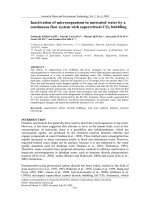
3 minutes. Besides studying the influence of discharge power, Liu et al. (Liu et al., 2008) also
examined the influence of other operating parameters on the inactivation of E. coli on
poly(tetrafluoroethylene) (PTFE) films. These authors found, in accordance with Boscariol et
al. (Boscariol et al., 2008), that the germicidal effect is higher at elevated discharge powers.
They also experimentally confirmed that the inactivation effect increases with increasing
plasma treatment time up to 50 seconds, after which it remains more or less stable. The
authors also studied the difference between direct and remote plasma exposure by
positioning the PTFE films at different distances varying from 0 to 80 cm from the centre of
the induction coil. Samples placed in the region 0-30 cm from the centre of the coil are
directly exposed to the plasma, while at larger distances, plasma treatment occurs in the
afterglow mode. Figure 9 shows the germicidal effect as a function of distance from the
www.intechopen.com
Inactivation of Bacteria by Non-Thermal Plasmas
37
induction coil and clearly shows that the oxygen plasma can effectively inactivate E. coli
within a distance of 0-40 cm. In the afterglow zone at 40 cm from the coil centre, the
concentration of ions and electrons is nearly zero, while the concentration of oxygen radicals
is still approximately 70 % of the initial value. This afterglow treatment could be interesting
to treat substrates prone to plasma-induced degradation.
Fig. 9. Germicidal effect of an inductively coupled RF plasma sustained in oxygen at
different distances from the induction coil [Reprinted with permission from (Liu et al.,
2008)].
Besides pure oxygen plasmas, also low pressure N
2
/O
2
plasmas have been widely
investigated for the inactivation of bacteria (Moisan et al., 2002, Philip et al., 2002, Moreau et
al., 2000, Rossi et al., 2006, Villeger et al., 2003, Villeger et al., 2005). Moreau et al. (Moreau et
al., 2000) and Moisan et al. (Moisan et al., 2002) carried out detailed studies on the effects of
a low pressure N
2
/O
2
microwave plasma with biological samples placed in the afterglow of
the discharge. Total inactivation of B. subtilis spores was achieved after 40 minutes of plasma
exposure at 100 W. In a following set of experiments, Philip et al. (Philip et al., 2002) studied
the influence of oxygen percentage added to the nitrogen carrier gas on B. subtilis
inactivation. Sterilization was achieved in less than 40 minutes with 0.7, 1.5 and 2% of added
oxygen. In contrast, sterility was only reached after 80 minutes with 4% of added oxygen,
while it was not even attained after 120 minutes with 7% of oxygen. The authors claim that
the short sterilization times (which occur at oxygen percentages below 2%) are due to the
high UV intensity of the discharge, since the spores are only slightly eroded. Increasing the
oxygen percentage in the mixture beyond this level increases the erosion rate of the spores,
hence reducing more and more the dimensions of the spores. However, although the spores
are heavily damaged, longer sterilization times are needed. The authors therefore state that
the spores are mainly being inactivated by UV photons and not by erosion caused by
reactive species. More recently, Rossi et al. (Rossi et al., 2006) claimed that the process
determining the time to reach complete spore inactivation is etching rather than UV action,
which is in contradiction with the paper written by Philip et al. (Philip et al., 2002). Rossi et
al. (Rossi et al., 2006) employed an RF plasma for the inactivation of G. stearothermophilus
spores and sterilization tests were performed in different N
2
/O
2
mixtures ranging from a
www.intechopen.com
Biomedical Engineering – Frontiers and Challenges
38
discharge in pure nitrogen to a pure oxygen one, which is a larger oxygen range than the
one examined by Philip et al. (Philip et al., 2002). It was found that the best sterilization
results were achieved in mixtures with high amounts of oxygen: complete sterilization was
obtained in the 95% O
2
- 5% N
2
mixture within 5 minutes of plasma exposure and significant
changes in the dimensions of the spores were observed for these discharges. In contrast,
mixtures with low oxygen amounts, which produce high UV emission, resulted in low
sterilization efficiencies and no variation in dimensions of the spores was found. The
authors therefore claim that erosion caused by reactive species might after all be significant
in the bacterial inactivation process. Recently, Kylian et al. (Kylian&Rossi, 2009)
demonstrated that the application of Ar/N
2
/O
2
plasmas offers the possibility to combine
the advantageous properties of the binary mixtures, namely, the capability of an N
2
/O
2
plasma to emit intense UV radiation needed for inactivation of bacterial spores together
with high removal rates of biological substances from Ar/O
2
discharges.
2.4 Atmospheric pressure plasmas
Although the use of vacuum plasmas can be considered as a significant improvement over
existing sterilization methods, there are still several drawbacks, including the need for batch
processing, long processing times and high costs. As a result, in the past decades, interest
has grown in applying atmospheric pressure plasmas for sterilization purposes and several
methods have been developed that allowed researchers to easily generate non-thermal
plasmas at high pressures, up to one atmosphere (Laroussi, 2005). In the following sections,
the most commonly used atmospheric pressure plasmas sources for sterilization purposes
will be briefly discussed together with some important results obtained for each of these
discharge types.
2.4.1 Dielectric barrier discharge (DBD)
A widely used atmospheric pressure discharge for bacterial inactivation is the dielectric
barrier discharge (DBD). In most cases, the plasma is created between closely spaced parallel
plates, where one or two plates are covered by a dielectric layer (Kogelschatz et al., 1997,
Kogelschatz, 2003). DBDs are generally driven by AC high voltages with frequencies in the
kHz range and power consumptions between 10 and 100 W (Stoffels, 2007). Usually, a DBD
operates in the filamentary mode: the breakdown starts at many points, followed by the
development of independent current filaments, named microdischarges (Kogelschatz et al.,
1997, Kogelschatz, 2003). However, it has been demonstrated that a homogeneous discharge
can be obtained under special, quite restrictive conditions [83,84]. DBDs can be used for
bacterial inactivation, but the narrow spacing between the electrodes makes it difficult to
introduce thick samples. For these samples, remote DBD treatment can however be
employed.
Laroussi et al. (Laroussi et al., 1999) demonstrated that a homogeneous helium/air DBD is
effective for the inactivation of E. coli and observed that exposure times of 2 to 20 minutes
can lead to complete inactivation. The authors also state that the treatment time necessary to
obtain a complete kill depends on several parameters, such as plasma power, working gas,
type of bacteria and type of medium. Using a similar pure air discharge, Kelly-Wintenberg
et al. (Kelly-Wintenberg et al., 1999) reported on the inactivation of a broad spectrum of
bacteria placed on a variety of surfaces. Experimental results have showed that at least a 5
log
10
CFU reduction in bacteria can be achieved between 15 and 90 seconds of plasma
www.intechopen.com
Inactivation of Bacteria by Non-Thermal Plasmas
39
exposure. An exception to these very short exposure times were experiments with solid
culture media in which 5 minutes of plasma exposure was necessary to achieve the same
reduction in CFU. To enable the exposure of large or complex three-dimensional samples,
Roth et al. (Roth et al., 2000) developed a so-called remote exposure reactor, in which
samples can be sterilized in the afterglow of an atmospheric pressure glow DBD. These
authors found that besides direct exposure, also remote plasma treatment can achieve
effective bacterial inactivation (Roth et al., 2000, Ben Gadri et al., 2000, Montie et al., 2000).
Besides DBDs in the homogeneous mode, also filamentary discharges have been employed
for the sterilization of various bacteria (Kostov et al., 2010, Heise et al., 2004, Trompeter et
al., 2002, Hahnel et al., 2010, Yasuda et al., 2008). Kostov et al. (Kostov et al., 2010) and
Yasuda et al. (Yasuda et al., 2008) were able to achieve sterilization of E. coli with their
filamentary air DBD, while Trompeter et al. (Trompeter et al., 2002) focussed on the
inactivation of B. subtilis and Aspergillus Niger with DBDs sustained in different working
gases. Heise et al. (Heise et al., 2004) observed that the spore reduction of B. subtilis depends
on which working gas is used to sustain the DBD: the efficiency of the discharge is the
lowest when air is employed and the highest with argon as discharge gas. More recently,
Hähnel et al. (Hahnel et al., 2010) observed that air humidity in a dielectric barrier surface
discharge has a significant influence on the bacterial inactivation rate: higher concentration
of water vapour in the process gas leads to higher killing rates of micro-organisms.
To enable the internal sterilization of complicated medical instruments, such as breathing
circuits, catheders and endoscopes, different authors have developed special DBD
configurations (Eto et al., 2008, Pointu et al., 2008, Sato et al., 2008). Eto et al. (Eto et al., 2008)
developed a so-called “linear DBD” with a diameter of 0.2-3 mm and a length of 40 cm and
found that 10
6
B. stearothermophilus spores could be killed inside a medical plastic tube at
room temperature after 12 minutes of air plasma exposure. Sato et al. (Sato et al., 2008) also
report on a special type of DBD and demonstrated the efficacy of their DBD for tube
sterilization. A photograph of their discharge in a long flexible narrow tube can be found in
Figure 10.
Fig. 10. Photograph of a DBD inside a tube for catheter sterilization [Reprinted with
permission from (Sato et al., 2008)][91].
2.4.2 Atmospheric pressure plasma jets
Since the 1990s, an increasing interest in plasma jets for bacterial inactivation has been
registered. Numerous plasma jets with different features have been developed and
www.intechopen.com
Biomedical Engineering – Frontiers and Challenges
40
described in literature (Herrmann et al., 1999, Laroussi et al., 2006, Shimizu et al., 2008,
Stoffels et al., 2006). These jets all differ in design, size, working gas, frequency of applied
voltage,… but the principle is the same. The plasma is ignited inside a nozzle equipped with
one or two electrodes and expands outside the nozzle via a gas flow. Atmospheric pressure
plasmas jets are very practical due to their small size and light-weight plasma generation
unit. They can be employed to treat small-sized objects, but can also be used for large-scale
treatments by moving the jet over the selected area or by applying multiple nozzles next to
each other. The following section will focus on the most important atmospheric pressure
plasma jets employed for bacterial inactivation, however, it is important to mention that also
other types of plasma jets have been developed for disinfection purposes (Shimizu et al.,
2010, Chiang et al., 2010, Ikawa et al., 2010, Lee et al., 2005, Liu et al., 2010, Kim et al., 2009,
Huang et al., 2007).
One type of plasma jet, which is actively employed for bacterial sterilization is the so-called
“atmospheric pressure plasma jet (APPJ)”. The design and operation of the APPJ have been
already discussed in detail elsewhere (Jeong et al., 1998, Park et al., 2000, Schutze et al., 1998)
and are only briefly summarized here for completeness. The APPJ consists of two coaxial
electrodes between which a feed gas (mixtures of helium, oxygen and other gases) flows at
high rate (50-100 litres per minute gas flow) and is schematically presented in Figure 11. By
applying RF power (50-100 W) at 13.56 MHz to the inner electrode, a spatially uniform
discharge is ignited. The chemically active species, such as excited atoms, excited molecules
and free radicals exit the nozzle at high velocity (jet length of 10 mm) and impinge on the
substrate being sterilized. Due to cooling by the high gas flow, the effluent temperature can
be kept at moderate levels (100 °C). The ions and electrons quickly recombine outside the jet
and are most likely not active in the bacterial inactivation process. Herrmann et al.
(Herrmann et al., 1999) used the APPJ in a helium-oxygen mixture to inactivate spores of B.
globigii and reported a reduction of seven orders of magnitude of the original concentration
of B. globigii in approximately 30 seconds.
Fig. 11. Schematic representation of the atmospheric pressure plasma jet (APPJ) [Reprinted
with permission from (Herrmann et al., 1999)].
A miniature jet was recently developed by Laroussi et al. (Laroussi et al., 2006, Lu&Laroussi,
2006), who named their new plasma source “plasma pencil”. The plasma pencil is basically a
2.5 cm diameter dielectric tube where two disk electrodes of about the same diameter as the
tube are inserted. The electrodes are separated by a gap that can be varied from 0.5 to 1.0 cm
www.intechopen.com
Inactivation of Bacteria by Non-Thermal Plasmas
41
and consist of a thin copper ring attached to the surface of a centrally perforated dielectric
disk. To ignite the plasma, sub-microsecond square high voltage pulses at repetition rates in
the 1-10 kHz range are applied between the two electrodes and a gas mixture (such as
helium/oxygen) is flown through the holes of the electrodes (flow rates between 1 and 10
litres per minute). When the discharge is ignited, a plasma plume with lengths up to 5 cm is
launched through the hole of the outer electrode in the surrounding air. Figure 12 shows a
photograph of the plume emitted by the plasma pencil. This plasma plume remains stable
and maintains at room temperature for extended operating times. As a result, the plume can
be touched by bare hands without any harm.
Preliminary experiments (Laroussi et al., 2006) have been recently carried out to test the
effectiveness of the plasma pencil to inactive bacteria. In this study, E. coli has been treated
with the plasma pencil in two different operating gases (helium and helium with 0.75%
admixture of oxygen) for different treatment times (30 seconds and 120 seconds). Figure 13
shows the results of these tests: from these photographs, it can be concluded that the area of
the inactivated region increases with increasing plasma exposure time and that the area of
the inactivated region is much greater when oxygen was added to helium, especially for
long treatment times.
Recently, the biomedical team at the Eindhoven University of Technology has introduced
another concept of a miniature atmospheric plasma jet: the “plasma needle” (Stoffels et al.,
2002, Stoffels et al., 2006). The portable plasma needle consists of a metal wire (0.3 mm
diameter) with a sharpened tip, confined in a Perspex tube (4 mm inner diameter). RF
power at 13.05 MHz ranging between 10 mW and several watts can be applied to the needle
resulting in the generation of a micro-plasma (0.1-2 mm glow size). In the 10-300 mW
regime, the gas temperature is close to body temperature, while above 1 W, it can increase to
100°C and more (Stoffels, 2007). Because the voltage necessary to ignite the discharge is the
lowest in helium, the plasma needle operates in helium-air mixtures. The Perspex tube is
filled with helium at flow rates between 0.5 and 2.0 litres per minute, while the air content
can be adjusted by pushing the needle in or out the Perspex tube: the more the needle
protrudes from the tube, the more air can enter the plasma region (Sladek&Stoffels, 2005). A
photograph of the portable plasma needle and a close-up of the metal wire inside the
Perspex tube can be seen in Figure 14.
Fig. 12. Photograph of the plasma plume in contact with human skin [Reprinted with
permission from (Lu&Laroussi, 2006)].
Sladek et al. (Sladek&Stoffels, 2005, Sladek et al., 2006) employed the plasma needle for the
inactivation of E. coli films and observed that plasma treatment results in the formation of a
bacteria-free void with a size up to 12 mm. 10
4
-10
5
colony forming units could be destroyed
after 10 seconds of plasma treatment at low power. The authors also state that prolongation
of treatment time or usage of higher power does not significantly improve the destruction
efficiency: short exposure at low plasma power is sufficient.
www.intechopen.com
Biomedical Engineering – Frontiers and Challenges
42
(a) (b)
Fig. 13. Photographs of Petri dishes showing the effects of the plasma pencil on E. coli cells
for the case of (a) pure helium; (b) helium + 0.75% oxygen. Top Petri dishes are untreated
samples, bottom Petri dishes were treated for 30 seconds (left) and 120 seconds (right)
[Reprinted with permission from (Laroussi et al., 2006)].
An atmospheric pressure plasma jet with a special design has been developed by Shimizu et
al. (Shimizu et al., 2008) and applied for the inactivation of E. coli. This so-called “microwave
plasma torch” consists of 6 stainless steel electrodes placed inside an aluminium cylinder.
The centres of the electrodes are equally distributed at a distance of 6 mm from the inner
surface of the cylinder. Microwave power of approximately 85 W at 2.45 GHz is applied to
the electrodes in flowing argon leading to the production of six plasma zones between each
of the electrode tips and the inner surface of the cylinder, as shown in Figure 15. The authors
observed that when an E. coli culture is placed for 2 minutes at 20 mm below the torch
(where the gas temperature is sufficiently cool), the E. coli bacteria were almost completely
killed within a 40 mm diameter.
Fig. 14. Photograph of the portable plasma needle (left) and normal glow operation of the
needle (right) [Reprinted with permission from (Sladek et al., 2006)].
www.intechopen.com
Inactivation of Bacteria by Non-Thermal Plasmas
43
Fig. 15. Photograph of the microwave plasma torch with six plasma zones [Reprinted with
permission from (Shimizu et al., 2008)].
3. Plasma sterilization of animal and human living tissue
3.1 Plasma sterilization in dentistry
Dental cavities, as the result of caries, are a common ailment and the improvement of
current treatment methods is a major issue in dentistry (Sladek et al., 2004). Preparation of
cavities prior to filling is done by removing necrotic, infected and non-remineralizable tissue
by means of mechanical drilling or laser techniques (Banerjee et al., 2000). In both methods,
heating takes place. Moreover, vibrations occur during mechanical drilling, which is usually
quite painful for the patient since both heating as vibrations can stimulate the nerve
(Banerjee et al., 2000). Moreover, these methods are often destructive: fractures can occur
and an excess of healthy tissue must be removed to ensure that the cavity is free of bacteria
(Sladek et al., 2004). A non-thermal atmospheric pressure plasma might offer a less
destructive and less painful method to prepare dental cavities. These plasmas operate at
room temperature and do not cause pain or bulk destruction of the tissue. Moreover,
although the plasma is superficial, the active plasma species it produces can easily reach the
inside of the cavity. Goree et al. (Goree et al., 2006b, Goree et al., 2006a) have employed the
plasma needle to kill S. mutans, which is the main micro-organism causing dental caries.
These authors found that the plasma needle can effectively kill these bacteria after 10
seconds of plasma treatment within a solid circle of 5 mm diameter, thereby demonstrating
its site specific capabilities. As a result, the plasma needle can provide an attractive
alternative for dental clinical treatment.
More recently, Jiang et al. (Jiang et al., 2009) have developed a hollow-electrode-based,
100ns-pulsed plasma dental probe that generates a tapered cylindrical plasma plume at
room temperature in ambient atmosphere. This plasma plume causes minimal heating of
biological materials and can be touched with bare hands without causing burning sensation
or pain. After the developing phase, Jiang et al. (Jiang et al., 2009) used the plasma dental
probe to disinfect root canals from extracted human teeth. For this purpose, two teeth were
placed 5 mm below the plasma dental probe nozzle. One tooth was exposed to the
helium/oxygen plasma for 5 minutes, while the other tooth, serving as control, was exposed
to a similar helium/oxygen flow for the same amount of time, but with the plasma switched
www.intechopen.com
Biomedical Engineering – Frontiers and Challenges
44
off. After treatment, the treated and control tooth were longitudinally and transversely split
with a dental burr and subsequently examined with SEM, as shown in Figure 16. It can be
clearly seen that in the case of the control tooth, biofilms cover the entire root canal surface.
However, in the plasma-treated root canal, a distinct zone without biofilms (1 mm in depth)
can be observed. However, the plasma failed to reach the lower zone. Nevertheless, the
authors state that greater sterilization depth can be achieved by optimizing the width and
the length of the plasma plume. Another solution to a better killing efficacy in root canals
was provided by Lu et al. (Lu et al., 2009), who constructed a cold plasma jet device which is
able to generate plasma inside the root canal. It was found that this device efficiently kills
Enterococcus faecalis (one of the main types of bacteria causing failure of the root canal)
within several minutes of plasma treatment. Although these preliminary results are very
encouraging, a lot of basic issues still need more in-depth investigation before atmospheric
pressure plasmas can be widely employed in dentistry.
(a) (b)
Fig. 16. (a) SEM image of the control split root canal and (b) SEM image of the split root
canal after 5 minutes of plasma treatment [Reprinted with permission from (Jiang et al.,
2009)].
3.2 Plasma sterilization of human/animal skin
Another interesting state-of-the-art application of non-thermal plasmas is the sterilization of
human or animal skin, however, this is a recently developed research topic and has up to
now only been examined by the research group of Alexander Fridman. In a hospital setting,
sterilization of living animal or human tissue with minimal damage to this tissue is of great
importance. Nowadays, chemical sterilization is commonly employed, however, this
technique does not always offer a solution. For example, it cannot be used for the
sterilization of open wounds, ulcers or burns due to the extent of damage they cause to
punctured tissues. Transporting chemicals for sterilization can also become a major logistics
problem for example in a military field hospital (Fridman, 2008). Non-thermal atmospheric
pressure plasma sources can offer an interesting alternative since it is quite a potent
disinfecting and sterilizing agent, as extensively discussed in the previous sections. As
previously mentioned, it is crucial that the living tissue does not get damaged during
www.intechopen.com
Inactivation of Bacteria by Non-Thermal Plasmas
45
plasma treatment, therefore, the discharge current should be limited below values
acceptable for treatment of living tissue. Moreover, the discharge itself should be
homogeneous enough to avoid local damage and discomfort (Fridman, 2008). The creation
of special atmospheric pressure discharges effectively solving these problems is an
important challenge and until now, only a few researchers succeeded in developing a non-
thermal plasma suitable for living tissue sterilization. Fridman et al. (Fridman et al., 2006)
especially developed a discharge for this purpose: the so-called floating-electrode DBD (FE-
DBD). Similar to the classical DBDs, the set-up consist of two electrodes: a dielectric-
protected powered electrode and a second active electrode, which can be human or animal
skin or an organ. This latter electrode is not grounded and remains at a floating potential.
The discharge ignites when the powered electrode approaches the surface to be treated at a
distance of less than 3 mm, depending on the form, duration and polarity of the driving
voltage. Figure 17 shows a photograph of the FE-DBD with human tissue as floating
electrode.
Fig. 17. FE-DBD for the sterilization of living tissue [Reprinted with permission from
(Fridman et al., 2006)].
Recently, the FE-DBD has been further optimized to minimize the DBD non-uniformities
and the related damaging effects. The best results were obtained when the FE-DBD was
employed in the pulsed mode with pulse durations below 30-100 ns, resulting in sufficient
uniformity and the possibility of non-damaging direct plasma treatment of living tissue
(Ayan et al., 2009). Fridman et al. (Fridman et al., 2006) investigated human tissue
sterilization by subjecting bacteria collected from cadaver skin containing Staphylococcus,
Streptococcus and yeast to their FE-DBD. The authors observed that sterilization generally
occurred within 4 seconds while in some cases a 6 second plasma treatment was found to be
necessary. To examine whether living tissue remains intact after FE-DBD treatment, cadaver
tissue was plasma-treated for up to 5 minutes showing no visible or microscopic changes in
the tissue (Fridman et al., 2006). Subsequently, an animal model (SKH1 hairless mouse) was
subjected to the plasma with varying doses to determine the damaging dose. First, an
animal was treated at what was deemed to be a damaging dose based on trials with cadaver
skin tissue (Fridman, 2008). Once the dose where the damage was visible was determined, a
new animal was treated at a lower dose. If no damage is observed at that dose, two more
animals were treated and if no damage occurred in all three cases, the dose was deemed
www.intechopen.com
Biomedical Engineering – Frontiers and Challenges
46
“maximum acceptable dose”. Once this maximum dose was determined, three animals were
treated at that dose and left alive under close observation for 2 weeks. These tests have
shown that the animals remain fine after a reasonably high plasma dose. Following the
investigation in mice, a similar investigation was carried out on pigs, achieving the same
results. Based on these findings, the authors concluded that atmospheric pressure non-
thermal plasmas can thus be an efficient tool for the sterilization of living tissue. This
conclusion opens interesting perspectives for non-thermal plasma applications in medicine,
including pre-surgical patient treatment, sterilization of wounds and burns as well as
treatment of internal organs.
4. Conclusions
During the last decades, the plasma community has witnessed a burst of research activity on
the germicidal properties of both low pressure as well as atmospheric pressure non-thermal
plasmas. While vacuum-based plasma sterilization is an already well-established
technology, the use of atmospheric pressure cold plasmas for the inactivation of bacteria is a
relatively recent event. As a result, there are still a lot of issues in atmospheric pressure
plasma sterilization that need more in-depth investigations. For example, there is still no
complete understanding of the biological pathways that cold atmospheric pressure plasmas
induce in cells and tissues during treatment. Although preliminary results are very
promising, a lot of work requiring collaborative efforts between plasma scientists,
microbiologists and biochemists remains to be done before plasma can establish itself as a
technique being routinely and effectively used for sterilization purposes. Despite all these
challenges, non-thermal plasmas have undoubtedly great potential as a novel method for
low temperature sterilization. Due to the growing amount of information and expertise on
this subject, it seems to be most likely that real life applications of non-thermal plasmas for
sterilization purposes will become a reality in the near future.
5. References
Ashman, L. E. & Menashi, W. P. 1972. Treatment of surfaces with low pressure plasmas.
Ayan, H., Fridman, G., Staack, D., Gutsol, A. F., Vasilets, V. N., Fridman, A. A. &
Friedman, G. 2009. Heating Effect of Dielectric Barrier Discharges for Direct
Medical Treatment. IEEE Transactions on Plasma Science, 37, 113-120.
Banerjee, A., Watson, T. F. & Kidd, E. A. M. 2000. Dentine caries excavation: a review of
current clinical techniques. British Dental Journal, 188, 476-482.
Ben Gadri, R., Roth, J. R., Montie, T. C., Kelly-Wintenberg, K., Tsai, P. P. Y., Helfritch, D.
J., Feldman, P., Sherman, D. M., Karakaya, F. & Chen, Z. Y. 2000. Sterilization and
plasma processing of room temperature surfaces with a one atmosphere uniform
glow discharge plasma (OAUGDP). Surface & Coatings Technology, 131, 528-542.
Birmingham, J. G. 2004. Mechanisms of bacterial spore deactivation using ambient
pressure nonthermal discharges. IEEE Transactions on Plasma Science, 32, 1526-
1531.
Bithell, R. M. 1982. Package and sterilizing process for same.
Bogaerts, A., Neyts, E., Gijbels, R. & van der Mullen, J. 2002. Gas discharge plasmas and
their applications. Spectrochimica Acta Part B-Atomic Spectroscopy, 57, 609-658.
www.intechopen.com
Inactivation of Bacteria by Non-Thermal Plasmas
47
Bol'shakov, A. A., Cruden, B. A., Mogul, R., Rao, M. V. V. S., Sharma, S. P., Khare, B. N. &
Meyyappan, M. 2004. Radio-frequency oxygen plasma as a sterilization source.
Aiaa Journal, 42, 823-832.
Borcia, G., Anderson, C. A. & Brown, N. M. D. 2006. Surface treatment of natural and
synthetic textiles using a dielectric barrier discharge. Surface & Coatings
Technology, 201, 3074-3081.
Boscariol, M. R., Moreira, A. J., Mansano, R. D., Kikuchi, I. S. & Pinto, T. J. A. 2008.
Sterilization by pure oxygen plasma and by oxygen-hydrogen peroxide plasma:
An efficacy study. International Journal of Pharmaceutics, 353, 170-175.
Boudam, M. K., Moisan, M., Saoudi, B., Popovici, C., Gherardi, N. & Massines, F. 2006.
Bacterial spore inactivation by atmospheric-pressure plasmas in the presence or
absence of UV photons as obtained with the same gas mixture. Journal of Physics
D-Applied Physics, 39, 3494-3507.
Bruce, R. L., Lin, T., Phaneuf, R. J., Oehrlein, G. S., Bell, W., Long, B. & Willson, C. G. 2010.
Molecular structure effects on dry etching behavior of Si-containing resists in
oxygen plasma. Journal of Vacuum Science & Technology B, 28, 751-757.
Cariou-Travers, S. & Darbord, J. C. 2001. Validation of plasma sterilization - The case of
Sterrad. Vide-Science Technique et Applications, 56, 34-46.
Chau, T. T., Kao, K. C., Blank, G. & Madrid, F. 1996. Microwave plasmas for low-
temperature dry sterilization. Biomaterials, 17, 1273-1277.
Chiang, M. H., Wu, J. Y., Li, Y. H., Wu, J. S., Chen, S. H. & Chang, C. L. 2010. Inactivation
of E. coli and B. subtilis by a parallel-plate dielectric barrier discharge jet. Surface
& Coatings Technology, 204, 3729-3737.
Choi, J. H., Han, I., Baik, H. K., Lee, M. H., Han, D. W., Park, J. C., Lee, I. S., Song, K. M. &
Lim, Y. S. 2006. Analysis of sterilization effect by pulsed dielectric barrier
discharge. Journal of Electrostatics, 64, 17-22.
Cvelbar, U., Vujosevic, D., Vratnica, Z. & Mozetic, M. 2006. The influence of substrate
material on bacteria sterilization in an oxygen plasma glow discharge. Journal of
Physics D-Applied Physics, 39, 3487-3493.
Daeschlein, G., von Woedtke, T., Kindel, E., Brandenburg, R., Weltmann, K. D. & Junger,
M. 2010. Antibacterial Activity of an Atmospheric Pressure Plasma Jet Against
Relevant Wound Pathogens in vitro on a Simulated Wound Environment. Plasma
Processes and Polymers, 7, 224-230.
De Geyter, N., Morent, R., Gengembre, L., Leys, C., Payen, E., Van Vlierberghe, S. &
Schacht, E. 2008. Increasing the Hydrophobicity of a PP Film Using a Helium/CF
4 DBD Treatment at Atmospheric Pressure. Plasma Chemistry and Plasma
Processing, 28, 289-298.
De Geyter, N., Morent, R., Van Vlierberghe, S., Dubruel, P., Leys, C., Gengembre, L.,
Schacht, E. & Payen, E. 2009. Deposition of polymethyl methacrylate on
polypropylene substrates using an atmospheric pressure dielectric barrier
discharge. Progress in Organic Coatings, 64, 230-237.
Denes, F. S. & Manolache, S. 2004. Macromolecular plasma-chemistry: an emerging field
of polymer science. Progress in Polymer Science,
29, 815-885.
www.intechopen.com
Biomedical Engineering – Frontiers and Challenges
48
Eto, H., Ono, Y., Ogino, A. & Nagatsu, M. 2008. Low-temperature internal sterilization of
medical plastic tubes using a linear dielectric barrier discharge. Plasma Processes
and Polymers, 5, 269-274.
Fraser, S. J., Gillette, R. B. & Olson, R. L. 1976. Sterilizing process and apparatus utilizing gas
plasma.
Fridman, A. 2008. Plasma Biology and Plasma Medicine. Plasma Chemistry. New York:
Cambridge University Press.
Fridman, G., Brooks, A. D., Balasubramanian, M., Fridman, A., Gutsol, A., Vasilets, V. N.,
Ayan, H. & Friedman, G. 2007. Comparison of direct and indirect effects of non-
thermal atmospheric-pressure plasma on bacteria. Plasma Processes and Polymers,
4, 370-375.
Fridman, G., Friedman, G., Gutsol, A., Shekhter, A. B., Vasilets, V. N. & Fridman, A. 2008.
Applied plasma medicine. Plasma Processes and Polymers, 5, 503-533.
Fridman, G., Peddinghaus, M., Ayan, H., Fridman, A., Balasubramanian, M., Gutsol, A.,
Brooks, A. & Friedman, G. 2006. Blood coagulation and living tissue sterilization
by floating-electrode dielectric barrier discharge in air. Plasma Chemistry and
Plasma Processing, 26, 425-442.
Gopal, N. G. S. 1978. Radiation Sterilization of Pharmaceuticals and Polymers. Radiation
Physics and Chemistry, 12, 35-50.
Goree, J., Liu, B. & Drake, D. 2006a. Gas flow dependence for plasma-needle disinfection
of S-mutans bacteria. Journal of Physics D-Applied Physics, 39, 3479-3486.
Goree, J., Liu, B., Drake, D. & Stoffels, E. 2006b. Killing of S-mutans bacteria using a
plasma needle at atmospheric pressure. IEEE Transactions on Plasma Science, 34,
1317-1324.
Gut Boucher, R. M. 1980. Seeded gas plasma sterilization method.
Hahnel, M., von Woedtke, T. & Weltmann, K. D. 2010. Influence of the Air Humidity on
the Reduction of Bacillus Spores in a Defined Environment at Atmospheric
Pressure Using a Dielectric Barrier Surface Discharge. Plasma Processes and
Polymers, 7, 244-249.
Heise, M., Neff, W., Franken, O., Muranyi, P. & Wunderlich, J. 2004. Sterilization of
polymer foils with dielectric barrier discharges at atmospheric pressure. Plasmas
and Polymers, 9, 23-33.
Henn, G. G., Birkinshaw, C., Buggy, M. & Jones, E. 1996. A comparison of the effects of
gamma-irradiation and ethylene oxide sterilization on the properties of
compression moulded poly-d,l-lactide. Journal of Materials Science-Materials in
Medicine, 7, 591-595.
Herrmann, H. W., Henins, I., Park, J. & Selwyn, G. S. 1999. Decontamination of chemical
and biological warfare, (CBW) agents using an atmospheric pressure plasma jet
(APPJ). Physics of Plasmas, 6,
2284-2289.
Huang, C., Yu, Q. S., Hsieh, F. H. & Duan, Y. X. 2007. Bacterial deactivation using a low
temperature argon atmospheric plasma brush with oxygen addition. Plasma
Processes and Polymers, 4, 77-87.
www.intechopen.com
Inactivation of Bacteria by Non-Thermal Plasmas
49
Hury, S., Vidal, D. R., Desor, F., Pelletier, J. & Lagarde, T. 1998. A parametric study of the
destruction efficiency of Bacillus spores in low pressure oxygen-based plasmas.
Letters in Applied Microbiology, 26, 417-421.
Ikawa, S., Kitano, K. & Hamaguchi, S. 2010. Effects of pH on Bacterial Inactivation in
Aqueous Solutions due to Low-Temperature Atmospheric Pressure Plasma
Application. Plasma Processes and Polymers, 7, 33-42.
Ishigaki, I. & Yoshii, F. 1992. Radiation Effects on Polymer Materials in Radiation
Sterilization of Medical Supplies. Radiation Physics and Chemistry, 39, 527-533.
Jacobs, P. T. & Lin, S. M. 1987. Hydrogen peroxide plasma sterilization system.
Jeong, J. Y., Babayan, S. E., Tu, V. J., Park, J., Henins, I., Hicks, R. F. & Selwyn, G. S. 1998.
Etching materials with an atmospheric-pressure plasma jet. Plasma Sources Science
& Technology, 7, 282-285.
Jiang, C. Q., Chen, M. T., Gorur, A., Schaudinn, C., Jaramillo, D. E., Costerton, J. W.,
Sedghizadeh, P. P., Vernier, P. T. & Gundersen, M. A. 2009. Nanosecond Pulsed
Plasma Dental Probe. Plasma Processes and Polymers, 6, 479-483.
Kalghatgi, S., Friedman, G., Fridman, A. & Clyne, A. M. 2010. Endothelial Cell
Proliferation is Enhanced by Low Dose Non-Thermal Plasma Through Fibroblast
Growth Factor-2 Release. Annals of Biomedical Engineering, 38, 748-757.
Kalghatgi, S. U., Fridman, G., Cooper, M., Nagaraj, G., Peddinghaus, M.,
Balasubramanian, M., Vasilets, V. N., Gutsol, A. F., Fridman, A. & Friedman, G.
2007. Mechanism of blood coagulation by nonthermal atmospheric pressure
dielectric barrier discharge plasma. IEEE Transactions on Plasma Science, 35, 1559-
1566.
Kelly-Wintenberg, K., Hodge, A., Montie, T. C., Deleanu, L., Sherman, D., Roth, J. R., Tsai,
P. & Wadsworth, L. 1999. Use of a one atmosphere uniform glow discharge
plasma to kill a broad spectrum of microorganisms. Journal of Vacuum Science &
Technology A-Vacuum Surfaces and Films, 17, 1539-1544.
Kelly-Wintenberg, K., Montie, T. C., Brickman, C., Roth, J. R., Carr, A. K., Sorge, K.,
Wadsworth, L. C. & Tsai, P. P. Y. 1998. Room temperature sterilization of surfaces
and fabrics with a One Atmosphere Uniform Glow Discharge Plasma. Journal of
Industrial Microbiology & Biotechnology, 20, 69-74.
Kim, S. J., Chung, T. H., Bae, S. H. & Leem, S. H. 2009. Characterization of Atmospheric
Pressure Microplasma Jet Source and its Application to Bacterial Inactivation.
Plasma Processes and Polymers, 6, 676-685.
Kogelschatz, U. 2003. Dielectric-barrier discharges: Their history, discharge physics, and
industrial applications. Plasma Chemistry and Plasma Processing, 23, 1-46.
Kogelschatz, U., Eliasson, B. & Egli, W. 1997. Dielectric-barrier discharges. Principle and
applications. Journal de Physique IV, 7, 47-66.
Kong, M. G., Kroesen, G., Morfill, G., Nosenko, T., Shimizu, T., van Dijk, J. &
Zimmermann, J. L. 2009. Plasma medicine: an introductory review. New Journal of
Physics, 11.
Kostov, K. G., Rocha, V., Koga-Ito, C. Y., Matos, B. M., Algatti, M. A., Honda, R. Y.,
Kayama, M. E. & Mota, R. P. 2010. Bacterial sterilization by a dielectric barrier
discharge (DBD) in air. Surface & Coatings Technology, 204, 2954-2959.
www.intechopen.com