Pulmonary inflammatory pseudo tumor in a severe superimposed pneumonia patient with Sars-Cov-2
Bạn đang xem bản rút gọn của tài liệu. Xem và tải ngay bản đầy đủ của tài liệu tại đây (1 MB, 6 trang )
MEDICAL SCIENCE l CASE REPORT
Medical Science
pISSN 2321–7359; eISSN 2321–7367
Pulmonary inflammatory
pseudo tumor in a severe
superimposed pneumonia
patient with Sars-Cov-2
To Cite:
Phan-Nguyen TV, Nguyen TA, Nguyen DM, Nguyen TV. Pulmonary
inflammatory pseudo tumor in a severe superimposed pneumonia
patient with Sars-Cov-2. Medical Science, 2022, 26, ms159e2213.
doi: />Author affliatian:
1
Thanh Van Phan-Nguyen1, The Anh Nguyen2, Duc Minh
Nguyen3, Tuan Vu Nguyen4*
Department of biochemistry, Pham Ngoc Thach University of Medicine,
Ho Chi Minh City, Vietnam
2
Department of Respiratory Medicine, Huu Nghi Hospital, Hanoi city,
ABSTRACT
Vietnam
3
Outpatient Department, National Hospital of Acupuncture, Hanoi city,
Vietnam
Background: COVID-19 is known to induce a wide range of symptoms, most
Cardiology department, Pham Ngoc Thach University of Medicine, Ho
likely as a result of fast respiratory deterioration, which leads to rapid
4
Chi Minh city, Vietnam
decompensation of the patient's clinical condition. Surprisingly, some patients
Corresponding author
*
Tuan Vu Nguyen,
have both the novel virus and a secondary bacterial infection, which makes
MD,PhD; Cardiology Department, Pham Ngoc Thach University of
disease management even more difficult. Case report: We reported a case of a
Medicine, Vietnam
patient with a positive polymerase chain reaction (PCR) test for SARS-CoV-2
Email:
presenting a rapidly worsening clinical course due to superimposed
Peer-Review History
Received: 07 April 2022
pneumonia diagnosed by laboratory markers and radiologic findings. The
Reviewed & Revised: 09/April/2022 to 27/April/2022
first Chest X-ray revealed a voluminous dense homogenous mass located in
Accepted: 29 April 2022
the middle lobe of the right lung and scattered alveolar opacities in the left
Published: 05 May 2022
lung field. Non-enhanced chest computed tomography (CT) scanner showed
Peer-review Method
External peer-review was done through double-blind method.
nonspecific imaging features of COVID-19 pneumonia by consolidation with
multifocal, diffuse, perihilar ground-glass opacities. Repeated chest X-ray
URL: />
showed this mass on the right is larger and more prominent of the alveolar
opacities scattered across the two lung fields. Conclusion: CT findings are
critical in assisting radiologists in quickly recognizing the characteristics of
This work is licensed under a Creative Commons Attribution 4.0
pulmonary lesions and their consequences. One of the imaging findings
International License.
consistent with lung super infection consequences is the advancement of
consolidation and multifocal nodular opacities, which presents the clinical
symptom and laboratory testing required in these individuals.
Keywords: SARS-CoV-2 variants, X-rays, Multidetector Computed
Tomography.
1. INTRODUCTION
COVID-19 was caused by SARS-CoV-2 which has resulted in a pandemic that
continues to have socioeconomic and health ramifications around the world.
The virus can present itself in a variety of ways, from asymptomatic infections
to severe acute respiratory syndrome that necessitates mechanical ventilation
DISCOVERY
SCIENTIFIC SOCIETY
Copyright © 2022 Discovery Scientific Society.
Medical Science, 26, ms159e2213 (2022)
in the intensive care unit (ICU). Bacterial superinfections and coinfections
have been seen in COVID-19, as they have in other respiratory viral infections.
Patients with COVID-19 are at risk for superimposed pneumonia, which
1 of 6
MEDICAL SCIENCE l CASE REPORT
affects about 10% of hospitalized patients (Huang et al., 2020; Huttner et al., 2020). The most prevalent cause of bacterial pneumonia
is Streptococcus pneumoniae (Clancy et al., 2021). Identifying bacterial superinfections and coinfections in COVID-19 patients might
be difficult due to overlapping symptoms, posing a risk to patient treatment. As a result of test indicators and radiologic anomalies,
a SARS-CoV-2 patient developed acute respiratory distress syndrome (ADRS) and bacterial pneumonia.
2. CASE REPORT
A 45-year-old male patient developed cough sputum, shortness of breath, and increase in the evening, and is easier to breathe when
sitting. The RT-PCR test findings for SARS-CoV-2 were positive on day 3 after symptom start, and he was moved to an isolation
area. History of arterial hypertention, type 2 diabetes, and chronic renal failure. Several months ago, a health check chest X-ray was
normal.
On day 9 at 17:20, the respiratory symptoms worsen; he was transferred to a hospital specializing in covid treatment. At the time
of admission, the patient presented contactable status, contraction breathing pulls the accessory respiratory muscles, jugular vein
distention, warm limbs, and radial pulse with 105 beats/minute. The temperature is 37 Cecilius degree, blood pressure 140/90
mmHg, and respiration rate 24 beats/minute, SpO2 77% / air. He was admitting diagnosis: SARS-CoV-2 infection 10 days after
symptom onset – Manifestations of decompensated heart failure / Hypertension – Chronic kidney failure – Type 2 diabetes. The
patient was treated immediately by lying with head elevated (Fowler's position); oxygen through cannula 6 L/min then 10 L/min;
NaCl 0.9% 500ml 1 bottle for IV LX drops per minute; AT – Furosemide 20mg 1 tube for IV; Dexamethasone 4mg 1.5 tubes for IV.
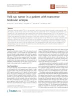
Chest X-ray at 20:40 showed a voluminous dense homogenous mass located in the middle lobe of the right lung and scattered
alveolar opacities in the left lung field (Figure 1).
Figure 1 Chest X-Ray of day 9 after symptom onset (A) prominently showed a voluminous dense homogenous mass located in the
middle lobe of the right lung and diffuse heterogenous reticulo-alveolar opacities in left lung. And day 9 after symptom onset (B)
shows that this mass on the right is larger and more prominent of the alveolar opacities scattered across the two lung fields. Note
that no sign of pleural effusion was seen on both plain.
On day 10, the clinical condition did not improve. White blood cells 9,02 x 10⁹/L (normal range 4.5 - 11.0 × 10⁹/L) with hight
neutrophils (84,1%). The patient was treated with antibiotics (ceftriaxone, moxifloxacin), dexamethasone, lovenox. Non-enhanced
chest CT scanner perfomed at 13:38 shows nonspecific radiologic findings of COVID-19 pneumonia by consolidation with
multifocal, diffuse, perihilar ground-glass opacities (Figure 2). Patient contactable but languid status.- Radial pulse 100
beats/minute;- Blood pressure: 120/70 mmHg;- SpO2: 92% (oxy mask 10L/min) - shortness of breath, mild exertion breathing, two
strokes, rate 30 breaths/min, no chest pain. Patient is referred to ICU and treated by 1. Conduct intubation, mechanical ventilation;
2. Paciflam 5mg x 5 ampules, Fentanyl 0,1mg x 2 ampules and NaCl 0,9% x 50ml, electric injection pump 5ml/h; 3. Rocuronium 5ml
x 2 ampules and NaCl 0,9% x 50ml, electric injection pump 10ml/h x 2. Repeated chest X-ray at 20:28, shows this mass on the right
is larger and more prominent of the alveolar opacities scattered across the two lung fields.
On day 11, patient with anesthetized/sedated patient; Radial pulse 140 beats/minute; Blood pressure: 110/60 mmHg; SpO2: 95%
(normal mechanical ventilation) and Placement of Central Venous Catheter > 30 cmH2O. Hight white blood cells counts 12,12 x
109/L (normal range 4.5 - 11.0 × 10⁹/L) with hight neutrophils (90,6%). Hight pro-calcitonin 0.33 ng/ml (normal <0,05 ng/ml). Hight C
– reactive protein 25,7 mg/L (normal < 3 mg/L). Hight NT-proBNP 228,1 pg/mL (normal <125 pg/mL).
Medical Science, 26, ms159e2213 (2022)
2 of 6
MEDICAL SCIENCE l CASE REPORT
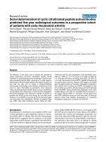
Figure 2 Chest non-enhanced CT scanner of day 10 after symptom onset. A, B: Thin-section axial (mediastinal window) images
shows a significant consolidation, as a mass-like with hypodensity of central necrosis, mesuring 98 x 120 x 125 mm (arrow) in the
middle lobar of the right lung, a finding more consistent with lobar pneumonia than COVID-19 (long arrow). Note that presented
pericardial effusion, localized anteriorly, the thickest 11mm (B, short arrow). Axial (C, D) and coronal multiplanar reformatted (E, F)
of the lung window images shows bilateral, multifocal consolidations distributing along the subpleural and perihilar area and a
partial round-glass opacities with superimposed inter- and intra-lobular septal thickening (crazy-paving pattern). According to the
RSNA chest CT classification system, the findings were classified as atypical appeance for COVID-19 pneumonia.
3. DISCUSSION
The Fleischner Society's consensus statement suggests imaging to discover underlying cardiopulmonary abnormalities in patients
with SARS-CoV-2 in the following situations: 1) mild symptoms (no evidence of significant pulmonary dysfunction or damage)
combined with risk factors of disease severity, such as age >65 years and comorbidities; and 2) moderate to severe symptoms (eg,
hypoxemia and dyspnea) presented by evidence of damage or pulmonary dysfunction or (eg, hypoxemia and dyspnea) (eg,
cardiovascular disease, diabetes, chronic bronchitis, hypertension, and immunocompromised individuals) (Rubin et al., 2020).
For SARS-CoV-2 patients who require chest imaging, this statement does not indicate whether radiography or CT should be
used. Chest radiographs are nonspecific on admission, and up to 41% of cases have normal results (Guan et al., 2020), but severe
patients admitting to ICU have 95% of cases are found abnormal radiographs (Arentz et al., 2020). As a result, radiography is the
most used method for determining disease progression and alternate diagnoses (eg, lobar pneumonia suggestive of bacterial
superinfection, pneumothorax, and pleural effusion) (Rubin et al., 2020). Early parenchymal lung illness and alternative diagnoses
such as abrupt heart failure due to COVID-19 myocardial injury are all more sensitive with chest CT (Driggin et al., 2020). Nonenhanced chest computed tomography (CT) should be done using a low-radiation-dose technique whenever possible to reduce
radiation exposure (Kwee and Kwee, 2020). For suspected pulmonary thromboembolism, an intravenous contrast substance may be
used (Woodard, 2021).
Medical Science, 26, ms159e2213 (2022)
3 of 6
MEDICAL SCIENCE l CASE REPORT
According to recent literature, the results of chest CT scan in pneumonia patients due to COVID-19 frequently exhibit three
main signs: ground-glass opacities (GGO), consolidation, and reticular (crazy-paving pattern); GGO is the most common sign,
accounting for up to 100% of cases (Guan et al., 2020). The frequency of occurrence of these signs varies over time form to symptom
onset and is roughly described in four stages: 1) early stage (0-4 days): mainly GGO; 2) progressive stage (5-8 days): GGO
predominantes with or without associated consolidation or reticular; 3) advanced stage (9-14 days): GGO with consolidation (as
well as our case) and possibly repairing signs on CT (bronchus distortion, subpleural line, and fibrotic strips); 4) Absorption stage
(>14 days): GGO and consolidation gradually decreased (Zhou et al., 2020b; Wang et al., 2020; Pan et al., 2020).
Typical appearance, ambiguous appearance, and atypical appearance are the four categories established by the RSNA for
reporting chest CT results that could be associated to COVID-19 pneumonia. Imaging characteristics with increased specificity for
COVID-19 pneumonia, such as peripheral, bilateral ground-glass opacities (GGO) with or without apparent crazy-paving pattern or
consolidation; or multifocal rounded GGO with/without consolidation with surrounding GGO, were widely reported by usual
impressions. Influenza pneumonia and organizing pneumonia are the most common differential diagnoses (such as can be seen
with drug toxicity and connective tissue disease). Indeterminate presentations in COVID-19 pneumonia include multifocal, diffuse,
perihilar, or unilateral GGO with or without consolidation that lack a defined distribution and are non-rounded or non-peripheral,
or a few very small GGO with a non-rounded and non-peripheral distribution. These imaging characteristics can be seen in a wide
range of infectious and noninfectious processes (such as acute hypersensitivity pneumonitis, pneumocystis infection, and diffuse
alveolar hemorrhage).
Atypical features include segmental consolidation of isolated lobar of bacterial pneumonia (as in our case), cavitation from
necrotizing pneumonia (Klebsiella or nontuberculous mycobacterial infection), and tree-in-bud opacities with centrilobular nodules,
which are more commonly associated with other diseases (Simpson and Newburger, 2020). Other nonspecific lesions such as
lymphadenopathy, pleural effusion, nodular lesions may suggest infectious pneumonia rather than COVID-19 (Kanne et al., 2020).
If secondary respiratory deterioration occurs with COVID-19 treatment, investigate the likelihood of superimposed pneumonia and
acquire lower respiratory tract cultures as well as chest imaging (Huttner et al., 2020).
In our case, although there were no signs of bacterial fever, no respiratory culture (this test is usually nonspecific because it can
be negative), but in symptomatic covid-19 pneumonia patient with progressive clinically to severe respiratory distress and
combined with two important parameters, as laboratory markers and radiologic findings, are sufficient to confirm the
superimposed bacterial pneumonia. At the last days of the patient, laboratory results presented elevated white blood cell and
neutrophils, and especially procalcitonin. Levels procalcitonin are normal in COVID-19 patients with mild disease, and may be
elevated in severe disease patients and levated levels correlate with a nearly 5-fold higher risk of severe SARS-CoV-2 infection
(Zhou et al., 2020a; Yang et al., 2020). As our case, pro-calcitonin 0.33 ng/ml (normal < 0.05ng/ml) and this level represent usually a
SIRS. CRP and ESR may both be elevated in pneumonia patients due to covid-19, which are nonspecific inflammatory markers and
therefore not useful in differentiating it from bacterial infection (Wu et al., 2020).
Typical radiographic features of superimposed bacterial pneumonia on chest radiography: lobar or segmental air-space
opacification, and on CT: segmental or lobar focal dense consolidation with or without ground-glass opacities (Figure 2) (Simpson
and Newburger, 2020; Wu et al., 2020). In an estimated 5.2 percent of COVID-19 patients, pericarditis is an inflammatory illness that
affects the sac surrounding the heart and is most typically caused by severe viral infections (Adams et al., 2020). A significant
tamponade, pericardial effusion, myopericarditis, and high CRP or NT-pro-BNP are also poor prognostic markers (Tung-Chen,
2020). Although nonspecific pericardial effusion, radiologists should investigate the possibility of COVID-19-related cardiac injury
when chest CT scans presenting pericardial effusion.
Finally, prognostic factors that increase the risk of death on admission in covid-19 patients pneumonia generally include older
age (>65 years) with comorbidities associated with organ function damage and developement of acute respiratory distress
syndrome (Zhou et al., 2020a; Yang et al., 2020). In which, pulmonary superinfection is the cause of death accounting for 16% of
potential cases and 3% of total patients with COVID-19 (Clancy et al., 2021).
4. CONCLUSION
Pneumonia in patients with SARS-CoV-2 can be severely developed, especially in patients with comorbidities (such as high blood
pressure and diabetes) and especially with pulmonary superinfection. To follow-up the disease, laboratory markers and radiologic
findings, especially on non-contrast CT, help radiologists efficiently identifying the characteristics of pulmonary lesions and their
complications. The progression of consolidation and multifocal nodular opacities is one of the imaging findings consistent with
pulmonary superinfection complications presenting the clinical manifestation and laboratory testing sought in these patients.
Medical Science, 26, ms159e2213 (2022)
4 of 6
MEDICAL SCIENCE l CASE REPORT
Patient consent statement
Written informed consent has been obtained from the patient for the publication of this case report and any accompanying
photographs. This case report is an incidental finding in the course of clinical work and has no ethical implications.
Author’s contribution
PNTV and NTA contributed equally to this article. Each author gave a substantial contribution in acquisition, analysis, and data
interpretation. Each author had a part in preparing article for drafting and revising it critically for important intellectual content.
Each author gave the final approval of the version to be published and agreed to be accountable for all aspects of the work, ensuring
that questions related to the accuracy or integrity of any part of the work are appropriately investigated and resolved.
Funding
This study has not received any external funding.
Conflicts of interest
The authors declare that there are no conflicts of interests.
Data and materials availability
All data associated with this study are present in the paper.
REFERENCES AND NOTES
1. Adams HJA, Kwee TC, Yakar D, Hope MD and Kwee RM
Chest CT Imaging Signature of Coronavirus Disease 2019
Infection: In Pursuit of the Scientific Evidence. Chest 2020;
158(5):1885-1895. 10.1016/j.chest.2020.06.025
M and Lee M Characteristics and Outcomes of 21 Critically
Ill Patients With COVID-19 in Washington State. JAMA
2020; 323(16):1612-1614. 10.1001/jama.2020.4326
Superinfections Among Persons With Coronavirus Disease
2019: A Comprehensive Review of Data From Postmortem
Forum
Infect
Dis
2021;
J
COVID-19:
don't
neglect
antimicrobial
stewardship principles! Clin Microbiol Infect 2020; 26(7):808810. 10.1016/j.cmi.2020.04.024
8. Kanne JP, Little BP, Chung JH, Elicker BM and Ketai LH
Essentials for Radiologists on COVID-19: An Update-
3. Clancy CJ, Schwartz IS, Kula B and Nguyen MH Bacterial
Open
6736(20)30183-5
7. Huttner BD, Catho G, Pano-Pardo JR, Pulcini C and
Schouten
2. Arentz M, Yim E, Klaff L, Lokhandwala S, Riedo FX, Chong
Studies.
China. Lancet 2020; 395(10223):497-506. 10.1016/S0140-
8(3):ofab065.
10.1093/ofid/ofab065
Radiology
Scientific
Expert
Panel.
Radiology
2020;
296(2):E113-E114. 10.1148/radiol.2020200527
9. Kwee TC and Kwee RM Chest CT in COVID-19: What the
Radiologist Needs to Know. Radiographics 2020; 40(7):18481865. 10.1148/rg.2020200159
4. Driggin E, Madhavan MV, Bikdeli B, Chuich T, Laracy J,
10. Pan F, Ye T, Sun P, Gui S, Liang B, Li L, Zheng D, Wang J,
Biondi-Zoccai G, Brown TS, Der Nigoghossian C, Zidar DA,
Hesketh RL, Yang L and Zheng C Time Course of Lung
Haythe J, Brodie D, Beckman JA, Kirtane AJ, Stone GW,
Changes at Chest CT during Recovery from Coronavirus
Krumholz
Disease 2019 (COVID-19). Radiol 2020; 295(3):715-721.
HM
and
Parikh
SA
Cardiovascular
Considerations for Patients, Health Care Workers, and
Health Systems During the COVID-19 Pandemic. J Am Coll
Cardiol 2020; 75(18):2352-2371. 10.1016/j.jacc.2020.03.031
10.1148/radiol.2020200370
11. Rubin GD, Ryerson CJ, Haramati LB, Sverzellati N, Kanne
JP, Raoof S, Schluger NW, Volpi A, Yim JJ, Martin IBK,
5. Guan CS, Lv ZB, Yan S, Du YN, Chen H, Wei LG, Xie RM
Anderson DJ, Kong C, Altes T, Bush A, Desai SR, Goldin O,
and Chen BD Imaging Features of Coronavirus disease 2019
Goo JM, Humbert M, Inoue Y, Kauczor HU, Luo F, Mazzone
(COVID-19): Evaluation on Thin-Section CT. Acad Radiol
PJ, Prokop M, Remy-Jardin M, Richeldi L, Schaefer-Prokop
2020; 27(5):609-613. 10.1016/j.acra.2020.03.002
CM, Tomiyama N, Wells AU and Leung AN The Role of
6. Huang C, Wang Y, Li X, Ren L, Zhao J, Hu Y, Zhang L, Fan
Chest Imaging in Patient Management during the COVID-19
G, Xu J, Gu X, Cheng Z, Yu T, Xia J, Wei Y, Wu W, Xie X, Yin
Pandemic: A Multinational Consensus Statement from the
W, Li H, Liu M, Xiao Y, Gao H, Guo L, Xie J, Wang G, Jiang
Fleischner
R, Gao Z, Jin Q, Wang J and Cao B Clinical features of
10.1148/radiol.2020201365
patients infected with 2019 novel coronavirus in Wuhan,
Society.
Radiol
2020;
296(1):172-180.
12. Simpson JM and Newburger JW Multisystem Inflammatory
Syndrome in Children in Association With COVID-19.
Medical Science, 26, ms159e2213 (2022)
5 of 6
MEDICAL SCIENCE l CASE REPORT
Circulation 2020; 142(5):437-440. 10.1161/CIRCULATIONAH
A.120.048726
13. Tung-Chen Y Acute pericarditis due to COVID-19 infection:
An underdiagnosed disease? Med Clin (Engl Ed) 2020;
155(1):44-45. 10.1016/j.medcle.2020.06.001
14. Wang Y, Dong C, Hu Y, Li C, Ren Q, Zhang X, Shi H and
Zhou M Temporal Changes of CT Findings in 90 Patients
with COVID-19 Pneumonia: A Longitudinal Study. Radiol
2020; 296(2):E55-E64. 10.1148/radiol.2020200843
15. Woodard PK Pulmonary Thromboembolism in COVID-19.
Radiology 2021; 298(2):E107-E108. 10.1148/radiol.2020204175
16. Wu CP, Adhi F and Highland K Recognition and
management of respiratory co-infection and secondary
bacterial pneumonia in patients with COVID-19. Cleve Clin J
Med 2020; 87(11):659-663. 10.3949/ccjm.87a.ccc015
17. Yang X, Yu Y, Xu J, Shu H, Xia J, Liu H, Wu Y, Zhang L, Yu
Z, Fang M, Yu T, Wang Y, Pan S, Zou X, Yuan S and Shang Y
Clinical course and outcomes of critically ill patients with
SARS-CoV-2 pneumonia in Wuhan, China: a singlecentered, retrospective, observational study. Lancet Respir
Med 2020; 8(5):475-481. 10.1016/S2213-2600(20)30079-5
18. Zhou F, Yu T, Du R, Fan G, Liu Y, Liu Z, Xiang J, Wang Y,
Song B, Gu X, Guan L, Wei Y, Li H, Wu X, Xu J, Tu S, Zhang
Y, Chen H and Cao B Clinical course and risk factors for
mortality of adult inpatients with COVID-19 in Wuhan,
China:
a
retrospective
cohort
study.
Lancet
2020a;
395(10229):1054-1062. 10.1016/S0140-6736(20)30566-3
19. Zhou S, Zhu T, Wang Y and Xia L Imaging features and
evolution on CT in 100 COVID-19 pneumonia patients in
Wuhan,
China.
Eur
Radiol
2020b;
30(10):5446-5454.
10.1007/s00330-020-06879-6
Medical Science, 26, ms159e2213 (2022)
6 of 6