The interaction between bsa modified nanodiamon with serum proteins
Bạn đang xem bản rút gọn của tài liệu. Xem và tải ngay bản đầy đủ của tài liệu tại đây (586.24 KB, 39 trang )
VIET NAM NATIONAL UNIVERSITY OF AGRICULTURE
FACULTY OF BIOTECHNOLOGY
GRADUATION THESIS
TITLE
THE INTERACTION BETWEEN BSA MODIFIED
NANODIAMOND WITH SERUM PROTEINS
Hanoi, March 2021
VIET NAM NATIONAL UNIVERSITY OF AGRICULTURE
FACULTY OF BIOTECHNOLOGY
TITLE
THE INTERACTION BETWEEN BSA MODIFIED
NANODIAMOND WITH SERUM PROTEINS
STUDENT:
Nguyen Tra My
MAJOR:
Biotechnology
CLASS:
K61CNSHE
610743
STUDENT CODE:
SUPERVISORS:
Phan Huu Ton, Ph. D
Vietnam National University of Agriculture
Pham Dinh Minh, Ph. D
Vietnam Academy of Science and Technology
Hanoi, March 2021
COMMITMENT
I assure this is my research. All the results are honestly collected and have not
been published in any article.
I accept responsibility for my commitment.
Hanoi, February 25th, 2021
Nguyen Tra My
i
ACKNOWLEDGEMENTS
The completion of this study could not have been possible without the expertise
of Ph. D Pham Dinh Minh, Ph. D Phan Huu Ton, my thesis adviser. I would also like
to thanks Ms. Quy, Ms. Hue, Mrs. Loan and Mr. Son for supporting me on my thesis.
A debt of gratitude in also owned to my beloved family and friends for their
belief and their help on the spirit.
Without all of you, none of this would indeed be possible.
Nguyen Tra My
ii
TABLE OF CONTENTS
COMMITMENT .............................................................................................................1
ACKNOWLEDGEMENTS ........................................................................................... ii
TABLE OF CONTENTS .............................................................................................. iii
LIST OF ABBREVIATIONS .........................................................................................v
SUMMARY................................................................................................................. viii
CHAPTER 1: INTRODUCTION....................................................................................1
CHAPTER 2: LITERATURE REVIEW.........................................................................2
2.1 Introduction to Nanodiamonds: Classification, Surface Modification, and
Biomedical Applications .............................................................................................. 2
2.2 Introduction to Proteomics and Its Application to Protein profiling ....................4
2.3 Interaction of Bare-ND and Surface-modified ND with Proteins/ Human Fluids/
Cell culture/ Immune System .......................................................................................6
2.3.1 Interaction of Bare-ND and Surface-modified ND with Proteins ..........................6
2.3.2 Interaction of Bare-ND and Surface-modified ND with Human fluids ................7
2.3.3 Interaction of Bare-ND and Surface-modified ND with Cell Culture ...................7
2.3.4 Interaction of Bare-ND and Surface-modified ND with Immune System: ........8
2.4 Introduction to BSA and previous study about BSA-conjungates NDs: ..............8
CHAPTER 3: MATERIAL AND METHOD ............................................................... 10
3.1 Materials ..............................................................................................................10
3.2 Methods ...............................................................................................................10
3.2.1 Production of Bare-Nanodiamonds ......................................................................10
3.2.2 Coating Nanodiamonds by Incubating with BSA ...............................................10
3.2.3 Interaction of Human Serum Proteins with Bare-ND and BSA-ND ..................11
3.2.3.1 Effect of Protein Amount and Incubating time ...................................11
3.2.3.2 Effect of Incubating Buffers .................................................................12
3.2.3.3 Effect of Washing buffers ...................................................................12
3.2.3.4 Size and Zeta-potential Measurements .................................................12
3.2.4 BSA-coated Nanodiamonds Interact with Human Serum Proteins for Nano LCMS/MS........................................................................................................................... 13
3.2.5 Liquid Chromatography coupled to Tandem Mass Spectrometry ......................13
iii
3.2.6 Bioinformatic Analysis .........................................................................................14
CHAPTER 4: RESULTS AND DISCUSSION ............................................................ 15
4.1 Synthesis and Characterization of Oxidative Nanodiamond (bare-NDs) and
Protein-modified Bare-NDs (BSA-NDs) ...................................................................15
4.1.1 Production of Bare-NDs .......................................................................................15
4.1.2 Synthesis and Characterization of BSA modified bare-NDs .............................. 16
4.2 Interaction of Human Serum Proteins with Bare-NDs and BSA-NDs ...............17
4.2.1 Effect of Protein Amount and Incubating time ....................................................17
4.2.2 Effect of Incubating Buffers .................................................................................19
4.2.3 Effect of Washing Buffers ....................................................................................20
4.2.4 Size Distribution and Zeta-potential of Bare-NDs and BSA-NDs Before and
After Conjuncgation with Human Serum Proteins........................................................21
4.3 Analysis of Human Serum Proteins Adsorbed onto the Surface of Bare-ND and
BSA-ND at Physiological pH ....................................................................................22
4.3.1 SDS-PAGE Analysis ............................................................................................ 22
4.3.2
Proteomics Analysis .........................................................................................23
4.4 Discussion: ..........................................................................................................25
CHAPTER 5: CONCLUSION AND RECOMMENDATION .....................................27
REFERENCES ..............................................................................................................28
iv
LIST OF ABBREVIATIONS
Abbreviations
Definitions
ND
Nanodiamond
BSA
Bovine serum albumin
BSA-ND
Bovine serum albumin conjungates to
Nanodiamond
HPHT
high pressure high temperature
CVD
chemical vapour deposition
DND
Detonation
MS
Mass spectrometry
ESI
electrospray ionization
TDP
Top-down proteomics
LC-MS/MS
Liquid Chromatography Mass
Spectrometry
cNDs
carboxylated nanodiamond
RBC
red blood cells
FND
flourescent nanodiamonds
FESEM
Field emission scanning eelectron
microscopy
DLS
dynamic light scattering
SDS-PAGE
Sodium dodecyl sulfatepolyacrylamide gel electrophoresis
v
LIST OF FIGURES
Figure 2. 1 Surface modification of nanodiamonds with various functional group
(Journal of Material Science & Engineering) ....................................................3
Figure 2. 2 The main components of a mass spectrometer (Application review on
Merckmilipore.com) ...........................................................................................5
Figure 4. 1 Production and physical characterization of bare-NDs. (a) The size
distribution of nanodiamonds suspension in H2O; (b) TEM image of bareNDs suspension in H2O ....................................................................................15
Figure 4. 2 Synthesis and physical characterization of BSA-ND. The
nanodiamonds suspension before coating (ND) and after coating with
BSA (BSA-NDs) (a) In H2O (b) Zeta-potential of BSA-ND and bare -ND
(c) The SDS-PAGE image shows the result of coating bare-ND with BSA.
The bare-ND protein band pattern is blank, showing the purity of ND
containing no contaminants. The BSA protein band pattern is shown as
the main band of over 66.2 kDa. .....................................................................16
Figure 4. 3 The SDS-PAGE image shows the result of the interaction between
bare-ND and human serum proteins at different incubating times and
different amounts of human serum proteins .....................................................18
Figure 4. 4 The SDS-PAGE image shows the resullt of the interaction between
BSA-ND and human serum proteins at different incubating times and
different amounts of human serum proteins. ...................................................18
Figure 4. 5 The SDS-PAGE image shows the result of the interaction of bare-ND
and BSA-ND with human serum proteins in different pH conditions .............19
Figure 4. 6 The SDS-PAGE image shows the result of the interaction of bare-ND
and BSA-ND with human serum proteins in different washing buffers. .........20
Figure 4. 7 Synthesis and physical charaterization of ND and BSA-ND before and
after interacting with serum proteins. a) Size distribution in H2O; b) Zetapotential ............................................................................................................21
Figure 4. 8 The SDS-PAGE image shows the result of interaction in human serum
with bare-ND and BSA-ND. The bare-ND protein band pattern is blank,
vi
showing the purity of ND containing no contaminants. The BSA protein
band pattern is shown as the the main band of 66.2 kDa. ................................ 22
Figure 4. 9 The bar chart show the most abundant serum proten groups found in
the sample of bare-ND interaction with serum. Abundancy was calculated
based on MS/MS intensity from MaxQuant ....................................................23
Figure 4. 10 The bar chart shows the 10 most abundant serum protein groups
found in the sample of BSA-ND interaction with serum. Abundancy was
calculated based on MS/MS intensity from MaxQuant ...................................24
vii
SUMMARY
Nanodiamonds (NDs) or nanocrystalline diamond is an immense term that
described a continuum of materials. It is recently considered as a promising material in
proteomics, in cell labeling and tracking, in drug delivery, and especially in vaccine
development. Numerous studies have shown the linking or conjugating of protein on
ND particles but there has not been work on how it interacts in human serum, which is
important for predicting their behavior in the human body. In this study, we have
examined the effect of surface modifications on the interaction of NDs and human
serum proteins. The adsorbed ability of proteins on the surface of NDs was
investigated and confirmed by the change in size before and after modification. The
interaction between bare-NDs and BSA-NDs with serum proteins is pH dependent and
depends on different proteins which are linked with. That leads to different behaviors
in the human body.
Keywords:
Nanodiamonds;
Surface-modified
Nanodiamonds; Nanomedicine; BSA
viii
Nanodiamonds;
Carboxylated
CHAPTER 1: INTRODUCTION
Over years of decades, many treatments against diseases are explored. There
are more and more new diseases that appeared, required continuing and strong
motivation in research to find the replacement for old treatment. In that circumstance,
researchers tend to focus on new material, that can help modified and control drugs in
the human body.
Nanodiamond (NDs) is considering a promising material in broad fields of
biotechnological and biomedical applications. Previous studies have demonstrated that
NDs are the least toxic of all carbon-based nanomaterials studied so far. Also, the
interactions of NDs with red blood cells and NDs surface modification by proteins in
blood have been investigated, which stated that NDs’s safe in organisms. Besides, with
a high affinity with a biomolecule, NDs have been developed to be drug carriers.
Many efforts have been dedicated to investigating the interactions of NDs with
biomolecules for achieving their application in the biomedical field. It is easily seen in
many articles of study using proteins such as lysozymes, bovine serum albumin
(BSA), cytochrome c, and myoglobin to physically adsorb on NDs and investigated
the binding properties. But there is no systematic research to examine how is its
behavior in human body fluid, considering the environment that influences the ability
of the complex in the immune system. Therefore, understanding the interaction with
human serum with protein binding NDs is essential for ND’s biotechnological
application.
In this study we focus on answering the question of how the effect conditions as
time, washing, and buffer to the BSA modified NDs and proteins of serum interact
with it, that is helpful for other researchers in the future.
Purpose: Study the interaction of BSA modified NDs with serum protein in
different conditions to see the effect on the behavior of complex in human serum, also
explore what proteins will be in the strong interaction with NDs for further application.
1
CHAPTER 2: LITERATURE REVIEW
2.1Introduction to Nanodiamonds: Classification, Surface Modification, and
Biomedical Applications
Nanodiamonds (NDs) are emerging as notable material widely researched in
biological and medical applications (M. Aramesh et al.,2012). NDs or diamond nanoparticles are stated as nano-scale carbon allotropes and exhibit characteristic
mechanical and optical properties (H. Tinwala et al.,2018). Following several research
recently, unlike other carbon materials, NDs are highly bio-compatible, non-toxicity at
both cellular and whole-organism levels, especially, the ND surface can be chemically
modified by absorbed biomolecules ability (M. Aramesh et al.,2012) (Tenzer et al.,
2013).
With these impressive properties, NDs have attracted the interest of the
scientific community in drug delivery, bioimaging, tissue engineering, and the latest in
vaccination (L. Fusco et al., 2020) (A. E. Garcia-Bennett et al., 2019) (L. Zhao et al.,
2013) .
By synthetic methods, NDs were classified into three main groups including
high-pressure high temperature (HPHT), chemical vapor deposition (CVD) formation,
and detonation (E. T. Sangiao et al., 2019). Depend on how it made, each of type has
its own significant properties. The HPHT-NDs gain their fluorescent properties from
nitrogen-vacancy centers and nowadays on the market have a size median of ~18nm (
S. Stehlik et al.,2015). Besides, the CVD method resultsnanocrystalline carbon film
thatcontains nanocrystalline layers with grain size in the range 10-200nm (E. Tamburri
et al., 2015). The last kind of NDs, the DND monocrystallites appear closely as round
particles with 4-5 nm in diameter, the primary particles form tightly and loosely bound
aggregates ( Shenderova, 2012) (in Ultananocrystalline Diamond second edition
2012). Following the detonation method, DNDs were revealed that reducing some
groups on the surface of the particle will form of C-C, C-X and C-N bonds ( A.
Schrand et al., 2009) .
DNDs are now widely applied in biomedical and diverse applications. Strong
covalent bonding between carbon atoms, fluorescent properties, size, and shape of ND
particles makes them ideal for applications involving the attach of sorbent molecules.
2
To take advantage of these properties, surface modification is an effective method to
raise the active work of biomolecules. There are two approaches to modification of
NDs include (1) surface functionalization and (2) doping o NDs. Using the property of
surface nanoparticles, surface functionalization widely used to modify and turn into
another complex. Take a deep look at surface functionalization, the surface chemistry
of NDs resulting platform for numerous functionalization schemes. The surface of ND
contains all the requirements for the biomedical application such as high colloidal
dispersibility, better solubility and stability in different biogical pH conditions, and
presence of specific moieties to interact with targeted biomolecules (H.Tinwala et,. al
2018). Since the method of synthesis and purify NDs, most of the carbon atoms on
surface ND are terminated with oxygen moieties such as carbonyl, or hydroxyl groups.
Therefore, by reaction chemistry, functional groups can be attached to the ND surface
and stay stable.
Figure 2. 1 Surface modification of nanodiamonds with a various functional
group (Journal of Material Science & Engineering)
In this work, we focus on bare-surface oxidized ND (bare-ND) that contain
carboxyl groups. Because of its affinity with proteins and peptides, this has been
applied in protein capture and proteomics study (Kong et al., 2005) (Aramesh et al,
2015). According to recent research, the strong and firm adsorption of proteins onto
ND is the result of the combination of different forces such as ionic interaction,
hydrogen bonding, Vander Waals interaction, and hydrophobic interaction (Huang and
Chang, 2004) (Chen et al., 2006) (Aramesh et al., 2015).
3
2.2 Introduction to Proteomics and Its Application to Protein profiling
Proteomics is the analysis of the entire protein components of a cell, tissue, or
organism at a specific time. Proteomics not only seeks to identify proteins potentially
present in a sample but also assesses protein abundance, localization, posttranslational
modification isoforms, and molecular interaction. Some of the main applications of
proteomics are disease diagnosis, prognosis and to moniter, the disease development
as well as in drug development.
Mass spectrometry (MS), among the variety of proteomics techniques, is emerging as
an outstanding method to determine and quantity proteins in a biological sample.This
method’s principle is measurements of ratio mass on a charge of the ions formed when
a sample is ionized then determine all of the isotopes of each element. The two
approaches using in MS are the “bottom-up” -which analyzes fragmentation in
proteolytic digestion, and “top-down” - which analyzes the intact proteins. The topdown proteomics (TDP) involves identifying proteins in the complex mixture without
prior digestion. Proteoforms of interest are first prepared for electrospray ionization
(ESI) to be charged and then directly infused into the mass spectrometer for data
analysis (T. Toby et al,.2016). Unlike TDP, in bottom-up proteomics or shotgun
proteomics - a name coined by the Yates lab, the peptide mixure are first undergone
proteolytic digestion before charged by ESI to enter an LC-MS/MS system. Peptide
identification is obtained by comparing the tandem mass spectra derived from peptide
fragmentation with theoretical tandem mass spectra resulted from in silico digestion of
a protein database (Y. Zhang et al,. 2016). Since the bottom-up proteomics allows to
attempt to combine a high number of protein identification with consistency and
accuracy in the quantification of protein levels, it is becoming widely applied in
protein profiling (E. Dupree et al,. 2020) .
4
Figure 2. 2 The main components of a mass spectrometer (Application review on
Merckmilipore.com)
The LC-MS/MS operates with a combination of chromatography and multiple
quadrupole mass spectrometers. Firstly, the sample is separated into the different
components, concentrating the amount of a single component that reaches the mass
spectrometer. The first quadrupole of the triple quadrupole configuration ionizes the
molecules of the analyte. Selected molecular ions are then fragmented in the second
quadrupole and selectively isolated by the third and final quadrupole for measurement
by the detector. The LC-MS/MS allows for the detection of low-level contaminants
with a high degree of specificity and sensitivity. Additionally, accurate quantitation is
possible for some sample matrices.
More specifically, the results of MS presented as a mass spectrum, a plot of
intensity as a function of the mass-to-charge ratio. Spectra in results are used for
determining the elemental or isotopic signature of a sample, and the mass of the
molecule is used to identify or determine the structure of molecules. The main parts of
a mass spectrometer consist of three components: an ion source, a mass analyzer, and a
detector (as Fig. 2.2). The ionizer is the first part of the system, that converts a portion
5
of the sample into ions. Depend on the phase (solid, liquid, gas) of the sample, the
method of ionization is used. Then the ions will be removed from the sample, then
move through the mass analyzer. In the last part of the system, the detector measures
the value of an indicator quantity and thus provides data to calculate the abundance of
each ion present.
With strong advanced in bottom-up proteomics techniques, a huge number of
proteins are profiled and quantifiedto serve for important research. In 2012,with a
study to define the protein content of matched samples of afferent prenodal lymph and
plasma-derived from healthy volunteers,
C. Clement et al. defined 253 proteins
obtained from the tandem mass spectrometry data. In combination with several
techniques, they discovered the common proteome between the two biological fluids
(144 out of 253 proteins) and this helped to highlight the principal biological activity
of these immunologically relevant body fluids.
In this study, shotgun proteomics is used as a method to identify and profile the
proteins from human serum that directly interact with nanoparticles and modified
nanoparticles. Besides, the interacted protein profiling is analyzed to determine how
the surface modifications of BSA on nanodiamonds affect the interaction with human
serum protein.
2.3Interaction of Bare-ND and Surface-modified ND with Proteins/ Human
Fluids/ Cell culture/ Immune System
2.3.1 Interaction of Bare-ND and Surface-modified ND with Proteins
Recently, there have been several studies about the interaction between bareND and some specific proteins, such as lysozyme,insulin, haemagglutinin...etc.To
analyze the changes in structural conformation and its enzymatic activity after
adsorption, E. Perevedentseva et al,.(2010) found that the activity of lysozyme
depends on nanodiamond particles’ size and properties but it does not alter the
lysozyme functional properties. In another research, bovine insulin was non-covalently
bound to detonated nanodiamondsin an aqueous solution and examine pH-dependent
desorption in alkaline environments of sodium hydroxide (R. Shimkunas et al,. 2009).
6
Following that, NDs may serve as an effective carrier for insulin cause the binding
activity plus the release of insulin under alkaline conditions. Moreover, nanodiamond
conjugated with haemagglutinin at a ratio of 1:2 (w/w) corresponding significant
immunization response (Pham NB at al,. 2017). That indicated the potential possibility
for the further modified surfaces on nanodiamond particles.
2.3.2Interaction of Bare-ND and Surface-modified ND with Human fluids
In 2019, a published article study on the interaction of red blood cells (RBC)
by nanoparticles shows that nanodiamonds results in a higher aggregation force that
leads to the formation of cell aggregates (T. Avsievic et,.al 2019). Interestingly,
polymeric particles do not cause unusual RBC aggregation. Besides, on the report
following J.Mona et al,. 2013
about the adsorption of human blood plasma on
various-sized carboxylated nanodiamond (cNDs), presents that there is no significant
change of blood plasma protein conformation. Furthermore, Wasdo et al,2008
identified 69 unique proteins that exhibited adsorption to the various nanoparticle and
also quantify the relative affinities with which these proteins bind. All of the
nanoparticles demonstrated a low relative affinity for albumin but the concentration is
so high, so there must be a bound protein fraction for all nanoparticles.
2.3.3Interaction of Bare-ND and Surface-modified ND with Cell Culture
To serve for further intracellular application, fluorescent nanodiamonds (FNDs)
are brought to interact with cell culture media (Hemelaar et al., 2017). The result show
that only some certain proteins and salts may cause aggregation, not all of them. That
also revealed FNDs have strongly binding to some proteins, and weakly with the
others in cell culture media. The method discussed to prevent aggregation is adding
proteins Foetal Bovine Serum before adding full medium to coat protein on the surface
of the nanodiamond. Moreover, the cytotoxicity of NDs is also under-researched (
Chen et al,. 2019). In a serum-free medium, the toxicity is obviously showed toward
the human lung epithelial cell line (BEAS-2B); however, in the presence of bovine
serum albumin, the toxicity of NDs was inhibited.
7
2.3.4 Interaction of Bare-ND and Surface-modified ND with Immune System:
Fluorescent nanodiamonds (FNDs) is revealed that in vitro resulted in immune
cell activation and have capabilities to stimulate innate immune cell anti-tumor
responses (Suarez-Kelly et al., 2017). Besides, nanoparticle which is coated by
trimeric Haemagglutinin (H7) elicited a significantly stronger H7-specific-IgG
response (Pham et al,.2017). These researches show a boost in the immune response
with the presence of nanodiamond particles. Furthermore, two NDsmodified by
carboxylic acid (ND-COOH) and amino-functionalized effect the immune response
(Fusco et al., 2020). In two, NDs-COOH showed more promise responses in the
regulation of immune-modulatory.
2.4 Introduction to BSA and previous study about BSA-conjugates NDs:
Bovine serum albumin (BSA) is a serum albumin protein derived from cows.
About chemical structure, BSA is a single polypeptide chain consisting of about 583
amino acid residues and no carbohydrate. BSA is considered a potential colloidal
delivery system because it is biodegradable, biocompatible, nontoxic, and
nonimmunogenic (E. Assadpour et al,.2019). Containing the drug binding sites, BSA
molecule combines with nanoparticles can load various drugs and bioactive
compounds (A. Elzoghby et al,. 2012). Moreover, due to their high content of charged
amino acids (e.g. lysine, arginine, aspartate, and glutamate ), albumin-based
nanoparticles could allow the electrostatic adsorption of positively or negatively
charged. The ability of albumin molecule to be a drug carrier was reviewed by F.
Kratz 2008. The author gives three-drug delivery technologies including encapsulation
of drugs into albumin nanoparticles which form nab-paclitaxel - was approved in 2005
for thetreatment of metastatic breast cancer.
BSA-conjugate NDs are applied in biomedical research. BSA nanoparticles are
conformed to be controlled release carriers for the isophethalaldoxime palladacycle
complex (K.Karami et al.,2020). Following that, the complex a four nuclear
palladacycle complex {Pd4[(N,C)(NOHCHC6H2CHNOH)(μ-Cl)2]2 was loaded on the
nanoparticle already synthesis with serum albumin, then by measurements of methods
of Field emission scanning electron microscopy (FESEM) and dynamic light scattering
(DLS) to explore the morphology, size, and zeta potential. Also, they do experiments
8
to see the release process and cytotoxicities of the synthesized nanoparticles. Therefore
BSA-conjugate NDs is the effective carrier to examine with a complex compound in
research.
In this study, we coat bare-NDs with BSA and examine the interaction of
human serum proteins with bare NDs and bare -ND coated with BSA.
9
CHAPTER 3: MATERIAL AND METHOD
3.1 Materials
Surface oxidized nanodiamond with a nominal size of 100 nm was obtained
by suspending the nanodiamond powder in 2X deionized water (AquaMAX Ultra 370
Series Water Purification System, Younglin, Korea) to ensure the concentration of the
stock solution is 10 mg/mL. Normal human sera were obtained among healthy
laboratory staff (N=5) via venipuncture (to be considered healthy, the blood donors
show no observable disease symptoms). Sodium dodecyl sulfate-polyacrylamide gel
electrophoresis (SDS-PAGE) chemicals were adopted from Bio-Rad (USA). Other
chemicals came from various sources, including Sigma Aldrich (Germany), Norgen
Biotek (Canada), Emsure (Germany), Mallinckrodt (England). Other equipment
includes a water bath (Jeio Tech, Korea), Ziptip C18 column (Millipore,USA),
Ultrasonic Sonicator (Branson 1210, Marshall Scientific, USA), Vacuum Evaporator
(SPD 1010 Speedvac system, Thermo Scientific, USA), Freeze Centrifuge (Thermo
Scientific, USA), Nanoparticle Size Analyzer (Zetasizer-Nano ZS, Malvern, UK).
3.2 Methods
3.2.1 Production of Bare-Nanodiamonds
According to the previous paper, starting with diamond powder, the surface of
NDs was modified by oxygen-containing groups in highly oxidative acid solution (the
H2SO4: HNO3ratio is 3: 1 (v/ v)) and high-temperature condition (about 100°C)
(Pham et al., 2013). The experiment procedure is conducted in a microwave reactor
(100 W, Model Discover, CEM) for 3 hours. At the end of the microwave heating,
precaution was carefully taken to ensure that the residual strong acids are diluted prior
to collection of the NDs. For safe operation, the microwave reactor is placed in a
chemical fume-hood to protect the operator from NO2 exposure.
3.2.2 Coating Nanodiamonds by Incubating with BSA
The coating process is described as follows. The amount of ND used was fixed at
60 µg (or 6 µL of stock solution) for all coating experiments. The PBS solution used
10
was PBS 1X, pH 7.4. According to Pham et al., at the protein: ND ratios (w/ w) is
1:12, the NDs presented the highest ability to bind with trimeric HA protein (Pham et
al., 2017). Therefore, in this study, the amount of BSA used was 5 µg to ensure the
ratio protein: ND (w/ w) is 1: 12.
First, add 200 µL of PBS 1X, pH 7.4 to microcentrifuge tubes, followed by the
addition of BSA with the amount mentioned above, vortex. Add 6 µL of stock ND to
the solution, mix thoroughly until the NDs completely suspend. The solution was then
put in a sonicator for 30 minutes to enhance coating efficacy before centrifuged
(13000 x g) to collect the pellet. The pellet then contained completely BSA-coated
NDs.
3.2.3 Preparation and determination concentration of human serum protein
The serum is prepared by transfer human blood to a effendorf tube, leave it 30
minutes for blood clotting. Then centrifuge at 2500-3000 rpm or 5-10 min at 4ºC.
Collect the upper solution to another tube. Divide into 20 μL/ tube to prevent
denaturation, store at -80ºC.
Prepare BSA solution with following concentrations: 0.125 ; 0.25 ; 0.5; 0.75;
1.0; 1.5; 2.0 mg/mL from BSA stock 10mg/mL. Dilute 20 times. Do Bradford
experiment by mixing 4 μL solution with 196 μL Bradfordsolution. Set up
spectrophometer at wavelength 595nm. Measure the absorbance of each solution.
Record results.
3.2.3 Interaction of Human Serum Proteins with Bare-ND and BSA-ND
3.2.3.1Effect of Protein Amount and Incubating time
The pellet derived from the coating step was resuspended in 200 µL PBS. A
volume of serum that corresponds to 600, 300, and 60 µg proteins was added to the
ND solution. For each amount of proteins, the samples are incubated at room
temperature in 5, 30, and 120 minutes. Then, the solution was centrifuged (13000 x g)
to collect the new pellet, which was then washed twice with 200 µL PBS. Finally,
SDS-PAGE was performed to test the efficacy of ND coating and interaction with
serum proteins.From the results of this experiment, all following experiments would
11
be done with 300 µg serum proteins and ND conjugates would be incubated with
serum proteins at room temperature in 30 minutes. The purpose of using excessive
amounts of serum is to mimic the high protein concentration present in normal blood.
3.2.3.2 Effect of Incubating Buffers
The interaction of bare-ND and BSA-ND with human serum proteins was
examined in acidic pH, physiological pH, and basic pH. First, resuspending the pellet
derived from the coating step in 200 µL 0.1% TFA; 200 µL PBS 1X (pH 7.4), and 200
µL Na2CO3
100mM to create the acidic pH, physiological pH, and basic pH,
respectively. Next, add 300 µg serum proteins to the ND solution, vortex, and leave at
room temperature for 30 minutes. After that, centrifuging the solution (13000 x g) to
collect the new pellet. Finally, washing the pellet twice with 200 µL PBS to remove all
the unbound proteins. The efficacy of interaction with serum proteins will be shown
via SDS-PAGE results
3.2.3.3 Effect of Washing buffers
The stability of the interaction between bare-ND, BSA-ND, and human serum
proteins was examined with three types of washing buffer: H2O, PBS 1X, and 2%
triton X100. First, the pellet derived from the coating step was resuspended in 200 µL
PBS 1X to form a homogeneous solution. Adding 300 µg serum proteins to the ND
solution. After being left at room temperature for 30 minutes, the solution was
centrifuged (13000 x g). The supernatant is discarded. The pellet is collected and
washed two times with 200 µL PBS. Similar to the above, running SDS -PAGE to
check the results.
3.2.3.4Size and Zeta-potential Measurements
Size distributions and Zeta-potentials measurements of bare-NDs and BSANDs before and after conjugation with serum proteins were done using a Nanoparticle
Size Analyzer (Zetasizer-Nano ZS, Malvern Instruments Ltd., UK). Before
12
measurements,
bare-NDs
and protein-coated NDs are re-suspended in 1.5 mL
deionized water
3.2.4 BSA-coated Nanodiamonds Interact with Human Serum Proteins for Nano
LC-MS/MS
To study the interaction of ND conjugates with serum proteins in the human
body, this experiment was done in physiological pH and washing buffer is PBS 1X.
The presentation of PBS 1X makes an excellent representation of native blood pH
(~7.4). The pellet derived from the coating step was resuspended in 200 µL PBS. A
volume of serum that corresponds to 300 µg proteins was added to the ND solution.
Incubating the solution in 30 minutes, room temperature. Then, centrifuging (13000 x
g) to collect the pellet, washing the pellet twice with 200 µL PBS.
3.2.5 Liquid Chromatography coupled to Tandem Mass Spectrometry
Protein digestion with trypsin was done directly on the surface of ND without
the need ofpre-digestion protein extraction from the ND surface. Initially, 200 µL H2O
was added to samples (pellet) and mixed with the aid of an ultrasonic sonicator until
the pellets were fully resuspended. For reduction, 5 μL of 200 mM Dithiothreitol was
added and incubated at 56°C for 30 minutes. For alkylation, add 20 μL 200 mM
Iodoacetamide and incubate the samples at room temperature in the dark for 30
minutes. To terminate the reaction, 100 μL FA 1% was added. The samples were then
centrifuged and pellets were collected. The pellets were washed twice by H2O to
remove chemical residue. For digestion, pellets were resuspended in H2O (50 μL),
added by 0.5 µg trypsin in cold NH4HCO3 50 mM, and incubated at 37°C,
overnight.After digestion, the solutions were centrifuged
to collect
the first
supernatants (80 µL each), which were decanted to new centrifuge tubes. Add 80 µL
peptide extraction buffer (10 mM NH4HCO3/ 50% Acetonitrile) to the previous pellets,
resuspend and centrifuge again to collect the second supernatant (100 µL) and add to
the first supernatants. All the mentioned supernatants (180 µL each tube) were
evaporated with a vacuum evaporator before being desalted by the Ziptip C18 column.
The protocol was adopted from the instruction of the manufacturer
13
3.2.6Bioinformatic Analysis
Tryptic
peptides
are then sequenced by nano LC-MS/MS methods.
Bioinformatic analysis is eventually used to identify serum proteins from sequenced
peptides using MaxQuant software.
14
CHAPTER 4: RESULTS AND DISCUSSION
4.1 Synthesis and Characterization of Oxidative Nanodiamond (bare-NDs) and
Protein-modified Bare-NDs (BSA-NDs)
4.1.1 Production of Bare-NDs
Size distribution of
re-suspended nanodiamonds in
H2 O
Number Percentage (%)
ND
25
20
15
10
5
0
0
200
400
600
800
-5
Diameter (nm)
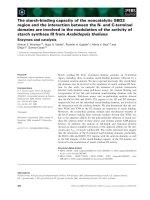
Figure 4. 1 Production and physical characterization of bare-NDs. (a) The size
distribution of nanodiamonds suspension in H2O; (b) TEM image of bare-NDs
suspension in H2O
Following previous research, in aqueous solution, the surface of bare-NDs are
oxidized in strong acid solution (the H2SO4: HNO3 ratio is 3:1 (v/v)) and the hightemperature condition (about 100ºC) (Pham et al,. 2013). As shown in Fig. 4.1a, the
original bare-NDs with a variety of sizes distribute unequally in solution. In the range
of size from ~50nm to ~400nm, they count different percentages but reach the peak at
100nm. Therefore, bare-NDs particles can be aggregate in a deionized water solvent to
form differently sized clusters. Furthermore, the TEM images showed that the
morphology of bare-NDs is not round (Fig. 4.1b).
15