mitochondrial disorders biochemical and molecular analysis
Bạn đang xem bản rút gọn của tài liệu. Xem và tải ngay bản đầy đủ của tài liệu tại đây (4.79 MB, 365 trang )
M
ETHODS
IN
M
OLECULAR
B
IOLOGY
™
Series Editor
John M. Walker
School of Life Sciences
University of Hertfordshire
Hatfield, Hertfordshire, AL10 9AB, UK
For further volumes:
/>
Mitochondrial Disorders
Biochemical and Molecular Analysis
Edited by
Lee-Jun C. Wong
Mitochondrial Diagnostic Laboratory, Department of Molecular and Human Genetics,
Baylor College of Medicine, Houston, TX, USA
ISSN 1064-3745 e-ISSN 1940-6029
ISBN 978-1-61779-503-9 e-ISBN 978-1-61779-504-6
DOI 10.1007/978-1-61779-504-6
Springer New York Dordrecht Heidelberg London
Library of Congress Control Number: 2011943084
© Springer Science+Business Media, LLC 2012
All rights reserved. This work may not be translated or copied in whole or in part without the written permission of the
publisher (Humana Press, c/o Springer Science+Business Media, LLC, 233 Spring Street, New York, NY 10013, USA),
except for brief excerpts in connection with reviews or scholarly analysis. Use in connection with any form of information
storage and retrieval, electronic adaptation, computer software, or by similar or dissimilar methodology now known or
hereafter developed is forbidden.
The use in this publication of trade names, trademarks, service marks, and similar terms, even if they are not identified
as such, is not to be taken as an expression of opinion as to whether or not they are subject to proprietary rights.
Printed on acid-free paper
Humana Press is part of Springer Science+Business Media (www.springer.com)
Editor
Lee-Jun C. Wong, Ph.D. FACMG
Clinical Molecular Genetics and Clinical Biochemical Genetics
Professor, Department of Molecular and Human Genetics
Director, Mitochondrial Diagnostic Laboratory
Baylor College of Medicine
One Baylor Plaza, NAB 2015
Houston, Texas 77030, USA
v
Preface
A major function of mitochondria is the production of energy molecule ATP, by the way of
electron transport chain and respiration, in a process called oxidative phosphorylation
(OXPHOS). In order to carry out OXPHOS, the assembly of fully functional mitochondria
requires the participation of approximately 1,500 genes encoded by both the mitochon-
drial and nuclear genomes. Thus, molecular defects in either of the two genomes may cause
mitochondrial dysfunction, giving rise to either Mendelian or Matrilineal disorders. Each
cell may contain hundreds to thousands of copies of the mitochondrial genome. Depending
on the specifi c genetic defect, the distribution of the affected tissues, and the proportion of
mutant to wild-type mitochondrial DNA (mtDNA) (termed heteroplasmy), the clinical
manifestations of the disease are remarkably variable and heterogeneous. Therefore, for any
given patient, establishing a diagnosis of a mitochondrial disorder can be very diffi cult. It
requires an evaluation of the family pedigree, in conjunction with a thorough assessment of
the clinical, histopathological, imaging, biochemical, and molecular features of the case.
Given the breadth and complexity of the problem, these studies are usually provided by
several different clinical specialties and/or laboratories; each focused on one or more par-
ticular areas. The laboratory and clinical methodologies used may vary widely, and to date
there has been no systematic presentation of the numerous protocols that are applied to the
assessment of these clinically and genetically heterogeneous mitochondrial disorders. It is
the main objective of this volume of Methods in Molecular Biology to provide such a collec-
tion of protocols.
This volume is divided into three parts. The fi rst part is the nonprotocol section that
contains three chapters describing the complexity of these dual genome disorders. Chapter
1 provides an overview of the mitochondrial syndromes caused by common point muta-
tions or deletions of the mtDNA, leading to the concepts and methods of analyzing muta-
tion heteroplasmy, tissue distribution, and the mtDNA content. Molecular defects in a
group of nuclear genes responsible for mtDNA biogenesis and the maintenance of mtDNA
integrity may cause mtDNA defects secondary to nuclear gene mutations. Chapter 2 focuses
on mitochondrial disorders caused by molecular defects in nuclear genes. The strategies
used to distinguish nuclear and mitochondrial etiologies of the disease, and approaches to
pinpoint an appropriate class of nuclear genes for further sequence analysis are described.
The third chapter presents useful diagnostic algorithms. Throughout these chapters, the
rationale for the application of the necessary diagnostic method included in this volume is
described.
The second part of this volume is devoted to biochemical protocols that are used to
study mitochondrial disorders. These include methods for mitochondrial functional studies
such as the assays of electron transport chain complex activities, the measurement of ATP
synthesis, oxygen consumption, and pyruvate dehydrogenase (Chapters 4 – 7 ); the analysis
of thymidine phosphorylase activity and measurements of unbalanced dNTP concentra-
tions (Chapters 8 and 9 ); assessment of CoQ by two different methods (Chapters 10
and 11 ); morphological and histochemical methods to evaluate mitochondrial dysfunction
(Chapter 12 ); blue native gel analysis of higher-order respiratory chain complexes and
vi Preface
mitochondrial protein translation (Chapters 13 and 14 ); and tools and novel technologies
used to study mitochondrial function and gene expression such as cybrids, fl uorescence-
activated cell sorting, and gene expression arrays (Chapters 15 – 17 ).
The third part of this volume focuses on the DNA-based approaches used to identify
molecular defects. This part includes screening of the known common mtDNA point muta-
tions and large deletions (Chapter 18 ); sequence analysis of both nuclear and mitochondrial
genomes (Chapter 19 ); the utility of oligonucleotide array comparative genome hybridiza-
tion to evaluate genomic deletions and copy number changes (Chapter 20 ); quantitative
analysis of mutant heteroplasmy and mtDNA depletions (Chapters 21 and 22 ); and, fi nally,
the interpretation of variants identifi ed by sequencing (Chapter 23 ).
There are a number of procedures that can be used to evaluate mitochondrial disorders,
such as electron microscopy and immunofl uorescence methods, that are not provided in
this volume. Furthermore, a novel one-step comprehensive molecular analysis by the enrich-
ment of all ~1,500 target genes followed by deep sequencing is being currently developed.
However, due to the limitations of space, a detailed exploration of these topics is not
included.
I am grateful to all contributing authors whose input made this volume, Mitochondrial
Disorders: Biochemical and Molecular Analysis , possible. I particularly appreciate the patience
of the authors who submitted their chapters on time.
Houston, TX, USA Lee-Jun C. Wong
vii
Contents
Preface. . . . . . . . . . . . . . . . . . . . . . . . . . . . . . . . . . . . . . . . . . . . . . . . . . . . . . . . . . . . . . v
Contributors. . . . . . . . . . . . . . . . . . . . . . . . . . . . . . . . . . . . . . . . . . . . . . . . . . . . . . . . . . ix
PART I MITOCHONDRIAL DISORDER: A COMPLEX DISEASE
OF THE TWO GENOMES
1 Mitochondrial DNA Mutations: An Overview of Clinical
and Molecular Aspects . . . . . . . . . . . . . . . . . . . . . . . . . . . . . . . . . . . . . . . . . . . . . . 3
William J. Craigen
2 Nuclear Gene Defects in Mitochondrial Disorders. . . . . . . . . . . . . . . . . . . . . . . . . . 17
Fernando Scaglia
3 Diagnostic Challenges of Mitochondrial Disorders: Complexities
of Two Genomes . . . . . . . . . . . . . . . . . . . . . . . . . . . . . . . . . . . . . . . . . . . . . . . . . . 35
Brett H. Graham
PART II BIOCHEMICAL ANALYSIS OF MITOCHONDRIAL DISORDERS
4 Biochemical Analyses of the Electron Transport Chain Complexes
by Spectrophotometry . . . . . . . . . . . . . . . . . . . . . . . . . . . . . . . . . . . . . . . . . . . . . . 49
Ann E. Frazier and David R. Thorburn
5 Measurement of Mitochondrial Oxygen Consumption Using
a Clark Electrode . . . . . . . . . . . . . . . . . . . . . . . . . . . . . . . . . . . . . . . . . . . . . . . . . . 63
Zhihong Li and Brett H. Graham
6 Mitochondrial Respiratory Chain: Biochemical Analysis and Criterion
for Deficiency in Diagnosis . . . . . . . . . . . . . . . . . . . . . . . . . . . . . . . . . . . . . . . . . . . 73
Manuela M. Grazina
7 Assays of Pyruvate Dehydrogenase Complex and Pyruvate Carboxylase
Activity. . . . . . . . . . . . . . . . . . . . . . . . . . . . . . . . . . . . . . . . . . . . . . . . . . . . . . . . . . 93
Douglas Kerr, George Grahame, and Ghunwa Nakouzi
8 Assessment of Thymidine Phosphorylase Function: Measurement of Plasma
Thymidine (and Deoxyuridine) and Thymidine Phosphorylase Activity . . . . . . . . . . 121
Ramon Martí, Luis C. López, and Michio Hirano
9 Measurement of Mitochondrial dNTP Pools. . . . . . . . . . . . . . . . . . . . . . . . . . . . . . 135
Ramon Martí, Beatriz Dorado, and Michio Hirano
10 Measurement of Oxidized and Reduced Coenzyme Q in Biological Fluids,
Cells, and Tissues: An HPLC-EC Method . . . . . . . . . . . . . . . . . . . . . . . . . . . . . . . 149
Peter H. Tang and Michael V. Miles
11 Assay to Measure Oxidized and Reduced Forms of CoQ by LC–MS/MS . . . . . . . . 169
Si Houn Hahn, Sandra Kerfoot, and Valeria Vasta
viii Contents
12 Morphological Assessment of Mitochondrial Respiratory Chain
Function on Tissue Sections . . . . . . . . . . . . . . . . . . . . . . . . . . . . . . . . . . . . . . . . . . 181
Kurenai Tanji
13 Blue Native Polyacrylamide Gel Electrophoresis: A Powerful Diagnostic
Tool for the Detection of Assembly Defects in the Enzyme Complexes
of Oxidative Phosphorylation . . . . . . . . . . . . . . . . . . . . . . . . . . . . . . . . . . . . . . . . . 195
Scot C. Leary
14 Radioactive Labeling of Mitochondrial Translation Products in Cultured Cells . . . . 207
Florin Sasarman and Eric A. Shoubridge
15 Transmitochondrial Cybrids: Tools for Functional Studies of Mutant
Mitochondria . . . . . . . . . . . . . . . . . . . . . . . . . . . . . . . . . . . . . . . . . . . . . . . . . . . . . 219
Sajna Antony Vithayathil, Yewei Ma, and Benny Abraham Kaipparettu
16 Fluorescence-Activated Cell Sorting Analysis of Mitochondrial Content,
Membrane Potential, and Matrix Oxidant Burden in Human
Lymphoblastoid Cell Lines . . . . . . . . . . . . . . . . . . . . . . . . . . . . . . . . . . . . . . . . . . . 231
Stephen Dingley, Kimberly A. Chapman, and Marni J. Falk
17 Molecular Profiling of Mitochondrial Dysfunction in Caenorhabditis elegans . . . . . . 241
Erzsebet Polyak, Zhe Zhang, and Marni J. Falk
PART III MOLECULAR ANALYSIS OF MITOCHONDRIAL DISORDERS
18 Analysis of Common Mitochondrial DNA Mutations by Allele-Specific
Oligonucleotide and Southern Blot Hybridization . . . . . . . . . . . . . . . . . . . . . . . . . 259
Sha Tang, Michelle C. Halberg, Kristen C. Floyd, and Jing Wang
19 Sequence Analysis of the Whole Mitochondrial Genome and Nuclear
Genes Causing Mitochondrial Disorders . . . . . . . . . . . . . . . . . . . . . . . . . . . . . . . . . 281
Megan L. Landsverk, Megan E. Cornwell, and Meagan E. Palculict
20 Utility of Array CGH in Molecular Diagnosis of Mitochondrial Disorders. . . . . . . . 301
Jing Wang and Mrudula Rakhade
21 Quantification of mtDNA Mutation Heteroplasmy (ARMS qPCR). . . . . . . . . . . . . 313
Victor Venegas and Michelle C. Halberg
22 Measurement of Mitochondrial DNA Copy Number . . . . . . . . . . . . . . . . . . . . . . . 327
Victor Venegas and Michelle C. Halberg
23 Determination of the Clinical Significance of an Unclassified Variant. . . . . . . . . . . . 337
Victor Wei Zhang and Jing Wang
Index. . . . . . . . . . . . . . . . . . . . . . . . . . . . . . . . . . . . . . . . . . . . . . . . . . . . . . . . . . . . . . . 349
ix
Contributors
KIMBERLY A. CHAPMAN
•
Department of Genetics , Children’s National Medical Center ,
Washington , DC , USA; Division of Human Genetics, Department of Pediatrics ,
The Children’s Hospital of Philadelphia , Philadelphia , PA , USA
M
EGAN E. CORNWELL
•
Medical Genetics Laboratories, Department of Molecular
and Human Genetics , Baylor College of Medicine , Houston , TX , USA
W
ILLIAM J. CRAIGEN
•
Department of Molecular and Human Genetics ,
Baylor College of Medicine , Houston , TX , USA
S
TEPHEN DINGLEY
•
Division of Human Genetics, Department of Pediatrics ,
The Children’s Hospital of Philadelphia , Philadelphia , PA , USA
B
EATRIZ DORADO
•
Department of Neurology , H. Houston Merritt Clinical
Research Center, Columbia University Medical Center , New York , NY , USA
M
ARNI J. FALK
•
Division of Human Genetics, Department of Pediatrics ,
The Children’s Hospital of Philadelphia and University of Pennsylvania School
of Medicine , Philadelphia , PA , USA
K
RISTEN C. FLOYD
•
Medical Genetics Laboratories, Department of Molecular
and Human Genetics , Baylor College of Medicine , Houston , TX , USA
A
NN E. FRAZIER
•
Murdoch Children’s Research Institute , Parkville , VIC , Australia
B
RETT H. GRAHAM
•
Department of Molecular and Human Genetics ,
Baylor College of Medicine , Houston , TX , USA
G
EORGE GRAHAME
•
Center for Inherited Disorders of Energy Metabolism,
University Hospitals Case Medical Center, Case Western Reserve University ,
Cleveland , OH , USA
M
ANUELA M. GRAZINA
•
Laboratory of Biochemical Genetics (CNC/UC),
Faculty of Medicine , University of Coimbra , Coimbra , Portugal
S
I HOUN HAHN
•
Seattle Children’s Hospital Research Institute , Seattle , WA , USA;
Department of Pediatrics , University of Washington School of Medicine ,
Seattle , WA , USA
M
ICHELLE C. HALBERG
•
Medical Genetics Laboratories, Department of Molecular
and Human Genetics , Baylor College of Medicine , Houston , TX , USA
M
ICHIO HIRANO
•
H. Houston Merritt Clinical Research Center, Department of
Neurology, Columbia University Medical Center , New York , NY , USA
B
ENNY ABRAHAM KAIPPARETTU
•
Department of Molecular and Human Genetics ,
Baylor College of Medicine , Houston , TX , USA
S
ANDRA KERFOOT
•
Seattle Children’s Hospital Research Institute , Seattle , WA , USA
D
OUGLAS KERR
•
Center for Inherited Disorders of Energy Metabolism,
University Hospitals Case Medical Center, Case Western Reserve University ,
Cleveland , OH , USA
M
EGAN L. LANDSVERK
•
Medical Genetics Laboratories, Department of Molecular
and Human Genetics , Baylor College of Medicine , Houston , TX , USA
x Contributors
SCOT C. LEARY
•
Department of Biochemistry , University of Saskatchewan ,
Saskatoon , SK , Canada
Z
HIHONG LI
•
Department of Molecular and Human Genetics , Baylor College
of Medicine , Houston , TX , USA
L
UIS C. LÓPEZ
•
Instituto de Biotecnologia, Centro de Investigacion Biomedica,
Parque Technologico de Ciencias de la Salud, Universidad de Granada ,
Armilla , Granada , Spain
Y
EWEI MA
•
Department of Molecular and Human Genetics , Baylor College
of Medicine , Houston , TX , USA
R
AMON MARTÍ
•
Laboratori de Patologia Mitocondrial , Institut de Recerca
Hospital Universitari Vall D’Hebron, Universitat Autonoma de Barcelona ,
Barcelona , Spain; Biomedical Network Research Centre on Rare Diseases
(CIBERER), Instituto de Salud Carlos III , Barcelona , Spain
M
ICHAEL V. MILES
•
Division of Pathology and Laboratory Medicine, Departments
of Pediatrics and Pathology & Laboratory Medicine , Cincinnati Children’s
Hospital Medical Center and University of Cincinnati College of Medicine ,
Cincinnati , OH , USA
G
HUNWA NAKOUZI
•
Center for Inherited Disorders of Energy Metabolism,
University Hospitals Case Medical Center, Case Western Reserve University ,
Cleveland , OH , USA
M
EAGAN E. PALCULICT
•
Medical Genetics Laboratories, Department of Molecular
and Human Genetics , Baylor College of Medicine , Houston , TX , USA
E
RZSEBET POLYAK
•
Division of Human Genetics, Department of Pediatrics ,
The Children’s Hospital of Philadelphia , Philadelphia , PA , USA
M
RUDULA RAKHADE
•
Mitochondrial Diagnostic Laboratory, Medical Genetics
Laboratories, Department of Molecular and Human Genetics , Baylor College
of Medicine , Houston , TX , USA
F
LORIN SASARMAN
•
Montreal Neurological Institute and Department of Human
Genetics , McGill University , Montreal , QC , Canada
F
ERNANDO SCAGLIA
•
Department of Molecular and Human Genetics , Baylor College
of Medicine , Houston , TX , USA
E
RIC A. SHOUBRIDGE
•
Department of Human Genetics , Montreal Neurological
Institute, McGill University , Montreal , QC , Canada
P
ETER H. TANG
•
Division of Pathology and Laboratory Medicine , Cincinnati
Children’s Hospital Medical Center , Cincinnati , OH , USA
S
HA TANG
•
Medical Genetics Laboratories, Department of Molecular
and Human Genetics , Baylor College of Medicine , Houston , TX , USA
K
URENAI TANJI
•
Neuromuscular Pathology Laboratory, Division of Neuropathology,
Department of Pathology and Cell Biology , Columbia University ,
New York , NY , USA
D
AVID R. THORBURN
•
Murdoch Childrens Research Institute and Victorian
Clinical Genetics Services Pathology, Royal Children’s Hospital , Melbourne , VIC ,
Australia; Department of Paediatrics , University of Melbourne , Melbourne , VIC ,
Australia
V
ALERIA VASTA
•
Seattle Children’s Hospital Research Institute , Seattle , WA , USA
xiContributors
VICTOR VENEGAS
•
Department of Molecular and Human Genetics , Baylor College
of Medicine , Houston , TX , USA
S
AJNA ANTONY VITHAYATHIL
•
Department of Molecular and Human Genetics ,
Baylor College of Medicine , Houston , TX , USA
J
ING WANG
•
Medical Genetics Laboratories, Department of Molecular
and Human Genetics , Baylor College of Medicine , Houston , TX , USA
V
ICTOR WEI ZHANG
•
Mitochondrial Diagnostic Laboratory, Medical Genetics
Laboratories, Department of Molecular and Human Genetics , Baylor College
of Medicine , Houston , TX , USA
Z
HE ZHANG
•
Center for Biomedical Informatics, The Children’s Hospital
of Philadelphia , Philadelphia , PA , USA
Part I
Mitochondrial Disorder: A Complex Disease
of the Two Genomes
sdfsdf
3
Lee-Jun C. Wong (ed.), Mitochondrial Disorders: Biochemical and Molecular Analysis, Methods in Molecular Biology, vol. 837,
DOI 10.1007/978-1-61779-504-6_1, © Springer Science+Business Media, LLC 2012
Chapter 1
Mitochondrial DNA Mutations: An Overview
of Clinical and Molecular Aspects
William J. Craigen
Abstract
Mutations that arise in mitochondrial DNA (mtDNA) may be sporadic, maternally inherited, or Mendelian
in character and include mtDNA rearrangements such as deletions, inversions or duplications, point muta-
tions, or copy number depletion. Primary mtDNA mutations occur sporadically or exhibit maternal inheri-
tance and arise due in large part to the high mutation rate of mtDNA. mtDNA mutations may also occur
because of defects in the biogenesis or maintenance of mtDNA, refl ecting the contribution of nuclear-
encoded genes to these processes, and in this case exhibit Mendelian inheritance. Whether maternally
inherited, sporadic, or Mendelian, mtDNA mutations can exhibit a complex and broad spectrum of disease
manifestations due to the central role mitochondria play in a variety of cellular functions. In addition,
because there exist hundreds to thousands of copies of mtDNA in each cell, the proportion of mutant
mtDNA molecules can have a profound effect on the cellular and clinical phenotype. This chapter reviews
the classifi cation of mtDNA mutations and the clinical features that determine the diagnosis of a primary
mtDNA disorder.
Key words: Mitochondrial DNA mutations , Electron transport chain , Heteroplasmy , MtDNA
deletion , MtDNA depletion
Mitochondria are essential organelles that are present in virtually
all eukaryotic cells and are the modern day remnants of the ancient
evolutionary symbiotic marriage of a protobacterium and progenitor
eukaryote. Historically, mitochondria have been viewed as simply a
source of cellular energy, yet mitochondria perform crucial roles in
a number of metabolic and developmental processes, including
ATP production via the oxidative phosphorylation (OXPHOS)
pathway, modulating apoptosis or programmed cell death, providing
a means to buffer and regulate calcium homeostasis, and participating
1. Introduction
4 W.J. Craigen
in cell cycle regulation through “retrograde signaling” ( 1, 2 ) .
Increasingly, signal transduction pathways are recognized to
converge on mitochondria in previously unrecognized ways,
including STAT3, AKT, PKA, and PKC signaling cascades (
3– 7 ) ,
although defi ning the functional signifi cance of these pathways is
an ongoing challenge. The complexity and centrality of mitochon-
drial functions means that mitochondria participate directly or
indirectly in an enormous variety of diseases, not just rare mono-
genic multisystem disorders but also common multifactorial disor-
ders such as diabetes, Alzheimer disease, and Parkinson disease.
Furthermore, progressive mitochondrial dysfunction has been
implicated in the normal aging process (
8 ) .
The term mitochondrial disorder generally refers to diseases
that are caused by disturbances in the OXPHOS system, and given
the dual genomes nature of the mitochondrial electron transport
chain (ETC), where 13 protein proteins are encoded by mitochon-
drial DNA (mtDNA) and the remainder by nuclear genes, there is
tremendous genetic, biochemical, and clinical complexity to this
heterogeneous group of often multisystem and fatal diseases.
A functional ETC leads to the coordinated transport of electrons
and protons, resulting in the production of ATP. The ETC is
embedded in the mitochondrial inner membrane and consists of
almost 90 proteins assembled into 5 multiprotein enzyme com-
plexes (complexes I–V) that can be assayed biochemically using
enzyme assays and functionally by measuring oxygen consumption,
ATP synthesis, or mitochondrial inner membrane electrochemical
potential. Other biophysical approaches such as evaluating the
integrity of the multiprotein complexes via blue native gel electro-
phoresis are increasingly employed for diagnostic purposes. Based
upon biochemical and molecular studies performed at major refer-
ral centers, around two thirds of ETC defects consist of isolated
enzyme defi ciencies, while one third of cases are due to multiple
enzyme complexes (
9 ) . Because of the dual genetic systems encoding
components of ETC and the need for a parallel system for the
synthesis of proteins within mitochondria (translation), in addition
to mechanisms required for the biosynthesis and maintenance of
mtDNA and the biogenesis of the organelle itself, there are remark-
ably diverse causes for mitochondrial disorders. Isolated OXPHOS
defi ciencies are generally caused by mutations in genes encoding
subunits of the OXPHOS system, whether nuclear or mtDNA-
encoded, or in genes encoding proteins required for the assembly
of specifi c OXPHOS enzyme complexes, whereas combined defi -
ciencies in the ETC complexes may refl ect the consequence of
mutations in mtDNA-encoded transfer RNAs or ribosomal RNAs,
or due to arrangements or depletion of mtDNA (
10 ) . Both heri-
table and sporadic (new mutation) forms of mtDNA mutations
occur, and mutations can be observed in either a mosaic composi-
tion within an individual (heteroplasmy) or in a uniform state
51 Mitochondrial DNA Mutations: An Overview of Clinical and Molecular Aspects
(homoplasmy), with the severity of pathogenicity infl uencing the
degree to which the proportion of mutant mtDNA molecules is
tolerated. This chapter focuses on disorders caused by primary
mutations of mtDNA, while disorders where mtDNA mutations
arise as a consequence of defects in nuclear-encoded genes necessary
for the replication and maintenance of mtDNA are discussed in the
following chapter.
The recognition of cytoplasmic inheritance dates to botanists of
the nineteenth century. However, the identifi cation of mtDNA
was not made until the early 1960s when Schatz reported its isola-
tion from yeast (
11 ) and Nass observed DNA fi bers within
mitochondria by electron microscopy (
12 ) . It was not until 1988
that heteroplasmic deletions of mtDNA in patients with mitochon-
drial myopathies were detected (
13 ) . Similar large deletions were
subsequently uncovered in patients with Kearns-Sayre syndrome
(
14, 15 ) , a multisystem sporadic disorder form of chronic progres-
sive external ophthalmoplegia (CPEO). Subsequently, an mtDNA
point mutation leading to a missense substitution of a histidine for
arginine in subunit 4 of NADH dehydrogenase (complex I) was
uncovered as the basis for Leber’s hereditary optic neuropathy
(LHON) (
16 ) . Soon thereafter, additional point mutations in
mtDNA-encoded tRNA genes were found to cause the mitochon-
drial syndromes myoclonic epilepsy with ragged-red fi bers
(MERRF) (
17 ) and mitochondrial encephalomyopathy, lactic aci-
dosis, and stroke-like episodes (MELAS) (
18 ) . Finally, the fi rst
example of a mitochondrial ribosomal RNA mutation associated
with nonsyndromic hearing loss and antibiotic-induced hearing
loss was described in 1993 (
19 ) . Thus, mutations in each func-
tional class of genes found in mtDNA; protein-coding genes,
tRNAs, and rRNAs, can be a cause of mitochondrial disease .
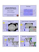
The human mtDNA genome is composed of 16,569 base pairs (bp),
encoding at total of 37 genes in a remarkably compact form. There
are 13 protein-coding genes, 22 tRNA genes, and 2 ribosomal genes,
with the overall organization of the genome shown in Fig.
1 . The
protein-coding genes contribute to the ETC complexes I, III, IV,
and V, with complex II being exclusively nuclear encoded. Preservation
of complex II (succinate dehydrogenase) activity can suggest the
2. History
3. mtDNA
Structure
6 W.J. Craigen
presence of an mtDNA-mediated disease, whether maternally inher-
ited or Mendelian. Of the 13 protein-coding genes, 7 contribute to
complex I (ND1, ND2, ND3, ND4, ND4L, ND5, and ND6), 1 is
a component of complex III (cytochrome b), 3 proteins form the
core of complex IV (cytochrome c oxidase; COX I, COX II, and
COX III), and 2 proteins are part of complex V (ATPase 6 and
ATPase 8). Based upon the exclusively maternal inheritance of
mtDNA, recombination between parental genomes would not be a
source of mtDNA variation, but, rather, variation refl ects both his-
torical population lineages and a high mutation rate (
20 ) . DNA
sequence variation at a population level has been categorized into
“haplogroups.” This variation has been used to reconstruct historic
population movements and has a variety of practical applications such
as forensics (
21 ) . Population variation also has effects on the patho-
genicity of particular mutations via still poorly understood interac-
tions between the mutation and the genetic “background” (
22 ) and
may predispose individuals to more common disorders (
23 ) .
F
12S rRNA
16S rRNA
V
L1
I
Q
M
W
A
N
C
Y
S1
D
K
G
R
H
S2
L2
E
T
P
ND1
ND2
ND3
ND4L
ND4
ND5
ND6
COI
COII
COIII
ATP6
ATP8
CytB
3243A>G (MELAS)
8344A>G (MERRF)
8356T>C (MERRF)
8363G>A (MERRF)
8993T>G/C (NARP)
1555A>G (Deafness)
11778G>A (LHON)
3460G>A (LHON)
14484T>C (LHON)
Complex I genes
Complex III gene
Complex IV genes
Ribosomal RNA genes
Transfer RNA genes
Complex V genes
Mitochondrial DNA
Common Mutations
Common
5 kb deletion
Fig. 1. A diagram of the human mitochondrial genome. The organization of genes encoded in mtDNA is shown, along with
the positions of the mutations referred to in the text.
71 Mitochondrial DNA Mutations: An Overview of Clinical and Molecular Aspects
Given the wide clinical variability and a lack of simple, defi nitive
testing, the prevalence of mitochondrial disorders is diffi cult to
accurately measure (
24 ) . However, estimates from laboratory refer-
ral centers and population screening have been reported (
25, 26 ) ,
and the overall frequency of ETC disorders has been estimated to
be approximately 1:5,000–8,000, including both primary mtDNA
disorders and Mendelian diseases. Studies of adult populations,
where primary mtDNA disorders are more common, suggest a
prevalence of about 1:10,000 for mtDNA mutations (
27, 28 ) .
Employing a small number of specifi c mutations as a screen in new-
borns, Elliott and colleagues demonstrated a remarkably high rate
of 1:200 newborns harboring an mtDNA mutation, with a corre-
spondingly high rate of new mutation (
29 ) . While it is likely that
the majority of these individuals will remain asymptomatic, these
high rates of detection refl ect the propensity for mutation in the
mitochondrial genome.
Factors that both defi ne and infl uence the inheritance and devel-
opment of mtDNA disease include maternal transmission, the degree
of heteroplasmy and the attendant threshold at which a tissue expe-
riences dysfunction, and the mitotic segregation of the mutation.
The mammalian oocyte contains over a 100,000 mitochondria,
while sperm do not contribute to the zygote mitochondrial popu-
lation. A single exception to this biological truism has been
reported, although it was identifi ed only in the setting of a mito-
chondrial disorder (
30, 31 ) . During subsequent embryonic devel-
opment, there is a gradual “dilution” in the number of mitochondria
per cell until mitochondrial biogenesis begins. In order to try to
explain the intergenerational changes in the degree of heteroplasmy
that can be observed, it has been debated whether the embryonic
reduction in mtDNA copy number in primordial germ cells leads
to shifts in heteroplasmy (
32 ) , whether it occurs postnatally in pri-
mordial germ cells (
33 ) , or whether other factors such as the pref-
erential replication of a subpopulation of mtDNA in germ cells
drive the rapid shifts in heteroplasmy that can be seen (
34 ) . A fi nal
answer remains to be conclusively determined.
Each somatic cell contains hundreds to thousands of copies of
mtDNA that during cell division distribute randomly among
daughter cells. In normal tissues, all mtDNA molecules are thought
to be identical. While some deleterious mutations are mild enough
to be tolerated in all mtDNA molecules, such as those causing
LHON, and thus are referred to as homoplasmic, many deleterious
mtDNA mutations impair mitochondrial functions to a degree that
is not compatible with cell survival. The relative proportion of
mutant to normal mtDNA genomes can vary among different
tissues, and similarly, different tissues exhibit varying sensitivity to
4. mtDNA
Mutations
8 W.J. Craigen
a disruption in mitochondrial function. This latter concept is
referred to as the threshold effect.
The segregation of heteroplasmic mtDNA to daughter cells,
referred to as mitotic segregation, also infl uences the development
of mitochondrial dysfunction. For pathologic mtDNA variants, the
exact mechanisms infl uencing the pattern of segregation are poorly
understood but may refl ect the survival of the resulting daughter
cells, the relative replication effi ciency of the two genomes, interac-
tions of the mtDNA genomes with nucleoid proteins that package
the mtDNA, or other mtDNA modifi cations. However, some
insights have been gleaned from studying the segregation patterns
of apparently neutral mtDNA sequence variants in model systems
such as the mouse, and these studies clearly reveal that mtDNA
segregation varies with age, is at least partially under the control of
nuclear genes (
35 ) , and depends on the tissue identity in which it
occurs (
36 ) . Recently, using heteroplasmic mouse strains, a nuclear
gene that infl uences mtDNA segregation in leukocytes was identi-
fi ed to be Gimap3 , a mitochondrial outer membrane GTPase
protein of unknown function (
37 ) .
With these concepts in mind, a brief review of the types of
mtDNA mutations is presented, categorized either as mtDNA
rearrangements or point mutations.
mtDNA deletions, duplications, and other more complex rear-
rangements are observed in disease states. In addition, multiple
deletions and mtDNA depletion can be observed in the context of
a Mendelian disorder (see Chapter 2 ). Patients harboring primary
mtDNA deletions (in contrast to those patients in whom the
deletion is a manifestation of a Mendelian disorder of mtDNA
integrity) generally exhibit one of three sporadic conditions. First,
Pearson syndrome is an often fatal disorder of infancy or early
childhood that is characterized by sideroblastic anemia and exo-
crine pancreas insuffi ciency and may be complicated by gastroin-
testinal problems and growth failure (
38 ) . Kearns-Sayre syndrome
is a multisystem disorder characterized by impaired eye movements
(chronic progressive external ophthalmoplegia (CPEO)), pigmentary
retinopathy, and a cardiac conduction defect. The signs and symp-
toms arise before 20 years of age. Other clinical problems may
include endocrinopathies such as diabetes mellitus, hypoparathy-
roidism, and short stature, progressive neurologic impairments
such as ataxia or dementia. Laboratory abnormalities are common,
including lactic acidosis, elevated cerebrospinal fl uid (CSF) protein,
and scattered cytochrome oxidase–negative RRF in skeletal muscle
biopsies. Finally, isolated CPEO with or without proximal muscle
5. mtDNA
Rearrangements
91 Mitochondrial DNA Mutations: An Overview of Clinical and Molecular Aspects
weakness is the mildest clinical syndrome associated with mtDNA
deletions (
39 ) . Patients with CPEO but without other symptoms
of Kearns-Sayre syndrome often develop neuromuscular symptoms
as they age, and conversely Pearson syndrome patients who survive
infancy may develop Kearns-Sayre syndrome at a later age (
40 ) .
In young patients with multisystem disease, mtDNA deletion testing
may be abnormal in blood samples since the deletion is more likely
to be a de novo germ line or early embryonic event, whereas in
older patients, the mtDNA deletion is more likely a somatic event
in the affected tissue. Thus, in patients with a delayed onset of
disease, the deletion is typically not detectable in blood specimens,
and it is necessary to use skeletal muscle for mtDNA deletion
testing (
41 ) .
At the molecular level, approximately 60% of mtDNA dele-
tions occur in a region of the mtDNA genome that is fl anked by
short direct repeat sequences, one of which is usually lost during
the deletion process, and these have been referred to as class I dele-
tions (
42 ) . Such repeats are thought to play a role in the formation
of mtDNA deletions. Approximately 30% of mtDNA deletions are
fl anked by imperfect repeats containing a few mismatches (class II
deletions), and about 10% have no repeats at the deletion fl anking
regions (
43 ) . The most common mtDNA deletion, which is present
in approximately one third of patients, is a 5-kb deletion (m.8470–
m.13447) that is fl anked by a 13-bp class I direct repeat (
42 ) .
While it has been speculated that defects in mtDNA replication
due to misalignment of direct repeats may cause mtDNA deletions
(
44 ) , an alternative mechanism involving the repair of mtDNA
damage has recently been proposed (
45 ) . A recent report summa-
rizing the molecular and clinical characteristics in 67 patients of
varying age reported that the deletion breakpoints found in the
youngest patients have signifi cantly lower breakpoint homology
relative to the older patients, with fewer class I breakpoints and an
almost threefold decreased incidence of the common 5-kb mtDNA
deletion relative to older patients, as well as increased heterogene-
ity in the breakpoint distribution. The severity of disease appears
not to be affected by the size of the mtDNA deletion or the
particular genes deleted (
46 ) . These fi ndings suggest that the
molecular events responsible for mtDNA deletions in young
patients may differ from those found in older patients.
Over 200 pathogenic point mutations have been identifi ed in
mtDNA from patients with a wide variety of disorders (
http://
mitomap.org/MITOMAP
), many of which are maternally inherited
6. mtDNA Point
Mutations
10 W.J. Craigen
and involve multiple organ systems but on occasion can be
sporadic and tissue specifi c. These can impair mtDNA-encoded
proteins, tRNAs, or rRNAs and potentially interfere with replica-
tion, transcription, or RNA processing. Examples of some of these
mechanisms of disease are provided by four of the most common
point mutations and their associated clinical syndromes.
Mitochondrial tRNAs are structurally distinct from other
tRNAs; they are shorter than bacterial or eukaryotic cytoplasmic
tRNAs and lack a variety of conserved nucleotides that are involved
in the prototypic tertiary interactions that create the canonical
L-shape of tRNAs, possibly resulting in a weaker tertiary structure.
In addition, in comparison to cytosolic tRNAs, posttranscriptional
base modifi cation appears to be more important for the proper
tertiary structure and function of mitochondrial tRNAs (
47 ) .
A pathogenic tRNA mutation leads to a combined OXPHOS
defect, in part through a decreased overall rate of mitochondrial
protein synthesis. Depending on which tRNA is mutated; there
will be varying effects on the individual ETC complexes based
upon the percentage of the corresponding amino acid in the differ-
ent ETC complex subunits. The pathogenic mechanisms leading
to defective translation caused by a tRNA mutation are numerous,
including impaired transcription termination, impaired tRNA mat-
uration, defective posttranscriptional modifi cation of the tRNA,
effects on tRNA folding and stability, reduced aminoacylation,
decreased binding to the translation factor mtEFTu or the mito-
chondrial ribosome, and altered codon decoding (
48 ) .
The tRNA
Leu(UUR)
gene ( MT-TL1 ) is particularly rife with
pathogenic mutations, with nearly 30 different mutations to date,
but mutations have now been detected in all 22 tRNA genes. The
prototypic tRNA mutation is the 3243A>G mutation in
tRNALeu
(UUR)
. This mutation causes a variety of clinical disorders, the
best known being MELAS (mitochondrial encephalomyopathy,
lactic acidosis, and stroke-like episodes) syndrome, which typically
becomes apparent in children or young adults after a normal early
developmental period (
49 ) . Signs and symptoms include recur-
rent vomiting, migraine-like headache, and stroke-like episodes
causing cortical blindness, hemiparesis, or hemianopia. MRI of
the brain shows regions of hypoperfusion that do not correspond
to a vascular distribution, and it has been suggested that the
underlying defect is one of endothelial function due to a func-
tional defi ciency in nitric oxide (
50 ) . Later features include hear-
ing loss, short stature, diabetes, retinopathy, muscle fatigue, and
lactic acidosis. The aberrant molecular mechanisms of the mutant
tRNA underlying the disorder are varied and somewhat contro-
versial, including a reduction in the aminoacylation of the tRNA
and a lack of wobble-base hypermodifi cation. This posttranscrip-
tional taurine modifi cation at the anticodon wobble position is
needed to restrict decoding to leucine
UUR
codons, and loss of this
111 Mitochondrial DNA Mutations: An Overview of Clinical and Molecular Aspects
modifi cation leads to a combination of a decoding defect of UUG
and UUA codons and amino acid misincorporation into proteins
(
51 ) . Additionally, the 3243A>G mutation has been shown to
diminish 16S rRNA transcription termination and alter processing
of the primary transcript (
52 ) . It is worth noting that the 3243A>G
mutation is a common, recurrent mutation that appears to arise
on a variety of haplogroup backgrounds and thus does not represent
a founder mutation. While the most common mtDNA mutation
causing MELAS syndrome is 3243A>G, and it is always found
in the heteroplasmic state, a number of other mutations have also
been associated with MELAS syndrome, including a missense
mutation in the ND5 gene that encodes subunit 5 of NADH
dehydrogenase (
53 ) and an intriguing mutation that abolishes the
binding site of the transcription termination factor MTERF1 to
the tRNA
Leu(UUR)
gene ( 54 ) .
A second common site for tRNA mutation is that of tRNA
Lys
( MT-TK ). The most common mutation is 8344A>G, which is
associated with MERRF (myoclonic epilepsy with ragged-red
fi bers) syndrome, and this mutation accounts for 80% of affected
individuals. The disorder is characterized by myoclonus, generalized
seizures, mitochondrial myopathy, and cerebellar ataxia. Other
clinical signs include short stature, dementia, hearing loss, a periph-
eral neuropathy, and cardiomyopathy with Wolff–Parkinson–White
syndrome, a cardiac conduction defect. Occasionally, pigmentary
retinopathy and lipomatosis are present. Similar to 3243A>G, it
has been reported to affect both aminoacylation and taurine modi-
fi cation of the wobble-base U, the latter disrupting codon-anticodon
pairing on the mitochondrial ribosome for both of the tRNA
Lys
codons (
55 ) . Two additional mutations in the tRNA
Lys
gene have
been associated with MERRF syndrome (8356T>C and 8363G>A),
and, like the 3243A>G mutation, MERFF mutations exist in the
heteroplasmic state.
A third common point mutation leads to a missense substitu-
tion in MT-ATP6 ; most commonly 8993T>G or 8993T>C, with
the former generally being clinically more severe. The clinical syn-
dromes associated with this mutation are defi ned by the degree of
heteroplasmy: lower mutation burdens cause NARP (neurogenic
muscle weakness, ataxia, retinitis pigmentosa) syndrome, which
usually affects young adults and causes retinitis pigmentosa, dementia,
seizures, ataxia, proximal muscle weakness, and a sensory neuropa-
thy (
56 ) . When there is a greater percentage of mutant mtDNA
molecules present, maternally inherited Leigh syndrome (MILS) is
observed, which is a severe infantile encephalopathy with charac-
teristic symmetrical lesions in the basal ganglia and the brainstem
and typically leads to early death (
57 ) .
An additional example of a class of mtDNA missense mutations,
in this case, mutations that are uniformly homoplasmic, causes
Leber’s hereditary optic neuropathy (LHON). The disorder is
12 W.J. Craigen
characterized by acute or subacute, painless loss of vision in young
adults due to bilateral optic atrophy, with reduced penetrance and
a four- to fi vefold greater frequency in males due to as yet unidenti-
fi ed nuclear gene modifi ers that have been mapped to the X chro-
mosome by linkage analysis (
58 ) . Three mtDNA point mutations
in complex I subunit genes account for more than 90% of LHON
cases. The causative mutations are 11778G>A in ND4, 3460G>A
in ND1, and 14484T>C in ND6. Because ETC bioenergetics
appears minimally impaired, it has been suggested that excess reactive
oxygen species in conjunction with a unique retinal ganglion cell
sensitivity accounts for the disease pathogenesis (
59 ) . In addition,
there is a clear effect of the mtDNA haplogroup on the penetrance
of specifi c mutations (
60 ) .
One fi nal example of an mtDNA point mutation that is repre-
sentative of a class of mutations is the 1555A>G mutation in the
12S rRNA ( MT-RNR1 ). Mammalian mitochondrial ribosomes
differ notably from cytosolic or bacterial ribosomes and even from
ribosomes from other mitochondria. They lack nearly half the
rRNA present in bacterial ribosomes and contain a correspondingly
higher protein content due to the incorporation of larger proteins
and numerous additional proteins, causing a greater molecular
mass and size than bacterial ribosomes (
61 ) . The 1555A>G muta-
tion is located in the decoding site of the mitochondrial small
subunit (SSU) ribosomal RNA and is predicted to cause a change
in the secondary rRNA structure to one that more closely resem-
bles the corresponding region of the bacterial 16S rRNA. This
alteration impairs protein synthesis and enhances an interaction
with aminoglycoside antibiotics, which further exacerbates the
translation defect. The mutation alone typically does not lead to
disease, but in combination with environmental modifi ers such as
the aminoglycosides or perhaps genetic modifi ers such as mito-
chondrial haplogroups (
62 ) , varying degrees of hearing loss is
observed. In addition to mtDNA-encoded modifi ers, nuclear
modifi er genes have been putatively identifi ed, making this class of
mutation currently unique. TFB1M , encoding a mitochondrial
rRNA methyltransferase, has been identifi ed as a possible nuclear
modifi er of the 1555A>G mutation (
63 ) , as has a second RNA
modifying enzyme TRMU (
64 ) . TRMU was recently identifi ed as
a tRNA 5-methylaminomethyl-2-thiouridylate methyltransferase
that when defi cient causes transient liver failure (
65 ) . Presumably,
alterations in RNA methylation due to malfunctioning TFB1M or
TRMU can diminish the deleterious effect of the 1555A>G muta-
tion on the ribosome conformation, although additional supporting
evidence is needed to fi rmly establish their role.
In summary, a variety of mutations can arise in mtDNA due in
large part to the high mutation rate, and these mutations can
exhibit a complex and broad spectrum of disease manifestations.
The properties of maternal inheritance, heteroplasmy, tissue- and