automated dna sequencing chemistry guide
Bạn đang xem bản rút gọn của tài liệu. Xem và tải ngay bản đầy đủ của tài liệu tại đây (2.92 MB, 246 trang )
Automated DNA
Sequencing
Chemistry Guide
© Copyright 2000, Applied Biosystems
For Research Use Only. Not for use in diagnostic procedures.
ABI PRISM and its design, Applied Biosystems, and MicroAmp are registered trademarks of Applera Corporation or its subsidiaries in the U.S. and
certain other countries.
ABI, BigDye, CATALYST, POP, POP-4, POP-6, and Primer Express are trademarks of Applera Corporation or its subsidiaries in the U.S. and certain
other countries.
AmpliTaq, AmpliTaq Gold, and GeneAmp are registered trademarks of Roche Molecular Systems, Inc.
Centricon is a registered trademark of W. R. Grace and Co.
Centri-Sep is a trademark of Princeton Separations, Inc.
Long Ranger is a trademark of The FMC Corporation.
Macintosh and Power Macintosh are registered trademarks of Apple Computer, Inc.
pGEM is a registered trademark of Promega Corporation.
Contents
i
1 Introduction. . . . . . . . . . . . . . . . . . . . . . . . . . . . . . . . . . . . . . . . . . 1-1
New DNA Sequencing Chemistry Guide . . . . . . . . . . . . . . . . . . . . . . . . . . . . . . . . . . . . . . . . . . 1-1
Introduction to Automated DNA Sequencing . . . . . . . . . . . . . . . . . . . . . . . . . . . . . . . . . . . . . . 1-2
ABI P
RISM
Sequencing Chemistries . . . . . . . . . . . . . . . . . . . . . . . . . . . . . . . . . . . . . . . . . . . . . 1-5
Applied Biosystems DNA Sequencing Instruments. . . . . . . . . . . . . . . . . . . . . . . . . . . . . . . . . . 1-7
Data Collection and Analysis Settings . . . . . . . . . . . . . . . . . . . . . . . . . . . . . . . . . . . . . . . . . . . 1-12
2 ABI P
RISM
DNA Sequencing Chemistries . . . . . . . . . . . . . . . . . . 2-1
Overview . . . . . . . . . . . . . . . . . . . . . . . . . . . . . . . . . . . . . . . . . . . . . . . . . . . . . . . . . . . . . . . . . . 2-1
Dye Terminator Cycle Sequencing Kits . . . . . . . . . . . . . . . . . . . . . . . . . . . . . . . . . . . . . . . . . . . 2-2
Dye Primer Cycle Sequencing Kits . . . . . . . . . . . . . . . . . . . . . . . . . . . . . . . . . . . . . . . . . . . . . . 2-8
Dye Spectra . . . . . . . . . . . . . . . . . . . . . . . . . . . . . . . . . . . . . . . . . . . . . . . . . . . . . . . . . . . . . . . 2-12
Chemistry/Instrument/Filter Set Compatibilities . . . . . . . . . . . . . . . . . . . . . . . . . . . . . . . . . . . 2-13
Dye/Base Relationships for Sequencing Chemistries . . . . . . . . . . . . . . . . . . . . . . . . . . . . . . . 2-14
Choosing a Sequencing Chemistry. . . . . . . . . . . . . . . . . . . . . . . . . . . . . . . . . . . . . . . . . . . . . . 2-15
3 Performing DNA Sequencing Reactions . . . . . . . . . . . . . . . . . . . 3-1
Overview . . . . . . . . . . . . . . . . . . . . . . . . . . . . . . . . . . . . . . . . . . . . . . . . . . . . . . . . . . . . . . . . . . 3-1
DNA Template Preparation . . . . . . . . . . . . . . . . . . . . . . . . . . . . . . . . . . . . . . . . . . . . . . . . . . . . 3-2
Sequencing PCR Templates . . . . . . . . . . . . . . . . . . . . . . . . . . . . . . . . . . . . . . . . . . . . . . . . . . . 3-10
DNA Template Quality. . . . . . . . . . . . . . . . . . . . . . . . . . . . . . . . . . . . . . . . . . . . . . . . . . . . . . . 3-15
DNA Template Quantity. . . . . . . . . . . . . . . . . . . . . . . . . . . . . . . . . . . . . . . . . . . . . . . . . . . . . . 3-17
Primer Design and Quantitation . . . . . . . . . . . . . . . . . . . . . . . . . . . . . . . . . . . . . . . . . . . . . . . . 3-18
Reagent and Equipment Considerations. . . . . . . . . . . . . . . . . . . . . . . . . . . . . . . . . . . . . . . . . . 3-20
Preparing Cycle Sequencing Reactions . . . . . . . . . . . . . . . . . . . . . . . . . . . . . . . . . . . . . . . . . . 3-21
Cycle Sequencing . . . . . . . . . . . . . . . . . . . . . . . . . . . . . . . . . . . . . . . . . . . . . . . . . . . . . . . . . . . 3-27
Preparing Extension Products for Electrophoresis . . . . . . . . . . . . . . . . . . . . . . . . . . . . . . . . . . 3-33
Removing Unincorporated Dye Terminators . . . . . . . . . . . . . . . . . . . . . . . . . . . . . . . . . . . . . . 3-34
Preparing Dye Primer Reaction Products for Electrophoresis . . . . . . . . . . . . . . . . . . . . . . . . . 3-46
Preparing and Loading Samples for Gel Electrophoresis. . . . . . . . . . . . . . . . . . . . . . . . . . . . . 3-50
Preparing and Loading Samples for Capillary Electrophoresis . . . . . . . . . . . . . . . . . . . . . . . . 3-53
4 Optimizing Gel Electrophoresis . . . . . . . . . . . . . . . . . . . . . . . . . . 4-1
Introduction . . . . . . . . . . . . . . . . . . . . . . . . . . . . . . . . . . . . . . . . . . . . . . . . . . . . . . . . . . . . . . . . 4-1
Reagents . . . . . . . . . . . . . . . . . . . . . . . . . . . . . . . . . . . . . . . . . . . . . . . . . . . . . . . . . . . . . . . . . . . 4-2
ii
Avoiding Problems with Sequencing Gels . . . . . . . . . . . . . . . . . . . . . . . . . . . . . . . . . . . . . . . . 4-4
5 Optimizing Capillary Electrophoresis . . . . . . . . . . . . . . . . . . . . . 5-1
Introduction . . . . . . . . . . . . . . . . . . . . . . . . . . . . . . . . . . . . . . . . . . . . . . . . . . . . . . . . . . . . . . . . 5-1
Capillary Electrophoresis Consumables . . . . . . . . . . . . . . . . . . . . . . . . . . . . . . . . . . . . . . . . . . 5-2
Optimizing Electrokinetic Injection . . . . . . . . . . . . . . . . . . . . . . . . . . . . . . . . . . . . . . . . . . . . . 5-4
Optimizing Electrophoresis Conditions. . . . . . . . . . . . . . . . . . . . . . . . . . . . . . . . . . . . . . . . . . . 5-7
Run Parameters for Specific Sequencing Chemistries. . . . . . . . . . . . . . . . . . . . . . . . . . . . . . . . 5-8
6 Optimizing Software Settings. . . . . . . . . . . . . . . . . . . . . . . . . . . . 6-1
Introduction . . . . . . . . . . . . . . . . . . . . . . . . . . . . . . . . . . . . . . . . . . . . . . . . . . . . . . . . . . . . . . . . 6-1
Choosing a Run Module . . . . . . . . . . . . . . . . . . . . . . . . . . . . . . . . . . . . . . . . . . . . . . . . . . . . . . 6-2
Choosing a Dye Set/Primer (Mobility) File. . . . . . . . . . . . . . . . . . . . . . . . . . . . . . . . . . . . . . . . 6-3
Choosing the Correct Basecaller . . . . . . . . . . . . . . . . . . . . . . . . . . . . . . . . . . . . . . . . . . . . . . . . 6-6
Creating an Instrument (Matrix) File. . . . . . . . . . . . . . . . . . . . . . . . . . . . . . . . . . . . . . . . . . . . . 6-7
Setting the Data Analysis Range . . . . . . . . . . . . . . . . . . . . . . . . . . . . . . . . . . . . . . . . . . . . . . . 6-15
7 Data Evaluation and Troubleshooting . . . . . . . . . . . . . . . . . . . . 7-1
Overview . . . . . . . . . . . . . . . . . . . . . . . . . . . . . . . . . . . . . . . . . . . . . . . . . . . . . . . . . . . . . . . . . . 7-1
Data Evaluation . . . . . . . . . . . . . . . . . . . . . . . . . . . . . . . . . . . . . . . . . . . . . . . . . . . . . . . . . . . . . 7-2
Practical Examples of Data Evaluation . . . . . . . . . . . . . . . . . . . . . . . . . . . . . . . . . . . . . . . . . . 7-10
Troubleshooting Sequencing Reactions. . . . . . . . . . . . . . . . . . . . . . . . . . . . . . . . . . . . . . . . . . 7-16
Troubleshooting DNA Sequence Composition Problems . . . . . . . . . . . . . . . . . . . . . . . . . . . . 7-30
Troubleshooting Sequencing Data. . . . . . . . . . . . . . . . . . . . . . . . . . . . . . . . . . . . . . . . . . . . . . 7-39
Troubleshooting Gel Electrophoresis on the ABI 373 and ABI P
RISM
377 . . . . . . . . . . . . . . 7-44
Troubleshooting Capillary Electrophoresis on the ABI P
RISM
310. . . . . . . . . . . . . . . . . . . . . 7-55
Troubleshooting Software Settings . . . . . . . . . . . . . . . . . . . . . . . . . . . . . . . . . . . . . . . . . . . . . 7-62
A Gel Preparation . . . . . . . . . . . . . . . . . . . . . . . . . . . . . . . . . . . . . . A-1
Introduction . . . . . . . . . . . . . . . . . . . . . . . . . . . . . . . . . . . . . . . . . . . . . . . . . . . . . . . . . . . . . . . . A-1
Protocol and Run Conditions for 19:1 Polyacrylamide Gels . . . . . . . . . . . . . . . . . . . . . . . . . . A-2
Protocol and Run Conditions for 29:1 Polyacrylamide Gels. . . . . . . . . . . . . . . . . . . . . . . . . . . A-6
Protocol and Run Conditions for Long Ranger and PAGE-PLUS Gels . . . . . . . . . . . . . . . . . A-10
Preparing APS, TBE Buffer, and Deionized Formamide . . . . . . . . . . . . . . . . . . . . . . . . . . . . A-15
iii
B IUB Codes . . . . . . . . . . . . . . . . . . . . . . . . . . . . . . . . . . . . . . . . . . . B-1
C References . . . . . . . . . . . . . . . . . . . . . . . . . . . . . . . . . . . . . . . . . . . C-1
D Technical Support . . . . . . . . . . . . . . . . . . . . . . . . . . . . . . . . . . . . .D-1
To Reach Us on the Web. . . . . . . . . . . . . . . . . . . . . . . . . . . . . . . . . . . . . . . . . . . . . . . . . . . . . . D-1
Hours for Telephone Technical Support . . . . . . . . . . . . . . . . . . . . . . . . . . . . . . . . . . . . . . . . . . D-1
To Reach Us by Telephone or Fax in North America. . . . . . . . . . . . . . . . . . . . . . . . . . . . . . . . D-1
Documents on Demand . . . . . . . . . . . . . . . . . . . . . . . . . . . . . . . . . . . . . . . . . . . . . . . . . . . . . . D-2
To Reach Us by E-Mail . . . . . . . . . . . . . . . . . . . . . . . . . . . . . . . . . . . . . . . . . . . . . . . . . . . . . . D-3
Regional Offices Sales and Service . . . . . . . . . . . . . . . . . . . . . . . . . . . . . . . . . . . . . . . . . . . . . D-3
E Part Numbers. . . . . . . . . . . . . . . . . . . . . . . . . . . . . . . . . . . . . . . . .E-1
ABI P
RISM
DNA Sequencing Kits and Reagents. . . . . . . . . . . . . . . . . . . . . . . . . . . . . . . . . . . .E-1
ABI P
RISM
310 Genetic Analyzer . . . . . . . . . . . . . . . . . . . . . . . . . . . . . . . . . . . . . . . . . . . . . . . E-5
ABI P
RISM
377 DNA Sequencer . . . . . . . . . . . . . . . . . . . . . . . . . . . . . . . . . . . . . . . . . . . . . . . .E-8
ABI 373 DNA Sequencer . . . . . . . . . . . . . . . . . . . . . . . . . . . . . . . . . . . . . . . . . . . . . . . . . . . . . .E-9
Documentation and Software . . . . . . . . . . . . . . . . . . . . . . . . . . . . . . . . . . . . . . . . . . . . . . . . . . E-10
Index
Introduction 1-1
Introduction 1
New DNA Sequencing Chemistry Guide
Purpose
Since the original
DNA Sequencing Chemistry Guide
was published in early 1995,
Applied Biosystems has released two new instrument platforms, five new sequencing
chemistries, and a new sequencing enzyme.
To accommodate this new information, we have written the
Automated DNA
Sequencing Chemistry Guide
. This updated guide provides the following:
♦
An introduction to automated DNA sequencing
♦
Descriptions of Applied Biosystems sequencing instruments, chemistries, and
software
♦
Detailed protocols for preparing DNA templates, performing cycle sequencing,
and preparing the extension products for electrophoresis
♦
Guidelines for optimizing electrophoresis and interpreting and troubleshooting
sequencing data
1
1-2 Introduction
Introduction to Automated DNA Sequencing
Sanger Dideoxy
Sequencing
DNA polymerases copy single-stranded DNA templates, by adding nucleotides to a
growing chain (extension product). Chain elongation occurs at the 3´ end of a primer,
an oligonucleotide that anneals to the template. The deoxynucleotide added to the
extension product is selected by base-pair matching to the template.
The extension product grows by the formation of a phosphodiester bridge between the
3´-hydroxyl group at the growing end of the primer and the 5´-phosphate group of the
incoming deoxynucleotide (Watson
et al.
, 1987). The growth is in the 5´
Æ
3´ direction
(Figure 1-1).
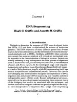
DNA polymerases can also incorporate analogues of nucleotide bases. The dideoxy
method of DNA sequencing developed by Sanger
et al.
(1977) takes advantage of this
ability by using 2´,3´-dideoxynucleotides as substrates. When a dideoxynucleotide is
incorporated at the 3´ end of the growing chain, chain elongation is terminated
selectively at A, C, G, or T because the chain lacks a 3´-hydroxyl group (Figure 1-1).
Figure 1-1
DNA strand synthesis by formation of phosphodiester bonds. The chain is
terminated by the use of dideoxycytidine triphosphate (ddC) in place of deoxycytidine
triphosphate (dCTP). The inset shows a schematic representation of the process.
3´ hydroxyl group
no 3´ hydroxyl group
TemplateExtension product
Introduction 1-3
Fluorescent
Sequencing
In the Applied Biosystems strategy for automated fluorescent sequencing, fluorescent
dye labels are incorporated into DNA extension products using 5´-dye labeled primers
(dye primers) or 3´-dye labeled dideoxynucleotide triphosphates (dye terminators).
The most appropriate labeling method to use depends on your sequencing objectives,
the performance characteristics of each method, and on personal preference.
Applied Biosystems DNA sequencers detect fluorescence from four different dyes that
are used to identify the A, C, G, and T extension reactions. Each dye emits light at a
different wavelength when excited by an argon ion laser. All four colors and therefore
all four bases can be detected and distinguished in a single gel lane or capillary
injection (Figure 1-2).
Figure 1-2
Four-color/one-lane fluorescent sequencing vs. one-color/four-lane method such
as radioactive sequencing
1-4 Introduction
Cycle Sequencing
Cycle sequencing is a simple method in which successive rounds of denaturation,
annealing, and extension in a thermal cycler result in linear amplification of extension
products (Figure 1-3). The products are then loaded onto a gel or injected into a
capillary. All current ABI P
RISM
DNA sequencing kits use cycle sequencing protocols.
See Chapter 3 for information on cycle sequencing protocols.
Figure 1-3
Cycle sequencing
Advantages of Cycle
Sequencing
♦
Protocols are robust and easy to perform.
♦
Cycle sequencing requires much less template DNA than single-temperature
extension methods.
♦
Cycle sequencing is more convenient than traditional single-temperature labeling
methods that require a chemical denaturation step for double-stranded templates.
♦
High temperatures reduce secondary structure, allowing for more complete
extension.
♦
High temperatures reduce secondary primer-to-template annealing.
♦
The same protocol is used for double- and single-stranded DNA.
♦
The protocols work well for direct sequencing of PCR products (see page 3-14).
♦
Difficult templates, such as bacterial artificial chromosomes (BACs), can be
sequenced.
Introduction 1-5
ABI P
RISM
Sequencing Chemistries
AmpliTaq DNA
Polymerase, FS
AmpliTaq
®
DNA Polymerase, FS is the sequencing enzyme used in ABI P
RISM
cycle
sequencing kits. It is a mutant form of
Thermus aquaticus
(Taq) DNA polymerase and
contains a point mutation in the active site, replacing phenylalanine with tyrosine at
residue 667 (F667Y). This mutation results in less discrimination against
dideoxynucleotides, and leads to a much more even peak intensity pattern (Tabor and
Richardson, 1995).
AmpliTaq DNA Polymerase, FS also contains a point mutation in the amino terminal
domain, replacing glycine with aspartate at residue 46 (G46D), which removes almost
all of the 5´
Æ
3´ nuclease activity. This eliminates artifacts that arise from the
exonuclease activity.
The enzyme has been formulated with a thermally stable inorganic pyrophosphatase
that cleaves the inorganic pyrophosphate (PP
i
) byproduct of the extension reaction
and prevents its accumulation in the sequencing reaction.
In the presence of high concentrations of PP
i
the polymerization reaction can be
reversed (Kornberg and Baker, 1992), a reaction called pyrophosphorolysis. In this
reaction, a nucleoside monophosphate is removed from the extension product with the
addition of PP
i
to form the nucleoside triphsphate.
In a sequencing reaction, if a dideoxynucleotide is frequently removed at a particular
position and replaced by a deoxynucleotide, eventually there is little or no chain
termination at that location. This results in a weak or missing peak in the sequence
data (Tabor and Richardson, 1990).
Dye-Labeled
Terminators
With dye terminator labeling, each of the four dideoxy terminators (ddNTPs) is tagged
with a different fluorescent dye. The growing chain is simultaneously terminated and
labeled with the dye that corresponds to that base (Figure 1-4).
Figure 1-4
One cycle of dye terminator cycle sequencing
Features of Dye-labeled Terminator Reactions
♦
An unlabeled primer can be used.
♦
Dye terminator reactions are performed in a single tube. They require fewer
pipetting steps than dye primer reactions.
♦
Four-color dye labeled reactions are loaded in a single gel lane or capillary
injection.
♦
False stops,
i.e.
, fragments that are not terminated by a dideoxynucleotide (see
page 7-30), go undetected because no dye is attached.
See Chapter 2 for information on ABI P
RISM
™
DNA sequencing kits.
1-6 Introduction
Dye-Labeled
Primers
With dye primer labeling, primers are tagged with four different fluorescent dyes.
Labeled products are generated in four separate base-specific reactions. The
products from these four reactions are then combined and loaded into a single gel
lane or capillary injection (Figure 1-5).
Figure 1-5
One cycle of dye primer cycle sequencing
Features of Dye-labeled Primer Reactions
♦
Dye primer chemistries generally produce more even signal intensities than dye
terminator chemistries.
♦
Labeled primers are available for common priming sites. Custom primers can also
be labeled.
♦
Four-color dye-labeled reactions are loaded onto a single lane or capillary
injection.
See Chapter 2 for information on ABI P
RISM
™
DNA sequencing kits.
Introduction 1-7
Applied Biosystems DNA Sequencing Instruments
ABI 373
DNA Sequencer
The ABI
™
373 DNA Sequencer is an automated instrument for analyzing fluorescently
labeled DNA fragments by gel electrophoresis. You can use three sizes of gel plates
for sequencing applications: 24-cm, 34-cm and 48-cm well-to-read lengths (see
Table 1-1 on page 1-10). The longer the well-to-read length, the better the resolution
of the gel.
Sequencing reaction products labeled with four different fluorescent dyes are loaded
into each lane of a 0.3-mm or 0.4-mm vertical slab gel made of polymerized
acrylamide or acrylamide derivatives. You can run up to 36 lanes simultaneously on a
single gel.
The dye-labeled DNA fragments migrate through the acrylamide gel and separate
according to size. At the lower portion of the gel they pass through a region where a
laser beam scans continuously across the gel. The laser excites the fluorescent dyes
attached to the fragments, and they emit light at a specific wavelength for each dye.
The fluorescence intensity is detected by a photomultiplier tube (PMT) and recorded
as a function of time. A moving stage contains the optical equipment (filter wheel and
photomultiplier tube). The PMT detects the fluorescence emission and converts it into
a digital signal. Each time the stage traverses across the gel (a scan) a different
bandpass filter is positioned in front of the PMT to detect each of the four dyes.
A single scan of the gel with one filter takes 1.5 seconds and measures signal in 194
channels. A complete scan with four filters takes 6 seconds and equals one data point.
The data is then transmitted to the Macintosh
®
computer and stored for processing.
The Sequencing Analysis software (see page 1-16) interprets the result, calling the
bases from the fluorescence intensity at each data point.
Refer to the
373 DNA Sequencing System User’s Manual
(P/N 902376) for more
information.
XL Upgrade
The ABI 373 DNA Sequencer with XL Upgrade increases the number of samples that
can be analyzed simultaneously. This increased throughput is made possible by
reengineering the instrument to collect data from 388 channels instead of 194. With
the XL Upgrade, the operation of the ABI 373 DNA Sequencer is controlled from the
Power Macintosh
®
computer supplied with the upgrade.
After the initial calibration by the Field Service Engineer, the instrument automatically
increases the PMT voltage to compensate for the smaller amount of signal generated
per lane when running 48- or 64-lane gels.
The XL Upgrade also includes new combs and spacers. For sequencing applications,
48-well and 64-well shark’ s tooth combs are available. You can still use 24-well or
36-well combs if desired.
Note
These combs are not interchangeable with combs for the ABI P
RISM
®
377 DNA
Sequencer.
Refer to the
373 DNA Sequencer With XL Upgrade User’s Manual
(P/N 904258) for
more information.
1-8 Introduction
Filter Sets
The ABI 373 and ABI 373 with XL Upgrade DNA Sequencers use filters mounted on a
filter wheel to separate light of different wavelengths. The instruments record the light
intensity in four regions, collectively called Filter Set A, centered at the following
wavelengths:
♦
Four-filter wheel: 540 nm, 560 nm, 580 nm, 610 nm
♦
Five-filter wheel: 531 nm, 560 nm, 580 nm, and 610 nm
Note
The five-filter wheel instruments also have Filter Set B (531 nm, 545 nm, 560nm, and
580 nm), but it is not used with existing Applied Biosystems sequencing chemistries. Filter Set
B was used for the T7 (Sequenase) terminator chemistries, which have been discontinued.
BigDye Filter Wheel
To use the new dRhodamine terminator, BigDye
™
terminator, and BigDye
™
primer
sequencing chemistries (see Chapter 2) on the ABI 373 and ABI 373 with XL Upgrade
DNA Sequencers, the ABI P
RISM
™
BigDye
™
Filter Wheel has been developed.
Its Filter Set A is as follows: 540 nm, 570 nm, 595 nm, and 625 nm.
Note The BigDye Filter Wheel also has Filter Set B (540 nm, 555 nm, 570, and 595 nm), but
it is not used with existing Applied Biosystems sequencing chemistries.
Refer to the Using the ABI 373 BigDye Filter Wheel User Bulletin (P/N 4304367) for
more information.
ABI PRISM 377
DNA Sequencer
The ABI PRISM 377 DNA Sequencer is a medium- to high-throughput, automated
instrument for analyzing fluorescently labeled DNA fragments by gel electrophoresis.
You can use two sizes of gel plates for sequencing applications: 36-cm and 48-cm
well-to-read lengths. The 48-cm well-to-read plates are used to obtain longer read
lengths.
Sequencing reaction products labeled with four different fluorescent dyes are loaded
into each lane of a 0.2-mm vertical slab gel made of polymerized acrylamide or
acrylamide derivatives. You can run up to 36 lanes simultaneously on one gel.
The dye-labeled DNA fragments migrate through the acrylamide gel and separate
according to size. At the lower portion of the gel they pass through a region where a
laser beam scans continuously across the gel. The laser excites the fluorescent dyes
attached to the fragments, and they emit light at a specific wavelength for each dye.
The light is collected in 194 channels during each scan and separated according to
wavelength by a spectrograph onto a cooled, charge-coupled device (CCD) camera,
so all four types of fluorescent emissions can be detected with one pass of the laser.
The data collection software collects the light intensities from the CCD at particular
wavelength bands (virtual filters) and stores them on a Power Macintosh computer as
digital signals for processing. The Sequencing Analysis software (see page 1-16)
interprets the result, calling the bases from the fluorescence intensity at each data
point.
Refer to the ABI P
RISM 377 DNA Sequencer User’s Manual (P/N 903433) for more
information.
Introduction 1-9
377-18
The ABI PRISM 377-18 DNA Sequencer is a lower-cost, lower-throughput version of
the ABI PRISM 377 DNA Sequencer. It can run up to 18 lanes on a single gel.
XL Upgrade
The ABI PRISM 377 DNA Sequencer with XL Upgrade increases the number of
samples that can be analyzed simultaneously. This increased throughput is made
possible by reengineering the instrument to collect data from 388 channels instead of
194 during each scan.
The XL Upgrade also includes new combs. For sequencing applications, 48-well and
64-well shark’ s tooth combs are available. You can still use 36-well or other lower lane
density combs if desired.
Refer to the ABI PRISM 377 DNA Sequencer XL Upgrade User’s Manual (P/N 904412)
for more information.
96-Lane Upgrade
The ABI PRISM 377 DNA Sequencer with 96-Lane Upgrade increases the number of
samples that can be run on each gel. The increased throughput is made possible by
reengineering the instrument to collect data from 480 channels instead of 388 for the
ABI PRISM 377 DNA Sequencer with XL Upgrade or 194 for the ABI PRISM 377 DNA
Sequencer.
The 96-lane upgrade includes new combs and new notched front glass plates. You
can still use lower lane density combs, but only with the original notched front glass
plates that were provided with the instrument.
The new notched front glass plate has a bevel in the loading region that increases the
thickness of the gel in this region from 0.2 mm to 0.4 mm. In addition, the scan region
has been increased from 6 inches to 7.5 inches. This makes sample loading easier
than for a 64-lane gel.
Refer to the ABI PRISM 377 DNA Sequencer 96-Lane Upgrade User’s Manual
(P/N 4305423) for more information.
1-10 Introduction
Gel Electrophoresis Instruments
ABI PRISM 310
Genetic Analyzer
The ABI PRISM
®
310 Genetic Analyzer is an automated instrument for analyzing
fluorescently labeled DNA fragments by capillary electrophoresis.
The sequencing reaction sample tubes are placed in an autosampler tray that holds
either 48 or 96 samples. The autosampler successively brings each sample into
contact with the cathode electrode and one end of a glass capillary filled with a
separation polymer. An anode electrode at the other end of the capillary is immersed
in buffer.
The sample enters the capillary as current flows from the cathode to the anode. The
short period of electrophoresis conducted while the capillary and cathode are
immersed in the sample is called electrokinetic injection. The sample forms a tight
band in the capillary during this injection. The end of the capillary near the cathode is
then placed in buffer. Current is applied again to continue electrophoresis.
When the DNA fragments reach a detector window in the capillary, a laser excites the
fluorescent dye labels. Emitted fluorescence from the dyes is collected once per
second by a cooled, charge-coupled device (CCD) camera at particular wavelength
bands (virtual filters) and stored as digital signals on a Power Macintosh computer for
processing. The Sequencing Analysis software (see page 1-16) interprets the result,
calling the bases from the fluorescence intensity at each data point.
Refer to the ABI P
RISM 310 Genetic Analyzer User’s Manual (P/N 903565) for more
information.
Table 1-1 Applied Biosystems Gel Electrophoresis Instruments
Instrument
Well-to-Read
Length (cm)
Number of
Lanes
Maximum
Throughput
(bases/hr)
a
a. Maximum throughput = maximum number of lanes ¥ maximum electrophoresis speed (50 bph for ABI 370 and ABI 373 models, 200 bph
for ABI PRISM 377 models)
Detection System Computer
ABI 370 24 16 800 PMT, 4-filter wheel HP Vectra
ABI 373 24 1200 Macintosh
ABI 373 Leon Model 6, 12, 24, 34 24, 36 1800 PMT, 5-filter wheel
ABI 373 Stretch Model 6, 12, 24, 34, 48
ABI 373 with XL
Upgrade
24 or
6, 12, 24, 34 or
6, 12, 24, 34, 48
24, 36, 48, 64 3200 Power
Macintosh
ABI 373 with BigDye
Filter Wheel
b
b. Allows use of dRhodamine-based chemistries on any ABI 373 or ABI 373 with XL Upgrade instrument with a 5-filter wheel. See page 1-8
for ABI 373 filter sets.
24, 36 or
24, 36, 48, 64
1800 or
3200
PMT, new 5-filter
wheel
Macintosh or
Power
Macintosh
ABI P
RISM 377 12, 36, 48 24, 36 7200 CCD camera,
spectrograph
Power
Macintosh
ABI P
RISM 377-18 18 3600
ABI P
RISM 377 with
XL Upgrade
24, 36, 48, 64 12,800
ABI P
RISM 377 with
96-Lane Upgrade
24, 36, 48, 64,
96
19,200
Introduction 1-11
Virtual Filter Sets ABI PRISM 310 and ABI PRISM 377 (All Models)
1
These instruments use virtual filter sets to detect light intensity in four non-overlapping
regions on a CCD camera. Each region corresponds to a wavelength range that
contains or is close to the emission maximum of an ABI PRISM dye.
The process is similar to using a physical filter to separate light of different
wavelengths. However, the filter sets are called “virtual filters” because the
instruments use no physical filtering hardware to perform the separation.
2
The exact positions of the CCD regions and the dye combinations appropriate to
these positions depend upon the virtual filter set used. For example, with Virtual Filter
Set E the instrument records the light intensity in four regions, or “windows,” centered
at 540 nm, 570 nm, 595 nm, and 625 nm. The window positions in each virtual filter
set have been optimized to provide the maximum possible separation among the
centers of detection for the different dyes while maintaining good signal strength.
The Data Collection Software color-codes the intensity displays from the four
light-collection regions. These appear as the blue, green, black (yellow on gel images),
and red peaks in the raw data.
The Sequencing Analysis Software uses the same four colors to color-code analyzed
data from all dye/virtual filter set combinations. The display colors represent the
relative, not the actual, detection wavelengths. For consistency, the software always
displays analyzed data with A as green, C as blue, G as black, and T as red in the
electropherogram view.
Table 1-2 shows the wavelengths of the “windows” in the virtual filter sets used in cycle
sequencing applications.
1. Includes the ABI PRISM 377, ABI PRISM 377-18, ABI PRISM 377 with XL Upgrade, and the
ABI PRISM 377 with 96-Lane Upgrade instruments.
2. The ABI PRISM 310 Genetic Analyzer and ABI PRISM 377 DNA Sequencer have a long-pass filter to
prevent light from the instrument’s argon ion laser from interfering with the detection of the dye signals.
Table 1-2 Wavelength Ranges of Virtual Filter Sets
Virtual
Filter Set Color
Wavelength Range of
Virtual Filter (nm)
A blue 530–541
green 554–564
yellow/black 581–591
red 610–620
E blue 535–545
green 565–575
yellow/black 590–600
red 620–630
1-12 Introduction
Data Collection and Analysis Settings
Overview This section is intended to provide an introduction to the data collection and analysis
settings, which are dealt with in more detail in Chapter 6.
Many users sequence DNA using more than one chemistry. Take care when entering
data collection and analysis settings in the software. If your data is analyzed with the
wrong software settings, the resulting electropherograms will show overlapping peaks
and gaps between peaks rather than the evenly spaced peaks characteristic of
correctly analyzed data.
Run Modules ABI 373 with XL Upgrade
A run module file contains all the parameters required for a particular function or
application. The parameters include the following:
♦ Electrophoresis power
♦ Current and voltage settings
♦ Laser settings
♦ Scanner settings
♦ PMT settings
There are three types of run module files. Not all of the parameters listed above are in
each module file.
♦ Plate Check
This module is for checking the cleanliness and alignment of the gel plates. Laser,
scanning, and PMT settings are associated with it.
♦ Pre Run
This module is for prerunning sequencing gels. Laser, scanning, electrophoresis,
and PMT settings are associated with it.
♦ Seq Run
This module is for running sequencing gels. Laser, scanning, electrophoresis, and
PMT settings are associated with it.
IMPORTANT When you select a run module, the filter set is chosen automatically. You must
edit the run module to change the filter set used to collect the data. Refer to the 373 DNA
Sequencer With XL Upgrade User’s Manual (P/N 904258) for more information.
Note The ABI 373 DNA Sequencer does not use run modules. Run parameters are set on
the instrument’s keypad. Refer to the 373 DNA Sequencing System User’s Manual
(P/N 902376) for information on setting run parameters.
ABI PRISM 310 and ABI PRISM 377 (All Models)
A run module file contains all the parameters required for a particular function or
application. The parameters include the following:
♦ Electrophoresis voltage
♦ Current and power settings
♦ Laser settings
♦ Scanner settings (ABI PRISM 377 DNA Sequencer only)
Introduction 1-13
♦ Virtual filters and CCD gain and offset
♦ Run temperature settings
♦ Injection time and voltage (ABI PRISM 310 Genetic Analyzer)
There are three types of module files. Not all of the parameters listed above are in
each module file.
♦ Plate check
These modules are for checking the cleanliness and alignment of the gel plates.
Laser, scanning, virtual filter, and CCD conditions are associated with these types
of files.
♦ Prerun
These modules are for prerunning the gel or polymer. Laser, scanning, virtual
filter, and electrophoresis, CCD, and gel temperature conditions are associated
with these types of files.
Note Plate check and prerun modules are not used with the ABI PRISM 310 Genetic
Analyzer.
♦ Run
These modules are for running the gel or polymer. Laser, scanning, virtual filter,
CCD, and electrophoresis parameters and gel temperature are associated with
these types of files.
IMPORTANT When you select a run module, the virtual filter set is chosen automatically.
You must be careful to select the correct run module for your sequencing chemistry.
The available run modules are listed in Table 6-1 on page 6-2.
Dye Set/Primer Files Mobility Correction
The different dyes affect the electrophoretic mobility of cycle sequencing extension
products. The relative mobility of the dye-labeled fragments is specific to each
sequencing chemistry (see page 6-4 for more information). Under the same set of
conditions, the mobilities are very reproducible.
The analysis software is able to compensate for these mobility differences by applying
mobility shifts to the data so that evenly spaced peaks are presented in the analyzed
data. The files that contain the mobility shift information are called dye set/primer files.
Dye set/primer files also tell the Sequencing Analysis software (see page 1-16) the
following:
♦ Which matrix file in the instrument file (see page 1-14) to use to analyze the data
♦ Dye/base relationships for converting raw data colors to base calls (see
page 2-14)
The dye set/primer files available are listed in Table 6-2 on page 6-5.
1-14 Introduction
Instrument Files Multicomponent Analysis
Multicomponent analysis is the process that separates the four different fluorescent
dye colors into distinct spectral components. Although each of these dyes emits its
maximum fluorescence at a different wavelength, there is some overlap in the
emission spectra between the four dyes (Figure 1-6). The goal of multicomponent
analysis is to isolate the signal from each dye so that there is as little noise in the data
as possible.
Figure 1-6 Spectral overlap of the dRhodamine dyes in the four virtual filters (vertical gray
bars) of Filter Set E
The precise spectral overlap between the four dyes is measured by running DNA
fragments labeled with each of the dyes in separate lanes of a gel or in separate
injections on a capillary. These dye-labeled DNA fragments are called matrix
standards.
The Data Utility software (see page 6-7) then analyzes the data from each of the four
matrix standard samples and creates an instrument file. The instrument file contains
three matrix files, which have tables of numbers with four columns and four rows
(Figure 1-7 on page 1-15). These numbers are normalized fluorescence intensities
and represent a mathematical description of the spectral overlap that is observed
between the four dyes.
The rows in the tables represent the virtual filters and the columns represent the dyes.
The top lefthand value, 1.000, represents the normalized fluorescence of the blue dye
in the blue filter. It follows that all matrix tables should have values of 1.000 on the
diagonal from top left to bottom right.
The other values in the table should all be less than 1. These values represent the
amount of spectral overlap observed for each dye in each virtual filter. For example,
the values in the third row reflect quantitatively the amount of each dye detected in the
third (“yellow”) virtual filter.
Introduction 1-15
Figure 1-7 Instrument file created in the Data Utility software, indicating the values obtained
with the dRhodamine matrix standards for Filter Set E on a particular ABI P
RISM 377 instrument
Note that the numbers decrease moving away from the diagonal in any direction. For
example, in the first column the amount of blue fluorescence seen through the red
filter (fourth row) should be less than that seen in the yellow filter (third row), which
should be less than that seen in the green filter (second row).
These values will vary between different instruments and between filter sets on a
single instrument. An instrument file must be made for each filter set used on each
instrument.
The instrument file is created for a specific filter set or virtual filter set when the
instrument is installed. Whenever a new filter set is used, a new instrument file must
be created for that filter set. Refer to your instrument user’s manual or the protocol for
the sequencing chemistry you are using for instructions on creating instrument files.
The appropriate instrument file can be applied to data on subsequent capillary runs or
gels on the same instrument, as long as the same filter set is used. This is because
the spectral overlap between the four dyes is very reproducible.
Multicomponent analysis of sequencing data is performed automatically by the
Sequencing Analysis software (see below), which applies a mathematical matrix
calculation, using the values in the instrument file, to all sample data.
See page 6-7 for instructions for creating instrument files.
1-16 Introduction
What Is In a Matrix File
The matrix files in an instrument file are used for specific types of chemistry, and
provide information to the Sequencing Analysis software to allow it to correct for
spectral overlap.
Matrix files also contain the following:
♦ Baselining algorithm for the chemistry being used
♦ Information that the Sequencing Analysis software uses to determine Peak 1
Locations and Start Points for data analysis
Sequencing Analysis
Software
The DNA Sequencing Analysis Software analyzes the raw data collected by the Data
Collection software:
♦ Tracks gel files (if using the ABI 373 or ABI P
RISM 377 DNA Sequencer)
♦ Extracts sample information from gel files (if using the ABI 373 or ABI PRISM 377
DNA Sequencer)
♦ Performs multicomponent analysis
♦ Applies mobility corrections
♦ Normalizes the base spacing
♦ Baselines data
♦ Determines analysis starting points
♦ Calls bases
See Chapter 7 for information on interpreting and troubleshooting sequencing data.
Refer to the ABI P
RISM DNA Sequencing Analysis Software User’s Manual for specific
information about the Sequencing Analysis software.
ABI PRISM DNA Sequencing Chemistries 2-1
ABI PRISM DNA
Sequencing Chemistries 2
Overview
In This Chapter This chapter describes the Applied Biosystems cycle sequencing chemistries, the
dyes used in them, and how to choose a sequencing chemistry.
Topic See page
Dye Terminator Cycle Sequencing Kits 2-2
Dye Primer Cycle Sequencing Kits 2-8
Dye Spectra 2-12
Chemistry/Instrument/Filter Set Compatibilities 2-13
Dye/Base Relationships for Sequencing Chemistries 2-14
Choosing a Sequencing Chemistry 2-15
2
2-2 ABI PRISM DNA Sequencing Chemistries
Dye Terminator Cycle Sequencing Kits
Rhodamine Dye
Terminators
The rhodamine dye terminators have the following dye labels. The structures of the
rhodamine dye terminators are shown in Figure 2-1.
Figure 2-1 Rhodamine dye terminators
Rhodamine Dye
Terminator Kits
The ABI PRISM
™
Dye Terminator Cycle Sequencing Kits combine AmpliTaq
®
DNA
Polymerase, FS, rhodamine dye terminators, and all the required components for the
sequencing reaction.
Note Throughout this manual, these kits will be referred to as “rhodamine dye terminators.”
The concentrations of the dye-labeled dideoxynucleotides and deoxynucleotides in
the dNTP mix have been optimized to give a balanced distribution of signal above 700
bases. The dNTP mix includes dITP in place of dGTP to minimize band compressions.
In the Ready Reaction format, the dye terminators, deoxynucleoside triphosphates,
AmpliTaq DNA Polymerase, FS, rTth pyrophosphatase, magnesium chloride, and
buffer are premixed into a single tube of Ready Reaction Mix and are ready to use.
These reagents are suitable for performing fluorescence-based cycle sequencing
reactions on single-stranded or double-stranded DNA templates, or on polymerase
chain reaction (PCR) fragments.
Terminator Dye Label
A R6G
CROX
G R110
T TAMRA
ABI PRISM DNA Sequencing Chemistries 2-3
In the Core Kit format, the reagents are supplied in individual tubes to maximize kit
flexibility. For convenience when sequencing large quantities of templates, the
reagents can be premixed and stored.
The cycle sequencing protocols are optimized for GeneAmp
®
PCR Instrument
Systems thermal cyclers, the CATALYST
™
800 Molecular Biology LabStation, and the
ABI PRISM
®
877 Integrated Thermal Cycler. For more information, refer to the
ABI PRISM Dye Terminator Cycle Sequencing Ready Reaction Kit Protocol
(P/N 402078) or the ABI PRISM Dye Terminator Cycle Sequencing Core Kit Protocol
(P/N 402116).
dRhodamine
Terminators
Applied Biosystems has designed new dichlororhodamine (dRhodamine) dye
terminators to give more even peak heights than the rhodamine dye terminators
(Rosenblum et al., 1997). The new dyes have narrower emission spectra, giving less
spectral overlap and therefore less noise (Figure 2-7 on page 2-12).
The new dRhodamine dye terminators have the following dye labels. The dye
terminator structures are shown in Figure 2-2.
Figure 2-2 dRhodamine terminators
Terminator Dye Label
A dichloro[R6G]
C dichloro[TAMRA]
G dichloro[R110]
T dichloro[ROX]