- Trang chủ >>
- Khoa Học Tự Nhiên >>
- Vật lý
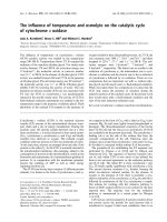
fundamentals of friction and wear on the nanoscale, 2007, p.713
Bạn đang xem bản rút gọn của tài liệu. Xem và tải ngay bản đầy đủ của tài liệu tại đây (20.51 MB, 713 trang )
NanoScience and Technology
NanoScience and Technology
Series Editors:
P. Avouris B. Bhushan D. Bimberg K. von Klitzing H. Sakaki R. Wiesendanger
The series NanoScience and Technology is focused on the fascinating nano-world,
mesoscopic physics, analysis with atomic resolution, nano andquantum-effect devices,
nanomechanics and atomic-scale processes. All the basic aspects and technology-
oriented developments in this emerging discipline are covered by comprehensive and
timely books. The series constitutes a survey of the relevant special topics, which are
presented by leading experts in the field. These books will appeal to researchers, engi-
neers, and advanced students.
Applied Scanning Probe Methods I
Editors: B. Bhushan, H. Fuchs, and
S. Hosaka
Nanostructures
Theory and Modeling
By C. Delerue and M. Lannoo
Nanoscale Characterisation
of Ferroelectric Ma terials
Scanning Probe Microscopy Approach
Editors: M. Alexe and A. Gruverman
Magnetic Microscopy
of Nanostructures
Editors: H. Hopster and H.P. Oepen
Silicon Quantum Integrated Circuits
Silicon-Germanium Heterostructure
Devices: Basics and Realisations
ByE.Kasper,D.J.Paul
The Physics of Nanotubes
Fundamentals of Theory, Optics
and Transport Devices
Editors: S.V. Rotkin and S. Subramo ney
Single Molecule Chemistry
and Physics
An Introduction
By C. Wang, C. Bai
Atomic Force Microscopy, Scanning
Nearfield Optical Microscopy
and Nanoscratching
Application to Rough
and Natural Surfaces
By G. Ka up p
Applied Scanning Probe Methods II
Scanning Probe Microscopy
Technique s
Editors: B. Bhushan, H. Fuchs
Applied Scanning Probe Methods III
Characterization
Editors: B. Bhushan, H. Fuchs
Applied Scanning Probe Methods IV
Industrial Application
Editors: B. Bhushan, H. Fuchs
Nanocatalysis
Editors: U. Heiz, U. Landman
Roadmap
of Scanning Probe Microscopy
Editors: S. Morita
Nanostructures –
Fabrication and Analysis
Editor: H. Nejo
Applied Scanning Probe Methods V
Scanning Probe Microscopy Techniques
Editors: B. Bhushan, H. Fuchs,
S. Kawata
Applied Scanning Probe Methods VI
Characterization
Editors: B. Bhushan, S. Kawata
Applied Scanning Probe Methods VII
Biomimetics and Industrial Applications
Editors: B. Bhushan, H. Fuchs
Enrico Gnec co
Ernst Meyer
Fundamentals
of Friction
and Wear
Wi th 300 Figures and 13 Tables
123
Editors:
Dr. Enrico Gnecco
Universität Basel
Institut für Physik
Klingelbergstr. 82, 4056 Basel, Switzerland
e-mail: enrico.gnecco@uni bas.ch
Professor Dr. Ernst M eyer
Universität Basel
Institut für Physik
Klingelbergstr. 82, 4056 Basel, Switzerland
e-mail:
Series Editors:
Professor Dr. Phaedon Avouris
IBM Research Division
Nano meter Scale Science & Technology
Thomas J. Watson Research Center, P.O . Box 218
Yorktown Heights, NY 10598, USA
Professor Bharat Bhushan
Nanotribology Laboratory for Inform a tion
Storage and MEMS/NEMS (NLIM)
W 390 Scott Laboratory, 201 W. 19th Avenue
The Ohio State University, Columbus
Ohio 43210-1142, USA
Professor Dr. Dieter Bimberg
TU Berlin, Fakutät Mathematik,
Naturwissenschaften,
Institut für Festkörperphysik
Hardenbergstr. 36, 10623 Berlin, Germany
Professor Dr., Dres. h. c.
Klaus von Klitzing
Max-Planck-Institut für Festkörperforschung
Heisenbergstrasse 1, 70569 Stuttgart, Germany
Professor Hiroyuki Sakaki
University of T okyo
Institute of Industrial Science,
4-6-1 Komaba, Meguro-ku, Tokyo 153-8505, Japan
Pr ofessor Dr. Roland Wiesendanger
Institut für Angewandte Physik
Universität Hamburg
Jungiusstrasse 11, 20355 Hamburg, Germany
ISBN-10 3-540-36806-X Springer Berlin Heidelberg New York
ISBN-13 978-3-540-36806-9 Springer Berlin Heidelberg New York
Library o f Congress Co ntrol Num ber: 2006934355
This work is subject to copyright. All rights are reserved, whether the whole or part of the material is concerned,
specifically the rights of translation, re printing, reuse of illustrations, recitation, broadcasting, reproduction on
microfilm or in any other way, and storage in data banks. Duplication of this publication or parts thereof is permitted
only under the provisions of the German Copyright Law of September 9, 1965, in its current version, and permission
for use must always be obtained from Springer. Violations ar e liable for prosecution under the German Copyright
Law.
Springer is a part of Springer Science+Business Media
springer.com
© Springer-Verlag Berlin Heidelberg 2007
The use of general descriptive names, registered names, trademarks, etc. in this publication does not imply, even in
the absence of a specific statement, that such names are exempt from the relevant protective laws and regulations and
therefore free for general use.
Product liability: The publishers cannot guarantee the accuracy of any information about dosage and application
contained in this book. In every individual case the user must check such information by consulting the relevant
literature.
Typesetting: LE-T
E
XJelonek,Schmidt&VöcklerGbR,Leipzig
Production: LE-T
E
XJelonek,Schmidt&VöcklerGbR,Leipzig
Cover: WMXDesign, Heidelberg
SPIN 11555315 57/3100/YL - 543210 Printedonacid-freepaper
Preface
Friction is an old subject of research and is certainly one of the most im-
portant ones from a practical point of view. The da Vinci-Amonton laws
are common knowledge (1. Friction is independent of apparent contact area,
2. Friction is proportional to the normal load 3. Friction is independent of
velocity). Experiments with small contacts have shown that these empiri-
cal laws of friction do not always hold. Reasons may be related to the large
surface-to-volume ratio and the greater importance of adhesion, surface struc-
ture and surface chemistry. Therefore, there is some need to get a better
understanding of the phenomenon of friction, to learn how to quantify and
eventually control it. In the first half of last century the school of Bowden
and Tabor have performed systematic, macroscopic experiments and have
related macroscopic friction to small contacting asperities. In the 1990’s ex-
periments performed with atomic force microscopy, surface force apparatus
and quartz microbalance, revealed interesting new physics on the nanometer
scale (atomic-scale stick-slip, confinement of liquid films, determination of
electronic and phononic contributions to dissipation). During the same time,
theoretical analysis of nanometer-sized contacts has been performed and gave
insight into the processes in the buried interface. Strong activities were pur-
sued in the US at Universities and corporate research laboratories. Similar
activities were pursued in Japan, where the main focus was on the under-
standing the tribology of hard disc drives and applications in automobile
industries. Europe has a long tradition in mechanical engineering sciences.
Activities at the University level were mainly driven by recent developments
in nanosciences (scanning probe microscopy, computer modelling). In 2001,
the European Science Foundation Programme “Nanotribology” (NATRIBO)
was started. The aim of this programme is to bring together experimentalists
and theoreticians to improve the understanding of nanometer-sized contacts.
The aim of this book is to give an overview of the status of resarch in this
field. Members of the NATRIBO-network and a selection of excellent inter-
national experts have contributed to this book. They made a strong effort to
give a deep insight into the complex phenomena of nanotribology.
The book is divided in seven sections. In the first section the instrumen-
tal setups most commonly used in nanotribology are introduced. The first
chapter presents the atomic force microscope (AFM), with a special empha-
sis on the force sensors and the ways to control the contact between tip and
VI Preface
surface. The interrelations between friction, load, material properties, tem-
perature, and the lateral forces detected in dynamic measurements, are also
discussed. The second chapter introduces the surface force apparatus (SFA),
as an independent tool and in combination with other techniques. A case
study of weakly adhesive surface under shear is discussed. The quartz crys-
tal microbalance is treated in Chapt. 3. After the acoustics of the crystal,
the driving circuits and the quality of the surface electrodes, the authors
present results obtained in ultra-high vacuum (UHV). Chapter 4 describes
the effects of normal and shear ultrasonic vibrations in AFM, focusing in par-
ticular on friction reduction and adhesion hysteresis. Finally, Chapt. 5 shows
how scanning probe microscopes can be combined with transmission electron
microscopes to image both tip and sample surface. Contact formation and
breaking, adhesion effects, electric conductivity and material transport are
consequently discussed.
Section 2 gives a detailed overview on friction phenomena occurring on
the atomic scale. Chapter 6 introduces the Tomlinson model and fundamen-
tal phenomena observed by AFM (atomic stick-slip, velocity dependence of
friction, superlubricity, and nanowear processes). The next chapter shows
how the rate theory has been applied to obtain general force-velocity rela-
tions. Analytical approximations are compared with precise numerical results.
Chapter 8 introduces the important problem of friction control. Mechanical
and chemical methods to achieve this goal are discussed from both theo-
retical and experimental points of view. Superlubricity is the main topic of
Chapts. 9–11. Chapter 9 shows how surface incommensurability and thermal
effects can lead to a strong reduction of friction, which was recently observed
experimentally. Lubrication by graphite, diamond-like carbon, fullerenes and
carbon nanotube is discussed within this frame. Chapter 10 presents theoret-
ical studies of superlubricity. Symmetry considerations, role of instabilities,
temperature effects, damping in the superlubric regime and long-range elas-
tic deformations are discussed, as well as generic models and applications to
layered materials, metal-metal contacts and hydrogen-terminated surfaces. In
particular, the presence of hydrogen is proved to be the key factor leading to
superlubricity between diamond surfacesas shown in the detailed theoretical
study presented in the last chapter of the section.
The third section of the book introduces contact mechanics on the
nanoscale. After a brief theoretical introduction, Chapter 12 describes the
main experimental methods to investigate elasticity on the nanoscale and re-
cent findings related to inorganic nano-objects and biological samples. Chap-
ter 13 addresses the special case on metallic nanocontacts, whose mechanical
properties cannot be separated from electron transport mechanisms. Fabri-
cation, elasticy, fracture, and shape of metal contacts are discussed, as well
as chains of gold atoms and metallic adhesion in atomic-sized tunneling junc-
tions. Quasi-crystals are the main subject of Chapt. 14. This leads the authors
to describe the surface roughness in relation to friction and adhesion. A par-
Preface VII
ticular emphasis is given on the roughness power spectrum, which is derived
from the surface height using optical and scanning probe microscopes. Chap-
ter 15 focuses on the roughness of self-affine fractal surfaces. The contact
morphology and the pressure distribution are estimated at different scales,
with and without adhesion, using molecular dynamics, and they are com-
pared with analytical contact models based on continuum mechanics. The
role of the elastic moduli of the underlying bulk is also treated here. Finally,
the last chapter of this section describes how nanoroughness is affected by
depth-sensing indentation. A special attention is given to elastomer probes
used in AFM investigations.
Section 4 describes dissipative mechanisms at finite separation under dif-
ferent points of view. Chapter 17 deals with the case of amplitude modulation
AFM, used to characterize surfaces in air or in liquids. In such case the en-
ergy dissipation accompanying the imaging process is given by the phase
shift signal acquired while scanning. The next chapter considers dissipation
in non-contact AFM. After a review of the experimental data at our dis-
posal, possible mechanisms of atomic-scale damping are discussed, as well
as detailed models developed to understand the effect of these mechanisms
on the imaging process. The theory of non-contact friction is the subject of
Chapt. 19. The fluctuating electromagnetic field which surrounds any solid
surface, and is responsible for radiative heat transfer and van der Waals inter-
action and friction, is examined under semiclassical and quantum theories. At
short separations, Van der Waals friction is greatly enhanced. Furthermore,
static charges on the surface are responsible of electrostatic friction around
a moving body, and possible applications to scanning probe spectroscopy are
discussed. This topic is extended in Chapt. 20, where the authors show how
the force sensitivity of free cantilevers is limited by thermal fluctuations and
material properties and how these problems are reduced by UHV annealing
or cooling to cryogenic temperatures.
Wear and fracture are treated in the fifth section of the book. Chapter 21
covers the mechanisms of surface damage down to micro- and nano-scales.
Both basic theories and experiments are considered, and a discussion on hard-
ness at different scales is also provided. Chapter 22 examines the relation be-
tween stress and chemical reactivity. Examples of single asperity tribochem-
ical wear include dissolution along monolayer steps in calicum carbonates
and phosphates, wear of the probing tip on reactive surfaces and tip induced
wear of silicate substrates. Chapter 23 gives an overview of stiction, friction
and wear phenomena affecting micro- and nano-electromechanical systems.
The tribological characterization of these devices is discussed together with
various solutions introduced to improve their reliability. The last chapter of
the section addresses nanotribological problems in automotive engineering.
Wear rates of few nanometers per hours are mandatory in internal combus-
tion engines, which requires exceptional finishing of the sliding surfaces in
the engine.
VIII Preface
Another growing field of nanotribology is the manipulation of nanoparti-
cles, which is treated in Sect. 6. Chapter 25 shows how the tip of a scanning
probe microscope operated in dynamic mode can be alternatively used to im-
age and move particles in a controlled way. With a proper calibration of the
excitation amplitude the energy dissipation and the frictional forces involved
in the manipulation process can also be estimated. Chapter 26 considers
a system of great interest in nanoscience, i. e. carbon nanotubes (CNTs).
In such case AFM can be used to test mechanical properties in dynamic
and quasi-static ways. Nanotube bundles, catalytically grown CNTs, and di-
ameter dependence of bending moduli are addressed as special cases. The
next chapter focuses on the manipulation of fullerene molecules on a silicon
surface. After summarizing the experimental results obtained with scanning
tunneling microscopes, the authors present a model which successfully inter-
pretes the mechanisms underlying adsorption, diffusion and manipulation of
the molecules.
The last section of the book deals with applications of nanotribology to
organic materials. Chapter 28 gives a detailed overview of friction on self-
assembled monolayers (SAMs). Homogenoues films are first addressed, and
the influence of chain length, terminal groups, packing states as well as en-
vironmental conditions on friction are discussed. The role of nanoscale het-
erogeneities on the nanoscale is considered in the second part of the chapter.
The next two chapters deal with polymers. In particular, Chapt. 29 consid-
ers the influence of hydrophobicity on the frictional forces experienced on
two different materials, whereas Chapt. 30 treats the molecular origins of
elastomeric friction. Both interfacial adhesion and internal friction are ther-
mally activated processes, and the competition between them gives a correct
interpretation of the experimental results. Finally, the last chapter of the
book describes the importance of friction and adhesion mechanisms in cell
dynamics, with particular emphasis on the adhesive forces experienced on
the substrates where the cells can spread and proliferate. This is of great
importance in the emerging field of tissue engineering.
In conclusion, we would like to thank all the authors for the time and the
energies that they have spent on this project, as well as all the participants to
the Nanotribo workshops for the interesting scientific discussions that they
have stimulated. A special thanks goes also to Claus Ascheron, Angela Lahee
and Steffi Hohensee from Springer-Verlag, who made possible the publication
of this book. Financial support from the European Science Foundation, the
Pico-Inside project, the Swiss National Center of Competence in Research
Nanoscale Science and the Swiss National Science Foundation is gratefully
acknowledged.
University of Basel Enrico Gnecco and Ernst Meyer
Contents
Experimental Techniques
1 Friction Force Microscopy
R. Bennewitz 1
2 Surface Forces Apparatus in Nanotribology
C.Drummond,P.Richetti 15
3 The quartz crystal microbalance
as a nanotribology technique
L. Bruschi, G. Mistura 35
4 Nanoscale Friction and Ultrasonics
M.T. Cuberes 49
5 Probing of Nanocontacts Inside
a Transmission Electron Microscope
D.Erts,A.L˜ohmus, J.D. Holmes, H. Olin 73
Friction on the Atomic Scale, Superlubricity
6 Stick-Slip Motion on the Atomic Scale
T. Gyalog, E. Gnecco, E. Meyer 101
7 Velocity dependence of atomic friction:
Rate theory and beyond
M. Evstigneev, P. Reimann 117
8 The Basic of Nanoscale Friction and Ways to Control it
J. Klafter, M. Urbakh 143
9 Experimental Observations of Superlubricity
and Thermolubricity
M. Dienwiebel, J.W.M. Frenken 159
XContents
10 Theoretical Aspects of Superlubricity
M.H. M¨user 177
11 First-Principles Atomic-Scale Study of Superlow Friction
S.Ciraci,S.Dag,O.Gulseren,T.Yildirim 201
Contact Mechanics on the Nanoscale
12 NanoMechanics: Elasticity in Nano-Objects
L.Merchan,R.Szoszkiewicz,E.Riedo 219
13 Mechanical Properties
of Metallic Nanojunctions
G. Rubio-Bollinger, J.J. Riquelme, N. Agra¨ıt, S. Vieira 255
14 Contact Mechanics, Friction and Adhesion
with Application to Quasicrystals
B.N.J. Persson, G. Carbone, V.N. Samoilov, I.M. Sivebaek,
U. Tartaglino, A.I. Volokitin, C. Yang 269
15 A Multiscale Molecular Dynamics Approach
to Contact Mechanics and Friction: From Continuum
Mechanics to Molecular Dynamics
U. Tartaglino, C. Yang, B.N.J. Persson 307
16 The Role of Nanoroughness in Contact Mechanics
R. Buzio, U. Valbusa 345
Dissipation Mechanisms at Finite Separations
17 Energy Dissipation and Nanoscale Imaging
in Tapping Mode AFM
R. Garc´ıa, N.F. Mart´ınez, C.J. G´omez, A. Garc´ıa-Mart´ın 361
18 Mechanisms of atomic scale dissipation at close approach
in dynamic atomic force microscopy
T. Trevethan, L. Kantorovich 373
19 Theory of Noncontact Friction
A.I. Volokitin, B.N.J. Persson 393
20 Dissipation at large Separations
S.Rast,U.Gysin,E.Meyer,D.W.Lee 439
Contents XI
Wear and Fracture on the Nanoscale
21 Surface-Damage Mechanisms: from Nano-
and Microcontacts to Wear of Materials
R. Cola¸co 453
22 Single Asperity Nanometer-Scale Studies
of Tribochemistry
J.T. Dickinson 481
23 Nanotribology of MEMS/NEMS
S. Achanta, J P. Celis 521
24 Nanotribology in Automotive Industry
M. Dienwiebel, M. Scherge 549
Manipulation of Nanoparticles
25 Nanotribological Studies by Nanoparticle Manipulation
U.D. Schwarz, C. Ritter, M. Heyde 561
26 Mechanical Properties of Carbon Nanotubes
A.J. Kulik, A. Kis, B. Lukic, K. Lee, L. Forr´o 583
27 Theory of Adsorption and Manipulation
of C
60
on the Si(001) Surface
N. Martsinovich, C. Hobbs, L. Kantorovich, P. Beton 601
Organic Materials
28 Nanoscale Friction of Self-assembled Monolayers
K. Mougin, H. Haidara 619
29 Sliding Friction of Polymers:
The Complex Role of Interface
S. Bistac, M. Schmitt, A. Ghorbal 647
30 Molecular Origins of Elastomeric Friction
S. Sills, K. Vorvolakos, K. Chaudhury, R.M. Overney 659
31 Nanotribological Perspectives in Tissue Engineering
M. D’Acunto, G. Ciardelli, A. Rechichi, F. M. Montevecchi, P. Giusti . 677
Index 709
1 Friction Force Microscopy
Roland Bennewitz
Department of Physic, McGill University, Montreal, Quebec, Canada
1.1 Introduction
Friction Force Microscopy (FFM) is a sub-field of scanning force microscopy
addressing the measurement of lateral forces in small sliding contacts. In line
with all scanning probe methods, the basic idea is to exploit the local in-
teractions with a very sharp probe for obtaining microscopic information on
surfaces in lateral resolution. In FFM, the apex of a sharp tip is brought into
contact with a sample surface, and the lateral forces are recorded while tip
and sample slide relative to each other. There are several areas of motivation
to study FFM. First, the understanding of friction between sliding surfaces in
general is a very complex problem due to multiple points of contact between
surfaces and the importance of lubricants and third bodies in the sliding pro-
cess. By reducing one surface to a single asperity, preparing a well-defined
structure of the sample surface, and controlling the normal load on the con-
tact the complexity of friction studies is greatly reduced and basic insights
into the relevant processes can be obtained. Furthermore, with the decrease
of the size of mechanical devices (MEMS) the friction and adhesion of small
contacts becomes a technological issue. Finally, the lateral resolution allows
to reveal tribological contrasts caused by material differences on heterogenous
surfaces.
The experimental field of FFM has been pioneered by Mate, McClelland,
Erlandsson, and Chiang [1]. The group built a scanning force microscope
where the lateral deflection of a tungsten wire could be measured through
optical interferometry. When the etched tip of the tungsten wire slid over
a graphite surface, lateral forces exhibited a modulation with the atomic
periodicity of the graphite lattice. Furthermore, a essentially linear load de-
pendence of the lateral force could be established.
In this chapter we will describe aspects of instrumentation and measure-
ment procedures. In the course of this description, a series of critical issues
in FFM will be discussed which are summarized in Fig. 1.1.
2 R. Bennewitz
Crosstalk between friction and
topography signals.
Wear during friction
measurement.
Calibration of the beam
deflection scheme.
Spring constant of normal and
torsional bending.
Displacement of tip position
parallel to the cantilever
direction with increasing load.
Sample surface quality.
Actual radius and constitution
of the tip.
Stiffness of the tip apex.
Environmental conditions, in
particular humidity.
Thermal fluctuations of
the cantilever.
Fig. 1.1. Critical issues in experimental friction force microscopy which are dis-
cussed in this chapter
1.2 Instrumentation
1.2.1 Force sensors
The force sensor in the original presentation of FFM by Mate et al. was
a tungsten wire [1]. Its deflection was detected by an interferometric scheme
where the wire constituted one mirror of the interferometer. A similar concept
was later implemented by Hirano et al., who optically detected the deflection
of the tungsten wire in a Scanning Tunneling Microscope when scanning
the tip in close proximity to the surface [2]. Mate and Hirano report lateral
spring constants from 1.5 to 2500 N/m, depending on the wire thickness and
length. Etching the wire to form a tip at its end, mounting the wire, aligning
of the light beam, and determination of the spring constant comprise some
experimental difficulties. These difficulties are greatly reduced by the use of
dedicated micro-fabricated force sensors. A very sophisticated instrumental
approach to the solution of those problems has been realized by Dienwiebel et
al. [3]. The group has attached a stiff tungsten wire to a micro-fabricated force
sensor made of silicon. The central part of the sensor is a pyramid holding the
tip. The position of the pyramid is detected in all three dimensions by means
of four optical interferometers directed towards the faces of the pyramid. It
is suspended in four symmetric high-aspect ratio legs which serve as springs
with isotropic spring constant in both lateral directions and a higher spring
constant in normal direction. The symmetric design of the instrument allows
1 Friction Force Microscopy 3
Fig. 1.2. Four design options for Friction Force Microscopy. a Concept of the
original instrument used by Mate et al. for their pioneering experiments [1]. The
deflection of a tungsten wire is detected by optical interferometry. The bent end of
the wire is etched into a sharp tip. b Beam-deflection scheme as devised by Marti
et al. [5]. Normal force F
N
and friction force F
F
cause bending and twisting of the
cantilever. The deflection of a reflected light beam is recorded by comparing currents
from four sections of a photodiode. c Cantilever device for the measurement of
lateral forces with piezoresistive detection [8]. Lateral forces acting on the tip cause
a difference in stress across the piezoresistors. d Micro-fabricated force detector for
isotropic measurements of friction forces. The block in the center holds a tungsten
tip, pointing upwards in this figure. The position of the block in all three dimensions
is recorded by four interferometric distance sensors which are indicated by the four
light beams below the devices [9]
for determination of normal and lateral forces acting on the tip with minimal
cross talk. An overview over different experimental realizations of FFM is
given in Fig. 1.2.
The most widely used form of micro-fabricated force sensors for FFM
is the micro-fabricated cantilever with integrated tip. The cantilever can be
either a rectangular beam or a triangular design based on two beams. The
lateral force acting on the tip is detected as torsional deflection of the can-
tilever. This scheme has been implemented in 1990 by Meyer et al. [4] and
4 R. Bennewitz
Marti et al. [5]. It is interesting to note that the triangular design is more
susceptible to deflection by lateral forces than the rectangular beam, contrary
to common belief and intuition [6]. However, triangular cantilevers are less
prone to the highly unwanted in-plane bending [7].
The deflection of cantilever-type force sensors is usually detected by means
of a light beam reflected from the back side of the cantilever at the position
of the tip. The reflected light beam is directed towards a position-sensitive
photodiode which detects normal and torsional bending of the cantilever as
a shift in the position of the light beam in orthogonal directions. Realis-
tically, there is always some cross-talk between the signals for normal and
torsional bending. It can be detected by exciting the cantilever to oscillate at
the fundamental normal and torsional resonance and measure the oscillation
amplitude in the orthogonal channels. The cross-talk can be minimized by
rotation of the position-sensitive photodiode or accounted for in the detection
electronics or software. Cross-talk can transfer topographic features into the
lateral force signal and create topographic artifacts from friction contrast,
the latter even amplified by the feedback circuit acting on the sample height.
Calibration of the beam-deflection scheme is not a simple task, however
very important in order to compare FFM results from different sources. Many
publications in the past have reported on relative changes in frictional prop-
erties, without providing any calibration at all. While such relative changes
certainly represent important physical findings, it is nevertheless of utmost
importance to provide all experimental information available, often allow-
ing for a rough quantitative estimate of the lateral forces. Lateral forces in
FFM can easily range from piconewton to micronewton, spanning a range of
very different situations in contact mechanics, and knowing at least the order
of magnitude of forces helps to sort the results qualitatively into different
regimes.
The calibration comprises two steps. First, the spring constant has to
be determined for the force sensor. Note that the beam-deflection scheme
actually determines the angular deflection of the cantilever. Nevertheless it
has become custom to quantify the force constant in N/m, where the length
scale refers to the lateral displacement of the tip apex relative to the unbent
cantilever. Second, a relation between the deflection of the cantilever and the
voltage readout of the instrument has to be established.
For the determination of the spring constant, several methods have been
suggested. The easiest is to calculate it from the dimensions of the can-
tilever. While width and thickness are easily determined by optical or electron
microscopy, thickness is better deduced from the cantilevers resonance fre-
quency. Alternatively, the spring constant can be determined from changes
in the resonances caused by the addition of masses to the free end of the
cantilever. Also, the analysis of a cantilever’s resonance structure in air can
provide the required quantities. The latter two methods have recently be
described and compared by Green et al. [10]. The relation between tip dis-
placement and voltage readout can be established by trapping the tip in
1 Friction Force Microscopy 5
a surface structure and displacing the sample laterally by small distances.
For a rough estimate one can also assume that the sensitivity of the position-
sensitive photodiode is the same for normal and torsional deflection. Taking
into account the geometry of the beam-deflection scheme, the torsional de-
flection sensitivity can be deduced from the normal deflection sensitivity (See
Ref. [11] and page 352 of Ref. [12]).
A method which provides a direct calibration of the lateral force with
respect to the readout voltage is the comparison with a calibrated spring
standard. Recent implementations of this approach suggest as calibrated
standards optical fibers [13] or micro-fabricated spring-suspended stages with
spring constants that can be traced to international standards [14]. A particu-
larly elegant method to calibrate FFM experiments is the analysis of friction
loops, i. e. lateral force curves from forward and backward scans, recorded
across surfaces with well-defined wedges [11, 15].
The torsional deflection of a cantilever can in principle be detected also by
optical interferometry, provided that the beam diameter is smaller than the
cantilever and the point of reflection is shifted off the torsional axis [16]. How-
ever, FFM results including normal and lateral force measurements require
the differential reading of multiple interferometers [3, 17].
An alternative to the detection of the cantilever bending via the beam-
deflection scheme is the implementation of piezoresistive strain sensors into
the cantilever. In order to measure both lateral and normal forces acting
on the tip in FFM, two such strain sensors need to be realized on one sen-
sor. Chui et al. have created a piezoresistive sensor which decouples the two
degrees of freedom by attaching a normal triangular cantilever to a series
of vertical ribs sensing lateral forces [18]. Gotszalk et al. have constructed
a U-shaped cantilever with one piezoresistive sensor in each arm, allowing
for the the detection of lateral forces at the tip [19]. While the publications
presenting these novel instrumental approaches contain experimental proofs
of concept, no further use of piezoresistive sensors in FFM experiments has
been reported. This is certainly due to a lack of commercial availability. Fur-
thermore, the signal-to-noise ratio in static force measurements using piezore-
sistive cantilevers seems not to reach that of optical detection schemes.
1.2.2 Control over the contact
The exact knowledge of the atomic configuration in the contact between tip
apex and surface is prerequisite for a complete understanding of the results
in Friction Force Microscopy. It is the most severe drawback in FFM that this
knowledge is not available in most cases. While sample surfaces can often be
prepared with atomic precision and cleanliness, the atomic constitution of
the tip apex is usually less controlled. Furthermore, in the course of sliding
atoms may be transferred from the tip to the surface or vice versa. Such
transfer processes occur even for very gentle contact formation, as shown in
experiments combining Scanning Probe Microscopy with a mass spectrome-
6 R. Bennewitz
try analysis of the tip apex [20–22]. The transfer of atoms may quite often not
only quantitatively but also qualitatively change the lateral forces encoun-
tered. In particular, the occurrence of atomic stick-slip motion can depend on
the establishment of a certain degree of structural commensurability between
tip and surface in the course of scanning [23, 24]. For atomic stick-slip mea-
surements on graphite surfaces, the role of small graphite flakes attached to
the tip has long been discussed and recently confirmed experimentally [1,25].
The best control over the atomic structure of the tip apex has been
achieved for metal tips in vacuum environments. By applying the established
procedures of Field Ion Microscopy (FIM), the tip structure can not only be
imaged but also conditioned on the atomic scale. Cross et al. have charac-
terized the adhesion between a tungsten tip and a gold surface and proved
the conservation of the atomic tip structure by means of FIM [26]. Even
with instruments of lower resolution, FIM can at least be used for cleaning
procedures and for a determination of the crystalline orientation of the apex
cluster [2].
The integrated tips at the end of micro-fabricated silicon cantilevers have
a well-defined crystalline orientation, usually pointing with the (100) direc-
tion along the tip. However, the tip surface and with it the whole tip apex
are at least oxidized and possibly contaminated through packaging, transport,
and handling. Furthermore, many tips are sharpened in a oxidation process
which introduces large stresses at the apex. While etching in hydrofluoric acid
can remove the oxide and for some time passivate silicon surface bonds by
hydrogen, a stable formation and reproducible characterization comparable
with FIM of metal tips has not yet been reported. Tips integrated into sili-
con nitride cantilevers are amorphous due to the chemical vapor deposition
process and may exhibit an even more complex structure and chemistry at
the tip apex.
One way of overcoming the uncertainty of the tip constitution is to use
methods of surface chemistry to functionalize the tip [27]. Specific interac-
tions between molecules attached to the tip and molecules on the surface can
be sensed by means of FFM [28]. At the same time, very strong adhesion
has been reduced by covering the tip with a passivating layer to allow for
lateral force imaging for example on silicon [29]. Numerous studies using this
method have been published, mainly concentrating on organic monolayers
on tip and surface. A recent review of the field has been given by Leggett
et al. [30]. Schwarz et al. have prepared well-defined tips for FFM by de-
position of carbon from residual gas molecules in a Transmission Electron
Microscope, keeping control of the tip radius for a quantitative analysis of
a contact mechanics study [31]. Force measurements explicitly aiming at in-
teractions between colloidal particles and a surface have been performed by
gluing micrometer-sized spheres of the desired size to the cantilever [32, 33].
As a final note, one should always be aware of the possible occurrence of
major tip wear which has been observed to happen in a concerted action of
mechanical and chemical polishing [34].
1 Friction Force Microscopy 7
1.3 Measurement procedures
The standard measurement in FFM is the so-called friction loop: The lateral
force acting on the tip is recorded for a certain distance of scanning in the di-
rection perpendicular to the long cantilever axis and for the reverse direction.
The area in the loop represents the dissipated energy, and the area divided
by twice the distance is the mean lateral force. It is always very instructive to
record the topography signal of forward and backward scan at the same time,
as differences will reveal cross-talk between normal and torsional bending of
the cantilever.
Whenever lateral forces are measured as a function of some experimen-
tal parameter, the influence of that parameter on adhesion should be studied
simultaneously. In order to interpret the experimental results in terms of con-
tact sizes versus dissipation channels the knowledge of adhesion is essential.
An excellent example is the jump in lateral forces observed on a C
60
crystal
when cooling to the orientational order-disorder phase transition, which was
fully explained by a change in adhesion [35]. For experiments carried out in
ambient environment, the dominant contribution to adhesion are usually cap-
illary forces which dependent greatly on the humidity and on the hydropho-
bicity of the surface [36]. The humidity dependence of FFM results itself
can depend again on the temperature [37, 38]. Consequently, an enclosure of
FFM experiments for humidity control greatly enhances the reproducibility
of results.
1.3.1 Friction as a function of load
One of the central experiments in tribology is the quantification of friction,
i. e. the change of lateral force with increasing normal load on the sliding
contact. One of the questions to be addressed is whether the relation be-
tween lateral and normal force is linear for FFM experiments, i. e. whether
Amontons’ law extends to the nanometer scale [39]. The number of FFM
studies reporting lateral force as a function of load is very large, and the
overall physical picture is multifaceted, to express it in a positive way. A col-
lection of results is shown in Fig. 1.3. From a procedural point of view it is
extremely important to measure the lateral forces for the full range of small
normal forces until the tip jumps out of contact, usually at a negative normal
force. In this way the adhesion in the system can be categorized, and possible
nonlinear characteristics at minimal loads are not overlooked. A useful way of
analyzing load dependence data from FFM experiments is the representation
in lateral force histograms, where for example friction on terraces and friction
at steps could automatically be distinguished [40].
When the normal load on the tip is varied the position of the contact may
be displaced along the long axis of the cantilever. This effect is caused by the
tilt of the cantilever with respect to the surface. On heterogeneous surfaces
such displacement may distort the friction measurement and, therefore, has
8 R. Bennewitz
Fig. 1.3. Examples for the diversity of friction vs. load curves measured by FFM.
a Amorphous carbon measured in an argon atmosphere [31]. The sub-linear charac-
teristic resembles the results of contact mechanics models. b Phenyltrichlorosilane
monolayer studied in ethanol [41]. A linear dependence is found until the mono-
layer collapses under the tip pressure. c Atomic friction on NaCl(100) recorded in
ultra-high vacuum [42]. A regime of vanishing friction is found for low loads. d Fric-
tion measurement on a hydrogen-terminated diamond surface with nanometer-scale
roughness [43]. The closed circles represent the erratic load dependence of FFM
results when the lateral displacement of the tip for increasing load is not compen-
sated. The open circles show the expected sub-linear characteristic after activating
the compensation
to be compensated [43]. Another effect that can seriously disturb friction
experimentsistheonsetofwearandtheconcomitantincreaseoflateral
forces. Wear thresholds in FFM can be as low as a few nanonewton normal
load, and wear at a constant low load may suddenly start after repeatedly
scanning the same area [44].
1.3.2 Friction as a function of material
On inhomogeneous surfaces Friction Force Microscopy can image contrasts
between different materials with high lateral resolution. Such contrast has
been found to arise from a difference in chemical interactions between differ-
ent molecular patches at the surface and the tip [45]. As mentioned above,
1 Friction Force Microscopy 9
it is crucial to complement lateral friction contrast with local measurements
of adhesion in order to elucidate whether adhesion and contact size or dif-
ferent channels of dissipation are dominating the contrast. Care has to be
taken regarding topographical artifacts, as different materials on heteroge-
neous surfaces are often found at different topographic heights. Interestingly,
friction contrast is also found between domains of identical molecular layers
with anisotropic lateral orientation [46–48]. Friction anisotropy on a given
surface has to be clearly distinguished from friction anisotropy for different
azimuthal orientations between the tip and the surface. In order to measure
the latter, the sample has to be rotated with respect to the tip [25].
1.3.3 Friction effects in normal force measurements
When the sample is approached towards the tip, the normal force can be
determined as a function of distance by measuring the normal bending of
the cantilever. In all beam-deflection type FFM the cantilever is tilted with
respect to the sample surface to make sure that the tip is the foremost pro-
trusion of the force sensor. Once the tip is in contact, the tilt causes a lateral
displacement of the tip position upon further approach. The friction forces
arising from this lateral displacement influence the normal force measure-
ment [33]. A detailed analysis of the process proves that one can actually
perform a calibrated friction experiment through normal force vs. distance
curves, in particular when using extended tips like colloid probes [49]. Even
when probing the surface in a dynamic intermittent contact mode these fric-
tional contributions can be detected as a phase shift between excitation and
cantilever oscillation [47].
1.3.4 Fluctuations in Friction Force Microscopy
Friction Force Microscopy is naturally subject to thermal fluctuations. Such
thermal fluctuations can influence the frictional behavior of sliding contacts,
as evident in the logarithmic dependence of friction on velocity at low scan-
ning velocities [50, 51] which has been linked to thermal fluctuations via its
temperature dependence [52]. Cantilever-type force sensors have a distinct
resonance structure which dominates the thermal noise spectrum. Typically,
oscillations at resonances with frequencies of several kHz are averaged out
in FFM experiments. However, these resonances influence the experimental
result and it is therefore very instructive to study the lateral force signal
with high bandwidth [53, 54]. Furthermore, the statistical distribution of lat-
eral forces in FFM experiments can be analyzed to reveal the role of thermal
fluctuations [55]. The limited scanning velocity of FFM normally separates
the frequency regimes of fast fluctuations and of slower occurrence of topo-
graphic or even atomic features. The velocity limitations of FFM have been
addressed by new designs combining the force sensor of an FFM with a ded-
icated sample stage [56,57].
10 R. Bennewitz
1.3.5 Friction as a function of temperature
The study of friction as a temperature is an obvious field of great interest.
However, the number of groups including a temperature dependence into
FFM studies is increasing only recently [35, 37, 52, 58, 59]. Thermal drift is
a severe problem in the design of Friction Force Microscopes working at
variable temperature, since the optical lever of the beam-deflection scheme
needs to have a certain length for sensitivity. Variable-temperature instru-
ments with thermal-expansion compensated design comparable to dedicated
Scanning Tunneling Microscopes [60] have not been reported so far. One in-
teresting approach to circumvent drift problems is the local heating of the
very tip [61].
1.3.6 Dynamic lateral force measurements
Dynamic friction force microscopy
When the sample is periodically displaced in lateral direction, the lateral
force acting on the tip and detected by the cantilever will be modulated with
the same periodicity. An early application of such a lateral modulation by
Maivald et al. was the enhancement of contrast at step edges [62]. Dynamic
Friction Force Microscopy detects the periodic lateral force signal by means of
a lock-in amplifier. This idea was implemented by G¨oddenhenrich et al., who
applied the periodic sample displacement along the long axis of the cantilever
and detected the lateral force as periodic buckling of the cantilever [63]. Si-
multaneously, their fiber-interferometric setup could statically measure the
deflection of the cantilever caused by normal forces. The same technique was
implemented by Colchero et al. for a beam-deflection instrument. The authors
provided a detailed analysis for the evaluation of the lateral forces when the
sample is displaced in a sinusoidal movement [64]. They also pointed to the
fact that using their method of Dynamic Friction Force Microscopy one will
obtain quantitative results when taking data, while static experiments need
subtraction of forward and backward scan before numbers can be obtained.
Carpick et al. have used a similar technique with very small sample dis-
placement amplitudes to avoid any slip of the tip over the surface [65]. In
such experiments, the amplitude of the lateral force provides a measure for
the contact stiffness. Dynamic friction force microscopy has been combined
with sophisticated versions of the pulsed-force mode for a simultaneous mea-
surement of all relevant properties of mechanical contacts [66]. In a recently
published study, Haugstad has analyzed the non-linear response of the lateral
force to the sinusoidal sample displacement in a Fourier analysis [67]. Using
this technique he was able to gain new insights into the transition from static
to kinetic sliding on a polymer blend.
Dynamic Friction Force Microscopy can gain sensitivity by tuning the
periodic excitation to resonances of the cantilever [68,69]. However, the cou-
pling between the mechanical properties of the contact and the flexural modes
1 Friction Force Microscopy 11
of the cantilever requires a complex analysis, as provided in a recent re-
view which also references previous work in the field of ultra-sonic force mi-
croscopy [70].
Dynamic non-contact lateral force experiments
The success of dynamic non-contact force microscopy in atomic resolution
imaging of insulating surfaces and its prospect of measuring dissipation phe-
nomena with the same resolution [71] has initiated projects which aim at
a dynamic non-contact microscopy using lateral oscillation of the tip. Jarvis
et al. have constructed a novel force sensor which allows to excite and detect
oscillations of the tip in normal as well as in lateral direction [72]. The in-
dependent oscillations were achieved by suspending the tip holder in hinges
at the end of two normally oscillating cantilevers. The group has controlled
the tip-sample distance by changes in the normal oscillation frequency, and
simultaneously recorded changes in the amplitude of the lateral oscillation
pointing to frictional tip-sample interactions.
A standard rectangular cantilever has been employed by Pfeiffer et al. for
the dynamic detection of interactions between a laterally oscillating tip and
a surface close to but not in contact [73]. In this study, the cantilever was
excited to oscillate at its first torsional resonance, making the tip oscillate lat-
erally. The distance between tip and a copper surface was controlled using the
tunneling current as feedback quantity. The lateral interaction between tip
and monatomic steps or single impurities could be detected as frequency shift
in the torsional oscillation. Giessibl et al. attached a tungsten tip to a quartz
tuning fork such that it would oscillate laterally over the surface. Again us-
ing tunneling as feedback, they were able to study dissipation in the lateral
movement with atomic resolution on a Si(111)7×7 surface, thereby tracing
friction to a single atom [74]. The damping of the lateral oscillation has been
explained in terms of a fast stick-slip process involving one adatom. The same
surface has recently been studied in dynamic lateral force microscopy using
a standard rectangular cantilever by Kawai et al. [75]. In this study a small
frequency shift in the torsional resonance frequency upon approach was used
to control the tip-sample distance. The torsional resonance was detected us-
ing a heterodyne interferometer scheme, where the focus of the light beam
was positioned on one side of the cantilever in order to be sensitive to the
torsional bending. This is actually a very informative method to study the
resonance structure of cantilevers which can show significant deviations from
ideal modeling due to extra masses and asymmetries [16].
The dynamic non-contact experiments introduced in this section are very
interesting tools to study conservative and dissipative interactions in lateral
motion even before a repulsive contact is established. Their full strength
might become evident once they are applied to the manipulation of atoms or
molecules on surfaces.
12 R. Bennewitz
1.4 Outlook
Friction Force Microscopy is now a widely distributed experimental method.
The experimental procedures and the calibration have been established to
allow for reproducible studies of frictional properties in single-asperity con-
tacts. The biggest drawback within the method is the lack of methods for
a reproducible preparation and characterization of tips on atomic scale, as
compared to the surface preparation by means of methods of Surface Science.
Such control over the atomic constitution of the contact area would greatly
advance our understanding of tribological processes on the nanometer scale.
Other instrumental challenges in the field include the further improvement
of FFM experiments at variable temperatures and in liquid environments.
References
1. C. Mate, G. McClelland, R. Erlandsson, and S. Chiang, Phys. Rev. Lett. 59,
1942 (1987).
2. M. Hirano, K. Shinjo, R. Kaneko, and Y. Murata, Physical Review Letters 78,
1448 (1997).
3. M. Dienwiebel, E. de Kuyper, L. Crama, J. Frenken, J. Heimberg, D J. Spaan-
derman, D. van Loon, T. Zijlstra, and E. van der Drift, Rev. Sci. Instr. 76, 43704
(2005).
4. G. Meyer and N. Amer, Appl. Phys. Lett. 57, 2089 (1990).
5. O. Marti, J. Colchero, and J. Mlynek, Nanotechnology 1, 141 (1990).
6. J. Sader and R. Sader, Applied Physics Letters 83, 3195 (2003).
7. J. Sader and C. Green, Review of Scientific Instruments 75, 878 (2004).
8. T. Gotszalk, P. Grabiec, and I. Rangelow, Ultramicroscopy 82, 39 (2000).
9. T. Zijlstra, J. Heimberg, E. van der Drift, D.G. van Loon, M. Dienwiebel,
L. de Groot, and J. Frenken, Sensors and Actuators, A: Physical 84, 18 (2000).
10. C. Green, H. Lioe, J. Cleveland, R. Proksch, P. Mulvaney, and J. Sader, Review
of Scientific Instruments 75, 1988 (2004).
11. D. Ogletree, R. Carpick, and M. Salmeron, Rev. Sci. Instr. 67, 3298 (1996).
12. E. Meyer, R. Overney, K. Dransfeld, and T. Gyalog, Nanoscience: Friction and
Rheology on the Nanometer Scale (World Scientific, Singapore, 1998).
13. N. Morel, M. Ramonda, and P. Tordjeman, Applied Physics Letters 86, 163103
(2005).
14. P. Cumpson, J. Hedley, and C. Clifford, Journal of Vacuum Science & Tech-
nology B (Microelectronics and Nanometer Structures) 23, 1992 (2005).
15. M. Varenberg, I. Etsion, and G. Halperin, Review of Scientific Instruments 74,
3362 (2003).
16. M. Reinstaedtler, U. Rabe, V. Scherer, J.A. Turner, and W. Arnold, Surface
Science 532–535, 1152 (2003).
17. G. Germann, S. Cohen, G. Neubauer, G. McClelland, and H. Seki, J. Appl.
Phys. 73, 163 (1993).
18. B. Chui, T. Kenny, H. Mamin, B. Terris, and D. Rugar, Appl. Phys. Lett. 72,
1388 (1998).
1 Friction Force Microscopy 13
19. T. Gotszalk, P. Grabiec, and I. Rangelow, Sensors and Actuators, A: Physical
123–124, 370 (2005).
20. U. Weierstall and J. Spence, Surface Science 398, 267 (1998).
21. T. Shimizu, J T. Kim, and H. Tokumoto, Appl. Phys. A 66, S771 (1998).
22. A. Wetzel, A. Socoliuc, E. Meyer, R. Bennewitz, E. Gnecco, and C. Gerber,
Review of Scientific Instruments 76, 103701 (2005).
23. A. Livshits and A. Shluger, Phys. Rev. B 56, 12482 (1997).
24. R. Bennewitz, M. Bammerlin, M. Guggisberg, C. Loppacher, A. Baratoff,
E. Meyer, and H J. G¨untherodt, Surf. Interface Anal. 27, 462 (1999).
25. M. Dienwiebel, G. Verhoeven, N. Pradeep, J. Frenken, J. Heimberg, and
H. Zandbergen, Phys. Rev. Lett. 92, 126101 (2004).
26. G. Cross, A. Schirmeisen, A. Stalder, P. Gr¨utter, M. Tschedy, and U. D¨urig,
Phys. Rev. Lett. 80, 4685 (1998).
27. T. Nakagawa, K. Ogawa, and T. Kurumizawa, Journal of Vacuum Science &
Technology B (Microelectronics and Nanometer Structures) 12, 2215 (1994).
28. C. Frisbie, L. Rozsnyai, A. Noy, M. Wrighton, and C. Lieber, Science 265, 2071
(1994).
29. L. Howald, R. L¨uthi,E.Meyer,P.G¨uthner, and H J. G¨untherodt, Z. Phys. B
93, 267 (1994).
30. G. Leggett, N. Brewer, and K. Chong, “Phys. Chem. Chem. Phys.” 7, 1107
(2005).
31. U. Schwarz, O. Zw¨orner, P. K¨oster, and R. Wiesendanger, Phys. Rev. B 56,
6987 (1997).
32. W. Ducker, T. Senden, and R. Pashley, Nature 353, 239 (1991).
33. J. Hoh and A. Engel, Langmuir 9, 3310 (1993).
34. W. Maw, F. Stevens, S. Langford, and J. Dickinson, Journal of Applied Physics
92, 5103 (2002).
35. Q. Liang, O. Tsui, Y. Xu, H. Li, and X. Xiao, Physical Review Letters 90,
146102 (2003).
36. E. Riedo, F. Levy, and H. Brune, Phys. Rev. Lett. 88, 185505 (2002).
37. F. Tian, X. Xiao, M. Loy, C. Wang, and C. Bai, Langmuir 15, 244 (1999).
38. R. Szoszkiewicz and E. Riedo, Physical Review Letters 95, 135502 (23 Sept.
2005).
39. J. Gao, W. Luedtke, D. Gourdon, M. Ruths, J. Israelachvili, and U. Landman,
Journal of Physical Chemistry B 108, 3410 (2004).
40. E. Meyer, R. L¨uthi, L. Howald, M. Bammerlin, M. Guggisberg, and H J. G¨un-
therodt, J. Vac. Sci. Technol. B 14, 1285 (1996).
41. M. Ruths, N. Alcantar, and J. Israelachvili, Journal of Physical Chemistry B
107, 11149 (2003).
42. A. Socoliuc, R. Bennewitz, E. Gnecco, and E. Meyer, Phys. Rev. Lett. 92,
134301 (2004).
43. R. Cannara, M. Brukman, and R. Carpick, Rev. Sci. Instr. 76, 53706 (2005).
44. A. Socoliuc, E. Gnecco, R. Bennewitz, and E. Meyer, Physical Review B (Con-
densed Matter and Materials Physics)
68, 115416 (2003).
45. R. Overney, E. Meyer, J. Frommer, D. Brodbeck, R. Luethi, L. Howald, H
J. Guentherodt, M. Fujihira, H. Takano, and Y. Gotoh, Nature 359, 133 (1992).
46. M. Liley, D. Gourdon, D. Stamou, U. Meseth, T. Fischer, C. Lautz,
H. Stahlberg, H. Vogel, N. Burnham, and C. Duschl, Science 280, 273 (1998).
47. M. Marcus, R. Carpick, D. Sasaki, and M. Eriksson, Physical Review Letters
88, 226103 (2002).