- Trang chủ >>
- Khoa Học Tự Nhiên >>
- Vật lý
nanobiotechnology ii. more concepts and applications, 2007, p.460
Bạn đang xem bản rút gọn của tài liệu. Xem và tải ngay bản đầy đủ của tài liệu tại đây (16.81 MB, 460 trang )
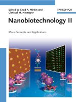
Nanobiotechnology II
Edited by
Chad A. Mirkin and
Christof M. Niemeyer
Each generation has its unique needs and aspirations. When Charles Wiley first
opened his small printing shop in lower Manhattan in 1807, it was a generation
of boundless potential searching for an identity. And we were there, helping to
define a new American literary tradition. Over half a century later, in the midst
of the Second Industrial Revolution, it was a generation focused on building
the future. Once again, we were there, supplying the critical scientific, technical,
and engineering knowledge that helped frame the world. Throughout the 20th
Century, and into the new millennium, nations began to reach out beyond their
own borders and a new international community was born. Wiley was there, ex-
panding its operations around the world to enable a global exchange of ideas,
opinions, and know-how.
For 200 years, Wiley has been an integral part of each generation’s journey,
enabling the flow of information and understanding necessary to meet their
needs and fulfill their aspirations. Today, bold new technologies are changing
the way we live and learn. Wiley will be there, providing you the must-have
knowledge you need to imagine new worlds, new possibilities, and new oppor-
tunities.
Generations come and go, but you can always count on Wiley to provide you
the knowledge you need, when and where you need it!
William J. Pesce Peter Booth Wiley
President and Chief Executive Officer Chairman of the Board
1807–2007 Knowledge for Generations
Nanobiotechnology II
More Concepts and Applications
Edited by
Chad A. Mirkin and Christof M. Niemeyer
The Editors
Prof. Dr. Chad A. Mirkin
Department of Chemistry
International Institute for Nanotechnology
Northwestern University
2145 Sheridan Road
Evanston, IL 60201
USA
Prof. Dr. Christof M. Niemeyer
Department of Chemistry
University of Dortmund
Chair of Biological and
Chemical Microstructuring
Otto-Hahn-Str. 6
44227 Dortmund
Germany
Cover
Cover images prepared
by Matthew Banholzer
and Rafael Vega
9 All books published by Wiley-VCH are
carefully produced. Nevertheless, authors,
editors, and publisher do not warrant the
information contained in these books,
including this book, to be free of errors.
Readers are advised to keep in mind that
statements, data, illustrations, procedural
details or other items may inadvertently be
inaccurate.
Library of Congress Card No.: applied for
Bibliographic information published by
the Deutsche Nationalbibliothek
Die Deutsche Nationalbibliothek lists this
publication in the Deutsche National-
bibliografie; detailed bibliographic data are
available in the Internet at h.
8 2007 WILEY-VCH Verlag GmbH & Co.
KGaA, Weinheim
All rights reserved (including those of
translation into other languages). No part of
this book may be reproduced in any form –
by photoprinting, microfilm, or any other
means – nor transmitted or translated
into a machine language without written
permission from the publishers. Registered
names, trademarks, etc. used in this book,
even when not specifically marked as such,
are not to be considered unprotected by law.
Printed in the Federal Republic of Germany
Printed on acid-free paper
Typesetting Asco Typesetters, Hong Kong
Printing betz-druck GmbH, Darmstadt
Binding Litges & Dopf GmbH, Heppenheim
Cover Design Adam-Design, Weinheim
Wiley Bicentennial Logo Richard J. Pacifico
ISBN 978-3-527-31673-1
Contents
Preface XV
List of Contributors XXI
I Self-Assembly and Nanoparticles: Novel Principles 1
1 Self-Assembled Artificial Transmembrane Ion Channels 3
Mary S. Gin, Emily G. Schmidt, and Pinaki Talukdar
1.1 Overview 3
1.1.1 Non-Gated Channels 3
1.1.1.1 Aggregates 4
1.1.1.2 Half-Channel Dimers 5
1.1.1.3 Monomolecular Channels 5
1.1.2 Gated Channels 6
1.1.2.1 Light-Gated Channels 7
1.1.2.2 Voltage-Gated Channels 7
1.1.2.3 Ligand-Gated Channels 9
1.2 Methods 10
1.2.1 Planar Bilayers 10
1.2.2 Vesicles 11
1.2.2.1
23
Na NMR 11
1.2.2.2 pH-Stat 11
1.2.2.3 Fluorescence 12
1.2.2.4 Ion-Selective Electrodes 12
1.3 Outlook 12
References 12
2 Self-Assembling Nanostructures from Coiled-Coil Peptides 17
Maxim G. Ryadnov and Derek N. Woolfson
2.1 Background and Overview 17
2.1.1 Introduction: Peptides in Self-Assembly 17
2.1.2 Coiled-Coil Peptides as Building Blocks in Supramolecular Design 18
2.1.3 Coiled-Coil Design in General 20
V
2.2 Methods and Examples 20
2.2.1 Ternary Coiled-Coil Assemblies and Nanoscale-Linker Systems 20
2.2.2 Fibers Assembled Using Linear Peptides 22
2.2.3 Fibers Assembled Using Protein Fragments and Nonlinear Peptide
Building Blocks 26
2.2.4 Summary: Pros and Cons of Peptide-Based Assembly of
Nanofibers 27
2.2.5 Assembling More-Complex Matrices Using Peptide Assemblies as
Linker Struts 30
2.2.5.1 Programmed Matrices Assembled Exclusively from Coiled-Coil
Building Blocks 30
2.2.5.2 Synthetic Polymer-Coiled-Coil Hybrids 31
2.2.6 Key Techniques 33
2.3 Conclusions and Perspectives 34
References 35
3 Synthesis and Assembly of Nanoparticles and Nanostructures Using
Bio-Derived Templates
39
Erik Dujardin and Stephen Mann
3.1 Introduction: Elegant Complexity 39
3.2 Polysaccharides, Synthetic Peptides, and DNA 40
3.3 Proteins 44
3.4 Viruses 48
3.5 Microorganisms 54
3.6 Outlook 56
Acknowledgments 58
References 58
4 Proteins and Nanoparticles: Covalent and Noncovalent Conjugates 65
Rochelle R. Arvizo, Mrinmoy De, and Vincent M. Rotello
4.1 Overview 65
4.1.1 Covalent Protein-Nanoparticle Conjugates 66
4.1.2 Noncovalent Protein–NP Conjugation 69
4.2 Methods 72
4.2.1 General Methods for Noncovalent Protein–NP Conjugation 72
4.2.2 General Methods for Covalent Protein–NP Conjugation 74
4.3 Outlook 75
References 75
5 Self-Assembling DNA Nanostructures for Patterned Molecular
Assembly
79
Thomas H. LaBean, Kurt V. Gothelf, and John H. Reif
Abstract 79
5.1 Introduction 79
5.2 Overview of DNA Nanostructures 80
VI
Contents
5.3 Three-Dimensional (3-D) DNA Nanostructures 84
5.4 Programmed Patterning of DNA Nanostructures 84
5.5 DNA-Programmed Assembly of Biomolecules 87
5.6 DNA-Programmed Assembly of Materials 89
5.7 Laboratory Methods 91
5.7.1 Annealing for DNA Assembly 92
5.7.2 AFM Imaging 93
5.8 Conclusions 93
Acknowledgments 94
References 94
6 Biocatalytic Growth of Nanoparticles for Sensors and Circuitry 99
Ronan Baron, Bilha Willner, and Itamar Willner
6.1 Overview 99
6.1.1 Enzyme-Stimulated Synthesis of Metal Nanoparticles 100
6.1.2 Enzyme-Stimulated Synthesis of Cupric Ferrocyanide
Nanoparticles 107
6.1.3 Cofactor-Induced Synthesis of Metallic NPs 107
6.1.4 Enzyme–Metal NP Hybrid Systems as ‘‘Inks’’ for the Synthesis of
Metallic Nanowires 113
6.2 Methods 115
6.2.1 Physical Tools to Characterize the Growth of Nanoparticles and
Nanowires 115
6.2.2 General Procedure for Monitoring the Biocatalytic Enlargement of
Metal NPs in Solutions 116
6.2.3 Modification of Surfaces with Metal NPs and their Biocatalytic Growth
for Sensing 116
6.2.4 Modification of Enzymes with NPs and their Use as Biocatalytic
Templates for Metallic Nanocircuitry 117
6.3 Outlook 117
References 118
II Nanostructures for Analytics 123
7 Nanoparticles for Electrochemical Bioassays 125
Joseph Wang
7.1 Overview 125
7.1.1 Particle-Based Bioassays 125
7.1.2 Electrochemical Bioaffinity Assays 125
7.1.3 NP-Based Electrochemical Bioaffinity Assays 126
7.1.3.1 Gold and Silver Metal Tags for Electrochemical Detection of DNA and
Proteins 126
7.1.3.2 NP-Induced Conductivity Detection 129
7.1.3.3 Inorganic Nanocrystal Tags: Towards Electrical Coding 130
7.1.3.4 Use of Magnetic Beads 133
Contents
VII
7.1.3.5 Ultrasensitive Particle-Based Assays Based on Multiple Amplification
Schemes 134
7.2 Methods 136
7.3 Outlook 137
Acknowledgments 138
References 138
8 Luminescent Semiconductor Quantum Dots in Biology 141
Thomas Pons, Aaron R. Clapp, Igor L. Medintz, and Hedi Mattoussi
8.1 Overview 141
8.1.1 QD Bioconjugates in Cell and Tissue Imaging 142
8.1.2 Quantum Dots in Immuno- and FRET-Based Assays 146
8.2 Methods 150
8.2.1 Synthesis, Characterization, and Capping Strategies 150
8.2.2 Water-Solubilization Strategies 151
8.2.3 Conjugation Strategies 151
8.3 Future Outlook 152
Acknowledgments 153
References 153
9 Nanoscale Localized Surface Plasmon Resonance Biosensors 159
Katherine A. Willets, W. Paige Hall, Leif J. Sherry, Xiaoyu Zhang, Jing Zhao, and
Richard P. Van Duyne
9.1 Overview 159
9.2 Methods 162
9.2.1 Nanofabrication of Materials for LSPR Spectroscopy and Sensing 162
9.2.1.1 Film Over Nanowells 163
9.2.1.2 Solution-Phase NSL-Fabricated Nanotriangles 164
9.2.1.3 Silver Nanocubes 166
9.2.2 Biosensing 167
9.3 Outlook 168
Acknowledgments 169
References 169
10 Cantilever Array Sensors for Bioanalysis and Diagnostics 175
Hans Peter Lang, Martin Hegner, and Christoph Gerber
10.1 Overview 175
10.1.1 Cantilevers as Sensors 176
10.1.2 Measurement Principle 177
10.1.3 Cantilevers: Application Fields 179
10.2 Methods 180
10.2.1 Measurement Modes 180
10.2.2 Cantilever Functionalization 181
10.2.3 Experimental Procedure 184
10.3 Outlook 186
VIII
Contents
10.3.1 Recent Literature 186
10.3.2 Challenges 188
Acknowledgments 189
References 190
11 Shear-Force-Controlled Scanning Ion Conductance Microscopy 197
Tilman E. Scha
¨
ffer, Boris Anczykowski, Matthias Bo
¨
cker, and Harald Fuchs
11.1 Overview 197
11.2 Methods 202
11.2.1 Shear-Force Detection 202
11.2.2 Ion Current Measurement 204
11.2.3 Shear-Force-Controlled Imaging 205
11.3 Outlook 207
Acknowledgments 209
References 209
12 Label-Free Nanowire and Nanotube Biomolecular Sensors for In-Vitro
Diagnosis of Cancer and other Diseases
213
James R. Heath
12.1 Overview 213
12.2 Background 213
12.3 Methods and Current State of the Art 216
12.3.1 Mechanisms of Sensing 216
12.3.2 The Role of the Sensing Environment 218
12.3.3 Nanosensor-Measured Antigen–Analyte On/Off Binding Rates 219
12.3.4 The Nanosensor/Microfluidic Environment 222
12.3.5 Nanosensor Fabrication 223
12.3.6 Biofunctionalizing NW and NT Nanosensors 226
12.4 Outlook 227
Acknowledgments 227
References 228
13 Bionanoarrays 233
Rafael A. Vega, Khalid Salaita, Joseph J. Kakkassery, and Chad A. Mirkin
13.1 Overview 233
13.2 Methods 234
13.2.1 Atomic Force Microscope-Based Methods 234
13.2.2 Nanopipet Deposition 237
13.2.3 Beam-Based Methods 238
13.2.4 Contact Printing 240
13.2.5 Assembly-Based Patterning 241
13.3 Protein Nanoarrays 242
13.3.1 Strategies for Immobilizing Proteins on Nanopatterns 243
13.3.2 Bio-Analytical Applications 244
13.3.3 Dynamic and Motile Nanoarrays 246
Contents
IX
13.3.4 Cell-Surface Interactions 246
13.4 DNA Nanoarrays 249
13.4.1 Strategies for Preparing DNA Nanoarrays 249
13.4.2 DNA-Based Schemes for Biodetection 250
13.4.3 Applications of Rationally Designed, Self-Assembled 2-D DNA
Nanoarrays 251
13.5 Virus Nanoarrays 253
13.6 Outlook 254
References 254
III Nanostructures for Medicinal Applications 261
14 Biological Barriers to Nanocarrier-Mediated Delivery of Therapeutic and
Imaging Agents
263
Rudy Juliano
14.1 Overview: Nanocarriers for Delivery of Therapeutic and Imaging
Agents 263
14.2 Basic Characteristics of the Vasculature and Mononuclear Phagocyte
System 263
14.2.1 Possible Interactions of Nanocarriers Within the Bloodstream 264
14.2.2 Transendothelial Permeability in Various Tissues and Tumors 264
14.2.3 Mononuclear Cells and Particle Clearance 267
14.3 Cellular Targeting and Subcellular Delivery 268
14.3.1 Targeting, Entry, and Trafficking in Cells 268
14.3.2 Biological and Chemical Reagents for Cell-Specific Targeting 271
14.3.3 Reagents that Promote Cell Entry 272
14.4 Crafting NPs for Delivery: Lessons from Liposomes 273
14.4.1 Loading 273
14.4.2 Release Rates 273
14.4.3 Size and Charge 274
14.4.4 PEG and the Passivation of Surfaces 274
14.4.5 Decoration with Ligands 275
14.5 Biodistribution of Liposomes, Dendrimers, and NPs 276
14.6 The Toxicology of Nanocarriers 277
14.7 Summary 278
References 278
15 Organic Nanoparticles: Adapting Emerging Techniques from the Electronics
Industry for the Generation of Shape-Specific, Functionalized Carriers for
Applications in Nanomedicine
285
Larken E. Euliss, Julie A. DuPont, and Joseph M. DeSimone
15.1 Overview 285
15.2 Methods 288
15.2.1 Bottom-Up Approaches for the Synthesis of Organic Nanoparticles
288
X
Contents
15.2.2 Top-Down Approaches for the Fabrication of Polymeric
Nanoparticles 291
15.2.2.1 Microfluidics 291
15.2.2.2 Photolithography 292
15.2.2.3 Imprint Lithography 294
15.2.2.4 IRINT 295
15.3 Outlook 297
References 299
16 Poly(amidoamine) Dendrimer-Based Multifunctional Nanoparticles 305
Thommey P. Thomas, Rameshwer Shukla, Istvan J. Majoros, Andrzej Myc,
and James R. Baker, Jr.
16.1 Overview 305
16.1.1 PAMAM Dendrimers: Structure and Biological Properties 306
16.1.2 PAMAM Dendrimers as a Vehicle for Molecular Delivery into
Cells 308
16.1.2.1 PAMAM Dendrimers as Encapsulation Complexes 308
16.1.2.2 Multifunctional Covalent PAMAM Dendrimer Conjugates 308
16.1.2.3 PAMAM Dendrimers as MRI Contrast Agents 312
16.1.2.4 Application of Multifunctional Clusters of PAMAM Dendrimer 312
16.2 Methods 313
16.2.1 Synthesis and Characterization of PAMAM Dendrimers 313
16.2.2 PAMAM Dendrimer: Determination of Physical Parameters 315
16.2.3 Quantification of Fluorescence of Targeted PAMAM Conjugates 315
16.3 Outlook 316
References 316
17 Nanoparticle Contrast Agents for Molecular Magnetic Resonance
Imaging
321
Young-wook Jun, Jae-Hyun Lee, and Jinwoo Cheon
17.1 Introduction 321
17.2 NP-Assisted MRI 322
17.2.1 Magnetic NP Contrast Agents 323
17.2.1.1 Silica- or Dextran-Coated Iron Oxide Contrast Agents 325
17.2.1.2 Magnetoferritin 327
17.2.1.3 Magnetodendrimers and Magnetoliposomes 327
17.2.1.4 Non-Hydrolytically Synthesized High-Quality Iron Oxide NPs:
A New Type of Contrast Agent 328
17.2.2 Iron Oxide NPs in Molecular MR Imaging 331
17.2.2.1 Infarction and Inflammation 332
17.2.2.2 Angiogenesis 333
17.2.2.3 Apoptosis 334
17.2.2.4 Gene Expression 335
17.2.2.5 Cancer Imaging 337
17.3 Outlook 340
Contents
XI
Acknowledgments 342
References 342
18 Micro- and Nanoscale Control of Cellular Environment for Tissue
Engineering
347
Ali Khademhosseini, Yibo Ling, Jeffrey M. Karp, and Robert Langer
18.1 Overview 347
18.1.1 Cell–Substrate Interactions 347
18.1.2 Cell Shape 350
18.1.3 Cell–Cell Interactions 351
18.1.4 Cell-Soluble Factor Interactions 351
18.1.5 3-D Scaffolds 352
18.2 Methods 354
18.2.1 Soft Lithography 354
18.2.2 Self-Assembled Monolayers 356
18.2.3 Electrospinning 357
18.2.4 Nanotopography Generation 358
18.2.5 Layer-by-Layer Deposition 358
18.2.6 3D Printing 358
18.3 Outlook 359
References 359
19 Diagnostic and Therapeutic Targeted Perfluorocarbon Nanoparticles 365
Patrick M. Winter, Shelton D. Caruthers, Gregory M. Lanza, and
Samuel A. Wickline
19.1 Overview 365
19.2 Methods 367
19.2.1 Diagnostic Imaging 367
19.2.2 Targeted Therapeutics 371
19.2.3 Other Imaging Modalities 373
19.3 Outlook 374
Acknowledgments 376
References 376
IV Nanomotors 381
20 Biological Nanomotors 383
Manfred Schliwa
20.1 Overview 383
20.2 The Architecture of the Motor Domain 388
20.3 Initial Events in Force Generation 388
20.4 Stepping, Hopping, and Slithering 390
20.5 Directionality 393
20.6 Forces 394
20.7 Motor Interactions 395
XII
Contents
20.8 Outlook 396
Acknowledgments 396
References 396
21 Biologically Inspired Hybrid Nanodevices 401
David Wendell, Eric Dy, Jordan Patti, and Carlo D. Montemagno
21.1 Introduction 401
21.2 An Overview 402
21.2.1 A Look in the Literature 402
21.2.2 Membrane Proteins and their Native Condition 403
21.3 The Protein Toolbox 404
21.3.1 F
0
F
1
-ATPase and Bacteriorhodopsin 404
21.3.2 Ion Channels and Connexin 406
21.4 Harvesting Energy 48
21.5 Methods 409
21.5.1 Muscle Power 409
21.5.2 ATPase and BR Devices 411
21.5.3 Excitable Vesicles 414
21.6 Outlook 414
Acknowledgments 416
References 416
Index 419
Contents
XIII
Preface
The broad field of nanotechnology has undergone explosive growth and develop-
ment over the past five years. In fact, no field in the history of science has experi-
enced more interest or larger government investment. Indeed, by the end of
2006, the worldwide government and private sector investment in nanotechnol-
ogy is projected to be approximately $9 billion. The enthusiasm researchers have
for this field is fueled by: 1) the desire to determine the unusual chemical and
physical properties of nanostructures, which are often quite different from the
bulk materials from which they derive, and 2) the potential to use such properties
in the development of novel and useful devices and materials that can impact
and, perhaps even transform, many aspects of modern life.
The subfield known as Nanobiotechnology holds some of the greatest promise.
This highly interdisciplinary field, which draws upon contributions from chemis-
try, physics, biology, materials science, medicine and many forms of engineering,
focuses on several important areas of research. Some of these include: 1) the de-
velopment of methods for building nanostructures and nanostructured materials
out of biological or biologically inspired components such as oligonucleotides,
proteins, viruses, and cells; these structures are intended for both biological and
abiological uses, 2) the utilization of synthetic nanomaterials to regulate and
monitor important biological processes, and 3) the development of synthetic
and soft matter compatible surface analytical tools for building nanostructures
important in both biology and medicine. Advances in this field offer novel and
potentially useful approaches to building functional structures including com-
putational tools, energy generation, conversion and storage materials, powerful
optical devices, and new detection and therapeutic modalities. Indeed, advances
in Nanobiotechnology have the potential to revolutionize the way the medical
community approaches modern disease management.
Although the field is still embryonic, major strides have been made. Powerful
new forms of signal amplification have been realized for both DNA and protein
based detection systems. Indeed, the first commercial molecular diagnostic sys-
tems that rely upon nanoparticle probes are expected to be available in 2007. Bio-
logical labels based upon nanocrystals are commercially available and used rou-
tinely for research purposes in laboratories worldwide. Many new nanomaterials
have boosted the efficacy and viability of several powerful pharmaceutical agents.
XV
Nanofabrication tools that allow one to routinely build oligonucleotide, protein,
and other biorelevant nanostructures on surfaces with extraordinary precision
have evolved to the point of commercialization and widespread use. These exam-
ples represent only a small part of this expansive field but are realized potential
and serve as motivators for future developments within it.
In 2004, we edited the book Nanobiotechnology: Concepts, Applications and Per-
spectives which was intended to provide a systematic and comprehensive frame-
work of specific research topics in Nanobiotechnology. Due to the great success
of this first volume, Nanobiotechnology II – More Concepts and Applications now
follows the notion of its precursor by combining contributions from bioorganic
and bioinorganic chemistry, molecular and cell biology, materials science and bio-
analytics to cover the entire scope of current and future developments in Nano-
biotechnology. The collection of articles in this volume again emphasizes the
high degree of interdisciplinarity necessarily implemented in the joint-venture of
biotechnology and nano-sciences. During the selection of potential chapters for
this volume we took into account, on the one hand, the progress by which partic-
ular areas had developed in the past three years. Because this occurred primarily
in two areas, namely the development of nanoparticle science and applications
as well as in the refinement of scanning probe microscopy related methods, the
majority of the chapters are concerned with these issues. On the other hand, ad-
ditional topics not yet covered in the first volume were identified, thus leading to
contributions from the area of small molecule- and peptide-based self-assembly
(chapters 1 and 2), the use of nanomaterials for medicinal applications (section
3), and the utilization of biomolecular machinery to create hybrid devices with
mechanical functionalities (section 4).
The current volume is divided into four main sections. Section I (Chapters 1–6)
concerns novel principles in self-assembly and nanoparticle-based systems. In
Chapter 1, Mary S. Gin, Emily G. Schmidt, and Pinaki Talukdar provide an over-
view of artificial transmembrane channels, attainable by organic synthesis and
the assembly of small molecule building blocks. This synthetic approach to ion
channels, initially aimed at elucidating the minimal structural requirements for
ion flow across a membrane, nowadays is focusing on the development of syn-
thetic channels that are gated, thus providing a means to control whether the
channels are open or closed. Such artificial signal transduction could lead to
novel sensing and therapeutic applications. The self-assembly of small molecules
also represents the underlying theme of Chapter 2, written by Maxim G. Ryadnov
and Derek N. Woolfson. They summarize current efforts to build nanoscopic and
mesoscopic supramolecular structures from short oligopeptides comprising the a-
helical coiled-coil folding motif. Examples of nanostructures and materials made
from such coiled-coil building blocks include programmable nanoscale linkers,
molecular switches, and fibrous and gel-forming materials which may be useful
for the production of peptide-polymer hybrids combining the advantages of both
natural and synthetic polymers. These structures also could lead to the design of
peptide-based switches that may render hybrid networks more controllable and
increase sensitivity and responses to local environments.
XVI
Preface
The notion of the first two chapters is connected with the area of nanoparticle
research in chapter 3, where Erik Dujardin and Stephen Mann illustrate how un-
raveling the specific interactions between bio-derived templates and inorganic
materials not only yields a better understanding of natural hybrid materials but
also inspires new methods for developing the potential of biological molecules,
superstructures and organisms as self-assembling agents for materials fabrica-
tion. In particular, the chapter describes the use of various types of bio-related
molecules, ranging from biopolymers, peptides, oligonucleotides to the complex
biological architecture of proteins, viruses and even living organisms, for the syn-
thesis and assembly of organized nanoparticle-based structures and materials.
Two additional chapters deal with the conversed approach, that is, the modifica-
tion of nanoparticles with biomolecules to add functionality to the inorganic com-
ponents. In Chapter 4, Rochelle R. Arvizo, Mrinmoy De, and Vincent M. Rotello
describe recent developments involving protein functionalized nanoparticles.
These conjugates, which are produced either by covalent or non-covalent coupling
strategies, hold potential for the creation of novel materials and devices in the
biosensing and catalysis fields. The combination of proteins and nanoparticles
also opens up novel approaches to the synthesis of nanoparticles, as summarized
in Chapter 6, written by Ronan Baron, Bilha Willner, and Itamar Willner. They
describe the application of biocatalysts, enzymes, such as oxidases and hydro-
lases, as active components for the synthesis and enlargement of nanoparticles
and for biocatalytic growth of nanoparticles, mediated by specific enzyme reac-
tions. This concept has strong implications in biosensor design, and the nano-
particle-enzyme hybrid systems also can be used as biocatalytic inks for the gen-
eration of metallic nanowires, and thus, bioelectronic devices.
The self-assembly behavior of another class of biomolecular recognition ele-
ments is described in Chapter 5. There, Thomas H. LaBean, Kurt V. Gothelf,
and John H. Reif summarize the current state-of-the-art of self-assembling DNA
nanostructures for patterned assembly of (macro)molecules and nanoparticles.
This field of research, which was initially covered in the previous volume of
Nanobiotechnology, has evolved significantly within the past three years. A large
number of groups are actively conducting research on such DNA-based nanoarch-
itectures. Although it is not yet clear whether commercially relevant applications
of DNA scaffolds will ever be realized, the basic exploration of design principles
based on the predictable Watson-Crick interaction of oligonucleotides opens
up long term perspectives in studying novel nanoelectronic structures, sensing
mechanisms, and materials research.
The increasing importance of nanostructures in analytical applications is re-
flected in the seven chapters of section II (Chapters 7–13). Developments of nano-
particle-based technologies are described in the first three chapters. As shown by
Joseph Wang, the large number of inorganic ions incorporated within nanopar-
ticles can be employed for signal amplification by electrochemical means. This
approach offers unique opportunities for electronic transduction of biomolecular
interactions, and thus, for measuring protein and nucleic acid analytes (Chapter
7). The peculiar optical properties of semiconductor nanoparticles are also used
Preface
XVII
for bioanalytical purposes. Hedi Mattoussi and colleagues describe in their contri-
bution the latest developments in this area (Chapter 8). This quantum dot bio-
labeling is rapidly moving towards sophisticated applications in cell and tissue
imaging as well as in FRET-based immuno-assays, thereby posing great demands
on the chemical and structural integrity of the hybrid probes. A more fundamen-
tal approach combining nanoparticle technologies and spectroscopy is described
in Chapter 9 by Richard P. Van Duyne and colleagues. The localized surface
plasmon resonance, occurring in optically coupled nanoparticles, coupled with
the size, shape, material and local dielectric environment dependence of the
nanostructures, forms the basis for a novel class of biosensors.
While the above mentioned chapters have the ‘‘bottom-up’’ assembly of func-
tional nanostructures in common, the following four chapters of section 2 take
advantage of micro- and nanosized probe structures fabricated by conventional
‘‘top-down’’ methodologies. The developments of micromechanical cantilever ar-
ray sensors for bioanalytical assays are described by Hans Peter Lang, Martin
Hegner, and Christoph Gerber in Chapter 10. The cantilever arrays respond me-
chanically to changes in external parameters, like temperature or molecule ad-
sorption, and thus, can be used to monitor binding events and chemical reactions
occurring at the sensors’ surfaces. In Chapter 12, James R. Heath reviews work
on nanotube-based sensors, which enable the label-free detection of biomarkers
for cancer and other diseases. It is pointed out here that the establishment of via-
ble carbon nanotube or semiconductor nanowire devices for routine diagnostics
will require a high-throughput fabrication method with an extraordinary high
level of integration of nanoelectronics, microfluidics, chemistry, and biology.
Chapter 11, written by Harald Fuchs and colleagues, reports on uses for shear
force-controlled scanning ion conductance microscopy. By using a local probe
that is sensitive to ion conductance in an electrolyte solution, gentle scanning of
delicate biological surfaces, allows one to obtain well resolved images of fine sur-
face structures, such as of membrane proteins on living cells. In Chapter 13,
Chad Mirkin and colleagues report on the preparation of arrays of nanoscale fea-
tures of biomolecular compounds by using dip-pen nanolithography. Arrays with
features on the nanometer length scale not only open up the opportunity to study
many biological structures at the single particle level, but they also allow one to
contemplate the creation of a combinatorial library, for instance, a complex pro-
tein array, underneath a single cell. This would open new possibilities for study-
ing important fundamental biological processes, such as cell-surface recognition,
adhesion, differentiation, growth, proliferation, and apoptosis.
Section III (Chapters 14–19) of this volume concerns the use of nanostructures
in medicinal applications. The six chapters focus on three major topics: the devel-
opment of nanoparticle-based drug delivery systems, the use of nanoparticles for
imaging, and the design of scaffolds for tissue engineering. Chapter 14, written
by Rudy Juliano, gives an introductory overview on biological barriers to nanocar-
rier-mediated delivery of therapeutic and imaging agents. This chapter also pro-
vides a brief assessment of the toxicology of nanomaterials, a subject which has
currently initiated widespread discussion because it is anticipated that nano(bio)-
XVIII
Preface
technology will be a key aspect of the future economy. With respect to the devel-
opment of suitable carrier systems, Larken E. Euliss, Julie A. DuPont, and Joseph
M. DeSimone in Chapter 15 summarize work on the development of biocompat-
ible organic nanoparticles, in particular, by means of top-down fabrication tech-
niques, so called lithographic imprinting, adapted from the electronics industry.
This method enables the inexpensive fabrication of monodisperse particles of var-
ious size and shape from a large variety of matrix materials, which have great po-
tential as functionalized carriers for applications in nanomedicine. An alternative
class of particles is described in Chapter 16, where Thommey P. Thomas and col-
leagues report on poly(amidoamine) dendrimer-based multifunctional nanopar-
ticles as a tumor targeting platform. The biocompatible dendrimer macromolecules
act as carriers of molecules for delivery into tumor cells and can achieve increased
drug effectiveness at significantly lower toxicity as compared to the free drug.
With respect to clinical imaging techniques, Young-wook Jun, Jae-Hyun Lee,
and Jinwoo Cheon review current work on the development of magnetic nanopar-
ticle-based contrast agents for molecular magnetic resonance imaging in Chapter
17. This research is aimed towards advances in cancer diagnosis because nano-
particle contrast agents promise in vivo diagnosis of early staged cancer with
sub-millimeter dimension, and might help to unravel fundamental biological pro-
cesses, such as in vivo pathways of cell evolution, cell differentiations, and cell-
to-cell interactions. A different class of nanoparticles is described in Chapter 19
by Samuel A. Wickline and colleagues. They have developed nanoparticles com-
prised of perfluorocarbon materials which are biologically and metabolically inert,
chemically stable, and non-toxic. These nanoparticles have been employed for
molecular imaging with MRI and targeted drug delivery.
Chapter 18 of this section, written by Robert L. Langer and colleagues reviews
methodologies for generating two- and three-dimensional scaffold architectures
for tissue engineering. The authors analyze the use of micro- and nanoscale engi-
neering techniques for controlling and studying cell-cell, cell-substrate and cell-
soluble factor interactions as well as for fabricating organs with controlled archi-
tecture and resolution.
Section IV of this volume is devoted to one of the most innovative topics of
Nanobiotechnology which concerns the fabrication of hybrid devices using or-
ganic and inorganic structures equipped with parts of Nature’s biomolecular ma-
chinery. To facilitate an introduction to natural molecular nanomotors, Manfred
Schliwa describes in Chapter 20 how these fascinating molecular machines are
built from amino acids and how they convert chemical energy into mechanical
motion. In Chapter 21, Carlo D. Montemagno and colleagues summarize current
approaches to fabricate biologically inspired hybrid nanodevices. In particular,
two lines of work are shown, protein-based mechanical devices and cellular power
generation devices, which both have in common the theme of combining biolog-
ical molecules with synthetic host structures.
Similar to the first volume, the purpose of Nanobiotechnology II – More Concepts
and Applications is to provide both a broad survey of the field as well as instruction
and inspiration to scientists at all levels from novices to those intimately engaged
Preface
XIX
in this new and exciting field of research. To this end, the current state-of-the-art
of the above described topics has been accumulated by international renowned
experts in their fields. Each of the chapters consists of three sections, (i) an over-
view which gives a comprehensive but still condensed survey on the specific
topic, (ii) a methods section which points the reader to the most important tech-
niques relevant for the specific topic discussed, and (iii) an outlook discussing
academic and commercial applications as well as experimental challenges to be
solved.
We are most grateful to the authors for providing this collection of high quality
manuscripts. We also would like to thank Dr. Sabine Sturm and the production
team of Wiley-VCH for continuous and dedicated help during the production of
this book.
Evanston and Dortmund, November 2006 Chad A. Mirkin
Christof M. Niemeyer
XX
Preface
List of Contributors
Boris Anczykowski
nanoAnalytics GmbH
Heisenbergstr. 11
48149 Mu
¨
nster
Germany
Rochelle R. Arvizo
Department of Chemistry
University of Massachusetts
710 North Pleasant St.
Amherst, MA 01003
USA
James R. Baker, Jr.
Michigan Nanotechnology
Institute for Medicine and
Biological Sciences
University of Michigan
Rm 9220C, MSRB III
Ann Arbor, MI 48109
USA
Ronan Baron
Institute of Chemistry
The Hebrew University of
Jerusalem
Jerusalem 91904
Israel
Matthias Bo
¨
cker
Center for Nanotechnology (CeNTech)
and Institute of Physics
University of Mu
¨
nster
Heisenbergstr. 11
48149 Mu
¨
nster
Germany
Shelton D. Caruthers
Department of Medicine and
Biomedical Engineering
Washington University School of
Medicine
4320 Forest Park Ave.
St. Louis, MO 63108
USA
Jinwoo Cheon
Department of Chemistry and Nano-
Medical National Core Research
Center
Yonsei University
134 Sinchon-dong
Seodaemun-gu
120-749 Seoul
South Korea
Aaron R. Clapp
Division of Optical Sciences
US Naval Research Laboratory
Washington, DC 20375-5320
USA
XXIXXI
Mrinmoy De
Department of Chemistry
University of Massachusetts
710 North Pleasant St.
Amherst, MA 01003
USA
Joseph M. DeSimone
Department of Chemistry
University of North Carolina at
Chapel Hill
Chapel Hill, NC 27599
USA
Julie A. DuPont
Department of Chemistry
University of North Carolina at
Chapel Hill
Chapel Hill, NC 27599
USA
Erik Dujardin
NanoSciences Group
CEMES, CNRS UPR 8011
BP 94347
29 rue Jeanne Marvig
31055 Toulouse Cedex 4
France
Eric Dy
Department of Bioengineering
University of California, Los
Angeles
420 Westwood Plaza
Los Angeles, CA 90095-1600
USA
Larken E. Euliss
Department of Chemistry
University of North Carolina at
Chapel Hill
Chapel Hill, NC 27599
USA
Harald Fuchs
Center for Nanotechnology (CeNTech)
and Institute of Physics
University of Mu
¨
nster
Heisenbergstr. 11
48149 Mu
¨
nster
Germany
Christoph Gerber
Institute of Physics
University of Basel
Klingelbergstrasse 82
4056 Basel
Switzerland
Mary S. Gin
Department of Chemistry
University of Illinois at Urbana-
Champaign
600 S. Mathews Ave.
Urbana, IL 61801
USA
Charles A. Goessmann
Department of Chemistry
University of Massachusetts
710 North Pleasant St.
Amherst, MA 01003
USA
Kurt V. Gothelf
Department of Chemistry
Aarhus University
Langelandsgade 140
8000 Aarhus C
Denmark
W. Paige Hall
Department of Chemistry
Northwestern University
2145 Sheridan Road
Evanston, IL 60208-3113
USA
XXII
List of Contributors
James R. Heath
Caltech Chemistry MC 127-72
and the NanoSystems Biology
Cancer Center
1200 East California Blvd.
Pasadena, CA 91125
USA
Martin Hegner
Institute of Physics
University of Basel
Klingelbergstrasse 82
4056 Basel
Switzerland
Rudy Juliano
Department of Pharmacology
School of Medicine
University of North Carolina at
Chapel Hill
Chapel Hill, NC 27599
USA
Young-wook Jun
Department of Chemistry and
Nano-Medical National Core
Research Center
Yonsei University
134 Sinchon-dong
Seodaemun-gu
120-749 Seoul
South Korea
Joseph J. Kakkassery
Department of Chemistry
International Institute for
Nanotechnology
Northwestern University
2145 Sheridan Road
Evanston, IL 60208
USA
Jeffrey M. Karp
Massachusetts Institute of Technology
77 Massachusetts Ave.
Cambridge, MA 02139-4307
USA
Ali Khademhosseini
Massachusetts Institute of Technology
65 Landsdowne St.
Cambridge, MA 02139
USA
Thomas H. LaBean
Departments of Computer Science and
Chemistry
Duke University
Durham, NC 27708
USA
Hans Peter Lang
Institute of Physics
University of Basel
Klingelbergstrasse 82
4056 Basel
Switzerland
Robert Langer
Massachusetts Institute of
Technology
77 Massachusetts Ave.
Cambridge, MA 02139-4307
USA
Gregory M. Lanza
Department of Medicine and
Biomedical Engineering
Washington University School of
Medicine
4320 Forest Park Ave., Campus
Box 8215
St. Louis, MO 63108
USA
List of Contributors
XXIII
Jae-Hyun Lee
Department of Chemistry and
Nano-Medical National Core
Research Center
Yonsei University
134 Sinchon-dong
Seodaemun-gu
120-749 Seoul
South Korea
Yibo Ling
Massachusetts Institute of
Technology
65 Landsdowne St.
Cambridge, MA 02139
USA
Istvan J. Majoros
Michigan Nanotechnology
Institute for Medicine and
Biological Sciences
University of Michigan
109 Zina Pitcher Place
Ann Arbor, MI 48109
USA
Stephen Mann
School of Chemistry
University of Bristol
Bristol BS8 1TS
UK
Hedi Mattoussi
Division of Optical Sciences
US Naval Research Laboratory
Washington, DC 20375-5320
USA
Igor L. Medintz
Division of Optical Sciences
US Naval Research Laboratory
Washington, DC 20375-5320
USA
Chad A. Mirkin
Department of Chemistry
International Institute for
Nanotechnology
Northwestern University
2145 Sheridan Road
Evanston, IL 60208
USA
Carlo D. Montemagno
Department of Bioengineering
University of California, Los Angeles
420 Westwood Plaza
Los Angeles, CA 90095-1600
USA
Andrzej Myc
Michigan Nanotechnology Institute for
Medicine and Biological Sciences
University of Michigan
109 Zina Pitcher Place
Ann Arbor, MI 48109
USA
Christof Niemeyer
Chair of Biological and Chemical
Microstructuring
University of Dortmund
Department of Chemistry
Otto-Hahn-Str. 6
44227 Dortmund
Germany
Jordan Patti
Department of Bioengineering
University of California, Los Angeles
420 Westwood Plaza
Los Angeles, CA 90095-1600
USA
Thomas Pons
Division of Optical Sciences
US Naval Research Laboratory
Washington, DC 20375-5320
USA
XXIV
List of Contributors


