Taxonomy and biology of calonectria in forest soil of vietnam
Bạn đang xem bản rút gọn của tài liệu. Xem và tải ngay bản đầy đủ của tài liệu tại đây (7.31 MB, 49 trang )
ACKNOWLEDGEMENTS
In order to complete this study course, with the consent of the Department of Forest
Resources and Environment Management (FREM), I conduct to do a research: “Taxonomy
and biology of Calonectria in forest soil of Vietnam”.
In the implementing research process, I have received the helps of Forest Protection
Research Centre (FPRC) - Vietnamese Academy of Forest Sciences (VAFS), everyone who
work in this centre, and especially the enthusiastic guidance of Associated Professor Ph.D
Phung Van Khoa - Vietnam National University of Forestry and Associate Professor Ph.D
Pham Quang Thu - Forest Protection Research Centre.
Therefore, I would like to express deep gratitude to Associated Professor Ph.D Phung
Van Khoa, Associated Professor Ph.D Pham Quang Thu who guide for me. Thanks to Forest
Protection Research Centre, and all members in here who have created favorable conditions for
me to complete this study. I also have a good eye for helping of my family, and my friends.
In the process of learning and doing research, I tried my best but time, knowledge, and
budget are limited. Therefore, this study could not avoid some faults. I am looking forward to
hearing comments from you to more complete the study.
Sincerely!
Hanoi, September- 2016
Nguyen Thi Loan
1
ABSTRACT
The research presents taxonomy and biology of Calonectria in forest soil of Vietnam.
Basing on data of 72 soil samples which were collected from some forests of Vietnam such as
Hoang Lien National Park, Tam Dao National Park, and Binh Phuoc province, the study
clearly described biology characteristics and pathogenicity of Calonectria. Furthermore,
taxonomy also is an important part of this research. Using method of sample collection in
collecting soil samples; method of isolation, culturing the fungus in finding fungi phylogeny;
method of pathogenicity in defining pathogenic ability; method of DNA technique in fungi
identification; method of using microscope in describing morphological characteristics of
fungi; and methods of culturing the fungus in different environment, temperature, moisture to
determine the biological characteristics of fungi. Data was collected in 2016 from 72 soil
samples in Hoang Lien National Park, Tam Dao National Park, and Binh Phuoc province.
The results of the study showed morphological characteristics and biological characteristics
of 41 isolates- not only this but also they were classified. Generally, this pathogenic fungi
need to be paid more attention in order to protect forest trees.
2
TABLE OF CONTENTS
ACKNOWLEDGEMENTS
ABSTRACT
TABLE OF CONTENTS
LIST OF FIGURES
LIST OF TABLES
1. INTRODUCTION ............................................................................................................ 1
1.1. Imperativeness ............................................................................................................... 1
1.2. Study site ....................................................................................................................... 1
1.3. Object of research .......................................................................................................... 5
1.4. Literature reviews .......................................................................................................... 5
2. GOALS AND (SPECIFIC) OBJECTIVES ........................................................................ 8
2.1. Goals .............................................................................................................................. 8
2.2. Specific objectives .......................................................................................................... 8
3. METHODS ....................................................................................................................... 9
3.1. Sampling methods: ......................................................................................................... 9
3.2. Isolation methods: ........................................................................................................ 10
3.3. Methods of pathogenicity testing: ................................................................................. 11
3.4. DNA technique: PCR amplification and sequencing: .................................................... 13
3.5. Method of describing the morphological characteristics of fungi: ................................. 14
3.6. Method of describing the biological characteristics of fungi: ........................................ 15
3.6.1. Growth of the mycelium on different nutrient media: ................................................. 15
3.6.2. Growth of the mycelium on PDA nutrient medium at different temperature scales:.... 16
3.6.3. Growth of the mycelium on PDA nutrient medium at different moisture scales: ........ 17
4. RESULTS ....................................................................................................................... 18
3
4.1. Result of sample collection. .......................................................................................... 18
4.2. Result of isolation, and culturing the fungus. ................................................................ 19
4.3. Pathogenicity testing. ................................................................................................... 22
4.4. Taxonomy by DNA technique: PCR amplification and sequencing. .............................. 24
4.5. Morphological characteristics of fungi. ......................................................................... 28
4.5.1. Morphological characteristics of Ca. follicola (DMC TN2.1) ..................................... 28
4.5.2. Morphological characteristics of Ca. pentaseptata (RTN BPP3.3) .............................. 28
4.5.3. Morphological characteristics of Ca. illicicola (LT DN 2.1 & LT DN2.2) .................. 29
4.6. Biological characteristics of fungi................................................................................. 29
4.6.1. Growth of the mycelium on different nutrient media .................................................. 29
4.6.2. Growth of the mycelium on PDA nutrient medium at different temperature scales:.... 32
4.6.3. Growth of the mycelium on PDA nutrient medium at different moisture scales.......... 33
5. DISCUSSION ................................................................................................................. 37
6. CONCLUSION ............................................................................................................... 39
REFERENCES
4
LIST OF FIGURES
Figure 1.1. Study site ............................................................................................................ 2
Figure 3.1. Soil samples ........................................................................................................ 9
Figure 3.2. Process of isolation and culturing the fungus ..................................................... 11
Figure 3.3. Confront sample and pathogenicity sample ....................................................... 12
Figure 3.4. Microscope and camera use to describe morphological characteristics ............... 15
Figure 3.5. Three types of nutrient media ............................................................................ 16
Figure 3.6. Fungus culturing................................................................................................ 16
Figure 3.7. Moisture experiment .......................................................................................... 17
Figure 4.1. Some strains under stereoscope ......................................................................... 19
Figure 4.2. Spore of Calonectria .......................................................................................... 20
Figure 4.3. Four groups of isolates ...................................................................................... 22
Figure 4.4. Pathogenic ability of fungal strains .................................................................... 23
Figure 4.5. The process of pathogenicity of Calonectria on Eucalyptus leaf ......................... 23
Figure 4.6. Classification tree of DMC TN2.1 ..................................................................... 25
Figure 4.7. Classification tree of RTN BP3.3....................................................................... 26
Figure 4.8. Classification trees of LT DN2.1, and LT DN2.2 ............................................... 27
Figure 4.9. Vesicle and spore of Ca. follicola ...................................................................... 28
Figure 4.10. Vesicle and spore of Ca. pentaseptata .............................................................. 29
Figure 4.11. Vesicle and spore of Ca. illicicola .................................................................... 29
Figure 4.12. Growth of the mycelium on different nutrient media ........................................ 30
Figure 4.13. Difference growth of mycelium in OSA, PDA, and MEA nutrient medium ..... 31
Figure 4.14. Growth of some mycelium in different temperatures ....................................... 32
Figure 4.15. Growth of mycelium at different temperature scales after 3 days ..................... 33
Figure 4.16. Growth of some mycelium in different moisture after 3 days ........................... 34
Figure 4.17. Growth of some mycelium in different moisture after 5 days ........................... 35
Figure 4.18. Growth of mycelium in different moisture scales ............................................. 36
5
LIST OF TABLES
Table 1.1. Calonectria species reported in Vietnam ............................................................................ 7
Table 3.1. Levels of pathogenic ability ............................................................................................ 13
Table 3.2. Formula to create moisture environment .......................................................................... 17
Table 4.1. List of soil samples is collected ....................................................................................... 18
Table 4.2. List of fungi strains were found ....................................................................................... 19
Table 4.3. Some characteristics of 41 fungi strains ........................................................................... 20
Table 4.4. Pathogenic strains and pathogenic levels of them............................................................. 24
6
1. INTRODUCTION
1.1. Imperativeness
In Vietnam, Cylindrocladium quinqueseptatum, anamoph of Calonectria species firstly was
found from Hue to the South as fungal pathogens to Eucalyptus spp. Calonectria
pentaseptata was announced in 2012 by Pham Quang Thu and coworkers (L.Lombard,
M.J.Wings, & Crous, sp. nov). And now, this fungus was found in most area of Vietnam.
Therefore, I conducted this research to find out the diversity and characteristics of
Calonectria. Hope that this study will be useful with disease management in the future in
order to protect flora.
1.2. Study site
As we know, Vietnam is located in the Indochina peninsula, belongs to South-East Asia
region. The Northern part of Vietnam is bordered by China; Cambodia and Laos in the West;
the Southwest is bordered by the Gulf of Thailand; East and South, Vietnam is bordered by
the East Sea. Vietnam has an area of 331 698 km², of which three quarters are hilly. In
Vietnam, about 327 480 km² is terrestrial, and 4,500 km² is an inland sea. Because Vietnam is
located along the coast, the climate is regulated in part by sea currents, so it brings more
marine climate factors. Average relative humidity is 84% throughout the year. Every year,
precipitation is from 1,200 to 3,000 mm, the number’s hours of sunshine is about 1,500 to
3,000 hours / year, and the temperature is between 5°C to 37°C.
With conditions such as terrain and climate were presented, Vietnam is a good place
for the development of diverse animals and plants, especially the forest fauna, and forest
flora. As is already known, mainly Vietnam area is hilly. Thus, we can see the forest has a
very important role in Vietnam. However, with hot, humid climate conditions and heavy
rainfall in some areas, these are favorable conditions for pests and harmful fungi grow. In this
1
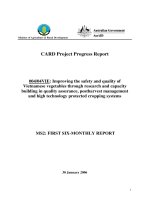
study, 3 sites with topographic features, the climate in Vietnam was chosen to study are
Hoang Lien National Park, Tam Dao National Park, and Binh Phuoc province.
Figure 1.1. Study site
2
First location is Hoang Lien National Park. This is a national park within Hoang Lien
Son Range. It has total area 28.509 ha, located in 4 communes of Sapa district (Lao Chai, Ta
Van, San Sa Ho, Ban Ho) in Lao Cai province and 2 communes (Phuc Khoa and Trung
Dong) of Tan Uyen district, Lai Chau province. This National Park has many differences
compared to other national parks in the special use forest (SUF) system of Vietnam where the
exchange of the two subtropical climates and alpine temperature happens. Terrain, this is
where is rugged terrain, high slopes, fragmented, peaks above 1,000 meters, including
Fansipan with 3.143m high. For specific characteristics about climate, weather, topographic,
Hoang Lien has incredibly rich of fauna – flora. According to the scientists, Hoang Lien
National Park is one of the most diverse biodiversity centers Vietnam, especially flora. There
exist here over 2,000 species of plants typically as Bo tree, azaleas, plum…, of which about
66 species are recorded in Vietnam’s Red Book, 32 rare species, and hundreds of precious
herbs. Besides, several ancient mushroom species were also detected here.
Second location is Tam Dao National Park which is a protected area zone in
North Vietnam.. The park is about 85km northwest of Hanoi. It runs 80 km from North- West
to South East, and has more than 20 peaks with altitudes of over 1000m. The highest summit
is Tam Dao North with an altitude of 1592m. Three other peaks with beautiful scenery are
Thien Thi at 1375m, Thach Ban at 1388m, and Phu Nghia at 1300m. Sharp peaks with
sloping sides and numerous, deep partitions are characteristic of the topology. Due to the tall
mountainous range that splits the area into two parts, the national park's climatic condition is
divided into two areas with different rainfalls. According to research, Tam Dao park has eight
kinds of forest types distributed in different topographic and climatic areas : the tropical
moist evergreen forest, the subtropical moist evergreen low mountain forest, the high
mountain short forest, the bamboo forest, the restored forest, the plantation forest, the bush
appears, the grass land. Out of over 2000 plant species, 904 species are considered useful to
3
humans. There are 42 species endemic to Tam Dao National Park and also 64 other species
considered rare. The national park has diverse animal species, with 840 species in total.
Eleven of these species are endemic to Tam Dao National Park including the snake species,
the amphibian and eight species of insects; 22 species are endemic to North Vietnam
including nine bird species, four reptiles, three amphibians and six species of insect; six are
endemic to Vietnam (five bird species and one species of amphibian).
Last location is Binh Phuoc province whichis located in the Southeast region of the
country, to the north of Ho Chi Minh City. It shares a border with Cambodia. Binh Phuoc
Province is relatively flat with elevations of between 50m and 200m throughout most of the
provinces. Elevations are gradually higher towards the east of the province and reach around
500m near parts of the border to Dak Nong Province of the Central Highlands. The highest
elevation is Ba Ra Mountain (736m) in the centre of the province. There are several hills
around the province with heights of up to around 200m in the west and 300m in the southeast.
Forestry land takes up 337,000 ha or 49 per cent of the province's total area. Forests are
located mostly in the northeast and southeast of the province as well as along the northern
border to Cambodia and the western border of Tay Ninh Province. Much of the rest of the
area is used to grow perennial cash crops. The total agricultural area is 293,700 ha. There is
21,900 ha of specially used land and 5700 ha of residential land. Average annual rainfall in
the province is 2280 mm / year, varying from 1900 to 2731 mm / year, is unevenly
distributed. In general, rainfall increases from the Southeast to the Northwest, in which the
area has average rainfall less than 2000 mm is 115,093.7 hectares (16.83%), an area with
average rainfall of 2,000 mm to 2500 mm is 301,964.3 hectares (44.2%), an area with
average rainfall of 2500 mm is greater than 266,666.3 ha (39%). The average annual
temperature is 26°C, ranges from 24.3°C to 28°C. December has lowest average monthly
4
temperature (24.3°C). April has maximum average monthly temperature (28°C), unevenly
distributed, tend to decrease from Southwest to Northeast.
1.3. Object of research
As we know, flora is the most important part in forest ecosystems. It not only reflects the
richness of the genetic biodiversity of forest resources, but also determines the diversity of
forests, scientific value, the value of landscape and environmental, economic benefit. All
above locations have suitable conditions for growth of flora. However, high temperature,
heavy rainfall was also favorable conditions for pests and diseases development, including a
pathogenic fungus –Calonectriawhich is object of this study. This is one kind of serious
pathogenic fungus in plant which we need to care if we want to protect the growth of tree in
there.
Most of Calonectria species are readily recovered from soil (Crous, 2002). Therefore, this
study also conducts isolate them from soil samples.
1.4. Literature reviews
The genus Calonectria (Ca.) was erected in 1867 by De Notaris, based on Ca.
daldiniana collected on leaves of Magnolia grandiflora that belong to Magnoliaceae, in Italy
(Rossman 1979a). Then Rossman (1979a) reduced Ca. daldiniana to synonymy under Ca.
pyrochroa. This nectrioid fungus was defined as having an ascocarp wall structure that is
brightly coloured, changing to blood-red in 3% KOH solution, warty to scaly and with a
Cylindrocladium (Cy.) anamorph (Rossman 1993, Rossman et al. 1999). However, due to the
restricted morphological characteristics of the teleomorph (Rossman 1979b, 1983),
specimens can in many cases only be identified to species level if the anamorph is present
(Schoch et al. 2000b, Crous 2002) (L. Lombard et al, 2010).
The anamorph genus Cylindrocladium, which is based on Cy. scoparium, was first
described by Morgan (1892) in the U.S.A., where it was found growing as saprobe on a pod
5
of Gleditsia triacanthos. Although Morgan (1892) failed to mention the stipe extension
terminating in a vesicle of characteristic shape, he defined the genus as having branched
conidiophores producing cylindrical conidia. This fungus has a wide distribution in subtropical and tropical regions of the world, and species are pathogenic to numerous plants
(Crous 2002) (L. Lombard et al, 2010).
Lombard also showed that Calonectria belongs to the Nectriaceae, one of three
families in Hypocreales, an order that has been reviewed extensively. The Nectriaceae is
circumscribed as having uniloculate ascomata that are orange to purple and not immersed in
well-developed stromata. The family includes approximately 20 genera of socio-economic
importance and Calonectria is most clearly distinguished from the others by its
Cylindrocladium anamorphs and relevance as plant pathogens.
The genus Calonectria was initially regarded as a saprobe as no disease symptoms
could be induced by inoculating a suspected host. The first proof of pathogenicity of these
fungi was provided by Massey in 1917, and subsequently by Anderson in 1919, who proved
pathogenicity of Ca. morganii (as Cy. scoparium). Then, Calonectria species have been
associated with a wide range of disease symptoms on a large number of hosts worldwide. In
the past, several authors have indicated that Calonectria species cause disease on plants
residing in approximately 30 plant families (Booth & Gibson 1973, French & Menge
1978, Peerally 1991a, Wiapara et al. 1996, Schoch et al. 1999). Upon closer inspection, the
number of plant families is actually closer to 100 and approximately 335 plant host species.
The plant hosts include important forestry, agricultural and horticultural crops and the impact
of these plant pathogens has likely been underestimated (L. Lombard et al, 2010).
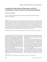
The majority of disease reports associated with Calonectria species in forestry include
hosts in 5 plant families, of which the most important are associated with Fabaceae (Acacia
spp.), Myrtaceae (Eucalyptus spp.) and Pinaceae (Pinus spp.). The symptoms may include
6
leaf spot, stem lesion, stem or crown canker, root rot, crown rot and stem rot. In Vietnam,
Ca.insularis and Ca. pauciramosa were also recorded for the first time, where they are
associated with leaf spot symptoms on Eucalyptus spp (Crous P.W et al, 2002).
Table 1.1. Calonectria species reported in Vietnam
FUNGAL
DISEASE
SPECIES
SYMPTOMS
Ca. insularis
HOSTS
REFERENCES
Leaf spot
Eucalyptus sp.
Crouset al. 2002
Ca. pauciramosa
Leaf spot
Eucalyptus sp.
Crouset al. 2002
Ca. reteaudii
Leaf blight
Eucalyptus spp.
Ca. pentaseptata
Leaf blight
Eucalyptus hybrid
Booth et al. 2000
Old et al. 2003
Crouset al. 2012
Stemming from this situation, I conducted a study "Research on Taxonomy and
Biology of Calonectria in forest soil of Vietnam". The aim of this research is to isolates and
identification of fungal species in the genus Calonectria through DNA sequence analysis and
morphological characteristic, and identifying, researching biological characteristics,
pathogenicity of isolation. Hopefully, this study is useful in order to manage plant diseases
and the conservation and development of flora biodiversity in Vietnam.
7
2. GOALS AND (SPECIFIC) OBJECTIVES
2.1. Goals
This study has two main points: Taxonomy and Biology. We also have 2 main goals:
- In Taxonomy: to identify the fungal species in the genus Calonectria through DNA
sequence analysis and morphological characteristic.
- In Biology: to identify and study biological characteristics, and pathogenicity ability
of fungal strains.
2.2. Specific objectives
- Collect soil samples in forests: Hoang Lien National Park, Tam Dao National Park,
and Binh Phuoc province. Isolates the fungal species belongs to genus Calonectria. Then, do
the DNA extraction and identification of the isolates by the DNA sequence analysis. Describe
the morphology characteristics of spores by using microscope Olympus BX 50.
- Determine the biological characteristics of fungal strains the pathogenicity of
isolation by inoculation tests.
8
3. METHODS
3.1. Sampling methods:
The disease is often compromised in the root; stem and fungal sources usually exist in
the soil. And most of Calonectria species are readily recovered from soil (Crous, 2002).
Extensive surveys will be taken in study sites. As was mentioned above, in Vietnam,
Ca.insularis and Ca. pauciramosa were also recorded with leaf spot symptoms on Eucalyptus
spp (Crous P.W et al, 2002). Therefore, we conduct to collect soil sample from Eucalyptus
plantations, and many difference places. Trying to find the place where the soil was not dry
for the Calonectria spp. likes the moist environment.
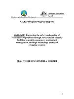
The soils samples will be collected from positions having symptomatic plants, natural
forest soil will also conclude. Soil samples will be kept in plastic bags, marked and wellinformed about the time and place of sampling, bring samples back to laboratory for
assessment.
Figure 3.1. Soil samples
9
3.2. Isolation methods:
Preparing:
Soil collected cup: can use the urine specimen collection cups or some kinds of cup
to hold food.
Sterilized alfalfa seeds : take the alfalfa seeds soak into the bleach solution
(2.5%), about 30 seconds later, and then rewash by distilled water 5 to 6 times.
Soil sample: firstly, blend the soil sample; then pick the root, leaf or other
substance out of the soil sample; and crush the large pieces of soil into small pieces.
Spray bottle of distilled water.
PDA nutrient medium (formula for 1 liter): 200g potatoes minced put in 1L water.
Boiling it in 30 minutes, filter the water. Then add enough water to 1 liter of water.
Thereafter, add 20g glucose and 18g agars, shake well to dissolve, steamed for 20 minutes at
121°C. Lastly, waiting to temperature of solution is about 50°C pouring them into petri dishes.
With antibiotic PDA nutrient medium, we add 0.2g streptomycin into 100ml distilled
water, shake well to dissolve. Put them into solution of potatoes, agar, and glucose when
temperature of solution is about 50°C. Then, shake again and pouring them into petri dishes.
Conducting:
Put soil sample into the cup (about 10mm).
Spray moderate sterilized water into the cup; make sure the surface of the
soil sample is moist (moisture is enough).
Sow the sterilized alfalfa seeds evenly on the soil sample; make sure the seeds
is not crowded, finally cover the lid, put the cup into a sealed chamber.
10
Figure 3.2. Process of isolation and culturing the fungus
After 2 to 4 days, the seeds will germinate, you can check it with a stereoscopic,
microscope. If there has Calonectria in the soil, you may find Calonectria infects the alfalfa
seeds or immature stems. Then you can isolate the Calonectria. Using the sharp needle to
pick up marcoconidiophore. Then transfer them into antibiotic PDA nutrient medium.
Subculture them into the PDA nutrient mediumto have pure fungal samples.
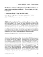
3.3. Methods of pathogenicity testing:
Pathogenicity test was conducted in leaves of Eucalyptus.
Preparing:
Fungi samples were pure in the petri dish;
Nest box lined with moist paper;
Leaves of Eucalyptus tree have similar size in one tree.
11
Instruments to implant fungus: knives, alcohol lamps, alcoholic, roll bandage, the
grid scale.
Conducting:
Putting leaves facing down into nest box lined with moist paper.
Disinfecting knife by alcohol lamps, alcoholic.
Cutting fungi in nutrient medium in similar size plugs, put them on the back of the
leaves; using roll bandage to sealed dish.
Tracking and measuring results after 4 days: using the grid to measure diseased
leaf area of the total leaf area.
Pathogenic ability = The percentage of diseased leaf area =
x100 (%)
Note: For control, doing similar but use nutrient medium plug without fungi.
Figure 3.3. Confront sample and pathogenicity sample
According to Pham Quang Thu about Eucalyptus disease and disease management (2005) having self adjustment to suitable, pathogenic ability is divided in 4 levels:
12
Table3.1. Levels of pathogenic ability
Level
Pathogen ability
Disease leaf area
1
None
0%
2
Low
< 20%
3
Medium
20- 40%
4
High
> 40%
3.4. DNA technique: PCR amplification and sequencing:
According toL. Hywel-Jones (2004), the isolation protocol of Lee & Taylor (1990)
was used to isolate genomic DNA from fungal mycelia. We also use it in this research.
Template DNA (approximately 20ng) was amplified in a 25 (.il PCR reaction mixture
consisting of 1 x PCR reaction buffer (10 mM Tris-HCl, 1.5 mM MgCl2, 50 mM KC1, pH
8.3) and 200 uM each of dATP, dCTP, dGTP and dTTP, with primer pairs EF1-728F and
EF2were used to amplified fragment of translation elongation factor gene region, and 1.25
units Taq DNA polymerase enzyme (Roche Diagnostics GmbH, Mannheim, Germany). The
reaction was set up as follows: initial denaturation at 96°C for 5 min, followed by 30 cycles
of denaturation at 94°C for 30 s, annealing at 52°C for 30 s, extension at 72°C for 90 s, and
final extension at 72°C for 7 min in a GeneAmp PCR System 2700 (PerkinElmer, Norwalk,
Connecticut)
A negative control, in which water was used instead of template DNA, was set up for
each experiment. PCR products were separated by electrophoresis at 75 V for 1 h in a 0.8%
(w/v) agarose gel in 0.5 xTAE buffer (0.4 M Tris, 0.05 M NaAc, 24©Verlag Ferdinand
Berger & Sưhne Ges.m.b.H., Horn, Austria, download under www.biologiezentrum.at and
0.01 M EDTA, pH 7.85) and visualised under UV light using a GeneGenius Gel
13
Documentation and Analysis System (Syngene, Cambridge, UK) following ethidium bromide
staining. PCR products were purified by using a NucleoSpin Extract 2 in 1 Purification Kit
(Macherey-Nagel GmbH, Germany). The cycle sequencing reaction with 20 to 40 ng of
purified PCR products and 10 pmol primers in a total volume of 10 |.il was carried out with
an ABI PRISM BigDye Terminator v3.0 Cycle Sequencing Ready Reaction Kit (PE
Biosystems, Foster City, CA, USA) containing AmpliTaq DNA Polymerase. The reaction
was set up as denaturing at 94°C for 5 min., followed by 25 cycles of 96°C for 10 s, 55°C for
10 s, and 60°C for 4 min., with a final incubation of 30 s at 60°C.
The resulting fragments were analyzed on an ABI Prism 3100 DNA Sequencer
(Perkin-Elmer, Norwalk, Connecticut). Then identification taxonomy of the isolates by the
DNA sequence analysis.
3.5. Method of describing the morphological characteristics of fungi:
Morphological or phenotypic characters have played a major role in the description of
fungal species (Brasier 1997, Taylor et. al. 2000) and form the basis of new fungal
descriptions as required by the ICBN (McNeill et al. 2005). In recent years, the use of
morphological characters alone to delimit new species has been set aside to a large extent,
with more focus being placed on biological and phylogenetic characters (Rossman 1996,
Brasier 1997, Taylor et al. 2000). This trend is also evident in recent studies on Calonectria
species (Crous et al. 2004b, 2006a). Observe the morphological characteristics of the fungus
under the microscope. Note the characteristics were seen to describe shape, bulkhead, and the
size. Then, using camera to measure the length of the spores and get pictures.
Characteristics of the anamorphs that are extensively employed in identifications
include vesicle shape, stipe extension length and macroconidial septation and dimensions.
The morphological characteristics of the teleomorph that is important for identifications are
ascospore septation and dimensions, ascospore number within the asci and perithecial color.
14
Figure 3.4. Microscope and camera use to describe morphological characteristics
3.6. Method of describing the biological characteristics of fungi:
The effects of different nutrient medium, levels of temperature, moisture to the
development of fungi, determine the optimum temperature, moisture level for growing.
Determine the optimum media for culture. The growth researching of the mycelium is conducted
with 26 strains which have pathogenic ability. All experiments were repeated 2 times and getting
the average diameter values represents experiments.
3.6.1. Growth of the mycelium on different nutrient media:
Firstly, preparing 3 kinds of media, including PDA, OSA, MEA (formula for 1 liter):
PDA nutrient medium: 200g potatoes minced in 1L water. Boiling in 30 minutes, filter
the water. Then add enough water to 1 liter of water. Thereafter, add 20g glucose and 18g agars.
OSA nutrient medium: 30g oatmeal put 1L water. Boiling it in 1hour, filter the
water. Then add enough water to 1 liter of water. Thereafter, add 20g glucose and 18g agars.
MEA nutrient medium: Add 20g malt extract and 18g agar in 1L water.
Shake well all solutions to dissolve, steamed for 20 minutes at 121°C. Lastly, when
temperature of solution is about 50°C, pouring them into petri dishes.
15
Secondly, the fungus was transferred into the middle of the petri dish. Each strain
implanted in 3 types of medium. After one day, 3days and 5 days- measure the diameter of the
mycelium growing on the medium once (measured perpendicular two-way and take the mean).
a
b
c
Figure 3.5. Three types of nutrient media
a. PDA nutrient medium
b.OSA nutrient medium
c. MEA nutrient medium
3.6.2. Growth of the mycelium on PDA nutrient medium at different temperature scales:
Fungus implanted into the middle of the dish containing PDA, this manipulation has to do
in put the box into the incubator cages with different temperature range 50C± 1; 150C±1; 250C ±
1; 350C ±1. After one day, 3days and 5 days measure the diameter of the mycelium growing on
the medium once (measured perpendicular two-way and take the mean).
Figure 3.6. Fungus culturing
16
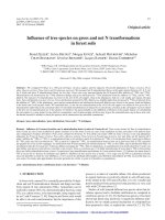
3.6.3. Growth of the mycelium on PDA nutrient medium at different moisture scales:
The experiment was conducted according to the method of Booth.C.P 1971 NaCl solution
is mixed with different concentrations in the desiccators make us get the moisture as follow:
Table 3.2. Formula to create moisture environment
NaCl (g/l)
0
16
32
48
64
RH (%)
100
90
80
70
60
Put mixed solutions into large desiccators. Put the lid on and put them in the
laboratory, in the temperature about 23-27°C. After 2 days, in different desiccators will have
different moisture, depend on NaCl concentration. When NaCl concentration is large,
moisture of environment is small. And vice versa, if the concentration of NaCl is smaller, the
environmental moisture will increase. PDA nutrient after sterilize autoclaving is poured into
sterile box with a thick layer 2 - 3 mm, implanted fungus into the middle of the petri dishes
containing the PDA, each desiccators put dishes.
Figure 3.7. Moisture experiment
After one day, 3 days, and 5 days- measure the diameter in two dimensions
perpendicular and take the average value.
17
4. RESULTS
4.1. Result of sample collection.
After collecting soil from 3 locations, 72 soil samples were collected.
Table 4.1. List of soil samples is collected
No.
Name
No.
Name
No.
Name
No.
Name
1
S01
19
S19
37
BB BD1
55
M2
2
S02
20
S20
38
CT BP2
56
M3
3
S03
21
S21
39
CT BP3
57
PG BD3
4
S04
22
S22
40
DMC TN1
58
PR BP1
5
S05
23
S23
41
DMC TN2
59
RTN BP1
6
S06
24
S24
42
DMC TN3
60
RTN BP2
7
S07
25
S25
43
DMC TN4
61
RTN BP3
8
S08
26
S26
44
DMC TN5
62
RTN BP4
9
S09
27
S27
45
DMC TN6
63
RTN BP5
10
S10
28
S28
46
DP BP5
64
SD1
11
S11
29
S29
47
DT BD6
65
SD2
12
S12
30
S30
48
DX BP4
66
SD3
13
S13
31
S31
49
LT DN2
67
TD1
14
S14
32
S32
50
LT ĐN2
68
TD2
15
S15
33
S33
51
LT ĐN3
69
TD3
16
S16
34
S34
52
LT ĐN6
70
TD4
17
S17
35
S35
53
LT ĐN7
71
TD5
18
S18
36
S36
54
M1
72
VC ĐN1
18
4.2. Result of isolation, and culturing the fungus.
Figure 4.1. Some strains under stereoscope
After isolation and culturing the fungus, 41 samples were isolated.
Table 4.2. List of fungi strains were found
No. Name
No. Name
No. Name
No. Name
1
BB BD1.1
12
DP BP5.2
22
RTN BP3.3
32
S15
2
BB BD1.2
13
DP BP5.3.1
23
RTN BP3.4
33
S16
3
BB BD1.3
14
DP BP5.3.2
24
RTN BP3.5.1
34
S24
4
BB BD1.4
15
DP BP5.4
25
RTN BP3.5.2
35
S29
5
DMC TN1.1
16
LT DN2.1
26
RTN BP3.6
36
S30.1.1
6
DMC TN1.2
17
LT DN2.2
27
RTN BP3.7
37
S30.1.2
7
DMC TN1.3
18
LT DN3
28
RTN BP5.1
38
S30.1.3
8
DMC TN2.1
19
RTN BP3.1.1
29
RTN BP5.2
39
S30.2
9
DMC TN2.2
20
RTN BP3.1.2
30
RTN BP5.3
40
S36.1
10
DMC TN2.3
21
RTN BP3.2
31
TD1
41
S36.2
11
DMC TN3
This fungus has cylindrical spore with septa are described in figure 4.2.
19