- Trang chủ >>
- Khoa Học Tự Nhiên >>
- Vật lý
mr bloomfields orchard the mysterious world of mushrooms molds and mycologists oct 2002
Bạn đang xem bản rút gọn của tài liệu. Xem và tải ngay bản đầy đủ của tài liệu tại đây (1.84 MB, 221 trang )
Mr. Bloomfield’s Orchard:
The Mysterious World of
Mushrooms, Molds,
and Mycologists
NICHOLAS P. MONEY
OXFORD UNIVERSITY PRESS
Mr. Bloomfield’s Orchard
This page intentionally left blank
Mr. Bloomfield’s Orchard
The Mysterious World of Mushrooms,
Molds, and Mycologists
NICHOLAS P. MONEY
1
2002
1
Oxford New York
Auckland Bangkok Buenos Aires Cape Town Chennai
Dar es Salaam Delhi Hong Kong Istanbul Karachi Kolkata Kuala Lumpur
Madrid Melbourne Mexico City Mumbai Nairobi São Paulo
Shanghai Singapore Taipei Tokyo Toronto
and an associated company in Berlin
Copyright © 2002 by Oxford University Press, Inc.
Published by Oxford University Press, Inc.
198 Madison Avenue, New York, New York 10016
www.oup.com
Oxford is a registered trademark of Oxford University Press
All rights reserved. No part of this publication may be reproduced,
stored in a retrieval system, or transmitted, in any form or by any means,
electronic, mechanical, photocopying, recording, or otherwise,
without the prior permission of Oxford University Press.
Library of Congress Cataloging-in-Publication Data
Money, Nicholas P.
Mr. Bloomfield’s orchard : a personal view
of fungal biology / Nicholas P. Money.
p. cm. Includes bibliographical references.
ISBN 0-19-515457-6
1. Fungi. I. Title.
QK603 .M59 2002
579.5—dc21 2002072654
1 3 5 7 9 8 6 4 2
Printed in the United States of America
on acid free paper
For Terence Ingold and his jewels
This page intentionally left blank
Contents
vii
Preface ix
CHAPTER 1 Offensive Phalli and Frigid Caps 1
CHAPTER 2 Insidious Killers 21
CHAPTER 3 What Lies Beneath 45
CHAPTER 4 Metamorphosis 65
CHAPTER 5 The Odd Couple 87
CHAPTER 6 Ingold’s Jewels 107
CHAPTER 7 Siren Songs 129
CHAPTER 8 Angels of Death 151
CHAPTER 9 Mr. Bloomfield’s Orchard 169
Notes 191
Index 203
This page intentionally left blank
Preface
It is indeed a singular and despised family to the history of which
we are about to dedicate this volume.
—M. C. Cooke, British Fungi (1871)
Some time ago, my colleague Jerry McClure told me that the most for-
tunate among us are faced with three options at the juncture in life once
valued as the midlife crisis: go insane, engage in an extramarital affair,
or write a book. In my own approach to this disconcerting landmark, all
but the third option vaporized under my wife’s guidance. The fruit of her
influence is in your hands.
Mr. Bloomfield’s Orchard is a personal reflection on the subject of
mycology, the scientific study of fungi. Many people giggle at the men-
tion of these organisms, drawing on vague notions about hallucinogens
and poisons, fairy tales, and the erectile behavior of mushrooms.
Although such peculiarities may draw people to this book, my primary
concern as its author is to explore our profound intimacy with fungi and
to articulate the most important consequences of these interactions.
Employing a flexible interpretation of that term interaction, this is a cel-
ebration both of the fungi (even the nasty ones) and of a selection of the
scientists obsessed with their study (none that I know of have been
exceptionally nasty). While I have written for a general audience, par-
ticularly those with some scientific education, I also hope to deepen the
appreciation of fungi among my biologist peers.
There are a number of people to whom I extend deep gratitude for
stimulating this book. As a teenager studying at Bristol University, my
first—and most inspiring—guide to mycology was Mike Madelin, and
my admiration for my doctoral mentor at Exeter, John Webster, grows
with every year. The dedication of this book to Terence Ingold is
ix
explained in the narrative. I also thank the staff of the Lloyd Library in
Cincinnati for maintaining the world’s supreme archive of mycological
publications. This book would not have been possible without the sanc-
tuary offered by the Lloyd. Speaking of sanctuaries, Frank Harold was
kind enough to offer me one in his laboratory in Colorado at a time when
I was lost in New England, and has now shown great generosity in
reviewing the Bloomfield manuscript. I thank my wife, Diana Davis, for
agreeing to marry me, and more pertinently in the context of this book,
for her invaluable service as my primary reader.
By discussing fungal processes that I have investigated (if only periph-
erally), this book has enabled me to revisit my twenty-year journey from
student to professional mycologist. I hope you have as much fun read-
ing about this odyssey as I have had recreating it.
Nicholas P. Money
Oxford, Ohio
January 2002
x PREFACE
Mr. Bloomfield’s Orchard
xi
This page intentionally left blank
CHAPTER 1
Offensive Phalli and Frigid Caps
I am . . . a mushroom
On whom the dew of heaven drops now and then.
—John Ford, The Broken Heart (1633)
All sound in the forest is damped by a morning mist trapped under the
pine trees on the edge of the moors in Devon, England. Three men are
tramping up a steep slope, their boots sinking into the soaking needles.
They are searching for eggs. A dead deer smell hangs in the watery air,
a hint of sweetness too, and even a suggestion of semen. This odor can-
not be ignored. Steamed glasses are wiped every few minutes. The old-
est of the men is wearing hunting pants that end at the knees, thick hik-
ing socks bridging the gaps to his red-laced boots. Webster stops, his
blue eyes bulging as he scans the forest floor. Squatting, he parts the
pine needles and uncovers five pure white eggs, somewhat larger than
golf balls. Each is attached to the soil by a branched umbilical cord that
snaps as it is tugged away from its siblings. The jelly-filled spheres have
cold skins. What monsters will hatch from such spawn? And what is
that smell?
A few feet from the nest is a very ugly penis. Poking 6 inches or more
from the pine needles, a full erection that arches a little, a pallid shaft pro-
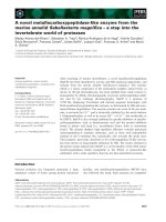
truding from a broken egg. Its head glistens with green-black syrup (Fig-
ure 1.1). This is the source of the smell. At the tip, a small hole is circled
by a raised ring. Some degenerate must be hiding under the needles and
is evidently aroused by the experience. But wait a moment; there are hun-
dreds of these apparitions higher up the slope. Have the collectors wan-
dered into a colony of sexual deviants fixated upon live burial?
1
But there are no horny corpses. Little Red Riding Hood’s chastity is safe.
The erections were accomplished by a fungus whose Latin name is Phal-
lus impudicus, the shameless penis, a type of “stinkhorn.” You must not
forego the spectacle offered by this beast. My first encounter with this
bizarre species was made during a foray with the mycologist John Web-
ster (Figure 1.2) and his Spanish assistant Henry Descals. The site on Dart-
moor was a favorite of John’s, a place he visited every year to collect spec-
imens for his undergraduate classes at the University of Exeter.
2 MR. BLOOMFIELD’S ORCHARD
Fig. 1.1 Erect fruiting body of Phallus impudicus.
Phallic mushrooms belong to the large group of fungi that includes the
more familiar organisms that generate brackets on trees and buttons and
portabella caps that end their lives sautéed in olive oil. These organisms are
members of a group of fungi called the Basidiomycota,
1
a name that refers
to a special kind of spore or microscopic seed called the basidiospore.
Thirty thousand species of basidiomycete have been described by scien-
tists, and seventy or so are phallic mushrooms and related fungi that man-
ufacture smelly cages. The phallic ones have proven impossible to ignore.
They are featured in Pliny the Elder’s thirty-seven-volume Natural History
written in the first century
A.D., a publication with the modest goal of
recording “all the contents of the entire world.” In his seventeenth-century
herbal, John Gerard pictured them in a modest, tip-down orientation, with
the following description: “Fungus virilis penis arecti forma, which wee Eng-
lish, [call] Pricke Mushrum, taken from his forme.” For Victorians in Eng-
land, sufficiently obsessed with sex to become excited by table legs, their
appearance was too much to bear. As a mature woman, Charles Darwin’s
daughter Etty so despised stinkhorns that she mounted an antifungal jihad
with the aid of gloves and a pointed stick. She burned the collections in
secret, thereby protecting the purity of thought among her female servants.
OFFENSIVE PHALLI AND FRIGID CAPS 3
Fig. 1.2 John Webster.
The transformation from egg to stinking horn is a slow erection that
often begins in the cool of the night and is not complete until sunrise. If an
unhatched egg is cut in half, the tissues of the expanded structure are dis-
played in prefabricated form (Figure 1.3). A hollow shaft of white spongy
material called the receptacle runs pole-to-pole through its center. The
receptacle is surrounded by the green-black cushion of spores called the
gleba, cased in a clear jelly veiled with white skin. When the egg hatches,
the receptacle expands by absorbing water and ruptures the skin, carrying
the spores on its tip into the air. The jelly lubricates the extending shaft and
helps keep the mass of spores in place. The spores are embedded in slime
that contains a cocktail of volatile chemicals, including hydrogen sulfide,
formaldehyde, methylmercaptan, and unique compounds called phallic
acids. Impersonating the smell of rotting flesh, the stinkhorn is irresistible
to flies, which swarm on the head, and to slugs, which glide for 20 or more
feet for the reward of the cadaverous confection. Within a few hours, the
head is cleaned down to the dimpled white surface of receptacle tissue, and
the shaft begins to wilt. Although the marathon erection is over, the
stinkhorn has been successful. Flies and slugs carry and defecate its spores,
whose stinkhorn genes contain the information needed to make more
stinkhorns. In common with humans, stinkhorns are here because they are
very good at making copies of themselves.
Stinkhorns and other mushrooms are the tips of mycological icebergs.
The umbilical cord at the bottom of the egg connects with the larger
organism that pulses unseen through leaf litter, crawls under the bark of
dying trees, and connects with the roots of healthier ones. This is the
feeding phase of the organism’s life, or life cycle, and grows as masses of
filamentous cells called hyphae. Only when these hyphae have gathered
a sufficient harvest of food, and when the subterranean fungus is fat-
tened and pumped full of water, can it surface to disturb our composure.
Biologists decipher the shape and structure of different organisms by
thinking about the functions for which they may be adapted, or the chal-
lenges that have been overcome by developing in a particular way. The
apparently ornamental figure of the phallic mushroom is really a very con-
servative structure. The top of the shaft is a sensible location for the spore
mass because its pungent slime is concentrated where it acts best as a bea-
con to flies. Stinkhorn receptacles are very delicate structures. They are built
4 MR. BLOOMFIELD’S ORCHARD
from masses of corrugated hyphae that are stretched into a weft of filaments
when the egg hatches. Most of the volume of the erect fruiting body is air.
But mechanically speaking, the stinkhorn is comparable with the mam-
malian penis because both erections are maintained by pressurized fluid
rather than a column of solid tissue. The penis contains flattened reservoirs
that become engorged with blood, while the tissue of the stinkhorn recep-
tacle is built to tear apart to make a honeycomb supported by pressurized
water within its hyphae. Despite these similarities, the origin of the pres-
surized fluid is fundamentally different in the two structures. Penile blood
pressure is generated by muscular activity; stinkhorn pressure is osmotic in
origin, something akin to the way that water is soaked up into a dry sponge.
While we can deconstruct the stinkhorn and explain its parts, the extraor-
dinary phallic resemblance remains a great surprise. I suppose that this
unusual fruiting body may be a jest by Satan—in its various stages of devel-
OFFENSIVE PHALLI AND FRIGID CAPS 5
Fig. 1.3 Cut egg of phallic mushroom. The central receptacle, which expands to
form the stalk, is surrounded by the green-black mass of developing spores called
the gleba. Jelly surrounds the gleba.
opment, Phallus has been identified as the devil’s eggs, devil’s horn, and
devil’s stinkpot
2
—but I’m putting my money on the Darwinian explanation.
At least for the fungus, fruiting bodies function to produce and disperse
spores, nothing else.
Mycologists have described thirty truly phallic-looking mushroom
species. As its common name suggests, the dog stinkhorn, Mutinus cani-
nus, is smaller in stature, has a pink shaft, and lacks the bulbous head. It
still smells awful and attracts flies. Species of Dictyophora are recognized
by a lacy veil that hangs down as a skirt beneath the head (see jacket
photo). The crinoline feminizes the phallic effect a little, and may offer a
ladder that allows wingless insects to reach the spores by crawling from
surrounding plants. The eggs of one species of Dictyophora are sold as
delicacies in China and are also marketed as aphrodisiacs. Inside the egg,
stinkhorn slime does not smell too awful, and some authors of mushroom
guidebooks claim that the whole thing can be consumed without much
suffering. In his book, In the Company of Mushrooms, Elio Schaechter
3
admitted to enjoying stinkhorn eggs and remarked that once filled with
cream, rings cut from the expanded receptacles were delicious. On a more
general note, it is a tragedy in a country as populous as China that any-
thing from tiger turds to whale afterbirths can be sold as long as the sug-
gestion is made that their consumption enhances erectile function.
The related cage fungi produce other kinds of flamboyant fruiting bod-
ies that share the seductive power that phallic mushrooms wield over
insects. Again, a preformed receptacle is packaged into an egg, and as this
structure absorbs water and expands, it carries a stinking spore mass into
the air. Rather than exiting the egg as a single shaft, the receptacles of cage
fungi unfold into more open structures. Clathrus forms a spherical cage
with spores spread on the inside of its bars (Figure 1.4 a). The receptacle
of Anthurus separates into four or more arms that curl back over the egg
to create a star (Figure 1.4 b). The arms are bright orange and their inner
surface is smeared with the spores. A time-lapse video that shows the
hatching of an Anthurus egg is quite shocking. It is difficult to describe
the performance delivered by this fungus. There is nothing comparable.
Here’s my best shot: as this fruiting body issues from the ground, its livid
arms simulate the agonized contortions of a horribly injured lobster.
Other cage fungi form stalks with chambered heads or claws at their sum-
mit, and Laternea elaborates long arms like Anthurus but fuses them at
6 MR. BLOOMFIELD’S ORCHARD
their tips and dangles a reeking lantern inside the resulting vault (Figure
1.4 c). Flies are the usual vectors for spore dispersal, but ants and sting-
less bees have also been seen feeding on some cages.
Ileodictyon (intestinal net) is a cage fungus that grows in New Zealand
and Australia. The Maori were quite taken with this fruiting body, accord-
ing it nine different names and barbecuing its eggs. When it escapes
human consumption, the white Ileodictyon cage expands from a buried
egg and disengages from its papery skin. The detached cage, smeared with
the usual excremental spore gunk, is then blown about on the surround-
ing grass. The Maori didn’t eat the repugnant hatchlings, denigrating
them as the “feces of ghosts or of the stars.” The quote is taken from an
intriguing article by the distinguished British mycologist Graham Goo-
day (a delightful scientist whose appearance evokes the stereotypical
image of a Royal Air Force fighter pilot from the Second World War) and
his friend John Zerning.
4
Zerning was struck by the shape of a dried spec-
imen of Ileodictyon displayed by Gooday at a meeting. He noticed the
resemblance between the cage and the geodesic homes designed by
Richard Buckminster Fuller (Bucky), which became popular in the
1960s. This polyhedral form is also characteristic of the carbon-based
molecules called buckminsterfullerenes, or buckyballs, which come close
to making organic chemistry seem interesting. The similarity of hippy
dwellings, buckyballs, and ghost feces is a reflection of the surprising
strength offered by their lightweight polyhedral structure. Any weight
saving is valuable for a fungus that, by necessity, makes conservative use
of building materials. The resistance to compression of the Ileodictyon
cage is important during its emergence from buried eggs and also when
it is blown around. By maintaining an open shape, the receptacle provides
a large surface area for exposure of the spores and their fetid scent.
Small changes in the details of receptacle development probably account
for the great variety of mature fruiting body shapes in stinkhorns and cage
fungi. For example, weakening of tissue along four or five tracks running
the length of the receptacle would cause the shaft to split like a banana skin
into four or five arms upon pressurized expansion. This would require
alterations in the arrangement of the receptacle tissues inside the egg, or
changes in the activity of specific enzymes during hatching. Then, with the
mobilization of some genes to control the orange coloration of the recep-
tacle, a Phallus-type fruiting body would be transformed into Anthurus.
OFFENSIVE PHALLI AND FRIGID CAPS 7
This is an oversimplification of the developmental differences between
these organisms, because there are other microscopic distinctions between
their structures. But research on other fungi does suggest that conspicuous
modifications in fruiting body morphology can be derived by surprisingly
minor changes in the expression of enzymes during development.
Given the similarities among all of the phallic and cage fungi, it seems
probable that natural selection may have sculpted the existing species in a
relatively short period of time, perhaps in as little as a few million years. But
why did all these structures evolve? Why did a phallus that divides at its tip
evolve from an ancestor that did not, or vice versa? The answer surely lies
in the relationships between these fungi and the insects and other inverte-
brates that disperse their spores. Different species of flies are lured by par-
ticular scents and personalized visual cues, so the various receptacles prob-
ably reflect distinctive solutions to the challenge of supporting and
8 MR. BLOOMFIELD’S ORCHARD
Fig. 1.4 Fruiting bodies of various gasteromycete fungi. (a–c) cage fungi: (a)
Clathrus ruber; (b) Anthurus archeri; (c) Laternea triscapa; (d) puffball, Lycoperdon
perlatum; (e) earth-star, Geastrum fornicatum; (f ) bird’s nest fungus, Cyathus stria-
tus. Not drawn to the same scale.
abc
de f
advertising spore slime. Biologists already recognize the significance of anal-
ogous characteristics in the origins of flowers among insect-pollinated
plants. While humans are seduced by many floral perfumes, colors, and
shapes, there are also numerous insect-pollinated flowers, such as the Suma-
tran giant Amorphophallus titanum, or corpse flower, which emit stinkhorny
smells.
5
Stinkhorns, cage fungi, and putrid flowers have all evolved parallel
features that attract insects that ordinarily congregate around carrion.
Along with the stinkhorns and cage fungi, other organisms including
puffballs, earth-balls, earth-stars, and bird’s nest fungi belong to the gas-
teromycete section of the basidiospore-producing fungi (Figure 1.4 d–f).
Surpassing the inventions of all other fungi, the gasteromycetes have
evolved a circus of mechanisms for dispersing their spores. Adapting an
image from Richard Dawkins, baby stinkhorns use insect wings to fly
away from their parents.
6
The offspring of puffballs, earth-balls, and
earth-stars are puffed into the air and are then carried away by wind.
Bird’s nest fungi also use a two-stage dispersal mechanism. Their tiny
fruiting bodies are shaped like champagne flutes and contain packets of
spores called peridioles. Raindrops splash the peridioles from these cups
onto surrounding blades of grass. Unsuccessful spores, those destined
for a swift passage to stinkhorn heaven or hell, wait, and wait longer, and
dehydrate, and die. Fortunate ones are consumed by herbivores grazing
around the fruiting bodies, are carried by the animals as they pass
through their digestive systems, and later deposited in a convenient pat
of warm manure. Cow feces offer perfect residence for a young bird’s nest
fungus (suggesting that stinkhorn hell lacks the excrement-filled ditch
of Dante’s Inferno). Finally, one gasteromycete fungus shoots a black ball
of spores from a fruiting body that operates as a tiny trampoline. This
organism, called Sphaerobolus, grows on wood mulch, and can ruin the
paintwork of a car parked close to a wet flower bed. The spore balls stick
to smooth surfaces with incredible tenacity, and even when they are
removed by vigorous cleaning, spots remain in the paint. Like the bird’s
nest fungi, this villain is adapted for an excursion through a herbivore
gut, but it doesn’t help to know that the intended targets of Sphaerobo-
lus are grass blades rather than my beloved Ford Probe.
The gasteromycetes are defined by the fact that their spores form inside
the fruiting body rather than on gills or other fertile surfaces exposed to
OFFENSIVE PHALLI AND FRIGID CAPS 9
the air. Their scientific name refers to this developmental feature: gastero
= stomach, mycetes = fungi, stomach fungi. They seem to have evolved
from different kinds of ancient fungi that produced conventional
umbrella-shaped mushrooms, and as such are regarded as a ragbag of
species rather than a natural grouping of organisms. The natural group is
an important concept in biology. Contrary to the delusions of Christian
fundamentalists, all animals with nipples and fur, for example, are
descendants of a single ancestral species. They belong to a natural group,
the mammals, from which every other living thing is excluded: without
nipples you don’t even merit an interview. Time is a crucial consideration
in this discussion, because, of course, every pair of species shares an
ancestor that could be found by delving back far enough into their respec-
tive evolutionary histories. Humans and stinkhorns are certainly related,
and far more closely (according to their genes) than either is to any plant.
But the natural group that includes Homo and Phallus also encompasses
every animal and every fungus, and as such is a pretty esoteric gathering.
In common with stinkhorns, gilled mushrooms are devices for spore pro-
duction and dispersal, nothing more or less. They have always held great
fascination for me, and I suppose my deepest professional roots lie in child-
hood tales involving mushrooms. My earliest memory of a mycological
experience comes from a dentist’s office. I was 5 years old and under gas for
multiple tooth extractions when I hallucinated a fairy ring with elves and
other phantasms dancing around the mushrooms. Then I awoke, tumbling
down the stairs from the torture chamber, bloody handkerchief pressed to
my mouth. I have remained captivated by the eeriness of mushrooms, and
have joined the ranks of mycologists who have become fascinated by trying
to understand how they operate. This is not a simple matter.
Umbrella- and bracket-shaped mushrooms maximize their spore-
producing capacity for a minimal investment in fruiting body tissue by
supporting massive numbers of spores with a single stalk. These fungi
spread their fertile tissues underneath the cap, folding a vast spore-
producing mat called the hymenium over the surface of gills, ripples, or
spines, or inside tubes. If a thin slice is cut from a mushroom cap with
a razor blade and viewed under a microscope, spore-producing cells
called basidia appear as projections from the hymenium (Figure 1.5).
Basidia are four-pronged crowns shaped like miniature cow udders and
bear a single basidiospore on each spike (or teat). One after the other,
10 MR. BLOOMFIELD’S ORCHARD
each of the spores in a quartet is catapulted horizontally, but only for a
short distance so that it does not hit the neighboring gill. After this mil-
lisecond journey, gravity assumes control of the flight path, and the
spore turns abruptly and accelerates straight down between the gills.
Once the spore falls beneath the cap it is swept away by air currents. If
an active mushroom is observed in the correct lighting, a dusty plume
of basidiospores is visible swirling away from the cap (Figure 1.6).
OFFENSIVE PHALLI AND FRIGID CAPS 11
Fig. 1.5 Microscopic view of spore expulsion from surface of a mushroom gill.
Note that the fluid drop is carried on the surface of the discharged spore. From
A.H.R. Buller, Researches on Fungi, vol. 2 (London: Longmans, Green, 1922).
Fig. 1.6 Cloud of spores dispersing from the horse mushroom, Agaricus arvensis.
Mushroom illustrated in section to expose the gills. An immature fruiting body is
connected to the same mycelium. From A.H.R. Buller, Researches on Fungi, vol. 1
(London: Longmans, Green, 1909).
The mechanism that catapults spores from the hymenium was solved
only recently. It relies upon the condensation of water on the surface of
the spore. A few seconds before discharge, a little bead of liquid devel-
ops at the base of the spore, grows until it becomes almost as wide as the
spore itself, and then, instantly, fluid and spore disappear (Figure 1.7).
John Webster, the egg hunter introduced earlier, tried to capture the dis-
charge process using high-speed cameras at a film institute in Germany.
The capacity for film wastage in this project was appalling. Webster
watched through the microscope, holding a trigger for the camera and
waiting for the appearance of the droplet of fluid. When the trigger was
squeezed, thousands of frames of film were pulled through the camera
in a couple of seconds by a deafening motor connected to the spool. But
even at very high speeds, the best sequences showed hundreds of frames
with a spore and its droplet, followed by hundreds of frames showing a
naked spike of a basidium from which the spore disappeared.
Now that the catapult mechanism is understood, the photographic
results are understandable. The spore is shot so fast from the gill that a
camera running at 20,000 frames per second would be needed to cap-
ture the event.
7
The final speed of the spore is only one meter per sec-
ond, compared with, for example, 7,800 meters per second for the Space
Shuttle. But the acceleration of the spore is quite astonishing. From a
standing start, this fungal cell covers a distance of one millimeter in a
thousandth of a second. This sounds more impressive when you con-
sider that the spore is only ten-millionths of a meter in length (10 µm),
so that its journey corresponds to a distance 100 times its own size. Scal-
ing up to human dimensions, this would be equivalent to vaulting from
a cliff edge and almost instantaneously reaching a speed of 400 miles per
hour. The spore pulls thousands of g’s when it is flung from the gill, 10
times more than a jumping flea. This feat would atomize a bungee
jumper.
The formation of the drop preceding spore discharge was first
described by a French scientist, Victor Fayod, in 1889, but more than a
century of research ensued before the discharge mechanism was
explained by John Webster.
8
It is important to recognize that the space
between the gills is saturated with water vapor that evaporates from the
mushroom’s tissues. Sugars and other molecules seep from the interior
12 MR. BLOOMFIELD’S ORCHARD