báo cáo hóa học:" Static platelet adhesion, flow cytometry and serum TXB2 levels for monitoring platelet inhibiting treatment with ASA and clopidogrel in coronary artery disease: a randomised cross-over study" potx
Bạn đang xem bản rút gọn của tài liệu. Xem và tải ngay bản đầy đủ của tài liệu tại đây (872.67 KB, 14 trang )
BioMed Central
Page 1 of 14
(page number not for citation purposes)
Journal of Translational Medicine
Open Access
Research
Static platelet adhesion, flow cytometry and serum TXB
2
levels for
monitoring platelet inhibiting treatment with ASA and clopidogrel
in coronary artery disease: a randomised cross-over study
Andreas C Eriksson*
1
, Lena Jonasson
2
, Tomas L Lindahl
3
, Bo Hedbäck
2
and
Per A Whiss
1
Address:
1
Division of Drug Research/Pharmacology, Department of Medical and Health Sciences, Linköping University, SE-581 85 Linköping,
Sweden,
2
Division of Cardiology, Department of Medical and Health Sciences, Linköping University, SE-581 85 Linköping, Sweden and
3
Department of Clinical Chemistry, Laboratory Medicine, University Hospital, SE-581 85 Linköping, Sweden
Email: Andreas C Eriksson* - ; Lena Jonasson - ; Tomas L Lindahl - ;
Bo Hedbäck - ; Per A Whiss -
* Corresponding author
Abstract
Background: Despite the use of anti-platelet agents such as acetylsalicylic acid (ASA) and clopidogrel in coronary heart
disease, some patients continue to suffer from atherothrombosis. This has stimulated development of platelet function
assays to monitor treatment effects. However, it is still not recommended to change treatment based on results from
platelet function assays. This study aimed to evaluate the capacity of a static platelet adhesion assay to detect platelet
inhibiting effects of ASA and clopidogrel. The adhesion assay measures several aspects of platelet adhesion
simultaneously, which increases the probability of finding conditions sensitive for anti-platelet treatment.
Methods: With a randomised cross-over design we evaluated the anti-platelet effects of ASA combined with clopidogrel
as well as monotherapy with either drug alone in 29 patients with a recent acute coronary syndrome. Also, 29 matched
healthy controls were included to evaluate intra-individual variability over time. Platelet function was measured by flow
cytometry, serum thromboxane B
2
(TXB
2
)-levels and by static platelet adhesion to different protein surfaces. The results
were subjected to Principal Component Analysis followed by ANOVA, t-tests and linear regression analysis.
Results: The majority of platelet adhesion measures were reproducible in controls over time denoting that the assay
can monitor platelet activity. Adenosine 5'-diphosphate (ADP)-induced platelet adhesion decreased significantly upon
treatment with clopidogrel compared to ASA. Flow cytometric measurements showed the same pattern (r
2
= 0.49). In
opposite, TXB
2
-levels decreased with ASA compared to clopidogrel. Serum TXB
2
and ADP-induced platelet activation
could both be regarded as direct measures of the pharmacodynamic effects of ASA and clopidogrel respectively. Indirect
pharmacodynamic measures such as adhesion to albumin induced by various soluble activators as well as SFLLRN-induced
activation measured by flow cytometry were lower for clopidogrel compared to ASA. Furthermore, adhesion to collagen
was lower for ASA and clopidogrel combined compared with either drug alone.
Conclusion: The indirect pharmacodynamic measures of the effects of ASA and clopidogrel might be used together with
ADP-induced activation and serum TXB
2
for evaluation of anti-platelet treatment. This should be further evaluated in
future clinical studies where screening opportunities with the adhesion assay will be optimised towards increased
sensitivity to anti-platelet treatment.
Published: 9 June 2009
Journal of Translational Medicine 2009, 7:42 doi:10.1186/1479-5876-7-42
Received: 27 February 2009
Accepted: 9 June 2009
This article is available from: />© 2009 Eriksson et al; licensee BioMed Central Ltd.
This is an Open Access article distributed under the terms of the Creative Commons Attribution License ( />),
which permits unrestricted use, distribution, and reproduction in any medium, provided the original work is properly cited.
Journal of Translational Medicine 2009, 7:42 />Page 2 of 14
(page number not for citation purposes)
Background
Anti-platelet drugs such as acetylsalicylic acid (ASA) and
clopidogrel are routinely used to prevent thrombosis in
cardiovascular disease. The benefits of ASA have been
clearly demonstrated by the Anti-platelet Trialists' Collab-
oration [1]. They found that ASA therapy reduces the risk
by 25% of myocardial infarction, stroke or vascular death
in "high-risk" patients. When using the same outcomes as
the Anti-platelet Trialists' Collaboration on a comparable
set of "high-risk" patients, the CAPRIE-study showed a
slight benefit of clopidogrel over ASA [2]. Furthermore,
the combination of clopidogrel and ASA has been shown
to be more effective than ASA alone for preventing vascu-
lar events in patients with unstable angina [3] and myo-
cardial infarction [4,5] as well as in patients undergoing
percutaneous coronary intervention (PCI) [6,7]. Despite
the obvious benefits from anti-platelet therapy in coro-
nary disease, low response to clopidogrel has been
described by several investigators [8-10]. A lot of attention
has also been drawn towards low response to ASA, often
called "ASA resistance". The concept of ASA resistance is
complicated for several reasons. First of all, different stud-
ies have defined ASA resistance in different ways. In its
broadest sense, ASA resistance can be defined either as the
inability of ASA to inhibit platelets in one or more platelet
function tests (laboratory resistance) or as the inability of
ASA to prevent recurrent thrombosis (i.e. treatment fail-
ure, here denoted clinical resistance) [11-13]. The lack of
a general definition of ASA resistance results in difficulties
when trying to measure the prevalence of this phenome-
non. Estimates of laboratory resistance range from
approximately 5 to 60% depending on the assay used, the
patients studied and the way of defining ASA resistance
[11,13]. Likewise, lack of a standardized definition of low
response to clopidogrel makes it difficult to estimate the
prevalence of this phenomenon as well [8]. The principles
of existing platelet assays, as well as their advantages and
disadvantages, have been described elsewhere [14-18]. In
short, assays potentially useful for monitoring treatment
effects include those commonly used in research such as
platelet aggregometry and flow cytometry as well as
immunoassays for measuring metabolites of thrombox-
ane A
2
(TXA
2
). Also, the PFA-100™, Multiplate™ and the
VerifyNow™ are examples of instruments commercially
developed for evaluation of anti-platelet therapy. How-
ever, no studies have investigated the usefulness of alter-
ing treatment based on laboratory findings of ASA
resistance [19]. Regarding clopidogrel, there are recent
studies showing that adjustment of clopidogrel loading
doses according to vasodilator-stimulated phosphopro-
tein phosphorylation index measured utilising flow
cytometry decrease major adverse cardiovascular events in
patients with clopidogrel resistance [20,21].
The current study used a randomised cross-over design in
order to investigate the effects on platelets of dual therapy
with ASA and clopidogrel as well as the effects of either
drug alone in patients with a recent acute coronary syn-
drome. Platelet function was assessed by means of flow
cytometry, serum TXB
2
-levels and by measuring static
platelet adhesion to proteins in microplates. The aim was
to evaluate the usefulness of the static platelet adhesion
assay for measuring the effects of ASA and clopidogrel.
Static adhesion is an aspect of platelet function that has
not been investigated in earlier studies of the effects of
platelet inhibiting drugs. Consequently, static platelet
adhesion is not measured by any of the current candidate
assays for clinical evaluation of platelet function. The
static platelet adhesion assay offers an opportunity for
simultaneous measurements of the combined effects of
several different platelet activators on platelet function. In
this study, platelet adhesion to albumin, collagen and
fibrinogen was investigated in the presence of soluble
platelet activators including adenosine 5'-diphosphate
(ADP), adrenaline, lysophosphatidic acid (LPA) and ris-
tocetin. Collagen, fibrinogen, ADP and adrenaline are
physiological agents that are well-known for their interac-
tions with platelets. Ristocetin is a compound derived
from bacteria that facilitates the interaction between von
Willebrand factor (vWf) and glycoprotein (GP)-Ib-IX-V
on platelets, which otherwise occurs only at flow condi-
tions [22]. The static nature of the assay therefore
prompted us to include ristocetin in order to get a rough
estimate on GPIb-IX-V dependent events [23]. LPA is a
phospholipid that is produced and released by activated
platelets and that also can be generated through mild oxi-
dation of LDL [24]. It was included in the present study
since it is present in atherosclerotic vessels and suggested
to be important for platelet activation after plaque rup-
ture. Finally, albumin was included as a surface since the
platelet activating effect of LPA can be detected when
measuring adhesion to such a surface [25]. Thus, by the
use of different platelet activators, several measures of
platelet adhesion were obtained simultaneously. This
means that the possibilities to screen for conditions
potentially important for detecting effects of platelet-
inhibiting drugs far exceeds the screening abilities of other
platelet function tests. Consequently, the static platelet
adhesion assay is very well suited for development into a
clinically useful device for monitoring platelet inhibiting
treatment. Also, it has earlier been proposed that investi-
gating the combined effects of two activators on platelet
activity might be necessary in order to detect effects of ASA
and other antiplatelet agents [26]. This is a criterion that
can easily be met by the static platelet adhesion assay.
Through the screening procedure we found different con-
ditions where the static adhesion was influenced by the
drug given. This suggests that the assay is able to detect
Journal of Translational Medicine 2009, 7:42 />Page 3 of 14
(page number not for citation purposes)
treatment effects, but further studies are needed in order
to refine the measurements.
Methods
Study design
The study was approved by the Research Ethics Commit-
tee of Linköping University, Linköping, Sweden and the
Medical Product Agency, Sweden (EudraCT Number
2005-003927-38). A total of 33 patients recently diag-
nosed with acute coronary syndrome were included on a
consecutive basis from the Department of Cardiology at
the University Hospital in Linköping, Sweden (Figure 1).
Exclusion criteria were type 1 diabetes, immunologic or
malignant disease, hepatic or kidney disease, heart failure
NYHA class III-IV, heart valve disease, thoracal epidural
anaesthesia or treatment with antibiotics, immunosup-
pressive drugs or continuous use of non-steroidal anti-
inflammatory drugs (NSAID). At the index event, 8
patients received a bare metal stent and 15 received a
drug-eluting stent following coronary angioplasty. During
the course of the study, two patients were lost because of
recurrent myocardial infarction and two left the study by
their own decisions. Thus 29 patients, 19 males and 10
females, completed the study. When entering the study
the male patients were on average 57 years old (range 40–
69 years), while mean age for the female patients were 60
years (range 52–66 years). In parallel we collected sam-
ples from 30 healthy controls matched for age and gender.
Only blood from controls declaring that they had not
used any anti-platelet medication for two weeks prior to
the study was used. For every control, samples were taken
at two occasions separated by 2–5.5 months (Figure 1).
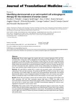
Flow chart showing the inclusion of patients and controlsFigure 1
Flow chart showing the inclusion of patients and controls. Patients and controls were included consecutively. Blood
samples from controls were drawn at two different occasions separated by 2–5.5 months. All patients entering the study
received ASA combined with clopidogrel and blood sampling was performed 1.5–6.5 months after initiating the treatment. This
was followed by a randomised cross-over enabling all patients to receive monotherapy with both ASA and clopidogrel. The
patients received monotherapy for at least 3 weeks and for a maximum of 4.5 months before performing blood sampling. A
total of 33 patients and 30 controls entered the study. In the end, 29 patients and 29 controls completed the study.
Contr ols fulfilling inclusion
criter ia at visit 1 (n=29)
Controls lost to blood
sampling (n=1)
Contr ols fulfilling inclusion
criter ia at visit 2 (n=29)
Patients r andomised to
clopidogr el treatment (n=16)
Patients lost to blood sampling
(n=2)
Patients r andomised to ASA
treatment (n=17)
Patients lost to blood sampling
(n=1)
Patients r andomised to
clopidogr el treatment (n=16)
Patients r andomised to ASA
treatment (n=14)
Patients lost to blood sampling
(n=1)
Patients r eceiving clopidogr el
+ ASA treatment (n=33)
Patients completing the study
(n=29)
Contr ols completing the study
(n=29)
Visit 1
Visit 2
Visit 3
Journal of Translational Medicine 2009, 7:42 />Page 4 of 14
(page number not for citation purposes)
One of the controls was excluded because of intake of
NSAIDs meaning that a total of 29 controls, 19 males and
10 females, completed the study. At study entry the mean
age of the male controls were 59 years (range 40–69
years), while mean for the female controls were 60 years
(range 51–65 years).
Blood was drawn from patients at three different occa-
sions (Figure 1). The first sample was drawn after all
patients had received combined treatment with ASA (75
mg/day) and clopidogrel (75 mg/day) for 1.5–6.5 months
after the index event. The study then used a randomised
cross-over design meaning that half of the patients
received ASA as monotherapy while half received only
clopidogrel (75 mg/day for both monotherapies). The
monotherapy was then switched for every patient so that
all patients in total received all three therapies. Samples
for evaluation of the monotherapies were drawn after
therapy for at least 3 weeks and at the most for 4.5
months. Most of the differences in treatment length can
be ascribed to the fact that the national recommendations
for treatment in this patient group were changed during
the course of the study. The allocation to monotherapy
was blinded for the laboratory personnel. In general, the
use of three different treatments for intra-individual com-
parisons in a cross-over design is different from previous
studies on ASA and clopidogrel, which have mainly been
concerned with only two treatment alternatives.
Whole blood was drawn from antecubital veins and col-
lected in (1) tubes containing sodium heparin (final conc.
17 units/mL) for platelet adhesion analysis, (2) tubes with
no additives for measurements of serum TXB
2
and (3)
tubes containing sodium citrate (final conc. 0.129 mol/L)
for flow cytometric analysis (patients only). To obtain
platelet rich plasma (PRP) for platelet adhesion analysis,
8 mL blood was transferred from sodium heparin tubes to
a single plastic centrifuge tube. This single tube was then
centrifuged for 20 min at 205 × g resulting in the produc-
tion of a PRP supernatant. Blood obtained in serum tubes
were allowed to clot at room temperature followed by
centrifugation for 10 min at 1000 × g. The serum was
transferred to eppendorf-tubes and stored at -70°C until
analysis of TXB
2
. For patients, blood samples were also
drawn into lithium heparin-tubes and K
2
EDTA-tubes for
biochemical analysis at the accredited Department of
Clinical Chemistry at the University Hospital in
Linköping, Sweden. The lithium heparin-tubes were used
for analysis of plasma concentrations of C-reactive protein
(CRP), cholesterol, triglycerides, LDL-cholesterol, HDL-
cholesterol, apolipoprotein-A1 (Apo-A1) and apolipopro-
tein-B (Apo-B), utilising the clinical chemistry analyzer
Advia 1650 from Roche. Concentrations of platelets and
leukocytes were determined from the K
2
EDTA-samples.
Static platelet adhesion
Static platelet adhesion was measured as previously
described [27]. Ninety-six well microplates (Nunc Max-
isorp, Roskilde, Denmark) were coated with proteins by
the addition of 100 μL/well of 2 mg/mL human albumin
(Octapharma AB, Stockholm, Sweden), 0.1 mg/mL
bovine collagen I (RnDsystems, Abingdon, UK) or 2 mg/
mL human fibrinogen (American Diagnostica Inc., Green-
wich, Connecticut, USA) followed by incubation at 4°C at
least overnight and for a maximum of 7 days. The micro-
plates were then washed two times in 0.9% NaCl by plate
inversion followed by the addition of 25 μL 0.9% NaCl or
25 μL MgCl
2
(5 mmol/L final concentration) and 25 μL of
platelet activators. The soluble platelet activators were
ADP and LPA from Sigma-Aldrich (St Louis, Missouri,
USA), adrenaline from Merck NM AB (Stockholm, Swe-
den) and ristocetin from Diagnostica Stago (Asnières-sur-
Seine, France) (Additional file 1: Variables). Experiments
were performed both in the absence and presence of
MgCl
2
since MgCl
2
has been shown to affect platelet adhe-
sion to the protein surfaces tested in this study [27,28].
The microplates were left for 20 min and then 50 μL PRP
diluted 4 times with 0.9% NaCl was added. Platelets were
then allowed to attach to the surfaces for 1 h at room tem-
perature without shaking. After incubation, unbound
platelets were removed by washing twice in 0.9% NaCl by
plate inversion and 140 μL of a sodium citrate/citric acid
buffer (0.1 mol/L, pH 5.4) containing 0.1% Triton X-100
and 1 mg/mL p-nitrophenyl phosphate (Sigma-Aldrich)
was added. Background absorbance was measured at 405
nm using a Spectramax microplate reader (Molecular
Devices, Sunnyvale, California, USA) and the microplates
were then incubated for 40 min at room temperature dur-
ing shaking. In parallel, 50 μL PRP as well as 50 μL 0.9%
NaCl were added to wells on a separate microplate. Both
PRP and NaCl wells were treated with 140 μL of the
sodium citrate/citric acid buffer described above followed
by background absorbance measurements and conse-
quently served as controls for 100% and 0% adhesion
respectively. During the 40 min incubation, an enzymatic
reaction occurred between added phosphatase substrate
and platelet acid phosphatase. Adding 100 μL 2 mol/L
NaOH to all wells (including 100% and 0%) stopped the
reaction and resulted in a colour change of the developed
product. Absorbance was measured at 405 nm with auto-
matic reduction of background absorbance and percent-
age platelet adhesion was calculated.
Flow cytometry
Platelet expression of P-selectin and binding of fibrinogen
were measured by flow cytometry as indicators of platelet
activation [29-32]. To tubes intended for fibrinogen bind-
ing analysis, 10 μL FITC-conjugated chicken anti-fibrino-
gen-antibodies (Diapensia, Linköping, Sweden) was
mixed with 100 μL Hepes buffer. Hepes buffer containing
Journal of Translational Medicine 2009, 7:42 />Page 5 of 14
(page number not for citation purposes)
EDTA was mixed with 10 μL of the same antibody for esti-
mation of background fluorescence. For P-selectin meas-
urements, 10 μL FITC-conjugated chicken anti-P-selectin-
antibodies (Diapensia) were added to 100 μL Hepes
buffer. Samples containing 10 μL anti-insulin-FITC (Dia-
pensia) and 100 μL Hepes buffer served as indicators of
background fluorescence. Whole blood (10 μL) was
added to all tubes followed by addition of 10 μL ADP, the
thrombin receptor PAR1 activating peptide SFLLRN (The
Biotechnology Centre of Oslo, Oslo University, Norway)
or vehicle (Hepes buffer) (Additional file 1: Variables).
After incubation for 10 minutes, the reaction was stopped
by addition of 1 mL Hepes buffer. Before flow cytometric
analysis, samples were diluted three times in Hepes buffer
and incubated for 30 min, while protected from light.
Flow cytometric analysis was performed with the instru-
ment Beckman Coulter Epics XL-MCL (Beckman Coulter
Inc., Fullerton, California, USA) with computer software
program (Expo 32 ADC, Beckman Coulter Inc.). The fluo-
rescence intensity was checked daily with fluorescent
beads (Flow set, Beckman Coulter Inc.). 5000 events were
collected based on their forward and side scatter proper-
ties.
TXB
2
Enzyme Immuno Assay
Serum levels of TXB
2
were measured with a commercial
enzyme immuno assay (EIA) kit according to the manu-
facturers' instructions (Cayman Chemical, Ann Arbor,
Michigan, USA). Amount of TXB
2
present in serum was
calculated with the use of a data analysis tool developed
by Cayman Chemical [33].
Statistics
The variables measured were subjected to Principal Com-
ponent Analysis (PCA) with direct obliminal rotation
using SPSS 14.0 software (SPSS Inc., Chicago, Illinois,
USA). This technique analyses to what extent different var-
iables are measuring the same concept and allows corre-
lating variables to be ordered into separate factors [34].
The PCA performed in this study included a total of 69
variables. Each variable were included in the PCA as a
composite of the results obtained from all data available
for the specific variable. Thus, variables measured in both
patients and controls (platelet adhesion and serum TXB
2
-
levels) consisted of data from three measurements on
patients and two on controls. All other variables were only
analysed on patients, which resulted in three measure-
ments that were included in the PCA. A variable was con-
sidered to be part of a factor when its loading was ≥ 0.4.
After finding distinct factors, the composite variables
included in the PCA were standardised according to Z-
scores. This procedure transforms all variables to the same
scale having a mean value of 0 and a standard deviation
of 1. For each individual, a mean was calculated from the
Z-scores of the variables that were found to belong to the
same factor. From the Z-mean of the individuals, a Z-
mean of the whole factor was calculated and further used
for statistical comparisons of means. The factors, as well as
some representative variables, were then analysed for
treatment effects and for intra-individual variations
within controls by Repeated Measures ANOVA. Differ-
ences between controls and patients were analysed by
One-sample t-test. Correlations between factors were
investigated with linear regression.
Results
Principal Component Analysis
In total the PCA grouped the initial 69 variables of platelet
activation and routine clinical chemistry analyses into 15
different factors that we renamed according to the aspects
they measured (Additional file 2: Factors). These names
and/or the factor numbers are used throughout the article
when describing and discussing the results of the present
study. This procedure including screening followed by sta-
tistical complexity reduction is unusual for this type of
study. Among the variables measuring platelet function,
platelet adhesion was represented by eight factors, flow
cytometry by two factors and serum TXB
2
formed a sepa-
rate factor. Visual inspection of the data of the healthy
controls for the initial factor solution revealed possibili-
ties for making the factors corresponding to platelet adhe-
sion even simpler. Attention was paid at (1) different
concentrations of the same soluble agonist on a specified
surface, (2) the effects of weak agonists compared to basal
adhesion and (3) the effect of an agonist compared to its
combination with another agonist.
The first scenario was found in factor 1. Since all surfaces
are represented with ADP at 1 and 10 μmol/L, it might be
possible that addition of 1 μmol/L ADP results in maxi-
mal platelet adhesion with 10 μmol/L not contributing
any further. In such a case it would be unnecessary to
include the high concentration of ADP since it would not
contribute any additional information. This was analysed
by paired analysis for the two doses of ADP on every sin-
gle surface. On all surfaces, ADP at 10 μmol/L was signif-
icantly different from 1 μmol/L ADP and all variables in
Factor 1 were therefore kept on this basis. However, four
of the variables in Factor 1 were excluded for other reasons
(see next section).
The second scenario regarding the effect of weak agonists
can be exemplified by Factor 5. It is possible that weak
agonists do not increase platelet adhesion significantly
compared to adhesion to the surface alone. As was the
case for different doses of ADP, the weak agonist will then
not contribute any relevant information regarding adhe-
sion and could therefore be excluded. For Factor 5, adren-
aline at 1 μmol/L was the only agonist that induced
significantly increased adhesion compared to the surface
Journal of Translational Medicine 2009, 7:42 />Page 6 of 14
(page number not for citation purposes)
alone and all others were consequently excluded from this
factor. As for Factor 1, other reasons motivated the exclu-
sion of adrenaline at 1 μmol/L as well from Factor 5 (see
next section).
A special case was observed for Factor 8. Pairwise analysis
of the data regarding adhesion to collagen in the presence
of Mg
2+
showed that both adrenaline and LPA induced a
weak albeit significant decrease in platelet adhesion. Since
both LPA and adrenaline are platelet agonists, the
decreased adhesion observed was considered irrelevant in
this case and the variables were excluded.
Factor 4, 6 and 7 belongs to the third scenario in which
comparisons were made between single agonist addition
and addition with the same agonist in the same concen-
tration combined with a second agonist. The combined
addition was excluded unless it resulted in significantly
increased adhesion compared to single agonist addition.
Finally, Factor 2 contained only variables that can be
regarded as negative controls resulting in no platelet adhe-
sion, as exemplified by albumin without any soluble acti-
vator. Such conditions can never detect inhibiting effects
of drugs, which prompted us to exclude the whole factor.
Intra-individual variation in healthy controls
Measurements of platelet adhesion and serum TXB
2
-levels
were performed on healthy controls on two separate occa-
sions (2–5.5 months interval) in order to investigate the
presence of intraindividual variation in platelet reactivity
and clotting-induced TXB
2
-production. The standardised
Z-scores from the simplified factors were used for analysis
by Repeated Measures ANOVA of the data from the
healthy controls. We found significantly decreased plate-
let adhesion at the second compared to the first visit for
ADP-induced adhesion (Factor 1, p = 0.012) and for adhe-
sion to fibrinogen (Factor 5, p = 0.012). This intra-indi-
vidual variability over time makes it difficult to draw any
conclusions regarding effects of anti-platelet treatment.
We therefore further analysed the individual variables
constituting Factors 1 and 5 with Repeated Measures
ANOVA in order to distinguish the variables that varied
significantly over time. Variables being significantly dif-
ferent between visit 1 and visit 2 were then excluded and
a new Repeated Measures ANOVA was performed on the
new factors. After this modification, none of the factors
corresponding to adhesion showed variation over time
and these factors were then used for analysis on patients.
Serum levels of TXB
2
, which constituted a separate factor,
varied significantly in healthy controls at two separate
occasions (Figure 2).
Effects of platelet inhibiting treatment in coronary artery
disease
When investigating possible effects of platelet-inhibiting
treatment with Repeated Measures ANOVA, significant
effects were seen for four of the factors corresponding to
platelet adhesion. The factors that were not able to detect
significant treatment effects were adrenaline-induced
adhesion (Factor 3), ristocetin-induced adhesion (Factor
4) and adhesion to fibrinogen (Factor 5). Regarding adhe-
sion factors detecting treatment effects, ADP-induced
adhesion (Factor 1, Figure 3A inset) was significantly
decreased by clopidogrel alone or by clopidogrel plus ASA
compared with ASA alone. Surprisingly, platelet adhesion
induced by ADP was lower for the monotherapy with
clopidogrel compared to dual therapy. ADP-induced
adhesion to albumin is shown as a representative example
of the variables of Factor 1 (Figure 3A). Ristocetin-induced
adhesion to albumin (Factor 6, Figure 3B inset) was signif-
icantly decreased by clopidogrel alone compared with
ASA alone. This difference was also seen for ristocetin
combined with LPA, which is shown as an example of a
variable belonging to Factor 6 (Figure 3B). In Factor 7
(Figure 3C inset), corresponding to LPA-induced adhe-
sion to albumin, we found clopidogrel to decrease adhe-
sion compared with ASA and compared with ASA plus
clopidogrel. These differences were reflected by the com-
bined activation through LPA and adrenaline, which was
a variable included in Factor 7 (Figure 3C). Finally, adhe-
sion to collagen (Factor 8, Figure 3D) was significantly
decreased by dual therapy compared with ASA alone or
clopidogrel alone. As can be seen from the above descrip-
tion, monotherapy with clopidogrel resulted in signifi-
cantly decreased adhesion compared to clopidogrel
combined with ASA for Factors 1 and 7. This was also
observed for the variable shown as a representative exam-
ple of Factor 6 (Figure 3B). The two factors corresponding
to flow cytometric measurements (Factors 14 and 15, Fig-
ure 4) both showed that ASA-treated platelets were more
active than platelets treated with clopidogrel alone or
clopidogrel plus ASA. Furthermore, serum TXB
2
-levels
(Figure 2) was significantly decreased by ASA alone or by
ASA plus clopidogrel compared with clopidogrel alone.
Regarding the other measurements not directly measuring
platelet function, significant differences were found for
Factor 10 including HDL and for platelet count (Factor
12) but neither for the factor corresponding to inflamma-
tion (Factor 9) nor for Factor 11 including LDL. Factor 10
including HDL was found to be elevated by both ASA and
clopidogrel monotherapies compared with dual therapy
(p = 0.003 for ASA, p = 0.019 for clopidogrel, data not
shown). Platelet count were found to be increased after
dual therapy compared with both monotherapies (p <
0.001, data not shown).
Journal of Translational Medicine 2009, 7:42 />Page 7 of 14
(page number not for citation purposes)
Comparisons between patients with coronary artery
disease and controls
The factors were further analysed by One-sample t-test for
differences between patients and controls. Thus, platelet
adhesion and serum TXB
2
-levels of patients were com-
pared to the mean of the two visits for controls included
in the present study. ADP-induced platelet adhesion (Fac-
tor 1) and ristocetin-induced adhesion to albumin (Factor
6) were significantly decreased for patients treated with
clopidogrel alone or in combination with ASA compared
to healthy controls (Figure 3A–B). Monotherapy with
clopidogrel resulted in significantly decreased platelet
adhesion for LPA-induced adhesion to albumin (Factor 7)
compared to controls (Figure 3C), while platelet adhesion
to collagen (Factor 8) was significantly decreased for dual
treatment (Figure 3D). Furthermore, adrenaline-induced
adhesion (Factor 3) and ristocetin-induced adhesion (Fac-
tor 4) were increased for platelets on dual treatment com-
pared to controls (p = 0.0002 and 0.0103 respectively,
data not shown). Serum TXB
2
-levels were significantly
decreased by dual therapy as well as by ASA alone com-
pared to controls (Figure 2). For the flow cytometric meas-
urements, patients were compared to historical reference
values produced from healthy controls during routine
clinical analysis. Consequently, we were not able to com-
pare the factors established in this study corresponding to
the flow cytometric measurements but instead compared
the individual variables. After in vitro activation, binding
of fibrinogen and expression of P-selectin were (with the
exception of ADP-induced P-selectin expression on ASA-
treated platelets) consistently decreased for patients com-
pared to the reference values (Table 1). In opposite, basal
levels of platelet activity were either equal, or slightly
increased, for patients compared to controls (Table 1).
Linear regressions
Linear regression analyses were primarily focused on
investigating possible correlations between any of the fac-
tors and (1) ADP-induced platelet adhesion and (2)
serum TXB
2
-levels. These analyses were motivated since
correlations with such pharmacodynamic measures of the
effect of clopidogrel and ASA might indicate if a particular
measure is dependent on ADP and/or TXB
2
. There was a
connection between ADP-induced platelet adhesion and
ADP-induced activation measured by flow cytometry (r
2
=
0.49, Figure 5). Other correlations with ADP-induced
adhesion were observed for Factors 5–8 with r
2
-values
ranging from 0.14–0.20. Furthermore, the two factors cor-
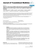
Effect of platelet inhibiting treatment on serum TXB
2
-levels (Factor 13)Figure 2
Effect of platelet inhibiting treatment on serum TXB
2
-levels (Factor 13). Serum TXB
2
-levels (Factor 13) for patients
(n = 29) and healthy controls (n = 29) are presented as mean + SEM. ASA alone or in combination with clopidogrel was signif-
icantly different from clopidogrel alone and compared to the mean of the controls (p < 0.001). Also, the difference between
controls at visit 1 and visit 2 was significant. ***p < 0.001, ns = not significant.
Journal of Translational Medicine 2009, 7:42 />Page 8 of 14
(page number not for citation purposes)
The influence of ASA and clopidogrel on platelet adhesionFigure 3
The influence of ASA and clopidogrel on platelet adhesion. The main figures are representative examples of the varia-
bles constituting the respective factors. The insets show the Z-scores for each factor. Also shown in the insets are the compar-
isons between the control means of visit 1 and 2 and treatment with ASA (A), clopidogrel (C) and the combination of ASA and
clopidogrel (A+C). The respective figures show the effect of platelet inhibiting treatment on ADP-induced adhesion (Factor 1,
Fig A), ristocetin-induced adhesion to albumin (Factor 6, Fig B), LPA-induced adhesion to albumin (Factor 7, Fig C) and adhe-
sion to collagen (Factor 8, Fig D) for patients (n = 29) and healthy controls (n = 29). All values are presented as mean + SEM.
*p ≤ 0.05, **p ≤ 0.01, ***p ≤ 0.001, ns = not significant.
Journal of Translational Medicine 2009, 7:42 />Page 9 of 14
(page number not for citation purposes)
The influence of ASA and clopidogrel on platelet activity measured by flow cytometryFigure 4
The influence of ASA and clopidogrel on platelet activity measured by flow cytometry. The effects of platelet
inhibiting treatment on platelet activation detected by flow cytometry induced by ADP (Factor 14, Fig A) and SFLLRN (Factor
15, Fig B) on patients (n = 29). The main figures are representative examples of the variables constituting the respective fac-
tors. The insets show the Z-scores for each factor. All values are presented as mean + SEM. ***p < 0.001, ns = not significant.
Journal of Translational Medicine 2009, 7:42 />Page 10 of 14
(page number not for citation purposes)
responding to platelet function measured by flow cytom-
etry (Factors 14 and 15), correlated with an r
2
-value of
0.28. Regarding TXB
2
, regression analyses were only per-
formed on samples with clopidogrel monotherapy since
levels of TXB
2
were totally suppressed when platelets were
treated with ASA. However, serum TXB
2
-levels did not cor-
relate with any of the other measurements.
Discussion
With the aim of finding variables sensitive to clopidogrel
and ASA-treatment, this study used a screening approach
and measured several different variables simultaneously.
To reduce the complexity of the material we performed
PCA in order to find correlating variables that measured
the same property. In this way the 54 measurements of
platelet adhesion were reduced to 8 factors. Visual inspec-
tion revealed that each factor represented a separate entity
of platelet adhesion and the factors could therefore be
renamed according to the aspect they measured. We thus
conclude that future studies must not involve all 54 adhe-
sion variables, but instead, one variable from each factor
should be enough to cover 8 different aspects of platelet
adhesion. In addition to the adhesion data, the remaining
15 variables also formed distinct factors that were possible
to rename according to measured property. It is notable
that serum TXB
2
formed a distinct group not correlated to
any of the other measurements.
It is important that laboratory assays used for clinical pur-
poses are reproducible and that they measure parameters
that are not confounded by other variables. Some of the
measurements performed in this study (clinical chemistry
variables and platelet function measured by flow cytome-
try) are used for clinical analysis at accredited laboratories
at the University hospital in Linköping. However, the
reproducibility of the platelet adhesion assay was mostly
unknown before this study [35]. Our initial results sug-
gested that the factors corresponding to ADP-induced
adhesion and adhesion to fibrinogen were not reproduci-
ble. We therefore excluded the most varied variables con-
stituting these factors, which resulted in no intra-
Table 1: Binding of fibrinogen and expression of P-selectin as measured by flow cytometry.
Type of measurement Activating agent Reference values ASA + Clopidogrel ASA Clopidogrel
Fibrinogen-binding Control 1 (0–3.4) 2.3 ± 0.3*** 5.0 ± 2.5
ns
2.4 ± 0.2***
ADP 0.1 38 (17–59) 9.9 ± 1.3*** 29.4 ± 3.6* 7.5 ± 1.4***
ADP 0.6 74 (60–89) 32.5 ± 2.7*** 62.1 ± 3.3** 22.9 ± 2.9***
SFLLRN 5.3 76 (55–98) 28.8 ± 4.3*** 48.5 ± 5.2*** 20.2 ± 4.0***
P-selectin expression Control 2 (0.9–3.1) 2.0 ± 0.2
ns
4.8 ± 0.9** 4.3 ± 0.6***
ADP 0.6 26 (10–42) 7.6 ± 0.8*** 24.8 ± 2.4
ns
10.9 ± 1.4***
SFLLRN 5.3 88 (70–100) 33.0 ± 3.7*** 55.4 ± 4.7*** 34.4 ± 3.7***
Platelets from patients (n = 29) were activated in vitro with adenosine 5'-diphosphate (ADP; 0.1 and 0.6 μmol/L) or SFLLRN (5.3 μmol/L) followed
by flow cytometric measurements of fibrinogen-binding or expression of P-selectin. Presented results are the mean-% of fibrinogen-binding and P-
selectin expression ± SEM. Reference values (obtained earlier during routine analysis at the accredited Dept. of Clinical Chemistry at the University
hospital in Linköping) are shown as mean with reference interval within parenthesis. Stars indicate significant differences for patients compared to
reference values. *p ≤ 0.05, **p ≤ 0.01, ***p ≤ 0.001, ns = not significant.
Correlation between static platelet adhesion and flow cytom-etryFigure 5
Correlation between static platelet adhesion and
flow cytometry. Correlation between ADP-induced plate-
let adhesion (Factor 1) and ADP-induced platelet activation
as measured by flow cytometry (Factor 14) for patients (n =
29) (r
2
= 0.49). Data included are from all three separate
anti-platelet treatments (ASA and clopidogrel alone as well as
ASA and clopidogrel combined).
Journal of Translational Medicine 2009, 7:42 />Page 11 of 14
(page number not for citation purposes)
individual effects for healthy controls in the platelet adhe-
sion assay. From this we conclude that many, but not all,
measures of platelet adhesion are reproducible. Moreover,
the static condition might limit the possibilities for trans-
lating the results from the adhesion assay into in vivo
platelet adhesion occurring during flow conditions. How-
ever, platelet adhesion to collagen and fibrinogen is
dependent on α
2
β
1
- and α
IIb
β
3
-receptors respectively in
the current assay [23]. This suggests that the static platelet
adhesion assay can measure important aspects of platelet
function despite its simplicity. Furthermore, vWf depend-
ent adhesion is not directly covered in the present assay
although ristocetin-induced adhesion appears to be
dependent on GPIb-IX-V and vWf [23].
We found that platelet adhesion to albumin, collagen and
fibrinogen induced by ADP was suppressed during treat-
ment with clopidogrel alone or in combination with ASA
compared to treatment with ASA alone. The same pattern
was also seen for the flow cytometric measures of ADP-
induced activation. Furthermore, clopidogrel treatment
suppressed both ADP-induced adhesion and ADP-
induced activity measured by flow cytometry below the
levels observed for healthy controls. These results are clear
indications that both assays are able to detect the direct
effect of inhibition of ADP-signalling by clopidogrel. It is
also interesting to note that, upon ADP-challenge, the dif-
ferent assays correlated relatively well to each other. The
variation between those factors not explained by the other
can probably be ascribed to the different aspects of plate-
let function that the two assays measure. Flow cytometry
measures the expression of activation-dependent recep-
tors when platelets are in solution, while the adhesion
assay measures the ability of platelets to adhere to a sur-
face [27].
Measurements of serum TXB
2
-levels in healthy controls
revealed significant intra-individual differences. This
effect may be attributed to varying stress levels for the con-
trols at the separate occasions since urinary levels of the
TXA
2
metabolite 2,3-dinor-TXB
2
has been shown to corre-
late with urinary cortisol [36]. Also, urinary TXB
2
follows
a seasonal variation pattern [37]. According to our discus-
sion above, this variation complicates the use of this
measure of platelet activity in clinical routine. However,
we also found that serum TXB
2
-levels were completely
suppressed after ASA treatment compared to both clopi-
dogrel treatment and controls. Consequently, despite the
intra-individual variations, this shows that serum TXB
2
is
a good indicator of the ability of ASA to inhibit the
cyclooxygenase pathway.
Some adhesion measures showed decreased adhesion by
clopidogrel alone compared with clopidogrel plus ASA.
Since dual therapy was always the initiating treatment,
this difference may reflect the presence of more active
platelets in the time frame closest to the index event and
coronary revascularization. It has been proposed that
reendothelialization after insertion of a bare metal stent is
complete after approximately 3 months [38] and that arte-
rial healing is even slower for a drug-eluting stent [39].
Consequently, it is possible that absence of endothelium
after stenting contributes to the high initial platelet activ-
ity found in this study. A sustained inflammatory
response after stenting may also partly explain the
decreased levels of the negative acute phase reactant HDL
cholesterol [40] during dual therapy compared with the
monotherapies. Similarly, the sustained inflammatory
response may explain the significant increase in platelet
count during combination therapy. Platelet counts may
initially decrease after surgical procedures followed by
recovery and increased platelet count within the following
period of time [41,42]. This would be represented by the
dual therapy group in the present study. The change in
platelet count is however complicated since it has also
been reported that patients with unstable angina had a
decreased platelet count compared with stable angina
patients and controls [43].
From this discussion it is evident that the adhesion assay
as well as flow cytometry can measure effects of clopidog-
rel when using ADP as activating stimuli. It is also evident
that serum-TXB
2
levels measure the effects of ASA. How-
ever, these measures focus on the primary interaction
between the drugs and the platelets, which could be prob-
lematic when trying to evaluate the complex in vivo treat-
ment effect. It has previously been found that only 12 of
682 ASA-treated patients (≈ 2%) had residual TXB
2
serum
levels higher than 2 standard deviations from the popula-
tion mean [44]. Measurements of the effect of arachidonic
acid on platelet aggregometry have also led to the conclu-
sion that ASA resistance is a very rare phenomenon [45].
Thus, our study supports these previous findings that
assays measuring the pharmacodynamic activity of ASA
(to inhibit the COX-enzyme) seldom recognizes patients
as ASA-resistant. This suggests that the cause of ASA-resist-
ance is not due to an inability of ASA to act as a COX-
inhibitor. Explanations for ASA resistance are diverse and
include e.g. patient non-compliance, interactions with
other drugs, platelet polymorphisms and sustained COX-
activity by other cells [12,13]. Several studies also propose
that ASA-resistant platelets have increased platelet activa-
tion through signalling pathways not directly involving
TXA
2
[46-49]. In line with these studies it has been pro-
posed that the presence of ASA resistance should be eval-
uated by combining measurements of TXB
2
-formation
with platelet aggregation [50]. We further suggest that
direct measurements of ADP and TXA
2
-effects (in our case
ADP-induced activation measured by adhesion or flow
cytometry and serum TXB
2
-levels) must be combined with
Journal of Translational Medicine 2009, 7:42 />Page 12 of 14
(page number not for citation purposes)
measures that are only partly dependent on ADP and
TXA
2
respectively. For instance, an adhesion variable
partly dependent on TXA
2
might be able to detect ASA
resistance caused by increased signalling through other
activating pathways. Such a scenario would be character-
ized by serum TXB
2
values showing normal COX-inhibi-
tion while platelet adhesion is increased. This study
employed a screening procedure in order to find such
indirect measures of the effects of ASA and clopidogrel.
Our results show inhibiting effects of clopidogrel com-
pared to ASA on adhesion to albumin in the presence of
LPA or ristocetin. This was also observed for our flow cyto-
metric measurements with SFLLRN as activator, which
confirms that SFLLRN is able to induce release of granule
contents in platelets [51,52]. SFLLRN- and ADP-induced
platelet activation, as measured by flow cytometry, was
moderately correlated to each other and adhesion
induced by LPA as well as ristocetin showed weak correla-
tions with ADP-induced adhesion. These results further
confirm that these measures of platelet activity are partly
dependent on ADP. We have earlier shown that adhesion
to albumin induced by simultaneous stimulation by LPA
and adrenaline (a variable belonging to the LPA-factor in
the present study) can be inhibited by inhibition of ADP-
signalling in vitro [25]. This strengthens our conclusion
that the effect on LPA-induced adhesion observed for
clopidogrel is caused by inhibition of ADP-signalling.
Also, the presence of LPA in atherosclerotic plaques and
its possible role in thrombus formation after plaque rup-
ture [24] makes it especially interesting for the in vivo set-
ting of myocardial infarction. The collagen surface is
different from the other stimuli since dual therapy results
in significantly depressed platelet adhesion compared to
both monotherapies. This indicates that adhesion to col-
lagen is dependent on both ADP and TXA
2
and this meas-
ure was the only adhesion-related factor that showed
potential for being partly dependent on TXA
2
. However,
the significant effects observed between treatments were
rather small. Nevertheless, it has earlier been shown that
platelet activation induced by collagen is reduced by
intake of ASA [26,53]. Regarding the flow cytometric
measurements there were no indications for platelet activ-
ity to be decreased for dual therapy compared to mono-
therapy with clopidogrel. However, platelet activation as
measured by flow cytometry was in general decreased for
patients having monotherapy with ASA compared to
healthy controls. This indicates that flow cytometry is also
able to detect effects of ASA.
Conclusion
In this study we employed different assays in order to eval-
uate platelet function in patients treated with different
anti-platelet regimens. Among these assays, the platelet
adhesion assay had a certain role since it had not been
used before for this clinical purpose. Actually, there are no
assays of static platelet adhesion that have been used in
previous studies aimed at investigating treatment effects
of platelet inhibiting drugs. Importantly, this study shows
that the static platelet adhesion assay is reproducible over
time. We also showed that the static platelet adhesion
assay as well as flow cytometry detected the ability of
clopidogrel to inhibit platelet activation induced by ADP.
Our results further suggest that other measures of platelet
adhesion and platelet activation measured by flow cytom-
etry are indirectly dependent on secreted ADP or TXA
2
.
One such measure is adhesion to a collagen surface, which
should be more thoroughly investigated for its ability to
detect effects of clopidogrel and ASA. Likewise, due to its
connection to atherosclerosis and myocardial infarction,
the LPA-induced effect should be further evaluated for its
ability to detect effects of clopidogrel. In conclusion, the
screening procedure undertaken in this study has revealed
suggestions on which measures of platelet activity to com-
bine in order to evaluate platelet function. Speculatively,
the ADP-mediated effects in the present adhesion assay in
combination with serum TXB
2
, may be combined with
LPA and collagen-induced adhesion for an optimal mon-
itoring of clopidogrel and ASA therapy.
Competing interests
The authors declare that they have no competing interests.
Authors' contributions
ACE carried out the analysis of static platelet adhesion and
serum TXB
2
-levels and performed the statistical analyses.
All authors participated in the design of the study, co-
operated in the drafting of the manuscript and read and
approved the final version of the manuscript.
Additional material
Acknowledgements
Margareta Hedbäck, research nurse at the Department of Cardiology, Uni-
versity Hospital in Linköping, is gratefully acknowledged for management of
the patients during the study. The excellent staff at the Department of
Transfusion Medicine and Clinical Immunology is acknowledged for skilful
Additional file 1
All variables measured in the study. A table showing all the variables
that were measured in the study.
Click here for file
[ />5876-7-42-S1.pdf]
Additional file 2
The final factors used for ANOVA analyses. A table showing the factors
used for ANOVA analyses.
Click here for file
[ />5876-7-42-S2.pdf]
Journal of Translational Medicine 2009, 7:42 />Page 13 of 14
(page number not for citation purposes)
help with blood sampling from the healthy controls. Kerstin M Gustafsson
at the Division of Clinical Chemistry is acknowledged for performing the
flow cytometric measurements. Professor Erland Svensson at the Swedish
Defence Research Agency, Division of Command and Control Systems,
Department of Man-System interaction is gratefully acknowledged for sig-
nificant statistical counselling. During the course of the research underlying
this study, Andreas C. Eriksson was enrolled in Forum Scientium, a multi-
disciplinary doctoral programme at Linköping University, Sweden. The
study was supported by grants from the Cardiovascular Inflammation
Research Centre and the Heart Foundation at Linköping University, the
County Council of Östergötland, Eleanore Demeroutis Foundation for
Cardiovascular Research at the University Hospital in Linköping, the Swed-
ish Research Council (K2007-64X-15060-04-3) and the Foundation for Old
Maidservants.
References
1. Antiplatelet Trialists' Collaboration: Collaborative overview of
randomised trials of antiplatelet therapy – I: Prevention of
death, myocardial infarction, and stroke by prolonged
antiplatelet therapy in various categories of patients. BMJ
1994, 308:81-106.
2. CAPRIE Steering Committee: A randomised, blinded, trial of
clopidogrel versus aspirin in patients at risk of ischaemic
events (CAPRIE). Lancet 1996, 348:1329-1339.
3. Yusuf S, Zhao F, Mehta SR, Chrolavicius S, Tognoni G, Fox KK:
Effects of clopidogrel in addition to aspirin in patients with
acute coronary syndromes without ST-segment elevation. N
Engl J Med 2001, 345:494-502.
4. Chen ZM, Jiang LX, Chen YP, Xie JX, Pan HC, Peto R, Collins R, Liu
LS: Addition of clopidogrel to aspirin in 45,852 patients with
acute myocardial infarction: randomised placebo-controlled
trial. Lancet 2005, 366:1607-1621.
5. Sabatine MS, Cannon CP, Gibson CM, Lopez-Sendon JL, Montalescot
G, Theroux P, Claeys MJ, Cools F, Hill KA, Skene AM, McCabe CH,
Braunwald E: Addition of clopidogrel to aspirin and fibrinolytic
therapy for myocardial infarction with ST-segment eleva-
tion. N Engl J Med 2005, 352:1179-1189.
6. Steinhubl SR, Berger PB, Mann JT 3rd, Fry ET, DeLago A, Wilmer C,
Topol EJ: Early and sustained dual oral antiplatelet therapy
following percutaneous coronary intervention: a rand-
omized controlled trial. JAMA 2002, 288:2411-2420.
7. Mehta SR, Yusuf S, Peters RJ, Bertrand ME, Lewis BS, Natarajan MK,
Malmberg K, Rupprecht H, Zhao F, Chrolavicius S, Copland I, Fox KA:
Effects of pretreatment with clopidogrel and aspirin fol-
lowed by long-term therapy in patients undergoing percuta-
neous coronary intervention: the PCI-CURE study. Lancet
2001, 358:527-533.
8. Angiolillo DJ, Fernandez-Ortiz A, Bernardo E, Alfonso F, Macaya C,
Bass TA, Costa MA: Variability in individual responsiveness to
clopidogrel: clinical implications, management, and future
perspectives. J Am Coll Cardiol 2007, 49:1505-1516.
9. Serebruany VL, Steinhubl SR, Berger PB, Malinin AI, Bhatt DL, Topol
EJ: Variability in platelet responsiveness to clopidogrel
among 544 individuals. J Am Coll Cardiol 2005, 45:
246-251.
10. Jaremo P, Lindahl TL, Fransson SG, Richter A: Individual variations
of platelet inhibition after loading doses of clopidogrel. J
Intern Med 2002, 252:233-238.
11. Sanderson S, Emery J, Baglin T, Kinmonth AL: Narrative review:
aspirin resistance and its clinical implications. Ann Intern Med
2005, 142:370-380.
12. Hankey GJ, Eikelboom JW: Aspirin resistance. Lancet 2006,
367:606-617.
13. Mason PJ, Jacobs AK, Freedman JE: Aspirin resistance and athero-
thrombotic disease. J Am Coll Cardiol 2005, 46:986-993.
14. Harrison P, Frelinger AL 3rd, Furman MI, Michelson AD: Measuring
antiplatelet drug effects in the laboratory. Thromb Res 2007,
120:323-336.
15. Michelson AD: Platelet function testing in cardiovascular dis-
eases. Circulation 2004, 110:e489-493.
16. Haubelt H, Anders C, Hellstern P: Can platelet function tests
predict the clinical efficacy of aspirin? Semin Thromb Hemost
2005, 31:404-410.
17. Cannon CP, McLean DS: Critical pathways using platelet testing
to potentially optimize the use of oral antiplatelet therapy.
Am J Cardiol 2006, 98(10A):33N-38N.
18. Rand ML, Leung R, Packham MA: Platelet function assays. Transfus
Apher Sci 2003, 28:307-317.
19. Michelson AD, Cattaneo M, Eikelboom JW, Gurbel P, Kottke-March-
ant K, Kunicki TJ, Pulcinelli FM, Cerletti C, Rao AK: Aspirin resist-
ance: position paper of the Working Group on Aspirin
Resistance. J Thromb Haemost 2005, 3:1309-1311.
20. Bonello L, Camoin-Jau L, Armero S, Com O, Arques S, Burignat-
Bonello C, Giacomoni MP, Bonello R, Collet F, Rossi P, Barragan P,
Dignat-George F, Paganelli F: Tailored clopidogrel loading dose
according to platelet reactivity monitoring to prevent acute
and subacute stent thrombosis. Am J Cardiol 2009,
103:5-10.
21. Bonello L, Camoin-Jau L, Arques S, Boyer C, Panagides D, Wittenberg
O, Simeoni MC, Barragan P, Dignat-George F, Paganelli F: Adjusted
clopidogrel loading doses according to vasodilator-stimu-
lated phosphoprotein phosphorylation index decrease rate
of major adverse cardiovascular events in patients with
clopidogrel resistance: a multicenter randomized prospec-
tive study. J Am Coll Cardiol 2008, 51:1404-1411.
22. Berndt MC, Shen Y, Dopheide SM, Gardiner EE, Andrews RK: The
vascular biology of the glycoprotein Ib-IX-V complex. Thromb
Haemost 2001, 86:178-188.
23. Eriksson AC, Whiss PA: Characterization of static adhesion of
human platelets in plasma to protein surfaces in micro-
plates. Blood Coagul Fibrinolysis 2009, 20:197-206.
24. Siess W: Athero- and thrombogenic actions of lysophospha-
tidic acid and sphingosine-1-phosphate. Biochim Biophys Acta
2002, 1582:204-215.
25. Eriksson AC, Whiss PA, Nilsson UK: Adhesion of human plate-
lets to albumin is synergistically increased by lysophospha-
tidic acid and adrenaline in a donor-dependent fashion. Blood
Coagul Fibrinolysis 2006, 17:359-368.
26. Graff J, Klinkhardt U, Harder S: Pharmacodynamic profile of
antiplatelet agents: marked differences between single ver-
sus costimulation with platelet activators. Thromb Res 2004,
113:295-302.
27. Eriksson AC, Whiss PA: Measurement of adhesion of human
platelets in plasma to protein surfaces in microplates. J Phar-
macol Toxicol Methods 2005, 52:356-365.
28. Eriksson AC, Nilsson UK, Whiss PA: The extracellular ion envi-
ronment modulates platelet adhesion after lysophosphatidic
acid treatment in vitro [abstract]. Atheroscler Suppl 2006, 7:s90.
29. Nylander S, Johansson K, Van Giezen JJ, Lindahl TL: Evaluation of
platelet function, a method comparison. Platelets 2006,
17:49-55.
30. Lundahl TH, Petersson J, Fagerberg IH, Berg S, Lindahl TL: Impaired
platelet function correlates with multi-organ dysfunction. A
study of patients with sepsis. Platelets 1998, 9:223-225.
31. Lindahl TL, Festin R, Larsson A: Studies of fibrinogen binding to
platelets by flow cytometry: an improved method for studies
of platelet activation. Thromb Haemost 1992, 68:221-225.
32. Berglund U, Lindahl T: Enhanced onset of platelet inhibition
with a loading dose of ticlopidine in ASA-treated stable cor-
onary patients. Int J Cardiol 1998, 64:215-217.
33. Cayman Chemical EIA Analysis Tools [man
chem.com/analysis/eia]
34. Bryman A, Cramer D: Aggregating variables Exploratory factor
analysis. In Quantitative Data Analysis with SPSS Release 8 for Windows:
For Social Scientists 1st edition. Edited by: Bryman A, Cramer D. Lon-
don: Routledge; 1999:271-285.
35. Eriksson AC, Jonasson L, Hedback B, Whiss PA: Monitoring plate-
let inhibiting treatment in coronary heart disease by static
platelet adhesion [abstract]. Atheroscler Suppl 2007, 8:s189.
36. Fimognari FL, Piccirillo G, Lama J, Paganica P, Monteleone G, Gianni
W, Cacciafesta M, Marigliano V: Associated daily biosynthesis of
cortisol and thromboxane A2: a preliminary report. J Lab Clin
Med 1996, 128:115-121.
37. Mustad V, Derr J, Reddy CC, Pearson TA, Kris-Etherton PM: Sea-
sonal variation in parameters related to coronary heart dis-
ease risk in young men. Atherosclerosis 1996, 126:117-129.
38. Grewe PH, Deneke T, Machraoui A, Barmeyer J, Muller KM: Acute
and chronic tissue response to coronary stent implantation:
pathologic findings in human specimen. J Am Coll Cardiol 2000,
35:157-163.
Publish with Bio Med Central and every
scientist can read your work free of charge
"BioMed Central will be the most significant development for
disseminating the results of biomedical research in our lifetime."
Sir Paul Nurse, Cancer Research UK
Your research papers will be:
available free of charge to the entire biomedical community
peer reviewed and published immediately upon acceptance
cited in PubMed and archived on PubMed Central
yours — you keep the copyright
Submit your manuscript here:
/>BioMedcentral
Journal of Translational Medicine 2009, 7:42 />Page 14 of 14
(page number not for citation purposes)
39. Joner M, Finn AV, Farb A, Mont EK, Kolodgie FD, Ladich E, Kutys R,
Skorija K, Gold HK, Virmani R: Pathology of drug-eluting stents
in humans: delayed healing and late thrombotic risk. J Am Coll
Cardiol 2006, 48:193-202.
40. Khovidhunkit W, Memon RA, Feingold KR, Grunfeld C: Infection
and inflammation-induced proatherogenic changes of lipo-
proteins. J Infect Dis 2000, 181 Suppl 3:S462-S472.
41. von Ruecker A, Hufnagel P, Dickerhoff R, Murday H, Bidlingmaier F:
Qualitative and quantitative changes in platelets after coro-
nary-artery bypass surgery may help identify thrombotic
complications and infections. Klin Wochenschr 1989,
67:1042-1047.
42. Moreau D, Timsit JF, Vesin A, Garrouste-Orgeas M, de Lassence A,
Zahar JR, Adrie C, Vincent F, Cohen Y, Schlemmer B, Azoulay E:
Platelet count decline: an early prognostic marker in criti-
cally ill patients with prolonged ICU stays. Chest 2007,
131:1735-1741.
43. Pizzulli L, Yang A, Martin JF, Luderitz B: Changes in platelet size
and count in unstable angina compared to stable angina or
non-cardiac chest pain. Eur Heart J 1998, 19:80-84.
44. Frelinger AL 3rd, Furman MI, Linden MD, Li Y, Fox ML, Barnard MR,
Michelson AD: Residual arachidonic acid-induced platelet acti-
vation via an adenosine diphosphate-dependent but cycloox-
ygenase-1- and cyclooxygenase-2-independent pathway: a
700-patient study of aspirin resistance. Circulation 2006,
113:2888-2896.
45. Rao GH, Michiels JJ: Aspirin resistance: does it exist? Semin
Thromb Hemost 2007, 33:210-214.
46. Kawasaki T, Ozeki Y, Igawa T, Kambayashi J: Increased platelet
sensitivity to collagen in individuals resistant to low-dose
aspirin. Stroke 2000, 31:591-595.
47. Macchi L, Christiaens L, Brabant S, Sorel N, Allal J, Mauco G, Brizard
A: Resistance to aspirin in vitro is associated with increased
platelet sensitivity to adenosine diphosphate. Thromb Res
2002, 107:45-49.
48. Rao GH, Escolar G, White JG: Epinephrine reverses the inhibi-
tory influence of aspirin on platelet-vessel wall interactions.
Thromb Res 1986, 44:65-74.
49. Hurlen M, Seljeflot I, Arnesen H:
Increased platelet aggregability
during exercise in patients with previous myocardial infarc-
tion. Lack of inhibition by aspirin. Thromb Res 2000, 99:487-494.
50. Violi F, Pignatelli P, Pulcinelli FM: Aspirin. J Thromb Haemost 2004,
2:338-339.
51. Lasne D, Donato J, Falet H, Rendu F: Different abilities of
thrombin receptor activating peptide and thrombin to
induce platelet calcium rise and full release reaction. Thromb
Haemost 1995, 74:1323-1328.
52. Kinlough-Rathbone RL, Perry DW, Guccione MA, Rand ML, Packham
MA: Degranulation of human platelets by the thrombin
receptor peptide SFLLRN: comparison with degranulation
by thrombin. Thromb Haemost 1993, 70:1019-1023.
53. Moshfegh K, Redondo M, Julmy F, Wuillemin WA, Gebauer MU, Hae-
berli A, Meyer BJ: Antiplatelet effects of clopidogrel compared
with aspirin after myocardial infarction: enhanced inhibitory
effects of combination therapy. J Am Coll Cardiol 2000,
36:699-705.