báo cáo hóa học:" Anti-tumor activity of patient-derived NK cells after cell-based immunotherapy – a case report" doc
Bạn đang xem bản rút gọn của tài liệu. Xem và tải ngay bản đầy đủ của tài liệu tại đây (791.9 KB, 18 trang )
BioMed Central
Page 1 of 18
(page number not for citation purposes)
Journal of Translational Medicine
Open Access
Research
Anti-tumor activity of patient-derived NK cells after cell-based
immunotherapy – a case report
Valeria Milani
1,2
, Stefan Stangl
3
, Rolf Issels
1,2
, Mathias Gehrmann
3
,
Beate Wagner
4
, Kathrin Hube
3
, Doris Mayr
5
, Wolfgang Hiddemann
1,6
,
Michael Molls
3
and Gabriele Multhoff*
3,7
Address:
1
Department of Internal Medicine, University Medical Center Großhadern, Ludwig-Maximilians-Universität München, Germany,
2
Clinical Cooperation Group (CCG) "Tumor Therapy by Hyperthermia", Helmholtz Zentrum München, German Research Center for
Environmental Health Munich, Germany,
3
Department of Radiation Oncology, Klinikum rechts der Isar, Technische Universität München,
Germany,
4
Transfusion Medicine and Haemostaseology, University Medical Center Großhadern, Ludwig-Maximilians-Universität München,
Germany,
5
Department of Pathology, University Medical Center Großhadern, Ludwig-Maximilians-Universität München, Germany,
6
Clinical
Cooperation Group (CCG) "Pathogenesis of Acute Leukemias", Helmholtz Zentrum München, German Research for Environmental Health,
Munich, Germany and
7
Clinical Cooperation Group (CCG) "Innate Immunity in Tumor Biology", Helmholtz Zentrum München, German
Research Center for Environmental Health, Munich, Germany
Email: Valeria Milani - ; Stefan Stangl - ;
Rolf Issels - ; Mathias Gehrmann - ;
Beate Wagner - ; Kathrin Hube - ; Doris Mayr -
muenchen.de; Wolfgang Hiddemann - ; Michael Molls - ;
Gabriele Multhoff* -
* Corresponding author
Abstract
Background: Membrane-bound heat shock protein 70 (Hsp70) serves as a tumor-specific recognition structure
for Hsp70-peptide (TKD) plus IL-2 activated NK cells. A phase I clinical trial has shown that repeated re-infusions
of ex vivo TKD/IL-2-activated, autologous leukapheresis product is safe. This study investigated the maintenance
of the cytolytic activity of NK cells against K562 cells and autologous tumor after 6 plus 3 infusions of TKD/IL-2-
activated effector cells.
Methods: A stable tumor cell line was generated from the resected anastomotic relapse of a patient with colon
carcinoma (pT3, N2, M0, G2). Two months after surgery, the patient received the first monthly i.v. infusion of his
ex vivo TKD/IL-2-activated peripheral blood mononuclear cells (PBMNC). After 6 infusions and a pause of 3
months, the patient received another 3 cell infusions. The phenotypic characteristics and activation status of
tumor and effector cells were determined immediately before and at times after each infusion.
Results: The NK cell ligands Hsp70, MICA/B, and ULBP-1,2,3 were expressed on the patient's anastomotic
relapse. An increased density of activatory NK cell receptors following ex vivo stimulation correlated with an
enhanced anti-tumoricidal activity. After 4 re-infusion cycles, the intrinsic cytolytic activity of non-stimulated
PBMNC was significantly elevated and this heightened responsiveness persisted for up to 3 months after the last
infusion. Another 2 re-stimulations with TKD/IL-2 restored the cytolytic activity after the therapeutic pause.
Conclusion: In a patient with colon carcinoma, repeated infusions of ex vivo TKD/IL-2-activated PBMNC initiate
an intrinsic NK cell-mediated cytolytic activity against autologous tumor cells.
Published: 23 June 2009
Journal of Translational Medicine 2009, 7:50 doi:10.1186/1479-5876-7-50
Received: 5 May 2009
Accepted: 23 June 2009
This article is available from: />© 2009 Milani et al; licensee BioMed Central Ltd.
This is an Open Access article distributed under the terms of the Creative Commons Attribution License ( />),
which permits unrestricted use, distribution, and reproduction in any medium, provided the original work is properly cited.
Journal of Translational Medicine 2009, 7:50 />Page 2 of 18
(page number not for citation purposes)
Background
Studies into the cellular basis of cancer immunosurveil-
lance demonstrate that lymphocytes of both adaptive and
innate immune compartments can prevent tumor devel-
opment [1]. In contrast to normal tissues, tumors fre-
quently express the stress protein heat shock protein 70
(Hsp70) on their plasma membrane, and this membrane-
associated form of the Hsp70 molecule acts as a tumor-
specific recognition structure for Hsp70-peptide activated
natural killer (NK) cells expressing CD94 [2,3]. More
recently, the glycosphingolipid globoyltriaosylceramide
(Gb3) was shown to enable the selective anchorage of
Hsp70 in plasma membranes of colorectal cancer cells [4].
The finding that Gb3 is predominantly found in choles-
terol-rich microdomains (CRM) of tumor, but not of nor-
mal cells might provide an explanation for the tumor-
specific Hsp70 membrane expression.
The region of the Hsp70 molecule which is exposed to the
extracellular milieu of tumors has been identified as the
14-mer peptide TKDNNLLGRFELSG (TKD), and this
resides in the amino acid sequence aa
450–463
of the C-ter-
minal domain substrate binding domain [5,6]. A combi-
nation of synthetically produced, GMP-grade Hsp70
peptide plus low dose IL-2 (TKD/IL-2) has been shown to
stimulate the cytolytic and migratory capacity of CD3
-
/
CD16/CD56
+
human [5,7] and mouse [8] NK cells. TKD/
IL-2-activated cells specifically kill allogeneic, Hsp70
membrane-positive tumor cell lines in vitro [9]. Moreover,
four repeated re-infusions of purified TKD/IL-2-activated
NK cells have been shown to eradicate the primary tumor
and prevent metastasis in a xenograft tumor mouse model
of human pancreatic cancer [10]. Importantly, the induc-
tion of NK cell cytotoxicity is also possible when PBMNC
rather than purified NK cells are incubated with TKD/IL-2
[11]. Furthermore, in the presence of other lymphocytes
and antigen presenting cells (APC), the cytotoxic response
against Hsp70 membrane-positive tumors has been
found to be selectively mediated by NK cells (unpub-
lished observations).
The enhanced cytolytic activity against Hsp70 surface-pos-
itive tumors is accompanied by, and correlates with an
increased expression density of NK cell receptors includ-
ing CD94/NKG2A/C, NKG2D and NCRs such as NKp30,
NKp44, NKp46 [2,3,12]. The expression density of the C-
type lectin receptor CD94 is associated with the capacity
of NK cells to bind Hsp70 protein and TKD [2], and cor-
relates with a strong lytic activity against Hsp70 mem-
brane-positive tumor target cells.
The mechanism of lysis of Hsp70 membrane-positive
tumors has been identified as being a perforin-independ-
ent, granzyme B-mediated apoptosis [13]. Previous stud-
ies have shown a high degree of correlation of the results
of a 4-h
51
chromium release assay and the granzyme B
ELISPOT assay for measuring the granzyme B mediated
killing of Hsp70 membrane-positive tumors by activated
NK cells. These findings indicate that the granzyme B
ELISPOT assay is a reliable test to determine Hsp70-reac-
tivity in NK cells.
An Hsp70 membrane-positive phenotype acts as a nega-
tive prognostic marker for patients with lower rectal carci-
nomas and non-small cell lung cancer (NSCLC), and the
overall survival of patients with Hsp70 membrane-posi-
tive cancer is significantly lower than that of their Hsp70
membrane-negative counterparts [14]. These findings
highlight the clinical significance of determining the
Hsp70 membrane status and the urgent need to treat
patients with Hsp70 membrane-positive tumors. A phase
I clinical study involving eleven patients with metastatic
colorectal cancer and one patient with non-small cell lung
cancer (NSCLC) has shown that the re-infusion of autolo-
gous, TKD/IL-2-activated leukapheresis products is feasi-
ble, safe and well-tolerated [15]. Furthermore, measurable
immunological responses in the form of an enhanced
expression of CD94 on NK cells and an increased NK cell
cytolytic capacity against an allogeneic, Hsp70 mem-
brane-positive colon carcinoma cell line CX+ were
induced in 10 of the 12 patients [15]. In line with previous
results from animal models [10], clinical responses fulfill-
ing formal staging criteria were observed in 2 patients,
who received more than 4 treatment cycles [15]. These
promising immunological data encouraged us to treat a
patient with an anastomotic relapse using a similar
approach to that in the phase I clinical trial mentioned
above. However, in this specific instance a tumor cell line
could be established from a biopsy of the patient's tumor
and its Hsp70 membrane-positive phenotype could be
confirmed.
Herein, we report the kinetics of the anti-tumor immune
responses in this patient who received a total of 9 re-infu-
sions of ex vivo TKD/IL-2-activated, autologous leukapher-
esis products over a 12-month period and the clinical
follow-up for 1 year. The kinetics of the initiation and
maintenance of an in vivo cytolytic response against
Hsp70-positive tumors within the first therapy cycles is in
line with our previous findings from the phase I clinical
trial. In this study an intrinsic NK cell activity was initiated
only in patients who received more than 4 repeated re-
infusion cycles of TKD/IL-2-activated, autologous
PBMNC. This finding was determined in 5 patients with
different tumor entities, stages and previous therapies.
This is also the first observation that the administration of
TKD/IL-2-activated PBMNC induces a sustained in vivo
NK cell cytolytic response against the patient's own,
Hsp70 membrane-positive tumor and the classical NK cell
target K562 which persists for at least 2 months. Further-
Journal of Translational Medicine 2009, 7:50 />Page 3 of 18
(page number not for citation purposes)
more, we demonstrate that a decline in the in vivo NK cell
activity can be restored by an additional 2 infusion cycles
with TKD/IL-2-activated, autologous PBMNC. This indi-
cates that the therapeutic intervention does not initiate an
irreversible state of immune tolerance.
Methods
Ethics
Signed informed consent was obtained from the patient
before the start of the first treatment and the clinical pro-
tocol was approved by the institutional ethical review
board of the University Medical Center Großhadern.
Patient characteristics and study setting
A 65 year-old male came to our attention in 03/05 at the
time of an anastomotic relapse of a colon carcinoma
which was initially diagnosed as being in stage IIIc (pT3,
pN2 (5/17), M0, G2) using the recently revised American
Joint Committee on Cancer (AJCC) Sixth Edition Cancer
Staging System [16,17]. The primary tumor had been sur-
gically removed in 02/03, but the patient had refused
standard systemic adjuvant chemotherapy at the time of
first diagnosis, having considered the "quality of life"
implications and being aware of the magnitude of the
incremental benefit.
The patient was in a good clinical condition at the time of
presentation (Karnofsky > 90%) and the resection of the
anastomotic relapse three months later (06/05) revealed a
high-grade colon carcinoma (rpT3, rpN0, M0, G2) (Figure
1, clinical history). Paraffin-embedded material of the pri-
mary tumor and the anastomotic relapse, as well as fresh
tumor biopsy material of the anastomotic relapse, were
available for laboratory use. The local tumor board rec-
ommended a post-operative systemic chemotherapy
which was again refused by the patient. Although fully
aware of the risk factors of his tumor disease and the rec-
ommended alternative chemotherapeutic options, the
patient decided to be treated with TKD/IL-2-activated,
autologous PBMNC.
In addition to the colon carcinoma the patient had a his-
topathologically proven highly differentiated prostate
cancer which had been diagnosed in 04/02. The patient
had refused resection and any pharmacological therapy of
the prostate carcinoma but the prostate specific antigen
(PSA) levels were determined regularly.
Ex vivo stimulation of patient-derived peripheral blood
mononuclear cells (PBMNC)
Two months after the surgical resection of the anasto-
motic relapse the experimental cell-based therapy was
started in 08/05 (Figure 1, study design) after having
received approval of the Institutional Ethical Committee
of the Medical Faculty of the Ludwig-Maximilians-Univer-
sität Munich and the patient's written informed consent.
In contrast to the phase I clinical trial, the whole proce-
dure was repeated up to 6 times on a monthly rather than
a 2-weekly basis. After a 3-month treatment pause, the
patient received another 3 leukapheresis and re-infusion
cycles within another 3 months. Vital and biological
parameters were measured every month during the cell-
based therapy and for another 12 months after the ther-
apy had been terminated. A scheme of the therapeutic
approach and the course of the disease are summarized in
Figure 1.
Identical to the protocol of the clinical phase I trial [15],
PBMNC concentrates were obtained by a 3–4 hour leuka-
pheresis processing approximately 2.5 times of the
patient's blood volume on a cell separator (COBE Spectra,
MNC program v6.1, Heimstetten, Germany). The first leu-
kapheresis product was aliquoted into two parts. Follow-
ing erythrocyte removal by density gradient centrifugation
(Ficoll-Hypaque, Life Technologies, Inc., Paisley, Scot-
land) in a GMP-grade closed cell culture bag and tubing
system (IBM 2997 cell washer), PBMNC were counted
and resuspended in GMP-grade Cellgro Stem Cell Growth
Medium (CellGro SCGM, Freiburg, Germany) at a density
of 10 × 10
6
cells/ml. The cell suspension was transferred
into 250-ml Teflon cell culture bags (Vuelife, Cellgenix)
and GMP-grade Hsp70-peptide TKDNNLLGRELSG (TKD,
purity > 96%, lot no. 0541026; Bachem, Bubendorf, Swit-
zerland) plus low dose IL-2 (100 IU/ml, Novartis, Nürn-
berg, Germany) were added.
The incubation of patient-derived PBMNC with TKD/IL-2
in an incubator (Binder, Tuttlingen, Germany) under gen-
tle rotation (cell shaker, Binder), at 37°C in a humidified
atmosphere (90%) containing 5% v/v CO
2
for 4 days was
performed to induce NK cell-mediated cytolytic activity
against Hsp70 membrane-positive tumors [5]. After
removal of the TKD peptide by 2 washing steps in Ringer's
lactate (Braun Melsungen, Germany), cells were resus-
pended in 500 ml Ringer's lactate and transferred into
infusion bags (600 ml, R2022, Baxter, Munich, Germany).
Aliquots of the PBMNC suspension were taken for sterility
tests prior to in vitro stimulation, on day 4 after stimula-
tion, and directly before re-infusion.
Ex vivo TKD/IL-2-activated PBMNC were re-infused by
intravenous (i.v.) injection within 30–60 min using an
infusion set consisting of syringe and a stem cell filter (2
μm diameter, Baxter). The patient's vital parameters were
monitored for 3 hours after the adoptive cell transfer.
Clinical and laboratory follow-up
Vital and routine laboratory parameters including white
blood counts, lymphocyte subpopulations, electrolytes,
creatinine, urea, bilirubin, C-reactive protein, serum alka-
Journal of Translational Medicine 2009, 7:50 />Page 4 of 18
(page number not for citation purposes)
line phosphatase, γ-glutamine transferase, alanine ami-
notranferease (ALT), aspartate aminotransferase (AST),
lactate dehydrogenase, Quick, and aPTT were determined
before each leukapheresis. Blood counts, electrolytes and
coagulation tests were measured before and after each
cycle of cell re-infusion. Differential blood counts and
lymphocyte subpopulations were assessed in peripheral
blood before each treatment cycle and in every PBMNC
concentrate on the day of leukapheresis. Prostate specific
antigen (PSA, Abbott, Germany) and carcinoembryonic
antigen (CEA, Abbott and Elecsys/Roche, Germany) levels
were determined approximately every 4 weeks during
therapy and in the follow-up period.
Clinical and radiological assessments of the disease,
including the proportion of the liver volume replaced by
tumor (LVRT) were performed every 3 months by colos-
copy, positron-emission tomography/computed tomog-
raphy (PET/CT) and prostate Magnetic Resonance
Imaging (MRI). Radiological responses were assessed by
"Response Evaluation Criteria In Solid Tumors"
(RECIST).
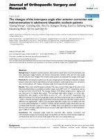
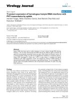
Study design upper panel) and clinical history of the patient (bottom panel)Figure 1
Study design upper panel) and clinical history of the patient (bottom panel). A 65 year old patient with an adenocar-
cinoma of the colon stage IIIc pT3, N2, M0, G2 (02/03) came to our attention at the time of an anastomotic relapse in 03/05.
After surgical resection of the colon carcinoma relapse in 06/05, a biopsy was provided to our laboratory for phenotypic char-
acterization. Two months later (08/05), the NK cell therapy was started. The patient received 6 sequential leukapheresis/re-
infusion cycles of autologous, ex vivo TKD/IL-2-activated PBMNC on a monthly basis. After a 3-month break, the patient
received another 3 cell re-infusions. The patient did not show any signs of metastases at the end of the NK cell therapy, as
determined by CT scan. The time interval between the beginning of the NK cell therapy and death was 27 months. Survival fol-
lowing recurrence and overall survival after first diagnosis was 32 and 58 months, respectively.
NK therapy-death: 27 months
Survival following recurrence: 32 months
Survival following primary tumor: 58 months
Study design
Clinical history
02/03 06/05 08/05 06/06 11/07
Sur ger y
adenocarcinom a
colon stage IIIc
pT3, pN2, M0, G2
Sur ger y
anastom otic
relapse
rpT3, rpN0, M0, G2
Death
CEA
NK cell therapy
Reinfusion
TKD/IL-2
stimulation
Leukapheresis
Journal of Translational Medicine 2009, 7:50 />Page 5 of 18
(page number not for citation purposes)
Hsp70 protein and Hsp70 antibody ELISA
The concentrations of Hsp70 protein and Hsp70 antibody
were measured in the patient's serum which was taken
before leukapheresis L7, L8, and L9 using a sandwich
ELSA kit (Duo Set IC; R&D Systems), according to the
manufacturer's instructions.
Generation of a tumor cell line
A 0.5 cm
3
tumor specimen from the patient's anastomotic
relapse was obtained from the Department of Pathology.
After washing, the tumor tissue was mechanically minced
in RPMI 1640 medium supplemented with 10% v/v fetal
calf serum (FCS), 1 mM sodium pyruvate, antibiotics (all
from Gibco-BRL, Eggenstein, Germany) and 2 mM L-
glutamine (PAN Systems, Aidenbach, Germany) and the
homogenate was passed through a sterile mesh. An aliq-
uot of the single cell suspension was immediately used for
flow cytometry analysis, and the other was seeded into T-
25 culture flasks in supplemented RPMI 1640 medium.
After 2 weeks, adherent cells were trypsinized (trypsin/
EDTA, Gibco-BRL), counted and 0.5 × 10
6
viable cells
were resuspended in 5 ml fresh medium for further flow
cytometric analyses. Aliquots of the established tumor cell
line from the first 5 cell passages were stored in liquid
nitrogen.
Flow cytometric analysis of tumor and effector cells
For flow cytometry of tumor cells, 2 × 10
5
propidium
iodide negative (viable) cells were incubated for 30 min at
4°C in the dark with the following monoclonal antibod-
ies (mAbs): anti-fibroblast (ASO2-PE, Dianova, Ham-
burg, Germany), anti-MHC class I (W6/32-FITC, IgG2a;
Cymbus Biotechnology, Eastleigh, UK), anti HLA-E
(MEM-E/06-PE, IgG1; Biozol Diagnostica, Eching, Ger-
many), anti-MICA/B (BAMO1, IgG1; BAMO2, IgG2a,
Bamomab, Munich, Germany, kindly provided by Dr.
Alexander Steinle, Tübingen), anti-ULBP-1,2,3 (AUMO2,
IgG2a; BUMO1, IgG1; CUMO1, IgG1; all purchased from
Bamomab), anti-human Hsp70 (cmHsp70.1-FITC,
mouse IgG1, multimmune GmbH, Munich, Germany).
The cmHsp70.1 mAb recognizes the sequence NLLGRFEL
(aa 454–461) in the C-terminal domain of Hsp70 which
is exposed to the extracellular milieu of tumor cells [5].
This sequence acts as a recognition structure for NK cells
that have been stimulated either with full length Hsp70
protein or with the 14-mer Hsp70 peptide TKDNNLL-
GRFELSG (aa 450–463) when combined with low dose
IL-2 [11,18,19]. The phenotypic characterization of the
tumor was performed at the Klinikum rechts der Isar,
Technische Universität München.
Unstimulated and stimulated PBMNC harvested from
leukapheresis products and from the peripheral blood
were incubated with the following mAbs as described
above: anti-CD3 and anti-CD16/56-tricolor-conjugated
(Caltag, Hamburg, Germany), anti-CD94-FITC (HP-3D9,
IgG1; Becton Dickinson Pharmingen, Heidelberg, Ger-
many) and anti-CD94-PE (Ancell Bayport, Minneapolis,
MN, USA); anti-CD56-FITC (Becton Dickinson), anti-
NKG2D-PE (149810, IgG1, R&D Systems, Minneapolis,
MN, USA). FITC and PE labeled IgG1 and IgG2a immuno-
blobulins were used as isotype-matched non-specific
binding controls (Caltag, Hamburg, Germany). Differen-
tial counts and determination of lymphocyte subpopula-
tions in leukapheresis products was done with a dual-
color lyse and wash method (Sumlset, BD). Flow cytomet-
ric analysis of unstimualted leukapheresis products were
performed at the Klinikum rechts der Isar, Technische
Universität München and at the LMU, the agreement of
the results between both laboratories was verified apply-
ing Rainbow Calibration Particles (BD). Stimulated effec-
tor cells were only analyzed by flow cytometry at the
Klinkum rechts der Isar, Technische Universität München.
After 2 washing steps in PBS containing 2% v/v FCS (PBS/
FCS) and the addition of propidium iodide (PI, Sigma-
Aldrich, Deisenhofen, Germany, stock solution 1 μg/ml),
the cells were immediately analyzed by flow cytometry
using a FACSCalibur™ instrument (Becton Dickinson,
Heidelberg, Germany). The cell population was identified
on the basis of their forward (FSC) and right angle light
scatter properties (FSC vs SSC) and the fluorescence char-
acteristics of 5,000 to 10,000 gated events were deter-
mined. Data acquisition and analysis were performed
using CellQuest™ Pro software (Becton Dickinson).
Measurement of phenotype and cytolytic activity of
patient-derived PBMNC
For the in vitro analysis of stimulated cell populations,
sterile aliquots of the leukapheresis products were incu-
bated under identical culture conditions as the sample
which was to be re-infused. The cytolytic activity of
patient-derived PBMNC, without any further enrichment
for NK cells, against the classical NK target cell line K562
and the autologous, Hsp70 membrane-positive tumor
before and after in vitro stimulation with TKD/IL-2, and of
freshly isolated, non-cultured patient-derived PBMNC
before and after re-infusion in vivo was assessed using a
standard 4-hour granzyme B ELISPOT assay and a
51
chromium release assay. As the lysis of Hsp70 mem-
brane-positive tumors by NK cells has previously been
identified as being perforin-independent, granzyme B
mediated apoptosis [13], this assay is suitable to deter-
mine the Hsp70-reactivity of NK cells.
For the ELISPOT assay, 96-well ELISPOT plates (Millipore
GmbH, Schwalbach, Germany) were coated with capture
antibody by overnight incubation at 4°C, after which they
were blocked using 10% v/v FCS. The effector and target
cells (3 × 10
3
) were added at different E/T ratios ranging
Journal of Translational Medicine 2009, 7:50 />Page 6 of 18
(page number not for citation purposes)
from 20/1 to 0.5/1. After 4 hours incubation at 37°C and
2 washes, a biotinylated detecting antibody (2 μg/ml) was
added. After an additional 2 washes, the presence of
granzyme B was visualized using 3-amino-9-ethly-carba-
zole substrate solution (25 min). Spots were counted and
data were analyzed using an Immuno Spot Series 3A Ana-
lyzer (CTL-Europe GmbH, Aalen, Germany).
Antibody blocking studies
For blocking of the cytolytic activity the NK specific anti-
bodies directed against NKp30, NKp44, NKp46 (Immu-
notech, Marseille, France) and the antibodies directed
against Hsp70 (cmHsp70.2, multimmune GmbH) and
MICA/B (BAMO1, IgG1; BAMO2, IgG2a, Bamomab,
Munich, Germany) on tumor cells were used. Briefly,
either effector or tumor cells were incubated with the rel-
evant antibodies at a final concentration of 5 μg/ml for 20
min at 4°C. Then the cells were used as targets for ELIS-
POT assays or a standard
51
chromium release assays, as
described elsewhere [9]. Briefly, K562 and autologous
tumor cells were labeled with sodium [
51
Cr] chromate
(100 μCi; NEN Dupont) and used as target cells. Three
thousand target cells were put into 96-well round-bot-
tomed plates and effector cells were added at indicated E/
T ratios. The cells were incubated for 4 hours at 37°C and
free
51
chromium was analyzed in a gamma counter (Coul-
ter). % spontaneous release was both target cells was
always less than 10%.
Immunohistochemistry
For the immunohistochemical analyses, paraffin-embed-
ded specimens were cut at 2–3 μm, using conventional
histological techniques and transferred to slides (Super
Frost Plus, Menzel, Germany). All staining was automati-
cally performed on a Ventanas Benchmark
®
XT using the
following antibodies at the indicated dilutions: CD1a
(Cat.1590, Immunotech, Tonsille); CD3 (SP7, NeoMark-
ers,1:300, Tonsille); CD4 (4B12, Novocastra,1:50, Ton-
sille); CD8 (C8/144B, NeoMarkers,1:50, Tonsille);
CD25–305 (Novocastra,1:50, Tonsille); CD45 (LCA,
2B11+PD7, Dako, 1:1000, Tonsille); CEA (TF-3H8-1,
1:100, Ventana, Darm); CD56 (123C3.D5, 1:50, Ven-
tana); Granzyme B (GrB-7, 1:25, Dako,); Perforin (5B10,
1:10, NeoMarkers); Hsp70 (6B3, antibody supernatant
was kindly provided by Dr. Elisabeth Kremmer, Helm-
holtz Center Munich).
Results and discussion
Phenotypic characterization of patient-derived tumor
The morphological appearance of the tumor cell line
derived from the anastomotic relapse under sub-conflu-
ent culture conditions is shown in Figure 2A. Following
regular twice weekly cell passages, the tumor cells formed
spheroids which could be suspended by a short trypsini-
zation step. The doubling-time of the patient-derived
tumor cell line was 22 hours. The phenotype was exam-
ined on single-cell suspensions of the tumor cell line
derived from the patient's tumor specimen by flow cytom-
etry and by immunohistochemistry. The percentage of
marker positive cells were determined on a minimum of
six separate occasions, and the findings are summarized in
Table 1. The tumor was found to be membrane MHC class
I positive, but negative for the expression of HLA-E. Fur-
thermore, the tumor revealed a strong membrane-positiv-
ity for the activatory NK cell ligands MICA/B, ULBP-3 and
Hsp70. The expression of ULBP-1 and -2 was lower than
that of ULBP-3. The percentage of contaminating connec-
tive tissue in the tumor cell culture, as determined using
the ASO2 mAb, always remained below 5% during pas-
sages 1 to 121 (Table 1). A comparative H&E immunohis-
tochemistry staining of the primary tumor biopsy (Figure
2B) and the anastomotic relapse (Figure 2C) revealed that
the cytosolic Hsp70 content is elevated in the anastomotic
relapse, thus indicating that Hsp70 levels might be associ-
ated with a more aggressive tumor stage. The antibodies
directed against MICA/B and ULBP-1,2,3, which were
used for flow cytometry did not stain paraffin-embedded
tumor specimens (data not shown).
Laboratory parameters
The total number of peripheral blood leukocytes, the per-
centage of lymphocytes, the hemoglobin content, the
number of thrombocytes, and the proportion of lym-
phocyte subpopulations such as CD3
+
, CD3
+
/CD4
+
and
CD3
+
/CD8
+
T cells, CD19
+
B cells, CD3
+
/CD16/56
+
NK-
like T cells, and CD3
-
/CD16/56
+
NK cells in the peripheral
blood were within normal levels throughout the 9-month
therapeutic intervention period (Table 2). The number of
CD4
+
/CD25
+
T cells and of CD3
+
/CD16/56
+
NK-like T
cells was always below 5%. Like in healthy human indi-
viduals the proportion of CD3
-
/CD16
+
CD56
+
NK cells in
the peripheral blood before the start of each leukapheresis
ranged between 14 to 21%. These data indicate that the
adoptive transfer of ex vivo TKD/IL-2-activated PBMNC
did not result in a significant numerical expansion or
depletion of a distinct lymphocyte subpopulation.
Table 1: Phenotype of the anastomotic relapse of an
adenocarcinoma of the colon as determined by flow cytometry
Cell marker Positively stained cells (%)
ASO2 2.1 ± 0.5
MHC I 89 ± 7
HLA-E 0.6 ± 1.2
MICA/B 73 ± 4.8
ULBP-1 33 ± 10
ULBP-2 64 ± 2.1
ULBP-3 98 ± 3.8
Hsp70 65 ± 1.8
Journal of Translational Medicine 2009, 7:50 />Page 7 of 18
(page number not for citation purposes)
The total number of nucleated cells and the total lym-
phocyte counts within the 9 leukapheresis products
ranged between 1.1 × 10
10
to 1.7 × 10
10
and 4.3 × 10
9
to
8.5 × 10
9
, respectively (Table 3). The number of NK cells
ranged from 0.9 × 10
9
(lowest value, 5
th
cycle) to 1.9 × 10
9
(highest value, 4
th
cycle), and this corresponded to 16% to
25% of the respective total lymphocyte population. These
parameters were not significantly different to those
obtained in the previous clinical phase I dose-escalating
study [15]. In this study the total lymphocyte counts in
the 12 cancer patients ranged from 0.7 × 10
9
to 8.5 ×
10
9
and the number of activated NK cells ranged from 0.1
× 10
9
to 1.5 × 10
9
.
In the follow-up period of approximately 1 year after ter-
mination of the cell-based therapy (06/06), which
included a chemoembolisation therapy consisting of
Gemcitabine (Gem), Irinotecan (Irino), Epirubicin (Epi),
and Oxaliplatin (Oxa), the leukocyte and lymphocyte
dropped below normal levels; hemoblobin levels and
thrombocyte counts remained within the normal range
(Table 4).
Similar to the phase I clinical trial, no acute or sub-acute
side effects occurred after 6 repeated infusion cycles [15].
Even after the 9
th
leukapheresis/re-infusion cycle (L9), the
therapy was well tolerated and the patient showed no
signs of toxic side effects. Both the leukapheresis and re-
infusion were performed in an out-patient setting on the
A- Photomicrograph view of the patient-derived cell line of the anastomotic relapseFigure 2
A- Photomicrograph view of the patient-derived cell line of the anastomotic relapse. Cells were cultured and pas-
saged twice a week. The picture was taken at sub-confluent stage at cell passage 26; the scale bar marks 10 μm. B/C: Compar-
ative immunohistochemical analysis of the cytosolic Hsp70 content in the primary colon carcinoma (B) and the anastomotic
relapse (C). Histological slides were stained with the Hsp70 specific antibody 3B3 which reacts with Hsp70 and does not cross-
react with Hsc70; the scale bar marks 100 μm.
Anastomotic relapse
10 μm
Primary colon carcinoma Anastomotic relapse
A
CB
Journal of Translational Medicine 2009, 7:50 />Page 8 of 18
(page number not for citation purposes)
patient's request. The patient reported a high quality of
life throughout the cell-based therapeutic period.
No treatment-associated changes in the standard labora-
tory parameters were observed during the cell-based treat-
ment procedure (data not shown). Although levels of the
tumor-associated marker PSA increased slightly from its
initial value at time of first diagnosis of the prostate cancer
(13.6 ng/ml) to the time point when the cell-based ther-
apy was started (15.3 ng/ml), they remained unchanged
during the cell-based therapy (Figure 3A). The level of the
tumor-associated marker CEA, which was 13.2 ng/ml (01/
03) before surgery of the primary colon tumor in 02/03,
and 9.5 ng/ml before surgery of the anastomotic relapse in
06/05, dropped to 5.4 ng/ml after the first and to 5.2 ng/
ml after the second tumor resection. During the first 6 cell
re-infusions the CEA levels remained almost unaltered
(L1, 4.4; L2, 4.6, L3, 4.2; L4, 4.3 L5, 3.6; L6, 3.9 ng/ml).
After the 3-month break in therapy the CEA values
increased to 5.6 ng/ml and after termination of the 9
th
therapy cycle the CEA value was 12.1 ng/ml (Figure 3B).
The Hsp70 protein levels in the serum of the patient
before the last three re-infusion cycles were found to be
elevated compared to that measured by commercially
available ELISA kits in healthy controls. Furthermore, the
Hsp70 antibody levels increased more than 20-fold dur-
ing the re-infusion cycles L7 and L8 and more than 10-
fold during L9, as compared to that of healthy human
individuals (Table 5). It remains unclear whether these
findings are related to the cell-based therapy or whether
these values reflect a spontaneous release of Hsp70 from
tumor cells.
Clinical response and the patient's clinical history
Magnetic resonance imaging (MRI) of the prostate
revealed that the prostate cancer remained unchanged
during the adoptive transfer with TKD/IL-2-activated NK
cells and the follow-up phase. The PSA levels did not sig-
nificantly alter during the observation period (Figure 3A).
With respect to the anastomotic relapse of the colon carci-
noma, the patient remained disease-free during the first 6
cell infusion cycles, during the 3-month break in therapy
and until the last cell infusion, as assessed by coloscopic
analyses every 3 months, and regular whole body MRI and
by PET-CT scans. These findings were in accordance with
the CEA values (Figure 3B).
However, the patient developed liver metastases in both
liver lobes with 20% of liver volume replaced by tumor
(LVRT) 11 months after the start of the adoptive transfer
of TKD/IL-2-activated effector cells and 13 months after
the resection of the anastomotic relapse. At this stage a
systemic chemotherapy was recommended which was
Table 2: White blood counts (WBCs), hemoglobin, thrombocytes and lymphocyte subpopulations in the peripheral blood after 9 re-
infusion cycles
Cycle 1.2.3.4.5.6.7.8.9.
WBCs, hemoglobin, thrombocytes in the peripheral blood
[Normal range] healthy donors (n = 6)
Leukocytes (G/l) [≥ 4] 4.1 6.3 5.0 5.0 5.2 5.2 4.0 4.9 6.5
Lymphocytes (%) [15–40%] (17) (22) (24) (16) (14) (16) (20) (29) (17)
Hemoglobin (g/dl) [≥ 11] 13.6 14.4 14.4 12.9 12.7 12.8 13.1 12.8 11.6
Thrombocytes (G/l) [≥ 100] 146 187 135 128 149 130 157 173 177
Lymphocyte subpopulations (%)
CD3
+
[55–95] 68 72 65 70 66 65 57 60 62
CD3
+
CD4
+
[35–65] 51 48 44 50 51 na* na na na
CD3
+
CD8
+
[21–45] 18 18 18 17 17 na na na na
CD19
+
[5-20] 14 18 18 16 17 15 14 21 12
CD3
+
CD16
+
CD56
+
134334nanana
CD3
-
CD16
+
CD56
+
[5-35] 19 17 21 16 14 15 23 15 19
* na, not analyzed
Table 3: Number of re-infused total nuclear cells, total lymphocytes and total NK cell counts
Cycle 1. 2. 3.4.5.6.7.8.9.
Total nuclear cells, lymphocytes, NK cells in the leukapheresis products
Total nuclear cells (×10
10
) 1.1 see 1. 1.5 1.2 1.4 1.7 1.7 1.3 1.2
Total lymphocytes (×10
9
) 7.6 8.5 8.3 4.3 5.8 6.3 5.1 6.9
Lymphocytes (%) (69) (57) (69) (31) (34) (37) (39) (58)
Total NK cells (×10
9
) 1.8 1.3 1.9 0.9 1.4 1.4 1.2 1.7
NK cells (%) (24) (16) (23) (20) (24) (23) (23) (25)
Journal of Translational Medicine 2009, 7:50 />Page 9 of 18
(page number not for citation purposes)
refused by the patient. In the absence of any therapeutic
intervention, the patient developed duodenum metas-
tases. Four months after the last infusion cycle the CEA
levels increased more than 10-fold from 12.1 (06/06) to
166.4 ng/ml (10/06) (Figure 3C). Systemic chemotherapy
was further refused by the patient but in 10/06 liver
lesions were treated with intra-arterial chemoembolisa-
tion consisting of Gemcitabine, Irinotecan, Epirubicin
and Oxaliplatin, every 6 to 8 weeks within the following
12 months (Figure 3C). Despite a transient drop of the
CEA levels from 353.4 (01/07) to 37.7 ng/ml (03/07) dur-
ing the treatment with Irinotecan, the general clinical con-
dition, liver function (cholestatic parameters), and CEA
levels gradually worsened (Figure 3C), and the patient
finally developed jaundice, malignant ascites and eventu-
ally died of progressive metastatic disease in 11/07.
In summary, the time interval between start of the cell-
based therapy and death was 27 months. The overall sur-
vival (time interval between first diagnosis of the colon
carcinoma and death) was 58 months and the survival fol-
lowing recurrence (time interval between anastomotic
relapse and death) was 32 months. An overview of the
clinical course is illustrated in the bottom panel of Figure
1.
Immunological responses
NK cell phenotype and in vitro cytolytic activity after TKD/IL-2
stimulation
In our previous phase I study, we reported that ex vivo
stimulation of PBMNC with TKD/IL-2 significantly
increases the cytolytic activity of NK cells against Hsp70
membrane-positive tumor cell lines in 10 of 12 patients
with advanced malignant disease [15]. T cells appeared
not to be affected by this therapeutic approach. Further-
more, IL-2 alone had no significant effect on the cytolytic
activity of PBMNC [15]. Concomitant with an increased
cytotoxicity, the mean fluorescence intensity (mfi) of the
NK cell receptor CD94 was found to be enhanced [15].
Here, we studied both, the percentage and the cell surface
density of T and NK cell marker positive cells in the leuka-
pheresis products before and after each of the 9 stimula-
tion cycles of freshly isolated, non-cultured PBMNC. The
percentage of CD3
+
T cells remained unaffected by the
stimulation with TKD/IL-2 however, between leukapher-
esis L3 and L6 the mean fluorescence intensity (mfi) of
CD3 appeared to be elevated above initial levels (Figure 4,
upper right panel). Within the three months therapy
break (L6+2, L6+8, L6+12 weeks after leukapheresis L6;
hatched bars) the CD3 mfi values dropped down to the
initial level and remained there during the last three re-
infusion cycles L7–L9, on freshly isolated, non-cultured
PBMNC of the patient.
With respect to the NK cell markers CD56 and the C-type
lectin receptor CD94, the percentage and the mfi values
were up-regulated in each treatment cycle, apart from leu-
kapheresis L4, when a maximum in the mfi value was
reached (Figure 4). The second re-infusion product was
identical to the first one which was aliquoted and cryo-
conserved in two parts. During the treatment pause (L6+2,
L6+8, L6+12 weeks after leukapheresis L6; hatched bars)
the levels of CD56 and CD94 gradually dropped but
could be enhanced by additional stimulation cycles.
In summary and in line with the data of the phase I clini-
cal trial, a comparative analysis of leukapheresis products
which were obtained before and after in vitro stimulation
with TKD/IL-2 revealed an increase in the surface densities
of CD94 and CD56. This was slightly decreased after the
3-month interruption of the therapy. The subsequent 3
treatment cycles again resulted in an enhanced density of
the indicated NK cell markers. Compared to unstimulated
cells the density of the activatory NK cell receptors was
also elevated following stimulation with TKD/IL-2. The
percentage of NKG2D positively stained cells and the
mean fluorescence intensity (mfi) values in the unstimu-
lated PBMNC was 21% (39) for leukapheresis L8 and
19% (42) for L9, respectively. Following TKD/IL-2 stimu-
lation the values increased up to 36% (52) for L8 and to
24% (45) for L9. Similarily the percentage of Natural
Cytotoxicity Receptor (NCR) positively stained cells and
the mfi in the TKD/IL-2-activated effector cells derived
from leukapheresis 9 was elevated from 1 (21) to 3%
(151) for NKp30, from 0.4 (15) to 1% (175) for NKp44,
and from 2 (45) to 8% (234) for NKp46. These activation
Table 4: Differential blood counts after termination of the cell-based therapy during chemoembolisation with Gemcitabine (Gem),
Irinotecan (Irino), Epirubicin (Epi), Oxaliplatin (Oxa)
Date 08/06 09/06 Gem 11/06 Gem 01/07 Irino 03/07 Epi 04/07 Epi 07/07 Oxa 09/07 10/07
WBCs, lymphocytes, hemoglobin, thrombocytes after cell-based therapy
[Normal range] healthy donors
(n = 6)
Leukocytes (G/l) [≥ 4] 6 7 7.6 9.9 6.4 6.3 3.4 3.3 3.0
Lymphocytes (%) [15–40%] (11) (9) (6) (7) (8) (7) (9) (13) (13)
Hemoglobin (g/dl) [≥ 11] 11.7 11.2 12.3 12.5 11.2 10.2 10.3 11.1 11.7
Thrombocytes (G/l) [≥ 100] 232 239 255 189 357 347 126 115 145
Journal of Translational Medicine 2009, 7:50 />Page 10 of 18
(page number not for citation purposes)
A – Kinetics of the prostate specific antigen (PSA)Figure 3
A – Kinetics of the prostate specific antigen (PSA). PSA values were determined in patients's blood before, during and
after adoptive transfer therapy with TKD/IL-2-activated PBMNC. The arrows indicate the time points of cell re-infusions. B –
Kinetics of the carcinoembryonic antigen (CEA). CEA values were determined in patient's blood before and during the adop-
tive transfer therapy with TKD/IL-2-activated PBMNC. The arrows indicate the time points of cell re-infusions. In 02/03 and in
06/05 primary tumor and anastomotic relapse was surgically removed. C – Kinetics of the carcinoembryonic antigen (CEA)
after completion of the cell-based therapy. CEA values were determined in patient's blood after the adoptive transfer therapy
with TKD/IL-2-activated PBMNC. In 10/06 a chemoembolisation of the liver metasases with Gemcitabine, Irinotecan, Epiru-
bicin and Oxaliplatin was initiated.
0
1
2
3
4
5
6
7
8
9
10
11
12
13
14
15
16
17
18
19
20
21
22
23
24
25
0
1
2
3
4
5
6
7
8
9
10
11
12
13
14
15
Surgery
primary tumor
Surgery
anastomotic
relapse
A
B
0
50
100
150
200
250
300
350
400
450
500
550
600
C
Gemcitabine
Irinotecan
Epirubicin
Oxaliplatin
Journal of Translational Medicine 2009, 7:50 />Page 11 of 18
(page number not for citation purposes)
markers were only determined in leukapheresis products
L8 and L9.
The cytolytic activity of the patient's leukapheresis prod-
ucts against the classical NK cell target line K562 (Figure
5A) and against the autologous, Hsp70 membrane-posi-
tive colon carcinoma (Figure 5B) before and after TKD/IL-
2 stimulation was measured by granzyme B ELISPOT
assay and by
51
chromium release assay. Before start of the
therapy up to the third leukapheresis no cytolytic activity
against K562 cells and autologous tumor cells was
detected in patient-derived non-stimulated PBMNC (Fig-
ure 5A/B, filled circles). The cytolytic activity against both
target cells could be significantly enhanced by TKD/IL-2
stimulation (Figure 5A/B, open circles). Remarkably, 1
month after re-infusion cycle 3 (before L4), freshly iso-
lated, non-cultured PBMNC of the patient exhibited an
initially increased anti-tumor activity against K562 cells
(Figure 5A) and autologous tumor (Figure 5B). These
effector cells also have shown a maximum in the expres-
sion density of the NK cell markers CD56 and CD94 (Fig-
ure 4).
Due to the fact that the increase in cytolytic activity fol-
lowing TKD/IL-2 stimulation in leukapheresis product L4
to L6 was not as pronounced as in leukapheresis L1 and
L3, the therapy was stopped for 3 months. Within these 3
months the high intrinsic cytolytic activity of patient-
derived PBMNC against K562 cells (Figure 5A) and autol-
ogous tumor (Figure 5B) eventually decreased but could
be restored completely by two further stimulation cycles
(L7, L8) with TKD/IL-2-activated leukapheresis products.
In the 9
th
stimulation cycle (L9) the in vitro anti-tumor
activity could not be increased. The cell-based therapy was
terminated at that stage.
The kinetics of the cytolytic activity of ex vivo stimulated
PBMC derived from leukapheresis L1–L6 and L7–L9
against K562 cells (left panel) and autologous tumor
(right panel) is summarized in Figure 6A. Compared to
the initial level a cytotoxic response was initiated after
each ex vivo stimulation cycle.
We have previously shown that data on the cytolytic activ-
ity of NK cells against Hsp70 membrane-positive leuke-
mic target cells obtained using the granzyme B ELISPOT
assay correlate with those obtained using a
51
chromium
release assay [20]. In line with these findings, also here the
51
chromium release assay corroborated the granzyme B
ELISPOT assay (data not shown).
Cytolytic activity of freshly isolated, non-cultured PBMNC after ex vivo
TKD/IL-2 stimulation and adoptive transfer
The kinetics of the cytolytic response of TKD-activated NK
cells within the patient was monitored by obtaining
peripheral blood of the patient immediately before each
cell re-infusion, 3 months after the sixth re-infusion and
every 4 weeks before the re-infusion of the activated leu-
kapheresis product L7, L8, and L9. Before start of therapy
the anti-tumor activity of patient-derived PBMNC against
K562 cells and Hsp70 membrane-positive autologous
tumor was < 5% and remained low during the first three
treatment cycles. Remarkably, one month after the third
cell infusion an intrinsically increased cytolytic response
against both tumor targets was firstly detected in the
patients blood (Figure 6B, upper panel). This activity
remained stably high during the next three re-infusion
cycles (data not shown). Therapy was interrupted for 3
months after the 6
th
re-infusion and the analysis of circu-
lating NK cells after the therapy break revealed that the
increased cytolytic capacity against K562 cells (left panel)
and autologous tumor (right panel) was reduced at that
time point but still elevated compared to the start of the
cell-based therapy. Before stimulation cycle L8 the anti-
tumor activity reached a maximum but started to decline
after the 9
th
stimulation cycle (L9, Figure 6B, lower panel).
A direct comparison of the kinetics of the cytolytic activity
of the in vitro stimulated leukapheresis product and of
freshly isolated, non-cultured PBMNC of the patient
against K562 (upper panel) and autologous tumor (lower
Table 5: Hsp70 protein and Hsp70 antibody levels in the serum of the patient within the last three treatment cycles, as determined by
standard commercial ELISA technique
Hsp70 protein serum levels (ng/ml) Hsp70 antibody levels (μg/ml)
Treatment cycle*
Before L7 10.9 ± 0.4 6,049 ± 129
Before L8 13.2 ± 0.8 5,380 ± 145
Before L9 13.3 ± 0.7 3,191 ± 122
Healthy individuals
(n = 60) [40] 2.07 ± 2.74 280 ± 58
(n = 95) [41] 4.93 207 ± 55
*The data of the patient represent mean values of at least 4 independent experiments, the healthy individuals were determined with commercial
ELISA kits.
Journal of Translational Medicine 2009, 7:50 />Page 12 of 18
(page number not for citation purposes)
Phenotypic changes of the effector cells before (black bars) and after (grey bars) in vitro TKD/IL-2 stimulationFigure 4
Phenotypic changes of the effector cells before (black bars) and after (grey bars) in vitro TKD/IL-2 stimulation.
The percentage (left panel) and mean fluorescence intensity (mfi, right panel) values of CD3
+
T cells and CD3
-
/CD56
+
and
CD3
-
/CD94
+
NK cells were determined before and after a 4 days in vitro TKD/IL-2 stimulation by flow cytometry. The hatched
bars indicate T and NK cell values derived from the patients blood during the therapeutic break 2 (L6+2), 6 (L6+6), 8 (L6+8),
and 12 (L6+12) weeks after re-infusion cycle L6. Only viable, propidium-iodide negative cells were gated and analyzed.
Leukapheresis/ reinfusion
Percentage positively stained cells
Mean fluorescence intensity (mfi)
L1 L3 L4 L5 L6 L6+2 L6+6 L6+8L6+12 L7 L8 L9
0
20
40
60
80
100
CD3 percentage d0
CD3 percentage d4
L1 L3 L4 L5 L6 L6+2 L6+6 L6+8L6+12 L7 L8 L9
0
10
20
30
CD56 percentage d0
CD56 percentage d4
L1 L3 L4 L5 L6 L6+2 L6+6 L6+8L6+12 L7 L8 L9
0
5
10
15
20
25
CD94 percentage d0
CD94 percentage d4
L1 L3 L4 L5 L6 L6+2 L6+6 L6+8L6+12 L7 L8 L9
0
200
400
600
800
1000
CD3 mfi d0
CD3 mfi d4
L1 L3 L4 L5 L6 L6+2 L6+6 L6+8 L6+12 L7 L8 L9
0
100
200
300
400
500
600
CD56 mf i d0
CD56 mf i d4
L1 L3 L4 L5 L6 L6+2 L6+6 L6+8 L6+12 L7 L8 L9
0
100
200
300
400
CD94 mfi d0
CD94 mfi d4
Journal of Translational Medicine 2009, 7:50 />Page 13 of 18
(page number not for citation purposes)
panel) during the whole therapeutic intervention (L1–L6
and L7–L9) at a distinct E/T ratio of 20/1 is summarized
in Figure 6C. This kinetics of initiation and maintenance
of the cytolytic response against Hsp70-positive tumors is
in line with our data from the phase I clinical trial [15]. It
shows that repeated re-infusions of TKD/IL-2 activated,
autologous PBMNC into patients with different tumor
entities, stages and previous therapies can result in NK cell
activity. Moreover, this is the first observation that ex vivo
activated NK cells can be sustained over longer periods in
the blood of a patient.
Blocking studies using antibodies against activatory NK
cell receptors NKp30, NKp44 and NKp46 and against the
NKG2D ligand MICA/B revealed that the cytolytic
response mediated by in vitro activated effector cells
derived from leukapheresis L9 against tumor cells was not
affected if compared to the effects which were mediated
by isotype-matched control antibodies (data not shown).
With respect to previous findings [3], we speculate that
lysis of Hsp70 membrane-positive tumor cells is rather
mediated through CD94/NKG2C.
In vitro cytolytic activity of patient-derived PBMNC derived from L1 to L6 and L7 to L9 against K562 cells (A) and autologous tumor (B)Figure 5
In vitro cytolytic activity of patient-derived PBMNC derived from L1 to L6 and L7 to L9 against K562 cells (A)
and autologous tumor (B). The lytic activity of patient-derived PBMNC before and after stimulation with TKD/IL-2 was
determined by standard granzyme B ELISPOT. Filled circles indicate the cytolytic activity of unstimulated PBMNC, open circles
that of TKD/IL-2 stimulated PBMNC. Due to technical problems data from L2 are not available. Viability of the tumor target
cells in each assay was > 95%.
E:T
0.5/1 1/1 2.5/1 5/1 10/1 20/1
counts
0
100
200
300
400
500
control
TKD/IL-2
L3
E:T
0.5/1 1/1 2.5/1 5/1 10/1 20/1
counts
0
100
200
300
400
500
control
TKD/IL-2
L5
E/T
0.5/1 1/1 2.5/1 5/1 10/1 20/1
counts
0
100
200
300
400
500
control
TKD/IL-2
L7
E:T
0.5/1 1/1 2.5/1 5/1 10/1 20/1
counts
0
100
200
300
400
500
control
TKD/IL-2
L3
E:T
0.5/1 1/1 2.5/1 5/1 10/1 20/1
counts
0
100
200
300
400
500
control
TKD/IL-2
L5
E:T
0.5/1 1/1 2.5/1 5/1 10/1 20/1
counts
0
100
200
300
400
500
control
TKD/IL-2
L1
E:T
0.5/1 1/1 2.5/1 5/1 10/1 20/1
counts
0
100
200
300
400
500
control
TKD/IL-2
L4
B
E/T
0.5/1 1/1 2.5/1 5/1 10/1 20/1
counts
0
100
200
300
400
500
control
TKD/IL-2
L9
E/T
0.5/1 1/1 2.5/1 5/1 10/1 20/1
counts
0
100
200
300
400
500
control
TKD/IL-2
L7
E/T
0.5/1 1/1 2.5/1 5/1 10/1 20/1
counts
0
100
200
300
400
500
control
TKD/IL-2
L9
E:T
0.5/1 1/1 2.5/1 5/1 10/1 20/1
counts
0
100
200
300
400
500
control
TKD/IL-2
L6
E/T
0.5/1 1/1 2.5/1 5/1 10/1 20/1
counts
0
100
200
300
400
500
control
TKD/IL-2
L8
L1
E:T
0.5/1 1/1 2.5/1 5/1 10/1 20/1
counts
0
100
200
300
400
500
control
TKD/IL-2
E:T
0.5/1 1/1 2.5/1 5/1 10/1 20/1
counts
0
100
200
300
400
500
control
TKD/IL-2
L4
E:T
0.5/1 1/1 2.5/1 5/1 10/1 20/1
counts
0
100
200
300
400
500
control
TKD/IL-2
L6
A
K562 autologous tumor
E/T
0.5/1 1/1 2.5/1 5/1 10/1 20/1
counts
0
100
200
300
400
500
control
TKD/IL-2
L8
Journal of Translational Medicine 2009, 7:50 />Page 14 of 18
(page number not for citation purposes)
Kinetics of the cytolytic activity of in vitro stimulated PBMNC (A) and freshly isolated, non-cultured PBMNC (B) of the patient derived from re-infusion cycle L1 to L6 and L7 to L9 against K562 cells (left panel) and autologous tumor (right panel)Figure 6
Kinetics of the cytolytic activity of in vitro stimulated PBMNC (A) and freshly isolated, non-cultured PBMNC
(B) of the patient derived from re-infusion cycle L1 to L6 and L7 to L9 against K562 cells (left panel) and autol-
ogous tumor (right panel). The lytic activity of patient-derived PBMNC was determined directly after in vitro stimulation
(A) and 1 month after the previous cell infusion without any further in vitro stimulation (B) by standard granzyme B ELISPOT at
E/T ratios ranging from 0.5/1 to 20/1. A direct comparison of the cytolytic activity of in vitro stimulated and freshly isolated,
non-cultured PBMNC at the distinct E/T ratio of 20/1 is illustrated in Figure 6C.
reinfusion
counts
13456 789
0
100
200
300
400
500
in vivo
in vitro
13456 789
0
100
200
300
400
500
in vivo
in vitro
counts
E/T
0.5/1 1/1 2.5/1 5/1 10/1 20/1
counts
0
100
200
300
400
500
L1
L3
L4
L5
L6
L1-6
E:T
0.5/1 1/1 2.5/1 5/1 10/1 20/1
counts
0
100
200
300
400
500
L1
L3
L4
L5
L6
L1-6
0.5/1 1.25/1 2.5/1 5/1 10/1 20/1
0
100
200
300
400
500
L7
L8
L9
L7, L9
K562 autologous tumor
0.5/1 1.25/1 2.5/1 5/1 10/1 20/1
0
100
200
300
400
500
L7
L8
L9
L8
L7
L8
L9
counts
0.5/1 1.25/1 2.5/1 5/1 10/1 20/1
0
100
200
300
400
500
L7
L8
L9
0.5/1 1.25/1 2.5/1 5/1 10/1 20/1
0
100
200
300
400
500
L7
L8
L9
0.5/1 1.25/1 2.5/1 5/1 10/1 20/1
0
100
200
300
400
500
L1
L3
L4
L5
L6
0.5/1 1.25/1 2.5/1 5/1 10/1 20/1
0
100
200
300
400
500
L1
L3
L4
L5
L6
L4-6L4-6
L1-3
L1-3
L7, L9
L8
L8
L7, L9
A
B
C
E/T
Journal of Translational Medicine 2009, 7:50 />Page 15 of 18
(page number not for citation purposes)
Immune reaction at the tumor site
Lymphocytes infiltrating colorectal cancers have been
shown to inhibit tumor growth and their presence is asso-
ciated with an improved prognosis [21,22] It has also
recently been shown that the presence of infiltrating
memory and effector T cells in human colorectal cancer
correlates with the signs of early metastatic invasion, a less
advanced pathological stage and an increased survival
[23] Furthermore, Galon et al [24] have shown that the
type, prevalence and location of immune cells within
human colorectal tumors has a prognostic value which is
superior to, and independent of, the histopathological
methods that are currently used to stage colorectal cancer.
Based on these findings, and with the consent of the
patient, paraffin-embedded specimens from the primary
colon adenocarcinoma (02/03), the anastomotic relapse
before start of the cell-based therapy (06/05) and a biopsy
of the duodenum metastases (04/07) were analyzed by
semi-quantitative immunohistochemistry. All specimens
were strongly positive for the carcino-embryogenic anti-
gen (CEA), which serves as a tumor marker for colon car-
cinoma. The presence of CD3
+
and CD45
+
cells was used
as an indicator of T cell infiltration and the prevalence of
CD4
+
, CD8
+
, CD56
+
cells as indicators of T helper, T cyto-
toxic and NK cells, respectively and CD1a was used as a
marker for antigen presenting cells (APC). The expression
of CD25 was considered here as a marker of lymphocyte
activation since it did not show any correlation with the
amount of CD4
+
cells which would reflect the presence of
regulatory T cells (CD4
+
/CD25
+
). The expression of per-
forin and granzyme B provided insight into the lytic activ-
ity of infiltrating T and NK cells. In the primary tumor and
in the anastomotic relapse there was a strong infiltration
of CD3
+
/CD4
+
T cells, but no infiltration of antigen pre-
senting cells, as determined by the marker CD1a (Table
6). The amount of T cells was lower in the metastatic tis-
sue. In all three tumor specimen hardly any CD8
+
cyto-
toxic lymphocytes were found (Table 4). A slight increase
in granzyme B-positive, CD56
+
NK cells was detectable in
the metastatic tissue which was taken after the cell-based
therapy, whereas perforin was absent (Table 4). This
might be related to the fact that TKD/IL-2 activated NK
cells kill their Hsp70 membrane-positive targets via a per-
forin-independent granzyme B mediated pathway.
Conclusion
A previous clinical phase I trial has demonstrated that up
to 6 repeated re-infusions of TKD/IL-2-activated, autolo-
gous PBMNC is safe and well-tolerated [15]. The observa-
tions that the administration of these cells induced NK
cell activity against tumor cell lines expressing Hsp70 on
the cell surface, as well as the unexpected clinical
responses that were induced prompted additional studies.
Herein, the maintenance of the cytolytic activity of ex vivo
TKD/IL-2-activated PBMNC against a classical NK target
and the autologous, Hsp70 membrane-positive tumor of
a patient with an anastomotic relapse of a colon adenocar-
cinoma was tested. In accordance with the protocol for the
clinical phase I trial, the patient received 6 cycles of ex vivo
TKD/IL-2-activated, autologous PBMNC, that were
derived form a leukapheresis, by i.v. injection. In contrast
to the phase I trial protocol, the cell re-infusions were
repeated every 4 instead of every 2 weeks.
No intrinsic NK cell activity was detected in patient-
derived PBMNC at the beginning of the therapeutic inter-
vention, nor was any apparent up to the third treatment
cycle. However, in vitro incubation of PBMNC with TKD/
IL-2 initiated a significant anti-tumor reactivity against the
classical NK target K562 and also the autologous tumor.
Most interestingly, after the fourth re-infusion cycle,
patient-derived PBMNC exhibited an intrinsically
enhanced NK cell activity. This finding is in line with the
kinetics of the NK cell activation in patients who received
more than 4 cell infusions in the phase I clinical trial [15].
Table 6: Semiquantitative immunohistological analysis of the tumor marker CEA, effector cell infiltration and effector cell function in
the primary colon carcinoma before start of the NK cell-based therapy, the anastomotic relapse before start of the NK cell-based
therapy and the duodenom metastases after finishing the NK cell-based therapy
Cell marker Primary colon tumor (02/03) Anastomotic relapse (03/05) Metastases (04/07)
CEA ++++* ++++ ++++
CD45 (lymphocytes) ++ ++ ++
CD3 (T cells) +++ +++ ++
CD4 (helper T cells) ++ ++ ++
CD8 (cytotoxic T cells) +/- +/- +/-
CD56 (NK cells) +
CD1a (APCs)
CD25 (IL-2 receptor) ++++++
Perforin (apoptosis inducer)
Granzyme B (apoptosis inducer) +
*The nomenclature means the amount of infiltrating marker-positive cells in a tumor section with a size of 2 cm
2
: -, < 50 cells; +/-, 50–80 cells; +,
80–150; ++, 150–200; +++, 200–300 cells; ++++, > 300.
Journal of Translational Medicine 2009, 7:50 />Page 16 of 18
(page number not for citation purposes)
Since the intervals of the cell infusions differed between
the phase I clinical trial and in the present study, it is more
likely to assume that the number of ex vivo stimulation
cycles is important for the initiation of the in vivo immune
response and not the kinetics. Phenotypic characteristics
and the lytic activity against K562 cells revealed that NK
cells and not T cells are responsible for the anti-tumor
activity. It currently remains unclear whether this activity
is due to the fact that the complete NK cell repertoire has
been activated after 4 stimulation cycles or whether ex
vivo-activated PBMNC have the capacity to activate other
NK cells in the circulation of the patient. A direct stimula-
tion of NK cells appears to be unlikely since soluble TKD-
peptide was not present in the infused cell suspensions.
However, it is possible that the cytolytic activity of TKD/
IL-2-activated NK cells might lead to the release of
cytosolic proteins [25] which enable a further secondary
stimulation of NK cells in vivo.
Due to the fact that in vitro TKD/IL-2 stimulation only
marginally increased the cytolytic anti-tumor activity in
PBMNC obtained from the leukapheresis L4 onwards, the
cell-based immunotherapy was interrupted for 3 months
after the sixth re-infusion cycle. The phenotype of the pre-
stimulated PBMNC that were derived from the patient's
blood reflected that of the in vitro stimulated effector cells.
Compared to the PBMNC, which were obtained before
the start of the therapy, the CD3
-
NK cells exhibited an
increased density of activatory NK cell markers such as
CD94/NKG2C, CD16/CD56, NKG2D, CD25 and the
NCRs NKp30, NKp44, NKp46, although the absolute
number of NK cells remained unaffected.
The elevated intrinsic cytolytic activity against K562 cells
and autologous tumor persisted for at least 2 months and
began to decline 3 months after the last cell infusion.
These data might provide an insight into the life-expect-
ancy and/or the cytolytic capacity of ex vivo-activated NK
cells following re-infusion into a patient. Another possi-
bility could be a numerical imbalance of active tumor-
controlling NK cells and seeding tumor cells which finally
results in a selection and an advantage of tumor cells with
metastatic potential. Moreover, we could show that the
patient's immune responses to Hsp70 membrane-positive
tumors could be restored by 2 additional re-infusion
cycles with TKD/IL-2-activated leukapheresis products.
A recent study from the Adjuvant Colon Cancer End
Points (ACCENT) data set examined prognostic factors
and survival rates following recurrence in stage II and III
colon cancer in a collection of individual patient data
from 18 trials testing FU-based adjuvant therapy con-
ducted between 1978 and 1999 [26]. In this study the
most important parameters were time from the initial
treatment to the recurrence of disease. The median sur-
vival following recurrence was 13.1 months and was 12.5
months for patients with an initial tumor stage III. Inter-
estingly, patients who had a recurrence following FU-
based adjuvant chemotherapy had a poorer prognosis
(median survival 11.5 months) than those who pro-
gressed after surgery alone (median survival 14.2
months). The patient described in the present study
remained disease-free for 15 months following recurrence
and died of progressive disease 32 months after diagnosis
of recurrence, a time interval which is more than double
that observed in the ACCENT study (32 months vs 12.5
months) [26]. Recent palliative systemic chemotherapy
with newer agents has been shown to be effective and to
substantially prolong survival [27-29], whereas locore-
gional treatments such as hepatic artery chemoembolisa-
tion currently do not provide a survival benefit for the
patient [30,31]. The time interval from progression with
liver lesions to death (16 months) and the overall survival
(58 months) of our patient who refused systemic chemo-
therapy was considerably greater than that which one
would expect after chemoembolisation [31], and that of
stage IIIc colorectal cancer patients that undergo surgery
alone (5-year median overall survival 20%) [20,32,33].
Explanations for the observed clinical outcome of the
patient may be related to the patient's individual tumor
disease, to the patient's immune status or to the applied
cell-based therapy. The patient's anastomotic relapse did
express a variety of ligands such as Hsp70, MICA-A/B,
ULBP-1,2,3 which are recognized by activatory NK cell
receptors. It is known that the DNA damage which is ini-
tiated in tumor cells by ionizing irradiation and certain
chemotherapeutic agents elicits anti-tumor immunity
[34]. These tumor cells can express "eat me" signals on
their cell surface, and they can secrete/release immunos-
timulatory factors, such as cytokines, which in turn stim-
ulate effector cells of the innate immune system [34].
The patient described in this report received several treat-
ments of intra-hepatic chemoembolisation which could
result in an overexpression of Hsp70 within the tumor
[35-38]. As a result of tumor cell necrosis or active release
Hsp70 might become available for the innate immune
system [31]. Regarding these results we hypothesize that
NK cells might be re-activated by stressed tumor cells.
Despite the high level of cytolytic activity over a period of
10 months, the patient died from metastatic liver disease
27 months after cell-based therapy, and 32 months after
recurrence. An explanation for this might be that the
tumor has escaped the control mediated by TKD/IL-2-acti-
vated NK cells in vivo. Furthermore, we cannot exclude
that the metastases succeeded to acquire an NK cell escape
mechanism such as a down-regulated activatory NK lig-
and expression such as Hsp70, MICA/B or ULBP-1,2,3 or
Journal of Translational Medicine 2009, 7:50 />Page 17 of 18
(page number not for citation purposes)
an up-regulation of inhibitory NK ligands such as HLA-E
molecules [23,39]. Unfortunately we are unable to
address these questions due to a lack of metastatic tumor
material from the patient. At the time point when meta-
static disease was histologically proven, the in vivo cyto-
lytic activity of patient-derived PBMNC had dropped.
Interestingly, the Hsp70 antibody levels and to a lower
extent also the Hsp70 protein levels in the serum were
found to be highly elevated above normal levels [40,41]
within the last three treatment cycles. Whether this
increase is associated with the stage of disease remains to
be determined by kinetic studies in a larger group of
patients.
In summary, we could demonstrate that 4 re-infusion
cycles of ex vivo TKD/IL-2-activated PBMNC initiate and
sustain an intrinsic NK cell-mediated cytolytic activity
against autologous tumor and the NK cell target K562.
This finding is in accordance to data derived from a clini-
cal phase I trial [15] and could be confirmed in a pilot
patient with malignant metastatic melanoma. An intrinsi-
cally enhanced cytolytic activity against Hsp70-positive
tumor cells was observed in all patients who received
more than 4 treatment cycles.
Competing interests
The authors declare that they have no competing interests.
Authors' contributions
GM contributed to conception and design, funding,
supervision, data interpretation, writing and final
approval of the manuscript. VM, SS, MG, BW, KH, and
DM contributed to data collection and assembly of data.
MM, WH, RI contributed to critical revision of the manu-
script. All authors read and approved the final version of
the manuscript.
Acknowledgements
This work was supported in part by grants from the Deutsche Forschungs-
gemeinschaft (DFG MU1238 7/2), the Bundesministerium für Forschung
und Technologie (BMBF, BioChance Plus, MOBITUM), EU-CARDIORISK
(#211403), EU-STEMDIAGNOSTICS (#037703), EU-TRANSNET
(#512253), and the multimmune GmbH, Munich.
References
1. Dunn GP, Old LJ, Schreiber RD: The immunobiology of cancer
immunosurveillance and immunoediting. Immunity 2004,
21:137-148.
2. Gross C, Hansch D, Gastpar R, Multhoff G: Interaction of heat
shock protein 70 peptide with NK cells involves the NK
receptor CD94. Biol Chem 2003, 384:267-279.
3. Gross C, Schmidt-Wolf IG, Nagaraj S, Gastpar R, Ellwart J, Kunz-
Schughart LA, Multhoff G: Heat shock protein 70-reactivity is
associated with increased cell surface density of CD94/CD56
on primary natural killer cells. Cell Stress Chaperones 2003,
8:348-360.
4. Gehrmann M, Liebisch G, Schmitz G, Anderson R, Steinem C, De MA,
Pockley G, Multhoff G: Tumor-specific Hsp70 plasma mem-
brane localization is enabled by the glycosphingolipid Gb3.
PLoS ONE 2008, 3:e1925.
5. Multhoff G, Pfister K, Gehrmann M, Hantschel M, Gross C, Hafner M,
Hiddemann W: A 14-mer Hsp70 peptide stimulates natural
killer (NK) cell activity. Cell Stress Chaperones 2001, 6:337-344.
6. Multhoff G, Botzler C, Issels R: The role of heat shock proteins
in the stimulation of an immune response. Biol Chem 1998,
379:295-300.
7. Gastpar R, Gehrmann M, Bausero MA, Asea A, Gross C, Schroeder
JA, Multhoff G: Heat shock protein 70 surface-positive tumor
exosomes stimulate migratory and cytolytic activity of natu-
ral killer cells. Cancer Res 2005, 65:5238-5247.
8. Zhang H, Liu R, Huang W: A 14-mer peptide from HSP70 pro-
tein is the critical epitope which enhances NK activity
against tumor cells in vivo. Immunol Invest 2007, 36:233-246.
9. Multhoff G, Botzler C, Jennen L, Schmidt J, Ellwart J, Issels R: Heat
shock protein 72 on tumor cells: a recognition structure for
natural killer cells. J Immunol 1997, 158:4341-4350.
10. Stangl S, Wortmann A, Guertler U, Multhoff G: Control of metas-
tasized pancreatic carcinomas in SCID/beige mice with
human IL-2/TKD-activated NK cells. J Immunol
2006,
176:6270-6276.
11. Moser C, Schmidbauer C, Gurtler U, Gross C, Gehrmann M, Thonigs
G, Pfister K, Multhoff G: Inhibition of tumor growth in mice
with severe combined immunodeficiency is mediated by
heat shock protein 70 (Hsp70)-peptide-activated, CD94 pos-
itive natural killer cells. Cell Stress Chaperones 2002, 7:365-373.
12. Gross C, Holler E, Stangl S, Dickinson A, Pockley AG, Asea AA, Mal-
lappa N, Multhoff G: An Hsp70 peptide initiates NK cell killing
of leukemic blasts after stem cell transplantation. Leuk Res
2008, 32:527-534.
13. Gross C, Koelch W, DeMaio A, Arispe N, Multhoff G: Cell surface-
bound heat shock protein 70 (Hsp70) mediates perforin-
independent apoptosis by specific binding and uptake of
granzyme B. J Biol Chem 2003, 278:41173-41181.
14. Pfister K, Radons J, Busch R, Tidball JG, Pfeifer M, Freitag L, Feldmann
HJ, Milani V, Issels R, Multhoff G: Patient survival by Hsp70 mem-
brane phenotype: association with different routes of metas-
tasis. Cancer 2007, 110:926-935.
15. Krause SW, Gastpar R, Andreesen R, Gross C, Ullrich H, Thonigs G,
Pfister K, Multhoff G: Treatment of colon and lung cancer
patients with ex vivo heat shock protein 70-peptide-acti-
vated, autologous natural killer cells: a clinical phase I trial.
Clin Cancer Res 2004, 10:3699-36707.
16. Macdonald JS: Adjuvant therapy of colon cancer. Cancer J Clin
1999, 49:202-219.
17. O'Connell JB, Maggard MA, Ko CY: Colon cancer survival rates
with the new American Joint Committee on Cancer sixth
edition staging. J Natl Cancer Inst 2004, 96:1420-1425.
18. Multhoff G, Mizzen L, Winchester CC, Milner CM, Wenk S, Eissner
G, Kampinga HH, Laumbacher B, Johnson J: Heat shock protein 70
(Hsp70) stimulates proliferation and cytolytic activity of nat-
ural killer cells. Exp Hematol 1999, 27:1627-1636.
19. Multhoff G, Pfister K, Botzler C, Jordan A, Scholz R, Schmetzer H,
Burgstahler R, Hiddemann W: Adoptive transfer of human natu-
ral killer cells in mice with severe combined immunodefi-
ciency inhibits growth of Hsp70-expressing tumors. Int J
Cancer 2000, 88:
791-797.
20. Stangl S, Gross C, Pockley AG, Asea AA, Multhoff G: Influence of
Hsp70 and HLA-E on the killing of leukemic blasts by
cytokine/Hsp70 peptide-activated human natural killer (NK)
cells. Cell Stress Chaperones 2008, 13:221-230.
21. Diederichsen AC, Hjelmborg JB, Christensen PB, Zeuthen J, Fenger
C: Prognostic value of the CD4+/CD8+ ratio of tumour infil-
trating lymphocytes in colorectal cancer and HLA-DR
expression on tumour cells. Cancer Immunol Immunother 2003,
52:423-428.
22. Naito Y, Saito K, Shiiba K, Ohuchi A, Saigenji K, Nagura H, Ohtani H:
CD8+ T cells infiltrated within cancer cell nests as a prognos-
tic factor in human colorectal cancer. Cancer Res 1998,
58:3491-3494.
23. Pages F, Berger A, Camus M, Sanchez-Cabo F, Costes A, Molidor R,
Mlecnik B, Kirilovsky A, Nilsson M, Damotte D, Meatchi T, Bruneval
P, Cugnenc PH, Trajanoski Z, Fridman WH, Galon J: Effector mem-
ory T cells, early metastasis, and survival in colorectal can-
cer. N Engl J Med 2005, 353:2654-2666.
24. Galon J, Costes A, Sanchez-Cabo F, Kirilovsky A, Mlecnik B, Lagorce-
Pages C, Tosolini M, Camus M, Berger A, Wind P, Zinzindohoue F,
Publish with BioMed Central and every
scientist can read your work free of charge
"BioMed Central will be the most significant development for
disseminating the results of biomedical research in our lifetime."
Sir Paul Nurse, Cancer Research UK
Your research papers will be:
available free of charge to the entire biomedical community
peer reviewed and published immediately upon acceptance
cited in PubMed and archived on PubMed Central
yours — you keep the copyright
Submit your manuscript here:
/>BioMedcentral
Journal of Translational Medicine 2009, 7:50 />Page 18 of 18
(page number not for citation purposes)
Bruneval P, Cugnenc PH, Trajanoski Z, Fridman WH, Pages F: Type,
density, and location of immune cells within human colorec-
tal tumors predict clinical outcome. Science 2006,
313:1960-1964.
25. Elsner L, Muppala V, Gehrmann M, Lozano J, Malzahn D, Bickeboller
H, Brunner E, Zientkowska M, Herrmann T, Walter L, Alves F, Mul-
thoff G, Dressel R: The heat shock protein Hsp70 promotes
mouse NK cell activity against tumors that express inducible
NKG2D ligands. J Immunol 2007, 179:5523-5533.
26. O'Connell MJ, Campbell ME, Goldberg RM, Grothey A, Seitz JF, Bene-
detti JK, Andre T, Haller DG, Sargent DJ: Survival following recur-
rence in stage II and III colon cancer: findings from the
ACCENT data set. J Clin Oncol 2008, 26:2336-2341.
27. Stangl R, Tendorf-Hofmann A, Charnley RM, Scheele J: Factors
influencing the natural history of colorectal liver metastases.
Lancet 1994, 343:1405-1410.
28. Schrag D: The price tag on progress – chemotherapy for
colorectal cancer. N Engl J Med 2004, 351:317-319.
29. Erlichman C, Sargent DJ: New treatment options for colorectal
cancer. N Engl J Med 2004, 351:391-392.
30. Chan R, Kerr D: Hepatic arterial chemotherapy for colorectal
cancer liver metastases: a review of advances in 2003. Curr
Opin Oncol 2004, 16:378-384.
31. Tellez C, Benson AB III, Lyster MT, Talamonti M, Shaw J, Braun MA,
Nemcek AA Jr, Vogelzang RL: Phase II trial of chemoemboliza-
tion for the treatment of metastatic colorectal carcinoma to
the liver and review of the literature. Cancer 1998,
82:1250-1259.
32. Greene FL, Stewart AK, Norton HJ: A new TNM staging strategy
for node-positive (stage III) colon cancer: an analysis of
50,042 patients. Ann Surg 2002, 236:416-421.
33. Gill S, Loprinzi CL, Sargent DJ, Thome SD, Alberts SR, Haller DG,
Benedetti J, Francini G, Shepherd LE, Francois SJ, Labianca R, Chen W,
Cha SS, Heldebrant MP, Goldberg RM: Pooled analysis of fluorou-
racil-based adjuvant therapy for stage II and III colon cancer:
who benefits and by how much? J Clin Oncol 2004, 22:1797-1806.
34. Tesniere A, Apetoh L, Ghiringhelli F, Joza N, Panaretakis T, Kepp O,
Schlemmer F, Zitvogel L, Kroemer G: Immunogenic cancer cell
death: a key-lock paradigm. Curr Opin Immunol 2008, 20:504-511.
35. Lindquist S, Craig EA: The heat-shock proteins. Annu Rev Genet
1988, 22:631-677.
36. Morimoto RI, Kline MP, Bimston DN, Cotto JJ: The heat-shock
response: regulation and function of heat-shock proteins and
molecular chaperones. Essays Biochem 1997, 32:17-29.
37. Botzler C, Ellwart J, Gunther W, Eissner G, Multhoff G: Synergistic
effects of heat and ET-18-OCH3 on membrane expression of
hsp70 and lysis of leukemic K562 cells. Exp Hematol 1999,
27:470-478.
38. Multhoff G, Meier T, Botzler C, Wiesnet M, Allenbacher A, Wilmanns
W, Issels RD: Differential effects of ifosfamide on the capacity
of cytotoxic T lymphocytes and natural killer cells to lyse
their target cells correlate with intracellular glutathione lev-
els. Blood 1995, 85:2124-2131.
39. Ulbrecht M, Martinozzi S, Grzeschik M, Hengel H, Ellwart JW, Pla M,
Weiss EH: Cutting edge: the human cytomegalovirus UL40
gene product contains a ligand for HLA-E and prevents NK
cell-mediated lysis. J Immunol 2000, 164:5019-5022.
40. Njemini R, Demanet C, Mets T: Comparison of two ELISAs for
the determination of Hsp70 in serum. J Immunol Meth 2005,
306:176-182.
41. Yeh CH, Tseng R, Zhang Z, Cortes J, O'Brien S, Giles F, Hannah A,
Estrov Z, Keating M, Kantarjian H, Albitar M: Circulating Hsp70
and progression in patients with chronic myeloid leukemia.
Leuk Res 2009, 33:212-217.