báo cáo hóa học:" Retinal pigment epithelial cells secrete neurotrophic factors and synthesize dopamine: possible contribution to therapeutic effects of RPE cell transplantation in Parkinson''''s disease" doc
Bạn đang xem bản rút gọn của tài liệu. Xem và tải ngay bản đầy đủ của tài liệu tại đây (1.13 MB, 9 trang )
BioMed Central
Page 1 of 9
(page number not for citation purposes)
Journal of Translational Medicine
Open Access
Research
Retinal pigment epithelial cells secrete neurotrophic factors and
synthesize dopamine: possible contribution to therapeutic effects of
RPE cell transplantation in Parkinson's disease
Ming Ming
1
, Xuping Li
1
, Xiaolan Fan
1
, Dehua Yang
1
, Liang Li
1
, Sheng Chen
2
,
Qing Gu
3
and Weidong Le*
1,2
Address:
1
Institute of Health Sciences, Shanghai Institutes for Biological Sciences, Chinese Academy of Sciences, and Shanghai Jiao Tong University
School of Medicine, Shanghai, 200025, PR China,
2
Institute of Neurology, Ruijin Hospital, Jiao Tong University School of Medicine, Shanghai,
200025, PR China and
3
Department of Ophthalmology, Shanghai First People's Hospital, Shanghai, 200025, PR China
Email: Ming Ming - ; Xuping Li - ; Xiaolan Fan - ; Dehua Yang - ;
Liang Li - ; Sheng Chen - ; Qing Gu - ; Weidong Le* -
* Corresponding author
Abstract
Background: New strategies for the treatment of Parkinson's disease (PD) are shifted from
dopamine (DA) replacement to regeneration or restoration of the nigro-striatal system. A cell
therapy using human retinal pigment epithelial (RPE) cells as substitution for degenerated
dopaminergic (DAergic) neurons has been developed and showed promising prospect in clinical
treatment of PD, but the exact mechanism underlying this therapy is not fully elucidated. In the
present study, we investigated whether the beneficial effects of this therapy are related to the
trophic properties of RPE cells and their ability to synthesize DA.
Methods: We evaluated the protective effects of conditioned medium (CM) from cultured RPE
cells on the DAergic cells against 6-hydroxydopamine (6-OHDA)- and rotenone-induced
neurotoxicity and determined the levels of glial cell derived neurotrophic factor (GDNF) and brain
derived neurotrophic factor (BDNF) released by RPE cells. We also measured the DA synthesis
and release. Finally we transplanted microcarriers-RPE cells into 6-OHDA lesioned rats and
observed the improvement in apomorphine-induced rotations (AIR).
Results: We report here: (1) CM from RPE cells can secret trophic factors GDNF and BDNF, and
protect DAergic neurons against the 6-OHDA- and rotenone-induced cell injury; (2) cultured RPE
cells express L-dopa decarboxylase (DDC) and synthesize DA; (3) RPE cells attached to
microcarriers can survive in the host striatum and improve the AIR in 6-OHDA-lesioned animal
model of PD; (4) GDNF and BDNF levels are found significantly higher in the RPE cell-grafted
tissues.
Conclusion: These findings indicate the RPE cells have the ability to secret GDNF and BDNF, and
synthesize DA, which probably contribute to the therapeutic effects of RPE cell transplantation in
PD.
Published: 28 June 2009
Journal of Translational Medicine 2009, 7:53 doi:10.1186/1479-5876-7-53
Received: 4 December 2008
Accepted: 28 June 2009
This article is available from: />© 2009 Ming et al; licensee BioMed Central Ltd.
This is an Open Access article distributed under the terms of the Creative Commons Attribution License ( />),
which permits unrestricted use, distribution, and reproduction in any medium, provided the original work is properly cited.
Journal of Translational Medicine 2009, 7:53 />Page 2 of 9
(page number not for citation purposes)
Background
Parkinson's disease is a neurodegenerative disorder which
affects approximately 1% population over the age of 60
[1]. The most motor symptoms of this disease are caused
by dysfunction of the nigro-striatal pathway. DAergic neu-
rons in the substantial nigral pars compacta (SNc) project
axons to striatum; when PD patients display symptoms,
more than half of the DAergic neurons in the SNc are lost.
In the last two decades, several different sources of DAer-
gic cells as transplantation therapy have been tried in ani-
mal models and in patients with PD [2-6]. RPE cell
transplantation has been applied in experimental and
clinical studies for its capability of producing L-dopa as
intermediate product of melanin [7,8]. RPE cell transplan-
tation therapy has many advantages: it does not require
immune suppression, the cells are relatively easy to
obtain, and the procedure has minimal ethic concern,
which make this approach attractive [9].
RPE cells are melanin containing cells that constitute a
monolayer between the neural retina and the choroid. In
RPE cells, tyrosine is catalyzed by tyrosinase to L-dopa
that is polymerized to form melanin [10]. It is hypothe-
sized that L-dopa in the RPE cells can be converted into
DA in the terminates of nigrostriatal DAergic neurons and
provide DA to nigro-striatal system directly after RPE cells
are transplanted [7]. However, such assumption has yet to
be verified.
RPE cells play a key role in maintaining the normal func-
tion of retina and can express several neurotrophic factors
such as platelet-derived growth factor (PDGF), epidermal
growth factor (EGF), vascular endothelial growth factor
(VEGF), and pigment-derived epithelial factor (PEDF)
[11], which nourish the neurosensory retina and also
probably provide trophic effects on the host DAergic neu-
rons.
In the present study, we attempt to determine whether the
neurotrophic effects of RPE cells play a role in restoring
the function of nigrostriatal system in the transplanted
model of PD, and to examine whether RPE cells have the
ability to synthesize and release DA in the cultures. Our
works provide the first evidence that RPE cells can secrete
the neurotrophic factors GDNF and BDNF, and synthesize
DA, which probably contribute to their beneficial effects
of RPE cells transplantation in animal model of PD.
Methods
Cell cultures
Human RPE cells were obtained from the RPE Cell Bank
at the Shanghai 1
st
Hospital. The method to collect the
RPE cells was similar to the previous description [12]. In
brief, human eyes were dissected by a circumferential inci-
sion above the ora serrata near the limbus; the anterior
segment and lens were separated and discarded. The neu-
ral retina was detached and layer of RPE cells were sepa-
rated from the choroid. The layer of RPE cells was
dissociated in 0.25% trypsin (Gibco-Invitrogen, USA), by
gentle titration, and the cells were collected by centrifuge
at 100 × g for 5 minutes. Then the cells were calculated
and seeded at the density of 10
5
per cm
2
. Growing
medium consisted of Dulbecco's modified Eagle's
medium (DMEM, Gibco-Invitrogen, USA), 10% fetal
bovine serum (FBS, heat-inactivated, Gibco-Invitrogen,
USA) and 100 unit/ml penicillin and streptomycin. At
confluence, cells were subcultured by trypsinization.
SH-SY5Y cells were cultured on poly-D-lysine (Sigma,
USA) precoated dishes in DMEM supplemented with 10%
FBS, and the medium was changed every 3 days.
To culture primary ventral mesencephalic (VM) cells,
pregnant Sprague-Dawley (SD) rats at gestation day 14
(Experimental Animal Center of Shanghai, China) were
anaesthetized with chloral hydrate (400 mg/kg, i.p.) and
VM tissues were dissected from embryonic brain and
trypsinized into single-cell suspension using sterilized
micropipette tips. The cells were resuspended in DMEM
and Ham's F12 at 1:1 (D-F12), supplemented with 10%
FBS and plated at a final density of 5 × 10
5
viable cells/cm
2
in 24-well plates (Nunc, Denmark) precoated with poly-
D-lysine. The cells were incubated at 37°C for 12 hours
and then switched to the serum-free medium, consisting
of D-F12 with 2% B27 supplement (Gibco-Invitrogen,
USA). For differentiation of VM cells, the cultured cells
were incubated in serum-free medium for 6 days, and the
culture medium was changed each 3 days.
Preparation of conditioned medium
CM by RPE cells (RPE-CM) was collected as previously
described [13]. Briefly, RPE cells were incubated with FBS-
deprived medium for 3 days, and the medium was col-
lected and centrifuged at 1000 × g for 10 minutes at 4°C
to remove cells and debris. The supernatant was concen-
trated 5-fold in an Amicon Ultra tube (Millipore, USA) by
centrifugation (4,000 × g, 2 hours) at 4°C. The concen-
trated medium was diluted by fresh DMEM to 1-fold con-
centration. The proteins which molecular weight is lower
than 10 kDa were removed by filtration.
Determine the protective role of RPE cells in vitro
SH-SY5Y cells were incubated in DMEM supplemented
with 10% FBS. Then the culture medium were replaced
with three different medium. One group was incubated
with the RPE-CM containing rotenone or 6-OHDA. The
second group was exposed to the normal medium con-
taining rotenone or 6-OHDA. The third group was cul-
tured in normal medium without toxins. After 24 hours of
incubation, 10 μl of the dye 3, [4,5-dimethylthiazol-2-yl]-
Journal of Translational Medicine 2009, 7:53 />Page 3 of 9
(page number not for citation purposes)
diphenyltetrazolium bromide (MTT) (5 mg/ml) was
added to make the final concentration at 0.5 mg/ml, and
then the plates were incubated for 4 hours at 37°C. After
medium were removed, 100 μl dimethyl sulfoxide per
well was added and the plate was incubated at 37°C for
15 minutes. Color intensity was assessed with a micro-
plate reader at the 570 nm wavelength. Each experiment
was performed in triplicate independently.
In order to test the protective role of CM against rotenone,
the VM cultures were treated under different conditions as
following for 8 hours. 1) RPE-CM with 25 nM rotenone;
2) normal medium with 25 nM rotenone; 3) normal
medium without rotenone. Then the neurons were immu-
nostained against TH and the number of TH-immunore-
active (TH-ir) neurons was counted in a blind manner by
an unrelated investigator. Ten fields per well (113 mm
2
surface area) were counted using a field lens, and the size
of field was 4 mm
2
and 10 fields consisted of about 35%
of the whole surface of the cultured well.
For 6-OHDA treatment, the VM cultures were treated
under different conditions as following for 24 hours: 1)
RPE-CM with 40 μM 6-OHDA; 2) normal medium with
40 μM 6-OHDA; 3) normal medium without 6-OHDA.
Measurement of GDNF and BDNF using Enzyme-linked
immunosorbent assay (ELISA)
After two days culture of 10
6
RPE cells, 2 ml serum-free
medium were collected and ultrafiltered using the Amicon
Ultra Tube (10 kDa). To determine in vivo neurotrophic
factors expression, 15 mg wet tissues with microcarriers-
RPE cells and tissues with microcarriers were lysed for
ELISA assay. The lysis buffer was prepared according to the
manual in a ratio of 1 mg tissue to 10 μl buffer. The con-
centrations of GDNF and BDNF were determined using
Emax ImmunoAssay System (Promega, USA) [14].
High performance liquid chromatography (HPLC) analysis
10
6
RPE cells were homogenized in 200 μl 0.4 M perchlo-
ric acid. Homogenates were centrifuged at 12,000 rpm for
20 minutes at 4°C and the supernatants were collected for
HPLC (Eicom HTEC-500, Japan), while the pellet was dis-
solved in 0.1 M NaOH for BCA protein analysis (Pierce,
USA). One liter mobile phage was consisted of 8.84 g cit-
ric acid monohydrate, 10 g sodium acetate anhydrate, 220
mg sodium octane sulfonate, 5 mg EDTA-2Na and 200 ml
methanol.
To analyze the DA release, the RPE cells was treated with
high potassium solution (56 mM K
+
) (84 mM NaCl, 55
mM KCl, 1 mM MgSO
4
, 1.25 mM KH
2
PO
4
, 2 mM CaCl
2
,
16 mM NaHCO
3
, and 10 mM glucose) as previously
described [15]. The high potassium solution collected
from RPE cells was mixed with 0.4 M perchloric acid in
the ratio of 1:1 and was centrifuged before HPLC assay.
Western blot
10
6
RPE cells were lysed in RIPA lysis buffer [(in mM):
Tris-HCl, 50, pH 7.4; NaCl, 150; 0.1% sodium dodecyl
sulphate (SDS), EDTA, 1; 1% Triton X-100, 1% sodium
deoxycholate, PMSF, 1; 5 μg/ml aprotinin, 5 μg/ml leu-
peptin]. Protein concentration was measured and 40 μg of
total proteins were loaded to SDS-polyacrylamide gel elec-
trophoresis (SDS-PAGE). The separated proteins were
transferred onto polyvinylidene difluoride (PVDF, Milli-
pore, USA) membrane, and incubated with anti-DDC
antibody (Proteintech Group, USA) or anti-dopamine
transporter (DAT) antibody (Santa Cruz, USA) overnight.
After incubation, the membrane was washed and incu-
bated with peroxidase-conjugated goat anti-rabbit IgG
(Pierce, USA), and developed with Super Signal West
Dura Extended Duration Substrate (Pierce, USA).
Reverse transcription PCR analysis
Total RNA from RPE cells was prepared using Trizol rea-
gent (Invitrogen, USA) and digested with RNase-free
DNase for 30 minutes to remove genomic DNA. 2 μg of
RNA were reverse transcribed into cDNA with the Reverse
Transcription System (Promega, USA) in 20 μl volume.
cDNA was used as template in the following PCR assay.
The primers used for PCR assays were as follows: (1)
DDC, forward: 5'-TTACTCATCCGATCAGGCACAC-3',
reverse: 5'-GGCAGAACAGTCAAAATTCACC-3'; (2) DAT,
forward: 5'-CGAGGCGTCTGTTTGGAT-3', reverse: 5'-
CAGGGAGTTGATGGAGGTG-3'; (3) GAPDH, forward:
5'-CCATGTTCGTCATGGGTGTGAACCA-3', reverse: 5'-
GCCAGTAGAGGCAGGGATGATGTTC-3'. PCR condi-
tions were 95°C for 10 minutes, followed by 35 cycles of
94°C for 45 seconds, 58°C for 45 seconds, 72°C for 1
minute, and a final extension step at 72°C for 5 minutes.
RPE cells-microcarriers attachment
The microcarriers which were dextran particles coated
with gelatin (Cytodex 3, Sigma, USA) were sterilized and
hydrated according to the manufacture manual (Sigma,
USA). Dry microcarriers were swollen in Ca
2+
, Mg
2+
-free
phosphate buffered saline (PBS) (50–100 ml/g Cytodex)
for at least 3 hours and the microcarriers were sterilized by
autoclaving (120°C, 20 minutes). The microcarriers were
rinsed using medium three times before mixed with RPE
cells, suspended at 2 × 10
6
density in 1 ml medium and
then mixed with 10
5
microcarriers. The mixture was
shaken in the rate of 60 rpm for 2 hours at 37°C, and then
was cultured for 24 hours [16].
Journal of Translational Medicine 2009, 7:53 />Page 4 of 9
(page number not for citation purposes)
6-OHDA lesion and RPE cell transplantation
SD rats were housed pre- and post-surgery in a tempera-
ture and humidity controlled room with a 12 hours light-
dark cycle. Food and water were freely available.
Experimental rats were anesthetized with chloral hydrate
(400 mg/kg) and received brain injection on a stereotaxic
frame (Myneurolab, USA). 6-OHDA (6 μg in 3 μl) was
dissolved in normal saline containing ascorbic acid (0.2
mg/ml), and injected into the right medial forebrain bun-
dles (MFB; anterior-posterior: -4.2 mm, lateral: -1.5 mm
from bregma, dorsal-ventral: -7.7 mm from dura, tooth-
bar set at -2.4 mm) via a 10 μl hamilton syringe with a
blunt-tip needle at a flow rate of 1 μl/minute. After injec-
tion, the needle was left in situ for 10 minutes and then
slowly withdrawn. A gelfoam plug was placed on the bro-
ken dura and the skin was sutured [17].
Microcarriers with RPE cells were washed three times with
Ca
2+
, Mg
2+
-free PBS and were kept at 4°C before trans-
plantation. The cell transplantation was performed by
stereotaxic injection into the right side of striatum (ante-
rior-posterior 1.5 mm, lateral -2.0 mm from bregma, dor-
sal-ventral -5.0 mm from dura, set toothbar at -3.3 mm)
as described previously [18]. Some of the rats were trans-
planted with the microcarriers alone as control.
Behavioural testing
SD rats were tested for the AIR behavior two weeks after 6-
OHDA lesion and four weeks after transplantation by
administration with apomorphine (0.2 mg/kg, i.p.). Only
the rats that exhibited a mean rotation toward the healthy
side at least 6.0 full body turns per minute were used for
transplantation. Four weeks after transplantation, rota-
tion behavior of rats was examined again. Rats trans-
planted with microcarriers alone were used as control for
the behavior test.
Histological procedure and immunostaining
Rats were deeply anesthetized with chloral hydrate and
sacrificed by transcardial perfusion with PBS (37°C) for
20 minutes followed by 4% paraformaldehyde (PFA)
(4°C) for 10 minutes. Brains were removed, postfixed for
2 hours in PFA and then cryoprotected for 24 hours in PBS
with 30% sucrose. Before frozen in -80°C, the brains were
embedded in embedding medium compound (Sakura,
USA). Coronal sections (10 μm) were made through the
striatum containing transplants and mounted to gelatin
coated slides. Adjacent sections were processed for hema-
toxylin-eosin (HE) stain and immunohistochemistry.
The sections were stained against cytokeratin antibody
(1:300, Sigma, USA), and the primary VM neurons were
stained against TH (1:3000, sigma, USA). A biotinylated
secondary rabbit anti-mouse antibody (Vector Laborato-
ries, UK) and peroxidase-coupled avidin-biotin staining
kit (ABC kit, Vector Laboratories, UK) were used.
For HE staining, the tissue sections were submerged into
the hematoxylin solution (0.5% hematoxylin, 5% alu-
minium ammonium sulphate, 1% ethanol, 0.1% sodium
iodate, 2% acetic acid and 30% glycerol) for 10 minutes
and washed by tap water. Place the sections in acid alco-
hol (0.3% concentrated hydrochloric acid in 70% etha-
nol) for several seconds and then in eosin solution (0.1%
eosin, 0.4% acetic acid in 95% ethanol) for 1 minute.
Then the sections were dehydrated and sealed.
Statistics
All data were expressed as means ± SEM. Independent t-
test followed by post hoc Bonferroni tests were used for the
analysis of other data via the SPSS 10.0 soft packages
(SPSS Inc., USA). The criterion for statistical significance
was set at p < 0.05.
Results
RPE-CM protects against rotenone and 6-OHDA toxicity
through GDNF and BDNF secretion
The neuroprotective ability of the RPE-CM was deter-
mined by adding CM into neurotoxins-treated DAergic
cell cultures. SH-SY5Y cultures were challenged by roten-
one or 6-OHDA. After exposure to 10 μM rotenone for 24
hours, the cell viability in the cultures was determined by
MTT assay. It was found that rotenone treatment resulted
in 43.1% decrease in cell viability as compared with con-
trol cultures (Fig 1A). Incubation with RPE-CM signifi-
cantly attenuated the rotenone-induced decrease in cell
viability by 72.9% (Fig 1A). When SH-SY5Y cells were
challenged by 50 μM 6-OHDA, RPE-CM showed a similar
protective ability on the cells viability. 6-OHDA decreased
the cell viability by 65.3%, treatment with RPE-CM pro-
tected the cell viability by 56.7% (Fig 1B).
BDNF and GDNF are believed to be the most important
neurotrophic factors in the survival of DAergic cells
[14,19,20]. So we focused on these two neurotrophic fac-
tors and measured the level of these two neurotrophic fac-
tors by ELISA assay to determine whether the protective
effect of RPE-CM is mediated by the secretion of GDNF
and BDNF. We found the RPE-CM contained high levels
of GDNF (0.019 pg/ml) and BDNF (0.49 pg/ml) (Table
1). Furthermore, adding antibodies of GDNF and BDNF
to abolish their biological effects demonstrated that
GDNF and BDNF were key elements in the neurotrophic
protection of RPE-CM. RPE-CM with antibodies against
GDNF or BDNF (1 μg/ml) was added into the SH-SY5Y
cultures in the presence of 10 μM rotenone or 50 μM 6-
OHDA. After 24 hours incubation, the cell viability of SH-
SH5Y cultures was measured by MTT assay. Application of
antibody against GDNF decreased the CM-mediated pro-
Journal of Translational Medicine 2009, 7:53 />Page 5 of 9
(page number not for citation purposes)
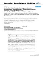
RPE-CM protect SH-SY5Y cells from injury in the presence of neurotoxinsFigure 1
RPE-CM protect SH-SY5Y cells from injury in the presence of neurotoxins. (A) SH-SY5Y cells cultured without
rotenone (Control), cells treated with 10 μM rotenone (Re), cells treated with 10 μM rotenone in RPE-CM (CM+Re) were
examined by MTT assay. Rotenone treatment produce significant cell lose in SH-SY5Y cultures (**p < 0.01 compared with con-
trol). RPE-CM significantly attenuated rotenone-induced cell loss (##p < 0.01 compared with rotenone group). (B) SH-SY5Y
cells were treated as in A in the presence of 50 μM 6-OHDA. 6-OHDA treatment produced significant cell lose in SH-SY5Y
cultures (**p < 0.01 compared with CM treated control). RPE-CM significantly attenuated 6-OHDA-induced cell loss (## p <
0.01 compared with 6-OHDA treated group). (C) Blockage of GDNF and BDNF by antibodies inhibited the protection of the
RPE-CM. RPE-CM was pretreated with 1 μg/ml GDNF antibody (Re+GDNFab+CM) or with 1 μg/ml BDNF antibody
(Re+BDNFab+CM) and incubated with SH-SY5Y cells in the presence of 10 μM rotenone. The protective effect of RPE-CM
could be partially blocked by GDNF and BDNF antibodies (*p < 0.05 compared with CM treated group). (D) Cells were
treated as in C in the presence of 50 μM 6-OHDA. The protective effect of RPE-CM could be partially blocked by GDNF and
BDNF antibodies when treated with 6-OHDA (*p < 0.05 compared with CM treated group; **p < 0.01 compared with CM
treated control). Data showed the mean ± SEM values from three independent experiments performed in triplicate.
Table 1: Neurotrophic factors secreted by RPE cells
Trophic factors BDNF GDNF
Concentration in medium (pg/ml) 0.49 ± 0.09 0.019 ± 0.005
Serum-free medium was incubated with RPE cells for two days, and subjected to ELISA assay.
Journal of Translational Medicine 2009, 7:53 />Page 6 of 9
(page number not for citation purposes)
tection on SH-SY5Y cells by 41.4% and 46.7% against rotenone and 6-OHDA induced injury, respectively (Fig
1C, D). While antibody against BDNF could reduce the
CM-induced protection on SH-SY5Y cells by 38.7% and
85.9% against rotenone and 6-OHDA induced injury,
respectively (Fig 1C, D).
To further support our findings, we then tested the neuro-
protection of RPE cells in primary VM DA neurons culture.
Exposure to 25 nM rotenone for 8 hours resulted in a sig-
nificant loss of the TH-positive cells by 50.6% as com-
pared with control cultures without rotenone treatment
(Fig 2B), while incubation with RPE-CM significantly
attenuated the rotenone-induced loss of TH-positive cells
by 44.3% (Fig 2A).
When DAergic neuron cultures were challenged by 6-
OHDA at 40 μM, RPE-CM showed a similar protective
ability on the TH-positive cells. 6-OHDA treatment
caused a 43.2% loss of TH-positive cells as compared with
non-toxin control cultures (Fig 2F), while RPE-CM atten-
uated the 6-OHDA-induced TH-positive cell loss by
63.1% (Fig 2E).
RPE cells express GDNF and BDNF after transplantation
As the role of GDNF and BDNF was demonstrated in the
neuroprotection of RPE-CM against 6-OHDA and roten-
one neurotoxicity in vitro, we measured the levels of
GDNF and BDNF in the RPE cell-grafted striatal tissues.
Four weeks after transplantation the striatal tissues with
microcarriers-RPE cells were taken out and homogenated,
followed by centrifugation at 12000 rpm for 20 minutes.
The striatal tissues transplanted with microcarriers were
used as control. ELISA assay showed that tissues with
microcarriers-RPE cells had 41.2% and 68.1% higher lev-
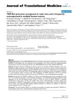
RPE-CM protects the DAergic neurons against the rotenone- and 6-OHDA- induced neuron loss in primary VM culturesFigure 2
RPE-CM protects the DAergic neurons against the
rotenone- and 6-OHDA- induced neuron loss in pri-
mary VM cultures. (A) VM neurons treated with CM in
the presence of 25 nM rotenone. Scale bar, 10 μm. (B) VM
neurons cultured in fresh medium in the presence of roten-
one. (C) VM neurons cultured in fresh medium without
rotenone. (D) The number of TH-ir neurons in the cultures
treated with fresh medium only (Control), with fresh
medium in the presence of 25 nM rotenone (Re) and with
CM in the presence of 25 nM rotenone (CM+ Re). Data rep-
resent the mean ± SEM. *p < 0.05. (E) VM neurons treated
with CM in the presence of 40 μM 6-OHDA. (F) VM neurons
cultured in fresh medium in the presence of 40 μM 6-OHDA.
(G) VM neurons cultured in fresh medium without 6-OHDA.
(H) The number of TH-ir neurons in the cultures treated
with fresh medium (Control), with fresh medium in the pres-
ence of 40 μM 6-OHDA (6-OH) and with CM in the pres-
ence of 40 μM 6-OHDA (CM+6-OH). Data represent the
mean ± SEM. *p < 0.05.
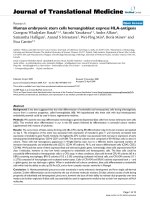
Determination of GDNF and BDNF from RPE cells after transplantationFigure 3
Determination of GDNF and BDNF from RPE cells
after transplantation. (A) Tissues with transplanted RPE
cells were lysed for GDNF determination and tissues with
transplanted microcarriers were used as control. (B) Tissues
with transplanted RPE cells were lysed for BDNF determina-
tion and tissues with transplanted microcarriers were used
as control. The concentrations of GDNF and BDNF were
determined using Emax ImmunoAssay System (Promega,
USA).
Journal of Translational Medicine 2009, 7:53 />Page 7 of 9
(page number not for citation purposes)
els of GDNF and BDNF as compared with the control
group which contained microcarriers only (Fig 3A, B).
RPE cells express DDC and synthesize DA
DDC is an enzyme that converts L-dopa to DA; the expres-
sion of DDC indicates the RPE cells have the ability to
produce DA. To determine whether the RPE cells can syn-
thesize DA, we measured the DDC mRNA by RT-PCR and
DDC protein by immunoblot, which showed that the RPE
cells could transcribe DDC mRNA and express abundant
DDC protein (Fig 4). But the mRNA and the protein of
DAT which transports DA through the membrane
couldn't be detected by RT-PCT and immunoblot (Fig 4).
Furthermore, we measured the content of DA and its
metabolite homovanillic acid (HVA) in RPE cells by
HPLC (Fig 5), which showed DA (peak time: 5.43 min-
utes) level as 29.13 ng/mg protein and HVA (peak time:
11.51 minutes) level as 267.89 ng/mg protein (Table 2).
However, DA release into the buffer was not detected after
56 mM potassium chloride treatment in the cultured RPE
cells, suggesting that the high potassium-depolarization
can not induce DA release from RPE cells (Table 2). It's
likely that RPE cells may have other mechanism to transfer
DA throughout the membrane.
Microcarriers-RPE cells survive in the host striatal tissues
and significantly improve AIR in 6-OHDA-lesioned rats
To demonstrate the RPE cells survival in the host striatum,
we performed HE staining and cytokeratin immunostain-
ing. HE staining showed that transplants were accurately
placed into the striatum (Fig 6A) and RPE cells were
attached outside the microcarriers (Fig 6B); immunostain-
ing demonstrated that these cells were cytokeratin-immu-
noreactive, a morphological marker of live RPE cells (Fig
6D).
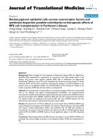
RPE cells express DDC but not DATFigure 4
RPE cells express DDC but not DAT. (A) DDC from
RPE cells was detected by western blot. RPE protein was
loaded and the equal level of C57 mouse striatum protein
was used as positive control; protein DDC was detected by
western blot and both samples displayed the same size pro-
tein bands. The protein DAT could not be detected in RPE
cells. (B) The cDNA of DDC but not DAT was detected by
PCR from the total cDNA of RPE cells.
Table 2: DA and HVA in RPE cells extract and DA release after
potassium treatment
DA HVA
Cells extract (ng/mg protein) 29.13 ± 4.11 267.89 ± 16.10
Release after potassium treatment None None
RPE cells were homogenated and centrifuged. The supernatant was
examined by HPLC for DA and its metabolic. To determine the DA
release, RPE cells were depolarized with high potassium (56 mM K
+
)
and the buffer was subjected to HPLC assay.
HPLC analysis of the synthesis and release of DA by RPE cellsFigure 5
HPLC analysis of the synthesis and release of DA by
RPE cells.(A) HPLC analysis of standard of DA, 3,4-dihy-
droxyphenylacetic acid (DOPAC) and HVA. (B) HPLC analy-
sis of RPE cells homogenate. The peaks of DA and HVA in
the RPE cells were detected but the DOPAC signal was
weak. (C) HPLC analysis of high potassium solution incu-
bated with RPE cells.
Journal of Translational Medicine 2009, 7:53 />Page 8 of 9
(page number not for citation purposes)
Before transplantation, AIR showed the basal level of rota-
tion in 6-OHDA lesioned rats. We selected the rats that
exhibited rotation toward the healthy side at least 6.0 full
body turns per minute for transplantation of microcarri-
ers-RPE or microcarriers alone as control. After transplan-
tation, microcarriers-RPE grafted animals displayed a
significant reduction in AIR behavior compared to control
rats that was transplanted with microcarriers alone (p <
0.05) (Fig 6F).
Discussion
Most of the cell-based therapies for PD are focused on two
goals: one is to provide a source of neurotrophic factors
which may modify disease course and the other is to pro-
vide a constant level of DA. RPE cell transplantation is a
promising therapy as shown in preliminary clinical trial.
In the present study, we attempt to elucidate the mecha-
nisms of this therapy by determining: whether RPE cells
exert protective effects on DAergic cells when challenged
by neurotoxins; whether RPE cells can produce and
release DA. Our results indicate that 1) RPE cells can
express and secrete GDNF and BDNF and protect DAergic
cells against neurotxoins-induced injury; 2) RPE cells can
express DDC and synthesize DA; 3) RPE cells attached to
microcarriers can survive in the host striatum and produce
high level GDNF and BDNF after transplantation; and 4)
RPE cells transplantation produce a statistically significant
improvement of AIR.
In the previous transplantation studies, microcarriers were
used to increase the survival of grafted cells [21]. Indeed,
microcarriers provide a substrate to which the cells can
establish a basal lamina and thus create a more favorable
microenvironment. Furthermore, cells attached to beads
may alter the immunogenic properties of cells, which may
prevent the recognition and immunological surveillance
[16]. In our experiments, we used cytodex 3 which con-
sists of a thin layer of denatured collagen chemically cou-
pled to a matrix of cross-linked dextran, and these
microcarriers facilitate the survival of transplanted RPE
cells.
We demonstrate that RPE cells can provide trophic effect
on DAergic cells, which may be one of the possible mech-
anisms underlying RPE cell therapy. Previous studies had
showed that RPE cells expressed several neurotrophic fac-
tors such as PEDF, PDGF, EGF, and VEGF [11]. Our results
elucidate that RPE cells can secrete BDNF and GDNF and
these two factors play important role in the neurotrophic
effects of RPE cells. Although RPE cells can express PEDF,
it accounts for only a portion of the neurotrophic effect
[22]. In this study we demonstrate that GDNF and BDNF
in RPE-CM contribute for the most part of trophic effect.
We also demonstrate that GDNF and BDNF are expressed
by grafted RPE cells.
Besides the neurotrophic effect of RPE cells, we document
that RPE cells can express DDC and produce DA. L-dopa
is a precursor of DA, and can be synthesized by RPE cells
as an intermediate product of melanin [23]. DDC, an
enzyme to convert L-dopa to DA, is found in the RPE cells
in our study. However, the depolarization-induced DA
release is not detected in the cells, indicating that the DA
release machinery as seen in most excitable cells is not
present in the RPE cells. It's possible that RPE cells may
have other mechanism to transfer DA throughout the
membrane. Previous report by Dalpiaz et al [24] showed
that DA could permeate the membrane of RPE cells, and
this permeation seems to be mediated by organic cation
transporter 3 [25]. The ability of DA synthesis in RPE cells
suggests RPE cells transplantation may be one of the
advantages for the cell replacement therapy to treat
advanced PD patients.
Microcarriers-RPE cells survive in the host striatum and transplantation with RPE cells significantly improve animal behavioursFigure 6
Microcarriers-RPE cells survive in the host striatum
and transplantation with RPE cells significantly
improve animal behaviours. (A) Low magnification
microphotogragh of the striatum with transplants. Arrow
indicates the transplants. The scale bar is 0.5 mm. (B) HE
staining of the corpus striatum of SD rat injected with micro-
carriers-RPE (M-RPE). 10 μm sections were stained with
hematoxylin-eosin. The scale bar is 20 μm. (C) The corpus
striatum of SD rat injected with microcarriers alone as con-
trol. Sections were stained as in B. (D) Corpus striatum of
SD rat injected with M-RPE was immunostained with cytok-
eratin antibody. The scale bar is 5 μm. (E) The corpus stria-
tum of SD rat injected with microcarriers only was stained as
in D. (F) AIR before transplantation and 4 weeks after trans-
plantation. Data are shown as mean ± S.E.M. *p < 0.05. N =
8.
Publish with Bio Med Central and every
scientist can read your work free of charge
"BioMed Central will be the most significant development for
disseminating the results of biomedical researc h in our lifetime."
Sir Paul Nurse, Cancer Research UK
Your research papers will be:
available free of charge to the entire biomedical community
peer reviewed and published immediately upon acceptance
cited in PubMed and archived on PubMed Central
yours — you keep the copyright
Submit your manuscript here:
/>BioMedcentral
Journal of Translational Medicine 2009, 7:53 />Page 9 of 9
(page number not for citation purposes)
Conclusion
RPE cells not only replenish L-dopa as elucidated by pre-
vious study, but can also synthesize DA and neurotrophic
factors which protect the intrinsic neurons after transplan-
tation. These findings make this cell replacement a more
viable and promising therapy for PD.
Competing interests
The authors declare that they have no competing interests.
Authors' contributions
The studies were designed by MM and WL and were per-
formed by MM, XL, and XF. Human RPE cells were sepa-
rated and cultured by QG. DY, LL and SC gave advises on
the work and helped in the interpretation of the data. WL
supervised all the work and wrote the paper together with
MM. All authors read and approved the final manuscript.
Acknowledgements
This work was supported by a grant from 973 National Project (NO.
2005CB724302), the National Natural Science Foundation (NO.
30730096), the National Basic Research Program of China from Science
and Technology Commission (NO. 2007CB947904) and the Technology
Commission (863 project 2007AA02Z460).
References
1. Twelves D, Perkins KS, Counsell C: Systematic review of inci-
dence studies of Parkinson's disease. Movement Disord 2003,
18:19-31.
2. Olanow CW, Goetz CG, Kordower JH, Stoessl AJ, Sossi V, Brin MF,
Shannon KM, Nauert GM, Perl DP, Godbold J, Freeman TB: A dou-
ble-blind controlled trial of bilateral fetal nigral transplanta-
tion in Parkinson's disease. Ann Neurol 2003, 54:403-414.
3. Minguez-Castellanos A, Escamilla-Sevilla F: Cell therapy and other
neuroregenerative strategies in Parkinson's disease (II). Rev
Neurol 2005, 41:684-693.
4. Shukla S, Agrawal AK, Chaturvedi RK, Seth K, Srivastava N, Sinha C,
Shukla Y, Khanna VK, Seth PK: Co-transplantation of carotid
body and ventral mesencephalic cells as an alternative
approach towards functional restoration in 6-hydroxy-
dopamine-lesioned rats: implications for Parkinson's disease.
J Neurochem 2004, 91:274-284.
5. Doudet DJ, Cornfeldt ML, Honey CR, Schweikert AW, Allen RC:
PET imaging of implanted human retinal pigment epithelial
cells in the MPTP-induced primate model of Parkinson's dis-
ease. Exp Neurol 2004, 189:361-368.
6. Wang R, Zhang J, Guo Z, Shen L, Shang A, Chen Y, Yao S, He T, Yin
D, Tian J: In-vivo PET imaging of implanted human retinal pig-
ment epithelium cells in a Parkinson's disease rat model.
Nucl Med Commun 2008, 29:455-461.
7. Watts RL, Raiser CD, Stover NP, Cornfeldt ML, Schweikert AW,
Allen RC, Subramanian T, Doudet D, Honey CR, Bakay RA: Stereo-
taxic intrastriatal implantation of human retinal pigment
epithelial (hRPE) cells attached to gelatin microcarriers: a
potential new cell therapy for Parkinson's disease. J Neural
Transm Suppl 2003:215-227.
8. Cepeda IL, Flores J, Cornfeldt ML, O'Kusky JR, Doudet DJ: Human
retinal pigment epithelial cell implants ameliorate motor
deficits in two rat models of Parkinson disease. J Neuropathol
Exp Neurol 2007, 66:576-584.
9. Ming M, Le W: Retinal pigment epithelial cells: biological prop-
erty and application in Parkinson's disease. Chin Med J (Engl)
2007, 120:416-420.
10. Aroca P, Urabe K, Kobayashi T, Tsukamoto K, Hearing VJ: Melanin
biosynthesis patterns following hormonal stimulation.
J Biol
Chem 1993, 268:25650-25655.
11. Subramanian T: Cell transplantation for the treatment of Par-
kinson's disease. Semin Neurol 2001, 21:103-115.
12. Seagle BL, Rezai KA, Kobori Y, Gasyna EM, Rezaei KA, Norris JR Jr:
Melanin photoprotection in the human retinal pigment epi-
thelium and its correlation with light-induced cell apoptosis.
Proc Natl Acad Sci USA 2005, 102:8978-8983.
13. Du F, Li R, Huang Y, Li X, Le W: Dopamine D3 receptor-prefer-
ring agonists induce neurotrophic effects on mesencephalic
dopamine neurons. Eur J Neurosci 2005, 22:2422-2430.
14. Peng C, Fan S, Li X, Fan X, Ming M, Sun Z, Le W: Overexpression
of pitx3 upregulates expression of BDNF and GDNF in SH-
SY5Y cells and primary ventral mesencephalic cultures. Febs
Lett 2007, 581:1357-1361.
15. Li X, Yang D, Li L, Peng C, Chen S, Le W: Proteasome inhibitor
lactacystin disturbs the intracellular calcium homeostasis of
dopamine neurons in ventral mesencephalic cultures. Neuro-
chem Int 2007, 50:959-965.
16. Cherksey BD, Sapirstein VS, Geraci AL: Adrenal chromaffin cells
on microcarriers exhibit enhanced long-term functional
effects when implanted into the mammalian brain. Neuro-
science 1996, 75:657-664.
17. Thomas J, Wang J, Takubo H, Sheng J, de Jesus S, Bankiewicz KS: A 6-
hydroxydopamine-induced selective parkinsonian rat model:
further biochemical and behavioral characterization. Exp
Neurol 1994, 126:159-167.
18. Subramanian T, Marchionini D, Potter EM, Cornfeldt ML: Striatal
xenotransplantation of human retinal pigment epithelial
cells attached to microcarriers in hemiparkinsonian rats
ameliorates behavioral deficits without provoking a host
immune response. Cell Transplant 2002, 11:207-214.
19. Kirik D, Georgievska B, Bjorklund A: Localized striatal delivery of
GDNF as a treatment for Parkinson disease. Nat Neurosci
2004, 7:105-110.
20. Petersen AA, Larsen KE, Behr GG, Romero N, Przedborski S, Brun-
din P, Sulzer D: Brain-derived neurotrophic factor inhibits
apoptosis and dopamine-induced free radical production in
striatal neurons but does not prevent cell death.
Brain Res Bull
2001, 56:331-335.
21. Tatard VM, Venier-Julienne MC, Saulnier P, Prechter E, Benoit JP,
Menei P, Montero-Menei CN: Pharmacologically active micro-
carriers: a tool for cell therapy. Biomaterials 2005, 26:3727-3737.
22. McKay BS, Goodman B, Falk T, Sherman SJ: Retinal pigment epi-
thelial cell transplantation could provide trophic support in
Parkinson's disease: results from an in vitro model system.
Exp Neurol 2006, 201:234-243.
23. Schraermeyer U, Kopitz J, Peters S, Henke-Fahle S, Blitgen-Heinecke
P, Kokkinou D, Schwarz T, Bartz-Schmidt KU: Tyrosinase biosyn-
thesis in adult mammalian retinal pigment epithelial cells.
Exp Eye Res 2006, 83:315-321.
24. Dalpiaz A, Filosa R, de Caprariis P, Conte G, Bortolotti F, Biondi C,
Scatturin A, Prasad PD, Pavan B: Molecular mechanism involved
in the transport of a prodrug dopamine glycosyl conjugate.
Int J Pharm 2007, 336:133-139.
25. Rajan PD, Kekuda R, Chancy CD, Huang W, Ganapathy V, Smith SB:
Expression of the extraneuronal monoamine transporter in
RPE and neural retina. Curr Eye Res 2000, 20:195-204.