báo cáo hóa học:" A practical approach for the validation of sterility, endotoxin and potency testing of bone marrow mononucleated cells used in cardiac regeneration in compliance with good manufacturing practice" docx
Bạn đang xem bản rút gọn của tài liệu. Xem và tải ngay bản đầy đủ của tài liệu tại đây (435.22 KB, 9 trang )
BioMed Central
Open Access
Page 1 of 9
(page number not for citation purposes)
Journal of Translational Medicine
Methodology
A practical approach for the validation of sterility, endotoxin
and potency testing of bone marrow mononucleated cells used in
cardiac regeneration in compliance with good manufacturing
practice
Sabrina Soncin, Viviana Lo Cicero, Giuseppe Astori*, Gianni Soldati,
Mauro Gola, Daniel Sürder and Tiziano Moccetti
Address: The Cell Therapy Unit, Cardiocentro Ticino, Via Tesserete 48, CH-6900 Lugano, Switzerland
Email: Sabrina Soncin - ; Viviana Lo Cicero - ;
Giuseppe Astori* - ; Gianni Soldati - ; Mauro Gola - ;
Daniel Sürder - ; Tiziano Moccetti -
* Corresponding author
Abstract
Background: Main scope of the EU and FDA regulations is to establish a classification criterion for advanced
therapy medicinal products (ATMP). Regulations require that ATMPs must be prepared under good
manufacturing practice (GMP). We have validated a commercial system for the determination of bacterial
endotoxins in compliance with EU Pharmacopoeia 2.6.14, the sterility testing in compliance with EU
Pharmacopoeia 2.6.1 and a potency assay in an ATMP constituted of mononucleated cells used in cardiac
regeneration.
Methods: For the potency assay, cells were placed in the upper part of a modified Boyden chamber containing
Endocult Basal Medium with supplements and transmigrated cells were scored. The invasion index was expressed
as the ratio between the numbers of invading cells relative to cell migration through a control insert membrane.
For endotoxins, we used a commercially available system based on the kinetic chromogenic LAL-test. Validation
of sterility was performed by direct inoculation of TSB and FTM media with the cell product following Eu Ph 2.6.1
guideline.
Results and discussion: The calculated MVD and endotoxin limit were 780× and 39 EU/ml respectively. The
1:10 and 1:100 dilutions were selected for the validation. For sterility, all the FTM cultures were positive after 3
days. For TSB cultures, Mycetes and B. subtilis were positive after 5 and 3 days respectively. The detection limit
was 1-10 colonies.
A total of four invasion assay were performed: the calculated invasion index was 28.89 ± 16.82% (mean ± SD).
Conclusion: We have validated a strategy for endotoxin, sterility and potency testing in an ATMP used in cardiac
regeneration. Unlike pharmaceutical products, many stem-cell-based products may originate in hospitals where
personnel are unfamiliar with the applicable regulations. As new ATMPs are developed, the regulatory framework
is likely to evolve. Meanwhile, existing regulations provide an appropriate structure for ensuring the safety and
efficacy of the next generation of ATMPs. Personnel must be adequately trained on relevant methods and their
application to stem-cell-based products.
Published: 8 September 2009
Journal of Translational Medicine 2009, 7:78 doi:10.1186/1479-5876-7-78
Received: 5 June 2009
Accepted: 8 September 2009
This article is available from: />© 2009 Soncin et al; licensee BioMed Central Ltd.
This is an Open Access article distributed under the terms of the Creative Commons Attribution License ( />),
which permits unrestricted use, distribution, and reproduction in any medium, provided the original work is properly cited.
Journal of Translational Medicine 2009, 7:78 />Page 2 of 9
(page number not for citation purposes)
Introduction
The European Union (EU) regulation on advanced ther-
apy medicinal products [1] (ATMP) is entered into force
in all European Member States on December 30, 2008,
and Food and Drug Administration (FDA) recently prom-
ulgated regulations on human cells, tissues, and cellular
and tissue-based products [2] issuing an appropriate regu-
latory structure for the wide range of stem-cell-based
products that may be developed to regenerate damaged
tissues. Main scope of the regulations is to establish clear
classification criteria for many new cell-based medicinal
products. In particular the European Regulation makes
reference to and is in coherence with the 2004/23/EC
directive on donation, procurement and testing of human
cells and tissues and with directive 2002/98/EC on
human blood and blood components. This means that
any use of human cells has to be in compliance with the
quality requirements therein described. The European
Regulation is also clear on requiring that all ATMP have to
be prepared according to the good manufacturing practice
(GMP) for medicinal products. Stem-cell-based therapies
have existed since the first successful bone marrow trans-
plantations in 1968 [3]. Among the ATMPs, bone mar-
row-derived mononuclear cells (BM-MNC), widely used
in cellular therapy protocols, include several populations
of stem cells able to restore vascularization or to transdif-
ferentiate into functional cardiac cells: hematopoietic
stem cells (HSC) which give rise to all mature lineages of
blood [4], mesenchymal stem cells (MSC) and endothe-
lial progenitor cells (EPC) which can be mobilized in the
peripheral blood and give rise to mature endothelial cells
in blood vessels [5]. The hematopoietic lineage is charac-
terized by the presence of the CD34 cell-surface antigen
(found in about 1% of human bone marrow mononucle-
ated cells); it has therefore been considered a useful cell
selection target for bone marrow progenitor cells. MSC
represent less than 0.1% of the bone marrow cell popula-
tion [6] and are able to generate non hematopoietic tis-
sues including adipocytes, chondrocytes, osteocytes,
myocytes [7,8] and cardiomyocites [9]. Angiogenesis and
vascuologenesis are responsible for the development of
the vascular system and are one of the main mechanisms
leading to improved cardiac function after the injection of
BM-MNC [10]. Among the CD34+ cells, the CD133 sur-
face antigen defines a subset of hematopoietic stem cells
enriched for Endotelial Progenitor Cells (EPCs) [11]. The
angiogenic potential of bone marrow cells has been tested
into hind limb ischemia animal models [12] and several
clinical studies are ongoing to evaluate the efficiency of
the intra-arterial administration of BMC into an ischemic
limb [13,14].
During the production of the BM-MNC as medicinal
products, variable amounts of impurities product and
process-related, are introduced into the final product: cells
enter in contact with buffers, reagents and plastics that
could be potentially harmful in humans. A safety assess-
ment of BM-MNC cells prepared using density gradient
centrifugation should be done in order to ensure that the
finished product do not contain any substance or impu-
rity that can have an adverse effect in the patient.
BM-MNC should be free from adventitious microbial that
could originate from the starting or raw materials or
adventitiously introduced during the manufacturing proc-
ess. In any case, a thorough testing must be performed at
the level of finished product in compliance with the meth-
odologies described in the EU or United States Pharmaco-
poeia (USP), in particular for endotoxin content, sterility
and cell potency.
Endotoxins are lipo-polysaccharides from gram-negative
bacteria and are the most common cause of toxic reactions
resulting from contamination with pyrogens: the absence
of bacterial endotoxins in a product implies the absence
of pyrogenic components, provided the presence of non-
endotoxin substrates can be ruled out. Endotoxins can be
detected by using the Limulus amoebocyte lysate (LAL)
test; unfortunately, it may be masked by factors interfering
with the reaction between the endotoxins and the LAL. As
a consequence, the suitability of the regents and materials
used and the product itself has to be established. The
endotoxin limit that can be accepted in a product is based
on the route of administration (intravenous or intrathe-
cal), the threshold pyrogenic dose and volume of the
injected product. Some endotoxin limits have been calcu-
lated and can be found in the Pharmacopoeia; for non-
compendial items and new drugs, the endotoxin limit
should be calculated by the user. The Maximum Valid
Dilution (MVD) provides an upper bound for dilution
that still provides for endotoxin detection at the endo-
toxin limit. To determine if any interfering characteristics
exist, each LAL assay must have a positive product control
(PPC) to ensure that endotoxin would be detected if it
were present in the sample.
Potency is the quantitative measure of biological activity
based on the attribute of the product, which is linked to
the relevant biological properties. The assay demonstrat-
ing the biological activity should be based on the
intended biological effect which should ideally be related
to the clinical response. Basically, two types of potency
assays can be envisioned: in vitro assays using cell systems
and in vivo assays using animal models. As concerning the
use of bone marrow mononucleated cells in cardiac
repair, the importance of characterizing the functionality
of injected cells was recently pointed out [15,16]: to eval-
uate the functional activity of the cells obtained after den-
sity gradient centrifugation, authors purposed both in
vitro and in vivo assays. Cells were evaluated for hemat-
Journal of Translational Medicine 2009, 7:78 />Page 3 of 9
(page number not for citation purposes)
opoietic colony-forming unit (CFU), and assessment of
mesenchymal stem cells colonies. Furthermore, based on
the observation that the migratory capacity of bone mar-
row mononucleated cells predicts the functional improve-
ment after cell transplantation in a hind limb ischemia
model [17] and in humans [18], authors purposed the
assessment of the migration capacity of the cells. At the
moment there is no consensus in establishing acceptance
criteria for the migration capacity of BM-MNC in cardiac
regeneration.
Cell migration and cell invasion assays measure the ability
of certain cell types to move through a porous membrane
toward a chemoattractant or growth factor. In contrast to
cell migration through an open pore, cell invasion
through an occluded pore is dependent on active enzy-
matic degradation of the matrix barrier. The Matrigel
Matrix consists of laminin, collagen IV, entactin, and var-
ious growth factors to mimic the basement membrane.
Endothelial cells express proteases MMP 2 and 9, which
actively digest the matrix. At the end-point of the assay,
invasive cells appear on the underside of the porous mem-
brane and can be quantified.
Guidelines for sterility testing of biologics is addressed in
the various worldwide pharmacopeias and in Section 21
of the Code of Federal Regulations (CFR), International
Conference on Harmonisation (ICH) and Food and Drug
Administration Points to Consider documents. ATMP
manufactured under GMP conditions require sterility test-
ing performed under GMP guidelines. There are two com-
mon types of sterility test methods: the membrane
filtration method that requires the test article to first pass
through a size exclusion membrane capable of retaining
microorganisms and the direct inoculation method
requires the sample to be inoculated directly into test
media. For the latter, sample is incubated for 14 days in
the test media. It is important to determine if the ATMP
under testing contains elements able to interfere with the
growth of microorganisms within the growth media used
for the assay.
Aim of this study is the validation of a commercial system
(Charles River Endosafe PTS) for the determination of
bacterial endotoxins in compliance with Eu Pharmaco-
poeia 2.6.14 (bacterial endotoxins), the validation of the
sterility testing in compliance with eu Pharmacopoeia
2.6.1 (sterility) and the validation of the potency assay in
an ATMP that is constituted of bone-marrow mononucle-
ated cells used in cardiac regeneration.
Materials and methods
Testing were performed in the quality control laboratory
of the cell therapy unit of the Cardiocentro Ticino. The
Laboratory is authorized and regularly inspected by the
Swiss competent authorities.
Sample Preparation
For the endotoxin testing and migration assay cells were
collected after informed consent from patients enrolled in
the "Swiss multicenter intracoronary stem cells study in
acute myocardial infarction" (SWISS-AMI,
NCT00355186). A total of 50 ml of bone marrow was
aspirated into heparin-treated syringes from the posterior
iliac crest under local anesthesia. Bone marrow was fil-
tered by using a 100 μm nylon mesh (BD Falcon TM Cell
Strainer, BD Biosciences), diluted 1:1 in Phosphate Buff-
ered Saline (PBS), and BM-MNC isolated by density gradi-
ent centrifugation on Ficoll-PAQUE PREMIUM (General
Electric). Cells were washed three times in PBS filtered
through a 70 μm nylon mesh (BD Falcon) and then resus-
pended in 10 ml of 5% v/v human albumin. One ml was
collected for migration and invasion assay and endotoxin
testing. For the sterility testing, peripheral blood mononu-
cleated cells were obtained from 50 ml of peripheral
blood collected from patients immediately after an acute
myocardial infarction (AMI) subjected to standard phar-
macological therapy.
Cell Characterization
For the immunophenotype, bone marrow and BM-MNC
cells were stained in quadruplicate with anti CD45 FITC
(Beckman Coulter, USA), anti CD34 PC7 (Becton Dickin-
son, San Jose, USA), anti CD133 PE (Miltenyi, Bergisch-
Gladbach, DE) and with 7-AAD (Beckman Coulter, USA)
for the cell viability test. Death cells were excluded from
the analysis. Analyses were performed using a Cytomics
FC 500 flow cytometer (Beckman Coulter) acquiring at
least 100.000 events. Isotype-matched murine FITC, PC-7,
and PE conjugated immunoglobulins were used as con-
trols. Cell phenotype was determined by using an ABX
Micros 60 (Horiba Diagnostics, France).
Migration and Invasion Assay
A total of 1 × 10
6
BM-MNC collected from acute myocar-
dial infarction patients subjected to standard pharmaco-
logical therapy were resuspended in 500 μl of 5% v/v
human albumin. For the migration assay, cells were
placed in the upper part of an 8.0 μm untreated polyeth-
ylene terephthalate membrane 24-well cell culture insert
(Becton Dickinson, CA). For the invasion assay cells were
placed in the upper part of a modified Boyden chamber
Matrigel Invasion Camber (BioCoat Matrigel invasion
chamber, Becton Dickinson, CA): the chamber consist of
a 24-well Cell Culture insert with an 8 μm pore size PET
membrane, uniformly coated with Matrigel Matrix. The
matrix provides a barrier to non-invasive cells while pre-
senting an appropriate protein structure for invading cells
Journal of Translational Medicine 2009, 7:78 />Page 4 of 9
(page number not for citation purposes)
to penetrate before passing through the membrane. Both
chambers were then placed in a 24-well culture dish con-
taining 500 μl of Endocult Basal Medium supplemented
with Endocult Single Quots (Stemcells Technologies, Van-
couver, Canada) and 20% fetal calf serum (Figure 1). After
24 hours of incubation at 37°C, 5% v/v CO
2
transmi-
grated cells were counted. Assays were run in duplicates.
Endotoxin Testing
Description of the PTS Endosafe system
The Endosafe portable test system is based on the kinetic
chromogenic LAL-test that is based on the cleavage of a
synthetic substrate by an enzyme produced in the reaction
of the lysate in the presence of endotoxin. The system con-
sists of LAL reagents and endotoxin controls in the form
of a single-use polystyrene cartridges. The cartridges are
potency tested, spike recovery is performed and the cali-
bration code is determined. The calibration code contains
the cartridge test parameters that were determined during
potency testing as well as the archived curve for that batch
of cartridges. The color intensity developed is propor-
tional to the endotoxin concentration. Each cartridge con-
sists of two sample channels and two spiked channels,
consistent with current Pharmacopoeia guidance for
licensed quantitative LAL methods. Each reservoir con-
tains a specific amount of LAL reagent, synthetic chro-
mogenic substrate, control standard endotoxin (CSE) and
buffers uniformly embedded in the cartridge. The car-
tridge is inserted into a dedicated reader and 25 μL of the
prepared sample are dispensed into the four reservoirs.
The reader draws, mixes and incubates the sample with
the various reagents at programmed time intervals before
transferring it to the optical chambers. The portable spec-
trophotometer then monitors the change in the optical
density and calculates the endotoxin level based on the
resulting kinetic values. Cartridges with 5-0.050 EU/mL
sensitivity were used in this study. Results are automati-
cally multiplied by the dilution factor entered into the
Endosafe system. With the correct dilution the unit
achieves results in approximately 15 min.
Preparation of the inhibition/enhancement test and preparation of
the cell therapy product dilution series
The calculated MVD and endotoxin limit for the ATMP
were 780× and 39 EU/ml respectively. The inhibition/
enhancement test was done by using the Charles River
R+D Inhibition/Enhancement cartridges (range 5-0.05
EU/ml ) and by testing the cell product undiluted and
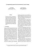
Schematic representation of the invasion assayFigure 1
Schematic representation of the invasion assay. BM-MNC cells were resuspended in 5% v/v human albumin and placed
in the upper part of a modified Boyden chamber Matrigel invasion chamber. The chamber consist of a 24-well cell culture insert
with an 8 μm pore size PET membrane, uniformly coated with Matrigel matrix. The matrix provides a barrier to non-invasive
cells while presenting an appropriate protein structure for invading cells to penetrate before passing through the membrane.
The chamber was then placed in a 24-well culture dish containing 500 μl of Endocult basal medium supplemented with Endoc-
ult single quots (Stemcells technologies, Vancouver, Canada) and 20% fetal calf serum. After 24 h of incubation transmigrated
cells were counted.
24 h
Matrigel Matrix occluding the 8.0 μm PET membrane
Chemoattractant
Invading
cells
Journal of Translational Medicine 2009, 7:78 />Page 5 of 9
(page number not for citation purposes)
diluted in pyrogen-free water as follows: 1:10; 1:100;
1:500; 1:700; 1:780.
This preliminary assay was performed with the aim to find
the dilution where the spiked endotoxin can be detected
without inhibiting or enhancing the test. Once prepared,
the cartridge was inserted in the Endosafe PTS and loaded
with 25 μl of the solution in each well. Results were scored
after 20 minutes of incubation at 37°C.
The ATMP was diluted in LAL reagent water (Charles River
) to 1:10 and 1:100 in pyrogen-free tubes and then loaded
in the system. All the tubes, water and pipette-tips were
pyrogen-free certified.
Sterility Testing
Sterility testing was carried out under aseptic conditions
regularly monitored by appropriate sampling of the work-
ing area and by carrying out appropriated controls as spec-
ified in on GMP documents.
Growth promotion test (GPT)
Sterility of the culture media Fluid thyoglicollate medium
(FTM) and soya-bean casein digest medium (TSB) used
for the culture of anaerobic and fungi/aerobic bacteria
(THIOC-T and TSB-T, bioMerieux SA, Switzerland) was
performed by incubating two vials of medium for 14 days
at 32.5°C and 22.5°C respectively. Growth promotion
test was performed by inoculating FTM media with 10-
100 colony-forming units (UFC) of Bacillus subtilis ATCC
6633; Staphylococcus aureus ATCC 6538; Pseudomonas aeru-
ginosa ATCC 9027; Clostridium sporogenes ATCC 19404
and TSB media with 10-100 UFC of Candida albicans
ATCC 10231; Aspergillus niger ATCC 16404 and Bacillus
subtilis ATCC 6633 (all from Quanti-Cult, Remel, Lenexa,
KS). Media were incubated as described for five and three
days respectively. Culture plates were inoculated in paral-
lel in order to check the viability of the micro-organisms.
Testing was also performed by using the following bacte-
rial strains isolated from bioburden in clean room: Staph-
ilococcus epdermidis 1, Micrococcus lylae and
Sphingobacterium multivorum. All testing were performed
in duplicate. Bacterial identifications were performed by
Gram-staining and by using the mini API detection system
(bioMerieux SA, Switzerland). The ID32 and ATB test
strips were used for the strain identification (bioMerieux
SA, Switzerland).
Validation test
Validation was performed by direct inoculation of TSB
and FTM media with 1% of the total volume of the prod-
uct under validation as stated in European Pharmaco-
poeia (2.6.27). For the latter, 500 μl of whole blood and
100 μl of the BM-MNC were inoculated together with 1-
10 UFC and 10-100 Colony-forming units of the bacterial
strains used in the growth promotion test and incubated
as above described. A growth promotion test was per-
formed as a positive control. If clearly visible growth of
micro-organisms is obtained after incubation in presence
of blood and the ATMP, the product possesses no antimi-
crobial activity under the conditions of the test, and the
sterility may be then carried out without further modifica-
tion.
Data Analysis
For the endotoxin testing, a test result was considered
valid when the percentage of spike recovery was between
50% and 200% with a coefficient of variation less than
25%.
For the sterility testing, the detection limit represent the
lowest bacterial concentration in the inoculums that the
system can evidence. The specificity of the system repre-
sent its ability to detect the single micro-organism in the
inoculums and the detection limit represent the lowest
micro-organism number in the sample that the system
can detect. The robustness of the system represent its abil-
ity to obtain identical results when using different prod-
ucts, medium from different lots in different working
days.
For the invasion assay, data were expresses as the percent
invasion through the Matrigel matrix and membrane rel-
ative to the migration through the 8.0 μm untreated Mem-
brane (invasion index). The Assay was considered positive
when at least ≥10% of the inoculate cells maintain their
invasion capacity.
Results
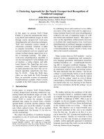
Cell phenotype
Cell phenotype of whole bone marrow and after density
gradient separation are reported in Figure 2 (mean ± SD,
n = 4).
Endotoxin testing
Testing was performed on three BM-MNC obtained from
different patients in three different days. Patient were sub-
jected to standard pharmacological treatment for acute
myocardial infarction. The mononucleated cells concen-
tration in the samples were 18.0 × 10
6
/ml; 15.2 × 10
6
/ml
and 16.2 × 10
6
/ml respectively (16.5 ± 1.2 × 10
6
mean ±
SD) with a pH of 6.5.
Results of the inhibition/enhancement test are reported in
Table 1. Based on the obtained results, the 1:10 and 1:100
dilutions were selected for the validation assay. An invalid
value, based on acceptance criteria, was observed in the
first run for the 1:10 dilution. The results of the validation
assay are reported in Table 2.
Journal of Translational Medicine 2009, 7:78 />Page 6 of 9
(page number not for citation purposes)
Sterility testing
Testing was performed on three whole peripheral blood
and the derived mononucleated fractions from different
patients in three different days. Patient were subjected to
standard pharmacological treatment for acute myocardial
infarction. The white blood cell concentration in the
mononucleated fraction were 13.0 × 10
6
/ml; 12.2 × 10
6
/
ml and 15.2 × 10
6
/ml respectively (13.5 ± 1.6 × 10
6
mean
± SD) with a pH of 6.5.
For the growth promotion test at the end of the incuba-
tion period, clearly visible growth of micro-organisms was
observed and identity confirmed for all bacterial strains.
As concerning the strains isolated from bioburden, S. epi-
dermidis 1 growth in both TSB and FTM media at both
concentrations whereas M. lylae and S. multivorum growth
at both concentrations in TSB medium only. For the vali-
dation test, all the FTM cultures resulted to be positive
after 3 days at both the concentration tested. For TSB cul-
tures, Mycetes were positive after 5 days and B. subtilis after
three. The detection limit of the system was then estab-
lished in 1-10 colonies. At the end of the incubation
period, subcultures in agar plates were performed for all
the microbial growth: all the identifications confirmed the
starting inoculum confirming the robustness of the sys-
tem.
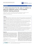
Migration and invasion assay
A total of four assays were performed in different days. For
all the samples a significant invasion index was observed:
28.89 ± 16.82% (mean ± SD). Complete results are
reported in Figure 3.
Discussion
Cellular therapy is an emerging field in medicine; all the
stem cell medicinal products must be in compliance with
principles and guidelines of good manufacturing practice
Phenotypical analysis of whole bone marrow cells and after density gradient centrifugation (bone marrow selected cells) (n = 4)Figure 2
Phenotypical analysis of whole bone marrow cells and after density gradient centrifugation (bone marrow
selected cells) (n = 4).
0.0
10.0
20.0
30.0
40.0
50.0
60.0
70.0
80.0
90.0
100.0
WHOLE BONE MA RROW
98.4 0.2 75.2 23.2 10.8 66.1
BONE MA RROW SELECTED CELLS
95.6 0.9 61.1 48.5 8.0 43.5
% V IA BILITY % CD34/ CD45
% COEXPR
CD34/CD133
% LY MPH % MON % GRA
Table 1: Results of the inhibition/enhancement test
SAMPLE DILUTION SPIKE RECOVERY
Undiluted 162%
1:10 53%
1:100 113%
1:500 132%
1:700 120%
1:780 98%
Journal of Translational Medicine 2009, 7:78 />Page 7 of 9
(page number not for citation purposes)
in respect of medicinal products and investigational
medicinal products for human use. When any new prepa-
ration or method of preparation is adopted, steps should
be taken to demonstrate its suitability for routine process-
ing: the defined process, using the materials and equip-
ment specified, should be validated in order to produce
cells of the required quality.
For certain ATMP that must be administered immediately
and that cannot be cryopreserved without damaging the
cell viability and quality, the availability of rapid testing
method for endotoxin and sterility testing is fundamental.
For the latter, traditional methods, including kinetic chro-
mogenic, kinetic turbidimetric and gel-clot LAL assay sys-
tems, have been widely used in the pharmaceutical
industry. Unfortunately, all of these methods are time-
consuming (several hours) and become problematic if
time-sensitive ATMPs products must be immediately
released. In the present paper, we have demonstrated that
Migration and invasion assay results for bone marrow derived mononucleated cellsFigure 3
Migration and invasion assay results for bone marrow derived mononucleated cells.
0
10
20
30
40
50
60
70
80
1234
SAMPLE
%
% MIGRATION
% INVASION
INVASION INDEX
Table 2: Results of the validation assay
1:10 DILUTION 1:100 DILUTION
1
st
run 2
nd
run 3
rd
run 1
st
run 2
nd
run 3
rd
run
Spike recovery (PPC) 122 119 121 115 76 95 143 178 176 163 142 183
PPC CV (%) 14.1 18.7 15.8 4.0 7.3 8.0 15.0 2.7 0.7 7.2 7.4 9.6
Sample CV 3.51.50000000000
Sample result (EU/mL) <0.532 <.513 <0.500 <0.500 <0.500 <0.500 <0.500 <0.500 <0.500 <0.500 <0.500 <0.500
Journal of Translational Medicine 2009, 7:78 />Page 8 of 9
(page number not for citation purposes)
the PTS endosafe system can be validated for the endo-
toxin testing of BM-MNC in compliance with European
and United States Pharmacopoeia. The time required by
the system was approximately 15 min, making it particu-
larly useful as an immediate release testing, where the aim
is to prepare and administer the product within a short
time period.
Sterility testing is regulated by USP 21CFR610.12 and by
Eu Pharmacopoeia 2.6.1. We have successfully validated
the sterility testing of a mononucleated cell preparation:
the sensitivity of the system for the ATCC and bioburden
bacterial strains here considered was 1-10 UFC in the
inoculums and cultures were positive after approximately
48 hours of incubation.
Recently, a rapid microbiological control strategy for cel-
lular products has been issued in EU and USP Pharmaco-
poeias based on the use of rapid detection systems as the
BacT/Alert 3D (bioMerieux, Durham, USA) or the Bactec
(Becton Dickinson, Franklin Lake, USA). Those systems
are in general non destructive, allowing a faster detection
when compared to TSB/FTM testing, and products can be
released after 7 days. Unfortunately, the microbial growth
of certain bacterial strains in those systems is still contro-
versial; as a consequence, those method should be strictly
validated both using the prescribed ATCC strains and by
using bioburden isolates.
All biological products must meet prescribed require-
ments of safety, purity and potency and no lot of any
licensed product may be released by the manufacturer
prior to the completion of tests for conformity with stand-
ards applicable to such product, including potency. The
current regulations allow for considerable flexibility in
determining the appropriate measurements of potency
that is necessary for product characterization testing; how-
ever, the complexity of an ATMP product can present sig-
nificant challenges in establishing a potency assays.
The migration assay of BM-MNC in response to endothe-
lial growth factors, seems to correlate with the beneficial
effects of the cell infusion after myocardial infarction
[15,16]: this assay has been then purposed as a quantita-
tive biological measure for the activity of the product
related to its specific ability to achieve the given result. In
particular, has been suggested that the correlation
between the "in vitro" data and the clinical efficacy may
be obtained by analyzing the outcomes from controlled
clinical studies [19,20]. In addition to the migration
assay, here we describe the use of the invasion assay as a
potency testing for BM-MNC cells: we purpose to define as
a minimal criteria to establish cell potency in cardiac
regeneration, the obtainment of an invasion index not
less than 10%. We are aware that the cell migration and
invasion results "in vitro" should be correlated with the
"in vivo" effect of the cells and this must be addressed
both in a suitable animal model and during a controlled
clinical trial of acute myocardial infarction.
Basic and clinical scientists, as well as scientists working in
the biotechnology and pharmaceutical industries, need an
increased awareness of the questions that must be
answered before a stem-cell-based product can be used
clinically. Unlike pharmaceutical products, many stem-
cell-based products may originate in academic laborato-
ries where researchers are unfamiliar with the applicable
regulations. As new stem-cell-based therapies are devel-
oped, the regulatory framework is likely to evolve. Mean-
while, existing regulations pertaining to biologic products
and human cells, tissues, and cellular and tissue-based
products provide an appropriate structure for ensuring the
safety and efficacy of the next generation of stem cell-
based medicinal products. As they conduct research on
stem cells, scientists should be aware of the relevant regu-
lations and their likely application to this products.
Competing interests
The authors declare that they have no competing interests.
Authors' contributions
GA wrote the manuscript, SS and VLC performed the
experiments, DS performed the sample collections as co-
investigator of the Swiss Ami clinical Trial, MG performed
literature search, GS and TM participated in study design
and coordination. All the authors read and approved the
final manuscript.
References
1. Regulation (EC) No 1394/2007 of the European Parliament
and of the Council of 13 November 2007 on advanced ther-
apy medicinal products and amending Directive 2001/83/EC
and Regulation (EC) No 726/2004. .
2. Food and Drug Administration 21 CFR 1271 (2006). .
3. Bach FH, Albertini RJ, Joo P, Anderson JL, Bortin MM: Bone-mar-
row transplantation in a patient with the Wiskott-Aldrich
syndrome. Lancet 1968, 2(7583):1364-1366.
4. Wilmut I, Schnieke AE, McWhir J, Kind AJ, Campbell KH: Viable off-
spring derived from fetal and adult mammalian cells. Nature
1997, 385(6619):810-813.
5. Asahara T, Murohara T, Sullivan A, Silver M, Zee R van der, Li T, Wit-
zenbichler B, Schatteman G, Isner JM: Isolation of putative pro-
genitor endothelial cells for angiogenesis. Science 1997,
275(5302):964-967.
6. Fibbe WE, Noort WA: Mesenchymal stem cells and hemat-
opoietic stem cell transplantation. Ann N Y Acad Sci 2003,
996:235-244.
7. Pittenger MF, Mackay AM, Beck SC, Jaiswal RK, Douglas R, Mosca JD,
Moorman MA, Simonetti DW, Craig S, Marshak DR: Multilineage
potential of adult human mesenchymal stem cells. Science
1999, 284(5411):143-147.
8. Colter DC, Class R, DiGirolamo CM, Prockop DJ: Rapid expansion
of recycling stem cells in cultures of plastic-adherent cells
from human bone marrow. Proc Natl Acad Sci USA 2000,
97(7):3213-3218.
9. Kawada H, Fujita J, Kinjo K, Matsuzaki Y, Tsuma M, Miyatake H, Mugu-
ruma Y, Tsuboi K, Itabashi Y, Ikeda Y, Ogawa S, Okano H, Hotta T,
Ando K, Fukuda K: Nonhematopoietic mesenchymal stem
Publish with Bio Med Central and every
scientist can read your work free of charge
"BioMed Central will be the most significant development for
disseminating the results of biomedical research in our lifetime."
Sir Paul Nurse, Cancer Research UK
Your research papers will be:
available free of charge to the entire biomedical community
peer reviewed and published immediately upon acceptance
cited in PubMed and archived on PubMed Central
yours — you keep the copyright
Submit your manuscript here:
/>BioMedcentral
Journal of Translational Medicine 2009, 7:78 />Page 9 of 9
(page number not for citation purposes)
cells can be mobilized and differentiate into cardiomyocytes
after myocardial infarction. Blood 2004, 104(12):3581-3587.
10. Kinnaird T, Stabile E, Burnett MS, Epstein SE: Bone-marrow-
derived cells for enhancing collateral development: mecha-
nisms, animal data, and initial clinical experiences. Circ Res
2004, 95(4):354-363.
11. Quirici N, Soligo D, Caneva L, Servida F, Bossolasco P, Deliliers GL:
Differentiation and expansion of endothelial cells from
human bone marrow CD133(+) cells. Br J Haematol 2001,
115(1):186-194.
12. Asahara T, Masuda H, Takahashi T, Kalka C, Pastore C, Silver M,
Kearne M, Magner M, Isner JM: Bone marrow origin of endothe-
lial progenitor cells responsible for postnatal vasculogenesis
in physiological and pathological neovascularization. Circ Res
1999, 85(3):221-228.
13. Tateishi-Yuyama E, Matsubara H, Murohara T, Ikeda U, Shintani S,
Masaki H, Amano K, Kishimoto Y, Yoshimoto K, Akashi H, Shimada
K, Iwasaka T, Imaizumi T: Therapeutic angiogenesis for patients
with limb ischaemia by autologous transplantation of bone-
marrow cells: a pilot study and a randomised controlled trial.
Lancet 2002, 360(9331):427-435.
14. Miyamoto K, Nishigami K, Nagaya N, Akutsu K, Chiku M, Kamei M,
Soma T, Miyata S, Higashi M, Tanaka R, Nakatani T, Nonogi H, Take-
shita S: Unblinded pilot study of autologous transplantation of
bone marrow mononuclear cells in patients with throm-
boangiitis obliterans. Circulation 2006, 114(24):2679-2684.
15. Dawn B, Bolli R: Bone marrow for cardiac repair: the impor-
tance of characterizing the phenotype and function of
injected cells. Eur Heart J 2007, 28(6):651-652.
16. Seeger FH, Tonn T, Krzossok N, Zeiher AM, Dimmeler S: Cell iso-
lation procedures matter: a comparison of different isolation
protocols of bone marrow mononuclear cells used for cell
therapy in patients with acute myocardial infarction. Eur
Heart J 2007, 28(6):766-772.
17. Heeschen C, Lehmann R, Honold J, Assmus B, Aicher A, Walter DH,
Martin H, Zeiher AM, Dimmeler S: Profoundly reduced neovas-
cularization capacity of bone marrow mononuclear cells
derived from patients with chronic ischemic heart disease.
Circulation 2004, 109(13):1615-1622.
18. Britten MB, Abolmaali ND, Assmus B, Lehmann R, Honold J, Schmitt
J, Vogl TJ, Martin H, Schächinger V, Dimmeler S, Zeiher AM: Infarct
remodeling after intracoronary progenitor cell treatment in
patients with acute myocardial infarction (TOPCARE-AMI):
mechanistic insights from serial contrast-enhanced mag-
netic resonance imaging. Circulation 2003, 108(18):2212-2218.
19. Food and Drug Administration: Guidance for Industry: Potency
tests for cellular and Gene Therapy Products. Center for Bio-
logics Evaluation and Research. US Department of Health and Human
Services; 2008.
20. Schächinger V, Erbs S, Elsässer A, Haberbosch W, Hambrecht R,
Hölschermann H, Yu J, Corti R, Mathey DG, Hamm CW, Süselbeck
T, Assmus B, Tonn T, Dimmeler S, Zeiher AM, REPAIR-AMI Investi-
gators: Intracoronary bone marrow-derived progenitor cells
in acute myocardial infarction. N Engl J Med 2006,
355(12):1210-1221.