Báo cáo hóa học: "One Step Nucleic Acid Amplification (OSNA) - a new method for lymph node staging in colorectal carcinomas" docx
Bạn đang xem bản rút gọn của tài liệu. Xem và tải ngay bản đầy đủ của tài liệu tại đây (559.11 KB, 6 trang )
METH O D O LOG Y Open Access
One Step Nucleic Acid Amplification (OSNA) - a
new method for lymph node staging in
colorectal carcinomas
Roland S Croner
1*
, Vera Schellerer
1
, Helene Demund
1
, Claus Schildberg
1
, Thomas Papadopulos
2
,
Elisabeth Naschberger
3
, Michael Stürzl
3
, Klaus E Matzel
1
, Werner Hohenberger
1
, Anne Schlabrakowski
4
Abstract
Background: Accurate histopathological evaluation of resected lymph nodes (LN) is essential for the reliable
staging of colorectal carcinomas (CRC). With conventional sectioning and staining techniques usually only parts of
the LN are examined which might lead to incorrect tumor staging. A molecular method called OSNA (One Step
Nucleic Acid Amplification) may be suitable to determine the metastatic status of the complete LN and therefore
improve staging.
Methods: OSNA is based on a short homogenisation step and subsequent automated amplification of cytokeratin
19 (CK19) mRNA directly from the sample lysate, with result available in 30-40 minutes. In this study 184 frozen LN
from 184 patients with CRC were investigated by both OSNA and histology (Haematoxylin & Eosin staining and
CK19 immunohistochemistry), with half of the LN used for each method. Samples with discordant results were
further analysed by RT-PCR for CK19 and carcinoembryonic antigen (CEA).
Results: The concordance rate between histology and OSNA was 95.7%. Three LN were histology+/OSNA- and 5
LN histology-/OSNA+. RT-PCR supported the OSNA result in 3 discordant cases, suggesting that metastases were
exclusively located in either the tissue analysed by OSNA or the tissue used for histology. If these samples were
excluded the concordance was 97.2%, the sensitivity 94.9%, and the specificity 97.9%. Three patients (3%) staged as
UICC I or II by routine histop athology were upstaged as LN positive by OSNA. One of these patients developed
distant metastases (DMS) during follow up.
Conclusion: OSNA is a new and reliable method for molecular staging of lymph atic metastases in CRC and
enables the examination of whole LN. It can be applied as a rapid diagnostic tool to estimate tumo ur involvement
in LN during the staging of CRC.
Introduction
Carcinomas of the colon and rectum (CRC) are the
fourth most common malignancy in the US and the sec-
ond most common cancer for woman (195,400 cases) as
well as the third most common cancer for men (217,400
cases) in Europe, with 207,400 associated deaths in 2006
[1,2]. One indepe ndent prognostic factor is the number
of affected lymph nodes (LN) which are examined on
the surgically resected specimen [3,4]. The number of
LN which are removed during lymphadenectomy and
submitted to posto perative histopathological examina-
tion can vary considerably. It is still under discussion
how many LN are necessary for accurate tumor staging
according to the TNM classification in CRC [5-10]. An
increasing number of LN harvested by the surgeon and
analysed by the pathologist has a positive influence on
the patients’ survival [5,11-13]. Nevertheless about 20%
of init ially node-negative stage UICC I and II CRC
patients suffer from recurre nt disease within five years
after surgery [14,15]. This scenario suggests that Haema-
toxylin and Eosin staining (H&E) as the current method
applied to assess the nodal status of CRC patients may
not be fully adequate. Small metastases (<5 mm) are
* Correspondence:
1
Department of Surgery, University of Erlangen-Nuremberg, Germany
Full list of author information is available at the end of the article
Croner et al. Journal of Translational Medicine 2010, 8:83
/>© 2010 Croner et al; licensee BioMed Central Ltd. This is an Open Access article distrib uted under the terms of the Creative Commons
Attribution License ( s/by/2.0), which permits unres tricted use, distribution, and reproduction in
any medium, provid ed the original work is properly cited.
quite frequent in CRC patients [16]. It has been pro-
posed that understaging in CRC is linked to the pre-
sence of occult tumour cells. Pathological investigation
including immunohistochemistry (IHC) and step sec-
tioning detected tumour deposits smaller than 2 mm in
20-30% of LN in stage UICC I and II CRC patients
[15,17,18]. M olecular analysis of sentinel LN in colon
carcinomas has resulted in the detection of micrometa-
static disease which was undetected by IHC [19]. This is
due to the fact that during RT-PCR the whole LN or at
least the biggest part of the LN can be analysed while
during histological work-up usually only a small part of
the LN is screened. For routine purposes the workload
of performing RNA extraction as a prerequisite to RT-
PCR is impract ical, especially when a high number of
LN must be investigated. In breast cancer a molecular
method called One Step Nucleic Acid Amplification
(OSNA) has recently been indicated as a fast molecular
diagnostic approach for the detection of LN metastases
[20,21]. The aim of our study was to apply this method
to LN staging in CRC patients in comparison to estab-
lished histological methods.
Materials and methods
Patient Samples
After informed consent 184 LN from 184 patients who
underwent surgery for the diagnosis of colon carcinoma
were harvested. LN were selected randomised from the
surgical resected specimen by the pathologist. The LN
were shock frozen in liquid nitrogen immediately after
surgery and stored at -80°C until further workup. The
mean follow up was 72 month (range 52-88 month).
Patients with a history of inflammatory bo wel disease
(Crohn’s disease, ulcerative colitis) were excluded from
the study. The study was carried out in concordance
with the guidelines of the ethical commission University
of Erlangen-Nuremberg and in compliance with the
Declaration of Helsinki. Patients’ characteristic s and his-
topathological criteria are listed in table 1.
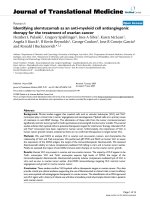
Study design
LN were cut into 4 slices (a, b, c, d) of each 1 or 2 mm
thickness with a special cutting device or cut in to halves
if the LN was too small for this cutting procedure [20].
Slices a and c were analysed by OSNA, slices b and d by
histology. In case discordant results were obtained, the
lysates of the discordan t samples were subjected to
quantitative RT-PCR (qRT-PCR) with CK19 and carci-
noembryonic antigen (CEA). If the result of this discor-
dant case investigation (DCI) was supportive of the
OSNA result it was concluded that the discordant result
waslikelytobecausedbytissueallocationbias(TAB),
meaning that tumor deposits were only contained in the
slices designated for OSNA or in the slices used for
histology. As a consequence these samples were
excluded from the sample cohort because comparison of
the two methods was not possible (figure 1).
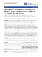
Histopathology
5 levels of histology were performed for each slice (b, d)
and results were recorded separa tely for each slice. Each
level consisted of 2 sections, one was stained with H&E
and the other one was used for CK19 IHC. Tissue was
cut in 4 μm slices and dried at 60 °C. Paraffin was sepa-
rated using xylol and ethanol (100%-70%). After 10 min-
utes of pronase digestion, the sections were incubation
with a CK19 antibody (Clone M0888 and clone No.
RCK 108, Dako, Glostrup, Denmark). A washing pro-
cess was carried out with Tris buffer and the secondary
antibody was added. Finally the staining with Fast Red
TR salt (Sigma-Aldrich, Germany) co mpleted the proce-
dure. The background was stained with Haematoxylin
(figure 2).
Table 1 Patients and tumour characteristics
patients
Age, years
Mean 66.3
Range 38-89
Sex
Male 116
Female 38
Grading
G1 1
G2 101
G3 70
Gx 12
Stage; TNM 6
th
edition
IA 10
IB 28
IIA 48
IIB 7
III 2
IIIA 14
IIIB 11
IIIC 23
IV 41
Nodal status
pN0 107
pN1 34
pN2 42
pN3 1
Croner et al. Journal of Translational Medicine 2010, 8:83
/>Page 2 of 6
One Step Nucleic Acid Amplification (OSNA)
To compare OSNA with histology, the CK19 mRNA
based OSNA procedure was performed as described
elsewhere [20]. CK 19 was identified as the most sensi-
tive marker for OSNA in CRC during pilot studie s (data
not shown). OSNA with beta-actin was carried out as
previously indicated and served as a RNA qual ity con-
trol [21]. The slices a and c were homogenised in 4 ml
of lysing buffer for 90 seconds (Lynorhag, Sysmex,
Kobe, J apan) and centrifuged for 1 minute at 10,000 g.
Afterwards CK19 or beta-actin mRNA was amplified by
reverse-transcription loop-mediated amplification (RT-
LAMP) i n the RD-100i (Lynoamp, Sysmex, Kobe) [22].
Automa ted amplification with a ready-to-use reagent kit
(Lynoamp, Sysmex, Kobe) was performed directly from
the sample homogenate, with no RNA purification
necessary, according to the manufacturer’s instructions.
The result was released after a total of 30-40 minutes
for 3-4 LN. During a pilot study in 136 LN from CRC
patients without lymph node metastases less than 250
copies/μl for CK19 were evalua ted as negative result for
OSNA (unpublished data). Therefore results were classi-
fied as (-) for CK 19 mRNA copies/μL less than 250 (for
beta-actin less than 1000 copies/μL), as (+) for CK19
mRNAbetween250-5000copies/μL (for beta-actin:
1000 - 5000 copies/μL), and (++) for mRNA copies/μL
higher than 5000.
Quantitative reverse-transcriptase polymerase chain
reaction as part of discordant case investigation (DCI)
DCI was performed after the original analysis. OSNA
runs were repeated from discordant sample homoge-
nates and afterwards RNA was isolated and subjected
toqRT-PCRforCK19,CEA,andbeta-actin.Condi-
tions for CK19, CEA and beta-actin qRT-PCR were
recently reported and described elsewhere [20,21].
Primer sequences for amplification of CEA were: 5’ -
AGACAATCACAGTCTCTGCGGA-3’ (forward) and
5’ - ATCCTTGTC CTCCACGGGTT-3’ (reverse). The
cut-off was set at cycle time = 29.6 for CK19, 28.5 for
CEA, and 30.0 for beta-actin.
Results
OSNA and histology
184 LN from 184 patients with colon cancer were inves-
tigated with both OSNA (CK19 mRNA as a m arker)
and intensive histological methods (H&E and CK19 IHC
on 5 levels for each of 2 LN slices). RNA quality was
assured by OSNA performed for beta-actin. 139 samples
gaveanegativeresultand37samplesgaveapositive
result with both methods (table 2). No isolated tumour
cells were found. In 10 out of 40 histology positive cases
the metastases was only found in one slice b ut not in
the other. Two positive samples contained a 10 mm and
Figure 1 Study design for lymph node workup.
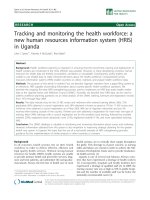
Figure 2 A) H&E staini ng and B) CK-19 immunohistochemistry
of lymph node metastases from colon carcinoma.
Croner et al. Journal of Translational Medicine 2010, 8:83
/>Page 3 of 6
a 5 mm macrometastasis and OSNA (++) results, but
were originally staged as IB and IIA, respectively, by
routine H&E staining. This means that LN were cut in
half and only one slice per of each half underwent H&E
staining. Three samples were histology+/OSNA -, and
5 samples were histology-/OSNA+, accounting for a
concordance rate of 95.7%, sensitivity of 92.5%, and spe-
cificity of 96.5% before DCI (table 2). Three patients
(3%) underwent LN upstaging during OSNA (table 3:
No 4, 5, 8) but were initially staged as LN negative dur-
ing routine histopathology (table 2: stage UICC IIA, IIA,
IB). During follow up one of these patients developed
metachronous distant metastases (DMS) in the liver
(table 4).
Discordant case investigation (DCI)
For all discordant samples additional OSNA runs as well
as qRT-PCR for CK19 an d CEA were conducted from
the remaining homogenate. However, since prolonged
storage in the homogenising buffer may adversely affect
RNA quality, data obtained by DCI may not fully reflect
the original condition and this may in particular become
evident in samples with CK19 mRNA copies near the
cut-off level of both OSNA and DCI.
Discordant case 1 contained a macrometastasis (8
mm), but only in slice b qRT-PCR and additional
OSNA runs performed from this sample during DCI
yielded positive and therefore concordant results so pos-
sibly the original negative value was due to a sample
mix-up. Sample 2 contained a 5 mm macrometastasis
with cal cified tissue. Quantitative RT-PCR was positive
forCK19only,withacycletimeclosetothecut-off
level, although all OSNA results for CK19 were negative.
Atthesametimethebeta-actinvaluewasrightonthe
cut-off level (not shown) which suggests that the RNA
concentration contained in the homogenate was very
low. Sample 3 contained a metastasis restricted to one
out of five levels whereas both OSNA and qRT-PCR
werenegative,stronglyindicativeoftissueallocation
bias (TAB), meaning that t umour deposits are restricted
to the slices either used for OSNA or histology. The
Table 2 Results of OSNA and histology (H&E Staining and
CK19 IHC) for 184 LN
OSNA HISTOLOGIC INVESTIGATION
Macrometastases Micrometastases Negative
++ 27 2 2 (1)
+ 8 - 3 (2)
- 3 (2) - 139
Total 38 (37) 2 144 (142)
Numbers in brackets indicate samples after discordant sample analysis, ++:
CK19 mRNA copies/μL higher than 5000, +:CK19 mRNA between 250 - 5000
copies/μL, -: CK 19 mRNA copies/μL less than 250
Table 3 Results of discordant case investigation between OSNA and histology (H&E Staining and CK19 IHC
No. Stage
UICC
Histology OSNA qRT-PCR Conclusion
size description original run
copies/μL
+/- second run
1
copies/μL
+/- beta-
actin
+/-
CK19
+/-
CEA
+/-
1 IV 8 mm Macrometastasis, only in
slice b, not d
<250 - 5470 ++ + + + Discordant
Sample mix-up
2 III 5 mm Macrometastasis,
calcified tissue
<250 - 0 - + + - Discordant
Beta-actin and CK19 value
near the cut-off level
3 IV 3 mm Macrometastasis, only
level 2
<250 - <250 - + - - Tissue allocation bias
4 IIA - negative 540 + <250 - + - - Discordant
Low copy
5 IIA - negative 1300 + <250 - + - - Discordant
6 IV negative 1300 + 290 + + - + Tissue allocation bias
7 IIIA 3.5 mm Positive with IHC in
level 1
25000 ++ 53700 ++ + + + Tissue allocation bias
8 IB - negative 27000 ++ <250 - + - - Discordant
Sample mix-up
OSNA: ++: CK19 mRNA copies/μL higher than 5000, +:CK19 mRNA between 250 - 5000 copies/μL, -: CK 19 mRNA copies/μL less than 250,
1
The indicated copy
number is the mean of three OSNA runs, QRT-PCR: +: positive, -: negative.
Table 4 Tumor characteristics and follow up of patients
which were histopathology LN negative and underwent
LN upstaging by OSNA
Histopathology OSNA Follow up
Tumor T N M N DMS dead/alive
Rectum 2 0 0 1 none alive
Rectum 3 0 0 1 Liver alive
Sigmoid colon 3 0 0 1 none alive
Mean follow up 72 month (range 52-88 month). DMS: distant meachronous
metastases.
Croner et al. Journal of Translational Medicine 2010, 8:83
/>Page 4 of 6
corresponding patient was formerly judged as pN0 by
routine histopathology performed outside this study.
Three out of the 5 histology-/OSNA+ samples (4 - 6)
contained CK19 mRNA copy numbers in the lower
range indicative of small tumour deposits located in the
slices analysed by OSNA. Two of these cases (4 and 5)
were originally node-negative and further CK19 OSNA
runs and qRT-PCR gave negative results. The beta-actin
OSNA copy numbers for these samples were seven and
four times lower at DCI, respectively, than the beta-
actin values from the original run (not shown). There-
fore we assume that RNA quality had suffered after a
couple months of storage so that the original results
could not be reproduced. In contrast to this, for sample
6, both OSNA and qRT-PCR s upported the output of
the original OSNA run. LN 7 exhibited (++) judgement
in all OSNA runs, and IHC gave positive results in only
level 1. Since this LN was rather small it was c ut into 2
pieces, and the metastasis was probably for the most
part located in the half used for OSNA and only with a
small part present in the other half used for histology.
Discordant case 8 was strongly positive in the first
OSNA run but negative upon OSNA repetition and
qRT-PCR so a sample mix-up cannot be excluded. In
summary, 3 out of 8 discordant samples were likely to
be caused by TAB. If these samples were excluded from
the sample cohort, the concordance rate was 97.2%
(176/181), sensitivity 94.9% (37/39), and specificity
97.9% (139/142).
Discussion
As lymphatic metastasis is a strong prognostic indicator
in CRC, the assessment of the nodal status is a key fac-
tor in CRC staging. 10-20% of n ode-negative stage
UICC I and II patients develop systemic disease within
less than 5 years. It has been suggested that using con-
ventional histological analysis, e.g. one or several H&E
sections, a certain proportion of micrometastases and
disseminated tumor cells remains undetected [17,23].
In the present study, 184 frozen LN from 184 CRC
patients were subjected to both, extensive histology and
OSNA, with al ternating slices of the LN used for each
method. The comparative evaluation showed 95.7% con-
cordance, 92.5% sensitivity, and 96.5% specificity. Lysates
of the 8 discordant samples were f urther analysed by
RT-PCR. In 3 out of 8 samples DCI supported the
OSNA finding. It was concluded that metastases were
strictly located in either the tissue used for OSNA or
the tissue used for histology which renders a compari-
son of the 2 methods impossible. By excluding these
samples concordance rate was 97.2%, sensitivity 94.9%,
and specificity 97.9% when compared to a very extensive
histological examination which is not routinely per-
formed in all institutions. Differences between CEA and
CK-19 qRT-PCR as detected in two cases reflect the
heterogeneity of marker expression in lymph node
metastases of CRC (table 3). These results are in con-
cordance with previous studies which also identified a
dis concordant expression between CEA and cytokeratin
in lymph node metastases of CRC in about 25% [24]. In
two cases there were differences between second run
OSNA and qRT-PCR (table 3). In case two there was
calcified tissue which mayhaveinfluencedOSNA.In
case six there was a low copy number of OSNA which
was slightly abov e the positive cut off value. The low
amount of CK19 mRNA in the sample was verified by
qRT- PCR. Nevertheless this borderline case reflects the
need of additional markers in specific cases such as
CEA.
In a different study carried out with an earlier prototype
of the RD-100i, 63 LN from 6 CRC patients were investi-
gated with both H&E staining and OSNA. None of the LN
was HE+/OSNA- and 3 of the 63 LN were HE-/ OSNA+,
resulting in upstaging of two patients [25]. The aspect of
upstaging was not the main focus of this investigation
since only one LN from one CRC patient was analysed.
Despite this, even in the 93 LN from stage UICC I or II
patients enrolled in this study tumour deposits were
detected in 3 cases (3%) by OSNA. One of these patients
developed hepatic DMS during follow up. Correct tumor
staging is essential to apply adequate adjuvant treatment.
If patients are under staged during routine histopathology
they will not receive necessary adjuvant chemotherapy
which may result in tumor progression during follow up.
More sensitive staging methods can reduce such pitfalls
and prevent tumor recurrence especially in stage UICC II.
For OSNA fresh tissue i s required and the lymph nodes
should be harvested by pathologists from the resected spe-
cimen. This could ca use a major change during clinical
practise. Therefore an interdisciplinary dialogue and plan-
ning is indispensable for this procedure. Furthermore
lymph node harvesting in fresh tissue is much harder
compared with formalin fixed material and requires a spe-
cial training. The OSNA lysate can be asservated and in
unclear cases RNA isolation for further diagnostics is
possible.
Our findings underline the requirement for a more
comprehensive diagnostic technique than H&E staining
of a limited numbers of sections has provided so far, in
particular the need to analyse the whole LN in order to
detect occult small tumour deposits. As opposed t o a
var iety of differen t histological approaches presented so
far for the identification of occult disease in LN of CRC
patients the OSNA method is a standardised technique
which includes a short homogenisation s tep and subse-
quent automated amplification of CK19 mRNA and
therefore ensures reproducible and objective judgement
[26]. In contr ast to RT-PCR for which RNA pur ification
Croner et al. Journal of Translational Medicine 2010, 8:83
/>Page 5 of 6
is mandatory, in the OSNA assay amplification directly
starts from the lysate and therefore allows analysis of
3-4 LN within 30-40 minutes and 12 LN within 2 hours.
For the reason that increasing numbers of investigated
LN correlate with a more accurate tumour staging, high
throughput methods are indispensable for future pur-
pose [5,13,14]. In conclusion, the CK19 mRNA based
OSNA is a new and reliable method to determine meta-
static disease in LN and can be applied as a rapid diag-
nostic tool during staging of CRC patients.
Acknowledgements
This study was supported the Interdisciplinary Centre for Clinical Research
(IZKF) of the University Erlangen-Nuremberg, the Federal Department of
Culture and Science (BMBF) Germany and the German Research Foundation
(DFG).
Author details
1
Department of Surgery, University of Erlangen-Nuremberg, Germany.
2
Department of Pathology, Vivantes Humboldt-Clinic, Berlin, Germany.
3
Department of Molecular and Experimental Surgery, University of Erlangen-
Nuremberg, Germany.
4
Department of Pathology, University of Erlangen-
Nuremberg, Germany.
Authors’ contributions
RSC participated in the design of the study, worked up the lymph nodes,
supported data workup, statistical analysis and drafted the manuscript, VS
was involved in technical assistance and in writing the manuscript, HD
carried out H&E staining and CK19 IHC, CS coordinated the study and
drafted the manuscript, TP participated in the study design and drafted the
manuscript, EN and MS added technical support and drafted the manuscript,
KEM and WH participated in patient recruitment and drafted the manuscript,
AS scored the H&E staining and CK19 IHC of the LN. All authors read and
approved the final manuscript.
Competing interests
The study was supported by the company Sysmex, Kobe, Japan. Otherwise
the authors declare that they have no competing interests.
Received: 24 March 2010 Accepted: 6 September 2010
Published: 6 September 2010
References
1. Ferlay J, Autier P, Boniol M, Heanue M, Colombet M, Boyle P: Estimates of
the cancer incidence and mortality in Europe in 2006. Ann Oncol 2007,
18:581-92.
2. Jemal A, Siegel R, Ward E, Murray T, Xu J, Thun MJ: Cancer statistics, 2007.
CA Cancer J Clin 2007, 57:43-66.
3. Hermanek P: Prognostic factor research in oncology. J Clin Epidemiol 1999,
52:371-4.
4. Radespiel-Troger M, Hohenberger W, Reingruber B: Improved prediction of
recurrence after curative resection of colon carcinoma using tree-based
risk stratification. Cancer 2004, 100:958-67.
5. Cianchi F, Palomba A, Boddi V, Messerini L, Pucciani F, Perigli G, Bechi P,
Cortesini C: Lymph node recovery from colorectal tumor specimens:
recommendation for a minimum number of lymph nodes to be
examined. World J Surg 2002, 26:384-9.
6. Greene FL, Sobin LH: The TNM system: our language for cancer care. J
Surg Oncol 2002, 80:119-20.
7. Leibl S, Tsybrovskyy O, Denk H: How many lymph nodes are necessary to
stage early and advanced adenocarcinoma of the sigmoid colon and
upper rectum? Virchows Arch 2003, 443:133-8.
8. Sobin LH, Hermanek P, Hutter RV: TNM classification of malignant tumors.
A comparison between the new (1987) and the old editions. Cancer
1988, 61:2310-4.
9. Turner J, Vollmer RT: Lymph nodes in colorectal carcinoma. The Poisson
probability paradigm. Am J Clin Pathol 2006, 125:866-72.
10. Yoshimatsu K, Ishibashi K, Umehara A, Yokomizo H, Yoshida K, Fujimoto T,
Watanabe K, Ogawa K: How many lymph nodes should be examined in
Dukes’ B colorectal cancer? Determination on the basis of cumulative
survival rate. Hepatogastroenterology 2005, 52:1703-6.
11. Hermanek P, Mansmann U, Staimmer DS, Riedl S: The German experience:
the surgeon as a prognostic factor in colon and rectal cancer surgery.
Surg Oncol Clin N Am 2000, 9:33-49, vi.
12. Hohenberger W, Merkel S, Weber K: Lymphadenectomy with tumors of
the lower gastrointestinal tract. Chirurg 2007, 78:217-25.
13. Goldstein NS: Lymph node recoveries from 2427 pT3 colorectal resection
specimens spanning 45 years: recommendations for a minimum
number of recovered lymph nodes based on predictive probabilities.
Am J Surg Pathol 2002, 26:179-89.
14. Berberoglu U: Prognostic significance of total lymph node number in
patients with T1-4N0M0 colorectal cancer. Hepatogastroenterology 2004,
51
:1689-93.
15. Davies M, Arumugam PJ, Shah VI, Watkins A, Roger Morgan A, Carr ND,
Beynon J: The clinical significance of lymph node micrometastasis in
stage I and stage II colorectal cancer. Clin Transl Oncol 2008, 10:175-9.
16. Rodriguez-Bigas MA, Maamoun S, Weber TK, Penetrante RB, Blumenson LE,
Petrelli NJ: Clinical significance of colorectal cancer: metastases in lymph
nodes <5 mm in size. Ann Surg Oncol 1996, 3:124-30.
17. Rosenberg R, Friederichs J, Gertler R, Hoos A, Mueller J, Nahrig J, Nekarda H,
Siewert JR: Prognostic evaluation and review of immunohistochemically
detected disseminated tumor cells in peritumoral lymph nodes of
patients with pN0 colorectal cancer. Int J Colorectal Dis 2004, 19:430-7.
18. Weitz J, Koch M, Kienle P, Schrödel A, Willeke F, Benner A, Lehnert T,
Herfarth C, von Knebel Doeberitz M: Ann Surg 2000, 232:66-72.
19. Bilchik AJ, Nora DT, Saha S, Turner R, Wiese D, Kuo C, Ye X, Morton DL,
Hoon DS: The use of molecular profiling of early colorectal cancer to
predict micrometastases. Arch Surg 2002, 137:1377-83.
20. Tsujimoto M, Nakabayashi K, Yoshidome K, Kaneko T, Iwase T, Akiyama F,
Kato Y, Tsuda H, Ueda S, Sato K, Tamaki Y, Noguchi S, et al: One-step
nucleic acid amplification for intraoperative detection of lymph node
metastasis in breast cancer patients. Clin Cancer Res 2007, 13:4807-16.
21. Visser M, Jiwa M, Horstman A, Brink AA, Pol RP, van Diest P, Snijders PJ,
Meijer CJ: Intra-operative rapid diagnostic method based on CK19 mRNA
expression for the detection of lymph node metastases in breast cancer.
Int J Cancer 2008, 122:2562-7.
22. Notomi T, Okayama H, Masubuchi H, Yonekawa T, Watanabe K, Amino N,
Hase T: Loop-mediated isothermal amplification of DNA. Nucleic Acids Res
2000, 28:E63.
23. Weitz J, Koch M, Lehnert T, Herfarth C, von Knebel Doeberitz M: Detection
of isolated disseminated tumor cells of colorectal carcinomas in lymph
nodes. Chirurg 2000, 71:410-6.
24. Rosenberg R, Hoos A, Mueller J, Nekarda H: Impact of cytokeratin-20 and
carcinoembryonic antigen mRNA detection by RT-PCR in regional lymph
nodes of patients with colorectal cancer. Br J Cancer 2003,
83(10):1323-1329.
25. Taniyama K, Motoshita J, Sakane J, Makita K, Akai Y, Daito M, Otomo Y,
Ono H, Mizunoe T, Takeuchi Y, Tominaga H, Koseki M: Combination
analysis of a whole lymph node by one-step nucleic acid amplification
and histology for intraoperative detection of micrometastasis.
Pathobiology 2006, 73:183-91.
26. Tsavellas G, Patel H, Allen-Mersh TG: Detection and clinical significance of
occult tumour cells in colorectal cancer. Br J Surg 2001, 88:1307-20.
doi:10.1186/1479-5876-8-83
Cite this article as: Croner et al.: One Step Nucleic Acid Amplification
(OSNA) - a new method for lymph node staging in colorectal
carcinomas. Journal of Translational Medicine 2010 8:83.
Croner et al. Journal of Translational Medicine 2010, 8:83
/>Page 6 of 6