Báo cáo hóa học: "Exhaustive expansion: A novel technique for analyzing complex data generated by higherorder polychromatic flow cytometry experiments" pot
Bạn đang xem bản rút gọn của tài liệu. Xem và tải ngay bản đầy đủ của tài liệu tại đây (1.46 MB, 15 trang )
METH O D O LOG Y Open Access
Exhaustive expansion: A novel technique for
analyzing complex data generated by higher-
order polychromatic flow cytometry experiments
Janet C Siebert
1*
, Lian Wang
2
, Daniel P Haley
3
, Ann Romer
2
, Bo Zheng
2
, Wes Munsil
1
, Kenton W Gregory
2
,
Edwin B Walker
3
Abstract
Background: The complex data sets generated by higher-order polychromatic flow cytometry experiments are a
challenge to analyze. Here we describe Exhaustive Expansion, a data analysis approach for deriving hundreds to
thousands of cell phenotypes from raw data, and for interrogating these phenotypes to identify populations of
biological interest given the experimental context.
Methods: We apply this approach to two studies, illustrating its broad applicability. The first examines the
longitudinal changes in circulating human memory T cell populations within individual patients in response to a
melanoma peptide (gp100
209-2M
) can cer vaccine, using 5 monoclonal antibodies (mAbs) to delineate
subpopulations of viable, gp100-specific, CD8+ T cells. The second study measures the mobilization of stem cells in
porcine bone marrow that may be associated with wound healing, and uses 5 different staining panels consisting
of 8 mAbs each.
Results: In the first study, our analysis suggests that the cell surface markers CD45RA, CD27 and CD28, commonly
used in historical lower order (2-4 color) flow cytometry analysis to distinguish memory from naïve and effect or
T c ells, may not be obligate parameters in defining central memory T cells (T
CM
). In the second study, we identify
novel phenotypes such as CD29+CD31+CD56+CXCR4+CD90+Sca1-CD44+, which may characterize progenitor cells
that are significantly increased in wounded animals as compared to controls.
Conclusions: Taken together, these results demonstrate that Exhaustive Expansion supports thorough interrogation
of complex higher-order flow cytometry data sets and aids in the identification of potentially clinically relevant
findings.
Background
Flow cytometry (FCM) is a powerful technology with
major scientific and public health relevance. FCM can
be used to collect multiple simultaneous light scatter
and antigen specific fluorescence measurements on cells
as each cell is excited by multiple lasers and emitted
fluorescence signals are passed along an array of detec-
tors. This technology permits characterization of various
cell subpopulations in complex mixtures of cells. Using
new higher-order multiparameter FCM techniques we
can simultaneously identify T and B cell subsets, stem
cells, and specific cell surface antigens, cytokines, che-
mokines, and pho sphorylated proteins produced by
these cells. Higher order FCM allows us to measure at
least 17 parameters per cell [1], at rates as high as
20,000-50,000 cells per second.
Increasing sophisticati on in FCM, coupled with the
inherent complex dimensionality of clinical and transla-
tional experiments, leads to data analysis bottlenecks.
While the literature documents a long h istory of auto-
mated approaches to gating events within a single sam-
ple [2-4], the gated data remains complex, with readouts
for tens to hundreds of phenotypes per sample, multiple
samples per patient, and multiple cohorts per study.
Unfortunately, there is a paucity of proven analytical
* Correspondence:
1
CytoAnalytics, Denver, CO, USA
Full list of author information is available at the end of the article
Siebert et al. Journal of Translational Medicine 2010, 8:106
/>© 2010 Siebert et al; licensee BioMed Central Ltd. This is an Open Access article distrib uted under the terms of the Creative C ommons
Attribution License ( which permits unrestricted use, distr ibution, and reproduction in
any medium, provided the original work is properly cited.
approaches that provide meaningful biological insight in
the face of such complex data sets.
Furthermore, interpretation of results from higher
order experiments may be biased by historical results
from simpler lower order experiments. Marincola [5]
suggests that modern high-throughput tools, coupled
with high-throughput analysis, provi de a more unbiased
opportunity to reevaluate the basis of human disease,
while advocates of cytomics [6,7] observe that exhaustive
bioinformatics data extraction avoids the inadvertent loss
of information associated with a priori hypotheses. Fun-
damentally, these authors underscore the distinction
between inductive (hypothesis-g enerating) and deductive
(hypothesis-driven) reasoning. This distinc tion is clearly
applicable to the interpretation of higher-order multi-
parameter flow cytometry data. Herein, we apply a
powerful inductive data analysis approach to two dis-
tinctly different studi es in order to demonstrate its broad
applicability. The first study examines human memory
T cell responses to a melanoma peptide cancer vaccine,
while the second inspects porcine stem cell phenotypes
associated with wound healing.
In a previously described melanoma booster vaccine
study [8], we used 8-color FCM to characterize the phe-
notypes of viable (7AAD
-
) melanoma antigen-specific
(gp100 tetramer
+
)CD8
+
T cells collected from periph-
eral blood. Memory and effector T cell subpopulations
responding to vaccine antigen were c haracterized using
5 additional monoclonal antibodies (mAbs) specific for
CCR7, CD45RA, CD57, CD27, and CD28. Samples were
collected from 7 donors at 3 time points: after (post)
the initial vaccine regimen (PIVR); at a long term me m-
ory (LTM) time point collected 18 to 24 month s after
the end of vaccine administration; and after two boost-
ing vaccines (P2B). Phenotypes for T
CM
have been
described based on lower-order 3-4 color staining with
different combinations of the above antibodies, with
data suggesting a consensus T
CM
phenotype of CCR7
+CD45RA-CD57-CD27+CD28+. W e demonstrated that
LTM gp100-specific CD8
+
T cells were enriched for this
consensus phenotype [8]. We also described a gp100-
specific T
CM
subset that retained CD45RA expression
(CCR7+CD45RA+CD57-CD27+CD28+), which we
terme d T
CMRA,
and which may represent a T
CM
precur-
sor population similar to that described in the mouse
[9]. Although this consensusphenotypehaspreviously
been used to primarily define naïve T cells, i t clearly
characterized a subpopulation of antigen-educated (i.e.
gp100 tetramer positive) long term memory CD8
+
T cells in the melanoma vaccine study. This phenotype
signature may delineate a T
CM
precursor population
that arises shortly after antigen activation of naïve
T cells. Thus, studies in the mouse demonstrate that
tumor-specific T
CM
and similar putative T
CM
precursors, referred to as central memory stem cells
(T
SCM
), which may derive from early daughter cell divi-
sion after antigen stimulation of naïve T cells, express
elevated levels of proliferation, enhanced survival in
vivo, and superior CTL function compared to effector or
effector-memory (T
EM
) T cells [9]. However, the origin
of T
CM
and T
SCM
precursors remains controversial,
since other data supports the hypotheses that such
memory subpopulations may also develop from effector
and effector-memory T cells [10]. Controversy aside,
enhanced proliferative and survival properties character-
istic of memory T cells have been correlated with anti-
tumor responses in mice and humans receiving adoptive
T cell-based therapies [11]. Thus, the use of higher-
order flow cytometry and comprehensive multipara-
meter data analysis could facilitate the identification and
expansion of T
CM
and T
CM
precursor subpopulations
(i.e. T
SCM
) for more effective cancer immunotherapy
regimens.However,suchatherapeutic strategy would
depend on first demonstrating memory T cell functional
properties by sorted cells exhibiting such putative mem-
ory phenotype signatures.
Our second study examines complex stem cell pheno-
types mobilized in response to wound healing. One use
of stem cell therapy may be that of repairin g damaged
tissues, since bone marrow stem and progenitor cells
can differentiate into muscle cells, endothelial cells, and
nerve cells in vitro and in vivo [12]. Extremity injuries
complicated by compartment syndrome (e.g. trauma-
related severe swelling that can lead to ischemia and
permanent tissue necrosis) are a common consequence
of battlefield trauma, crush injuries that have been
report ed in recent earthquakes, and many sport injuries.
While faciotomy can reduce the injury, there is no treat-
men t that replaces or regenerates muscle and nerve tis-
sues, leaving the patient with a permanent disability
[13]. Human studies have demonstrated that injection of
bone marrow stem cells into ischemic muscle may
reduce the damage to the muscle and the loss of muscle
function [14-18]. We have hypothesized that healthy,
autologous bone marrow stem cells could be used to
treat compartment syndrome. Our init ial investigation
focused on determining the optimal time to harvest
bone marrow stem and progenitor cells after injury in
the event that injury might amplify the mo bilization of
stem cell populations in the bone marrow. Bone marrow
samples were collected from 8 injured swine and 8 con-
trol swine at pre-injury (baseline) and at 4 consecutive
one-week intervals. Bone marrow was characterized by 5
different staining panels consisting of 8 mAbs each, as
presented in Table 1. In total, 12 differ ent monoclonal
antibodies (CD29, ckit, CD56, CXCR4, CD105, CD90,
Sca-1, CD44, CD31, CD144, CD146, and VEGFR2) were
used. Others have used more restrictive lower order
Siebert et al. Journal of Translational Medicine 2010, 8:106
/>Page 2 of 15
combinations of these markers to delineate mesenchy-
mal stem cells (CD29, CD90, and CD44) [19,20], primi-
tive stem cells (ckit, CXCR4, and S ca-1) [21-23],
myoblasts (CD56 and CXCR4) [24,25], and vascular-
relative cells ( CD146, CD31, CD144, CD105, and
VEGFR2) [26-29]. However, to date, there has been no
description of the combined use of all of these putative
progenitor cell set descriptors in higher order staining
panels.
Our multipa rameter studies allow the identification of
hundreds to thousands of phenotypes of cells, based on
combinations of positive or negative expression of the
included mAbs. For example, in the melanoma vaccine
study, we initially consider ed all 32 (2
5
) possible pheno-
types defined by positive and negative combinations of
all 5 variable markers, e.g. CCR7+CD45-CD57-CD27
+CD28+ [8]. This type of analytical strategy is used by
many researchers [30-32]. However, it focuses on popu-
lations defined by exactly the number of variable para-
meters in the staining panel (5, in the case of the
vaccine study). Thus, to more thoroughly explore the
data, we exhaustively expanded the data sets to include
all possible phenotypes defined by combinations of 0, 1,
2, 3, 4, and 5 markers, e.g. CCR7+ and CCR7+CD57-
CD27+CD28+. When each marker can as sume one of
two values (positive or negative), the number of possible
cell subsets in an M-marker study is 2
M
. When each
marker can assume one of three possible values (posi-
tive, negative, or unspecified), the number of possible
cell sets is 3
M
,or3
5
(243) in this 5 marker study, as illu-
strated in Table 2. In the w ound healing study, bone
marrow was characterized by 5 different 8 color panels.
Exhaustive Expansion of these 8 marker sets to include
all possible 0, 1, 2, 8 marker sets resulted in 6,561 (3
8
)
sets per panel, for a total of 32,805 (6,56 1 × 5 panels)
cell subpopulations per sample.
Since we could not manually analyze data from hun-
dreds to thousands of phenotypes efficiently, we first
identified numerically interesting phenotypes by com-
puting metrics for all derived sets. For example, in the
melanoma vaccine study, the middle of three time
points represented a long term me mory time point, col-
lected 18 to 24 mo nths after exposure to the vaccine
antigen. Consequently, one feature of interest was the
delineation of phenotypes that peaked at this long term
memory time point. In the wound healing study, since
there were both wounded animals and control animals,
we could identify phenotypes in which the expression
levels for the wounded animals were greater than t he
levels for the control animals. In each case, simple
visualizations, such as those presented in the Results,
illustrated the patterns of r esponse and helped us vet
the numerically interesting phenotypes for biological
relevance. In both studies we identified results with pos-
sible important clinical implications that would have
been very difficult to find using standard analyt ical tech-
niques. Using Exhaustive Expansion we were able to
define a putative minimum obligate phenotype for cen-
tral memory T cells, and delineate multiple bone-mar-
row-derived putative myogenic MSC subpopulations
that may be mobilized in response to myonecrotic
injury.
Methods
Melanoma Vaccine Study
The clini cal trial protocol and the flow cytometry stain-
ing and analysis procedures used to acquire data in this
study have been described in detail elsewhere [8,33].
Briefly, early s tage melanoma patients were vacc inated
every second or every third week over six months with
a modified, HLA-A2 restricted melanoma associated
peptide, gp100
209-2M
. Leukophereses we re collected
before the vaccine regimen, a fter (post) the initial vac-
cine regimen (PIVR); at a long term memory (LTM)
time point 18-24 months later; and following two addi-
tional boosting vaccines (P2B) given at one month inter-
vals following the LTM leukopak collection. The
protocol was reviewed by NCI’s CTEP and approved by
the Providence Health System institutional review
board. All patients gave written informed consent. Cryo-
preserved PBMCs from PIVR, LTM and P2B time points
were stained simultaneously with gp100 tetramers and
with mAbs specific for CD8b, CCR7, CD45RA, CD57,
CD27, CD28, and with 7AAD to discriminate live from
dead cells. All samples were analyzed on a 9 color Beck-
man Cyan ADP flow cytometer. Viable lymphocytes
were gated for positive CD8b and gp100 tetramer stain-
ing, and gp100-specific CD8b
+
T cells were further
interrogated for expression of the remaining five cell
surface markers (CCR7, CD45RA, CD57, CD27 , and
CD28) to determine their subphenotypes. At least 5,000
gp100-specific CD8b
+
T cells were colle cted per sample.
Table 1 Five monoclonal antibody panels for stem
cell study
Panel Main CD31 CD144 CD146 VEGFR2
Antibody CD29 CD29 CD29 (CD146) CD29
ckit (CD31) (CD144) ckit ckit
CD56 CD56 CD56 CD56 CD56
CXCR4 CXCR4 CXCR4 CXCR4 CXCR4
CD105 CD105 CD105 CD105 CD105
CD90 CD90 CD90 CD90 (VEGFR2)
Sca-1 Sca-1 Sca-1 Sca-1 Sca-1
CD44 CD44 CD44 CD44 CD44
Each of the 5 panels consists of 8 mAbs. The differences from the main panel
are indicated both in the name of the panel and by the antibody listed in
parentheses.
Siebert et al. Journal of Translational Medicine 2010, 8:106
/>Page 3 of 15
All data was acquired in FCS format (Summit 4.2) and
analyzed using the FCOM format of Winlist 5.0 Soft-
ware (Verity House Software). “Fluorescence minus one”
(FMO) controls were u sed to define positive and nega-
tive histogram staining regions for each fluorescent
variable.
Porcine Stem Cell Study
All protocols were approved by the IACUC of Legacy
Research and Technology Center. A bilateral compart-
ment syndrome injury was produced in the anterior
tibialis muscles by infusing porcine plasma directly into
the muscles. A standardized bone marrow collection
procedure was used as previously described [34], with
bone marrow harve sted from the tibia of anesthetized
swine. Bone marrow was transferred to an automated
cell processing system, BioSafe SEPAX cell separating
system (Biosafe S A, Bern, Sw itzerland), within 60 min-
utes of collection, and mononuclear ce lls were isolated.
Each sample was divided into 5 aliquots, which were
stained for surface marke r expression as summarized in
Table 1. All samples w ere acquired using a BD™ LSR II
flow cytometer.
To identify ckit (a.k.a stem cell factor (SCF)) expres-
sion, a porcine SCF ligand conjugated with biotin, kindly
provided by Dr. Christene Huang (Transplantation Biol-
ogy Research Center at Massachusetts General Hospi-
tal), was used together with a streptavidin-PE (Jackson
Immunoresearch, W est Grove, PA) for secondary bind-
ing. The antibodies for the other markers were all com-
mercial monoclonal antibodies which were specific for
porcine antigens or were anti-human or anti-mouse
which cross react with the designated epitopes in swine:
CD29-FITC, CD146-FITC and CD105 (GeneTex Inc.,
Irvine, CA), CD90-APC and CD44-APC-Cy7 (BioLe-
gend, San Diego, CA), C D56-PE-TR (Inv itrogen,
Carlsbad, CA), Sca-1-Alexa Fluor 700 (Sca-1-AF700),
CXCR4-PE-Cy7 (eBioscience, San Diego, CA), CD31-PE
(AbD Serotec, Raleigh, NC), CD144-PE (Santa Cruz
Biotechnology, Santa Cruz, CA), and VEGFR2-APC
(R&D Systems, Minneapolis, MN). The anti-CD105 anti-
body was conjugated with Pacific Blue using a monoclo-
nal antibody labeling kit (Invitrogen, Carlsbad, CA),
following manufacturer’s protocol.
Systems and Software
While the details of the data analysis approach are
provided in the Results, we highlight the system com-
ponents below. The “ Expander” program for deriving
all possible phenotypes or sets is implemented in the
Java programming language, and is freely available
upon request. Input consists of a comma-delimited file
containing fields for absolute set or phenotype names,
3 additional qualifiers, and the percentage of cells in
the set specified by the name and the qualifiers. Out-
put consists of a comma-delimited file containing
fields for 3 qualifiers, the relative set name, and the
derived data value. The three qualifiers from the input
are passed to corresponding rows in the output with-
out modification. These qualifiers support downstream
analysis based on characteristics such as donor, time
point, and treatment protocol. Representative input
and output formats are shown in Table 3. Relative set
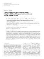
names and their derivation are illustrated in Figure 1
and described in the associated results. The derived
data values are simply the sum of the frequencies of
the relevant subsets. The output was then loaded into
a relational database (MySQL), and standard SQL
statements and graphing utilities were used to interro-
gate the data. Statistical tests were performed using
the R software environment for statistical computing
() .
Table 2 Combinations of positive/negative phenotypes in a 5-marker panel
Number of
markers
(M)
Number of +/- gates
given M markers
(G)
Combinations Number of combinations of M markers
in a 5 marker panel (C)
Number of gates
times number
of combinations
(G × C)
02
0
= 1 No markers specified 1 1
12
1
= 2 A, B, C, D, E 5 10
22
2
= 4 AB, AC, AD, AE, BC, BD, BE, CD,
CE, DE
10 40
32
3
= 8 ABC, ABD, ABE, ACD, ACE, ADE,
BCD, BCE, BDE, CDE
10 80
42
4
= 16 ABCD, ABCE, ABDE, ACDE, BCDE 5 80
52
5
= 32 ABCDE 1 32
TOTAL = 243
This table illustrates the total number of positive/negative gates in a 5-marker panel, with hypothetical markers A, B, C, D and E. There are five possible 1-marker
combinations, ten 2-marker combinations, ten 3-marker combinations, five 4-marker combinations, and one 5-marker combination. For each combination, there
are 2
M
positive/negative gates where M is the number of markers in the combinations. Thus, there are 243 possible phenotypes in a 5 marker experiment. This
generalizes to 3
M
.
Siebert et al. Journal of Translational Medicine 2010, 8:106
/>Page 4 of 15
Statistical Methods
In the mela noma vaccine study, the Wilcoxon signed-
rank test was used to identify either increased expres-
sion between time points or decreased expression
between time points, depending on t he pair of time
points under consideration. The p-values were then
used to screen populations for biologically meaningful
results. These p-values provided a simple, well-under-
stood metric to encapsulate the differences between the
two time points. An alte rnative metric, such as 4 of 7
donors showing at least a 5% change between time
points, would have been more verbose and would have
required more detailed justification. In the p orcine
wound healing study, the Wilcoxon rank sum test was
used to identify phenotypes in which the wounded
cohort showed a greater change from baseline than did
the control cohort.
Results
Exhaustive Expansion
In both studies, standard FCM analysis software was
used to establish positive and negativ e gates based on
the use of “fluorescence-minus- one” (FMO) controls for
the included markers. In the case of the 5 memory mar-
kers used in the melanoma vaccine study, 32 (2
5
)sets
were subsequently generated using WinList ’s™ (http://
www.vsh.com) FCOM function. Such combinatio n gates
also can be generated with other flow cytometry
analytical software such as FlowJo (wjo.
com) and FCS Express (ovosoftware.
com). The gating strategy for this study is illustrated in
Figure 1. By inspecting a series of two-dimensional scat-
ter plots, positive and negative gating boundaries were
set, dividing the cells into subpopulat ions. Each of the 4
quadrants in do t plots 1 through 4 illustrates the fre-
quencies of phenotypes of gp100 tetramer
+
CD8
+
T
cells that are defined by positive and negative combina-
tions of CCR7, CD45RA, CD57, CD27, and CD28.
Next we derived the percentage of cells in the more
comprehensive analysis of all 243 (3
5
) possible pheno-
types, as defined by 0, 1, 2, 5 parameters, using a cus-
tom Java program as described in the Methods. We
utilize a shorthand notation for phenotypes by introdu-
cing a placeholder (”.”) to represent an unspecified para-
meter. These concep ts are also illustrated in Figure 1, in
which the callout table shows the shorthand notation
for 2 populat ions sp ecified by 5 markers, CCR7
+CD45RA-CD57-CD27+CD28+ (+– ++) and CCR7
+CD45RA-CD57-CD27+CD28- (+–+-). The table also
shows the notation for the 4 marker phenotype (+ – +.)
resulting from the summation of the frequencies of the
two 5 marker phenotypes. Notice that CD28 assumes 3
values, “+“, “-“,and“.“. The phenotype +–+. repre-
sents the combination or union of two subphenotypes
or subsets (+–++ and +– +-), Hereafter, subphenotype
signatures will be referred to as either sets or
phenotypes.
The universal set ( ) contains 100% of the cells
in the population of interest (e.g. viable, antigen-positive,
CD8
+
cells), and thus serves as an internal control. All
other sets are proper subsets of the universal set. As
presented here, Exhaustive Expansion applies to binary
classification systems (e.g. positive and negative gating),
but extension to n-ary classification systems (e.g. dim,
intermediate, bright) is possible. After derivation of fre-
quencies for all sets, data was loaded into a relational
database (MySQL) and analyzed with SQL statements
and graphing utilities.
Melanoma Vaccine Study
Average CV Suggests Stable CD27, CD28, and CD45RA
Expression Over Time
Having derived the percentage of cells in all 243 0-
through 5-parameter sets in the melanoma vaccine
study, we generated longitudinal profiles for all sets as
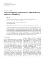
shownbytheexampleinFigure2.Thisenabledusto
clearly see the responses of each donor over time. Addi-
tionally, these profiles allow each donor to serve as his
or her own control. Next, we looked for sets that were
interesting based on coefficient of variation (CV, stan-
dard deviation divided by mean). We computed Average
CV by calculating CVs for each donor across 3 time
Table 3 Representative input and output for the
“Expander” program
Representative Input
CCR7+CD45+CD57-CD27+CD28-, panel, EA02, LTM,2.48
CCR7+CD45+CD57-CD27+CD28+, panel, EA02, LTM,5.41
CCR7+CD45+CD57+CD27-CD28-, panel, EA02, LTM,1.47
CCR7+CD45+CD57+CD27-CD28+, panel, EA02, LTM,0.22
CCR7+CD45+CD57+CD27+CD28-, panel, EA02, LTM,0.34
CCR7+CD45+CD57+CD27+CD28+, panel, EA02, LTM,1.34
Representative Output
panel, EA02, LTM,+++++,1.34
panel, EA02, LTM,++++-,0.34
panel, EA02, LTM,++++.,1.68
panel, EA02, LTM,+++-+,0.22
panel, EA02, LTM,+++–,1.47
panel, EA02, LTM,+++ ,1.69
panel, EA02, LTM,+++.+,1.56
panel, EA02, LTM,+++ ,1.81
The Expander program derives aggregate sets or supersets from input data,
and outputs both the relative set name and the percentage of cells in both
the newly derived sets and the original sets. The percentage of cells in the
derived sets is calculated by adding together the percentages in the subsets,
as illustrated in Figure 1. The rows below illustrate the format of both input
and output, but not direct correspondence between input and output. Output
is loaded into a relational database for further analysis.
Siebert et al. Journal of Translational Medicine 2010, 8:106
/>Page 5 of 15
points, and then averaging the 7 CVs. We then sorted
the longitudinal profiles both by ascending average CV
and descending average CV. In this data, the sets with a
low average CV, as shown in Figure 2, were particularly
interesting because of their common use in lower order
flow cytometry analysis to dis tinguish central memory
and effector memory T cells [35,36]. At 8.59%, the
CD45RA+ phenotype has the lowest Average CV of all
242 non-universal sets (those with at least one marker
specified). In this case, even though there is inter-donor
variation, the values are relatively stable over time for
each individual donor. There are 4 donors with rela-
tively low levels of CD45RA expression, 2 donors w ith
relatively high levels, and 1 donor with an intermediate
level. Thus, inspection reveals that the low Average CV
was associated with donor stratification. Profiles for
CD27+ and CD28+ are also shown in Figure 2, and
similarly suggest overall low average CVs for individual
patient phenotype frequencies over all 3 time points, but
do not indicate inter-donor variation. Notably, all three
Figure 1 Representative gating strategy and additional phenotype set calculations. This figure illustrates a gating strategy in which CCR7
+
cells are further categorized by positive or negative expression of CD45RA and CD57. Cells in each resulting quadrant (dot plot B) are then
categorized based on CD27 and CD28 staining frequencies (dot plots 1-4). The callout table illustrates how the two phenotypes CCR7+CD45RA-
CD57-CD27+CD28+ (+–++) and CCR7+CD45RA-CD57-CD27+CD28- (+–+-), marked by dotted lines, are aggregated to form a superset
population, CCR7+CD45RA-CD57-CD27+ (+–+.), in which CD28 expression is unspecified.
Siebert et al. Journal of Translational Medicine 2010, 8:106
/>Page 6 of 15
of these m arkers are associated with the T
CM
consensus
phenotype (CCR7+ CD45RA- CD57- CD27+ CD28+)
predicted from l ower order 3- and 4-marker flow cyto-
metry analysis, yet individually show low to moderate
frequencychangesoverthetimecourseofthevaccine
study, even though our previous data suggested T
CM
increased at LTM for most patie nts [8]. Since several
studies have shown that early effector-memory T cells
(T
EM
) are also CD45RA- CD27+ CD28+ [8,35,36], the
stability in expression of each of these single markers
over time may reflect the redistribution o f gp100-speci-
fic memory CD8
+
T cells from the T
EM
to the T
CM
phe-
notype compartment at LTM. Conversely, by this line of
reason ing, higher frequencies of memory T cells may be
expected to be distributed in the T
EM
phenotype com-
partment after antigen challenge at PIVR and P2B.
Peak Finding Algorithm Highlights Central-Memory-Like
Phenotype
Arguably, in situations of acute primary antigen chal-
lenge, such as the gp-100 v accine regimen, central
memory phenotypes (T
CM
) should be more predominant
18 to 24 months after antigen ex pos ure, represented by
a peak frequency at time point B (LTM). Both effector
and early and late stage effector memory phenotypes
should be more predominant after recent secondary
antigen exposure, represented by an increase in these
phenotypes (and a concomitant decrease in T
CM
)fol-
lowing boosting immunizatio ns at time point C (P2B).
Thus, to identify specific patterns of longitudinal
changes, we computed p-values (Wilcoxon signed-rank
test, a paired test) between pairs of time points for each
phenotype.
To identify t he T
CM
peaks, we looked for phen otype s
that showed a statistically significant increase from A to
B, and a concomitant decrease from B to C. Twenty
three sets met these criteria with p-values less than 0.05.
Eleven sets met these criteria with p-values less than
0.01. We inspected the longitudinal profiles for all 11
sets to verify the presence of reasonable peaks. We did
not correct for multipl e comparisons because we s imply
Figure 2 Longitudinal single parameter frequency profiles for 7 patients across 3 time points . Frequencies of CD45RA+, CD27+, and
CD28+ gp100-specific CD8
+
T cells are shown for each patient (EA02, EA07 ) for each of 3 time points (PIVR, LTM, P2B). The Average CV (CV
computed for each patient, then all 7 patients averaged) is shown for each phenotype. All 3 Average CV values are less than 16%, suggesting
stable expression over time for each of these cell surface parameters.
Siebert et al. Journal of Translational Medicine 2010, 8:106
/>Page 7 of 15
used the p-values as a numeric indicator of changes
across the population, giving us direction for visual
inspection. Furthermore, we did not make family-wide
conclusions about the statistic al significance of the
peaks. We call the algorithm used in this analysis a
“peak finding algorithm.” A similar approach could be
used to find valleys.
Eight of the 11 sets with p-values less than 0.01 were
supersets of the consensus T
CM
phenotype CCR7
+CD45RA-CD57-CD27+CD28+ (+–++). These sets and
the relationships between them are illustrated in the
directed acyclic graph (DAG) shown in Figure 3. Since
we derived supersets of cells by combini ng sets, this set
inclusion hierarchy provides a tool to visualize the rela-
tionships between these sets. The terminal node of the
DAG is the consensus T
CM
phenotype of CCR7
+CD45R A-CD57-CD27+CD28+ (+–++). Figures 4A, 4B,
and 4C illustrate the behavior of this phenotype over
time. Figure 4A illustrates the changes from time point
A to B for all 7 donors, while Figure 4B illustrates the
changes f rom B to C. Figure 4C shows the longitudinal
profile for all donors. The 4 CD45RA+ “low” donors,
identified in Figure 2, exhibited correspo ndingly similar
higher frequencies of the consensus T
CM
phenotype at
time point B (LTM), and are show n on the left side of
Figure 4C.
One of the phenotypes identified by the peak-finding
algorithm was CCR7+CD57-CD27+CD28+ ( + ++), in
which CD45RA is unspecified, and therefore includes
both the CD 45RA+ putative T
CM
precursor phenotype
(T
CMRA
) and the CD45RA- T
CM
phenotype. The longi-
tudinal profile for this set is shown in Figure 4C, and
shows that 6 of 7 patients clearly peak at time point B.
If the basic assumption that circulating gp100 specific
CD8
+
T cells which are maintained 1-2 years after initial
antigen exposure are both T
CM
and T
CMRA
is correct,
this data confirms that CD45RA staining may not be
obligate in ide ntifying all long term central memory T
cell subpopulations. This interpretation is reinforced by
the donor-level consistency in CD45RA expression over
time as illustrated in Figure 2. Fundamentally , i f 3
donors (e.g. EA02, EA07, EA29) have relatively consis-
tently high/intermediate frequencies of CD45RA staining
over time, they are unlikely to show a peak in the 5-
marker consensus phenotype characterized by negative
expression of CD45RA at the LTM time point when fre-
quencies of central memory subpopulations should be
elevated. Similarly, CD27+ and CD28+ staining may not
be obligate descriptors for T
CM
/T
CMRA
subpopulations
since staining frequencies for both remain relatively
stable (low average CVs - Figure 2) over time, and may
simply reflect memory T-cell redistribution between
T
EM
and T
CM
/T
CMRA
phenotype compartments. Conco-
mitant CCR7+CD57- staining may prove to be a more
definitive minimal obligate phenotype signature for
T
CM
/T
CMRA
subpopulations. This is suggested by the
observations that 6 of 7 p atients show CCR7+CD57-
peaks at LTM (Figure 4C), and that 7 of the 9 sets in
Figure 3 are subsets of the CCR7+CD57- (+ )
phenotype.
Porcine Stem Cell Study
Screening of Thousands of Subpopulations Identifies Novel
Stem Cell Phenotype
In the porcine wound-healing study, Exhaustive Expan-
sion was applied to 5 different 8-parameter data sets
Figure 3 Phenotype hierarchy of central-memory like sets. The graph shows the family or hierarchy of 9 sets that match the criteria for long
term memory peaks (statistically significant increases from time point A to time point B, and decreases from time point B to time point C, with P
< 0.01 for each comparison), and are supersets or parent sets of the consensus central memory phenotype of CCR7+CD45RA-CD57-CD27+CD28
+(+–++).
Siebert et al. Journal of Translational Medicine 2010, 8:106
/>Page 8 of 15
generated using WinList’s FCOM functi on, after setti ng
positive and negative staining regions for each marker
with FMO controls. This resulted in delineation of 6,561
(3
8
) sets per samp le per panel. Next, we comp uted
changes from baselin e (e.g. week 1 results min us week 0
results) for all phenotypes for all donors for weeks 1
through 4. We did not see clear kinetic changes in this
data over the 4 we ek period, perhaps because these
changes occurred much earlier, during the interval
between week 0 and week 1, when no samples were
drawn. Thus, to look for changes from baseline across
the time frame of the study, we averaged the change
from baseline data for each donor for each cell popula-
tion over the 4 observations made in week 1 through
week 4. Hereaft er, we refe r to this metric as the average
delta value.
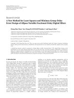
Figure 4 Long-term frequency changes for the T
CM
consensus phenotype, CCR7+CD45RA-CD57-CD27+CD28+ (+–++) and two
associated supersets. (A) Plot illustrating the statistically significant increase in the T
CM
consensus phenotype frequency between PIVR and LTM
for all 7 patients. (B) The concomitant decrease between LTM and P2B for the frequency of the consensus T
CM
phenotype. (C) The longitudinal
expression profile for the T
CM
consensus phenotype showing LTM peaks for 4 of 7 patients; longitudinal profile for the CD45RA unspecified
superset, CCR7+CD57-CD27+CD28+ (+ ++), showing LTM peaks for 6 of 7 patients; and longitudinal profile for the CD45RA, CD27, and CD28
unspecified superset, CCR7+CD57- (+ ), also showing LTM peaks for 6 of 7 patients. Data suggests CD45RA, CD27, and CD28 may not be
obligate descriptors for central memory T cells.
Siebert et al. Journal of Translational Medicine 2010, 8:106
/>Page 9 of 15
Additionally, we defined a process control range,
based on analysis of 6 aliquots from a single animal
drawn at a single point in time. For each phenotype, the
process control range w as defined as the maximum fre-
quency value of the 6 replicates minus the minimum
frequency v alue. This provided a conservative approach
to quantifying the precision of our assay, and allowed us
to focus on phenotypes with readouts exceeding the
process control range.
Next, to identify populations of numeric interest, we
identified sets in which 6 or more (out of 8) wounded
animals had an av erage delta greater than the process
control range, and 6 o r more control animals had an
average delta less than or equal to the process control
range. The resulting 122 se ts (0.4% of the total 32,805
sets) came from three of the five panels, with two panels
having no sets that matched these criteria. Of the 122
sets, 76 had p-values (Wilcoxon rank sum, one-sided)
less than 0.05. Twenty-three of these 122 phenotypes
were positive for CD29 (b1-integrin) and CXCR4, which
are indicative of mus cle progenitor cells in mouse mod-
els [25,37]. All of these CD29+CXCR4+ sets were f rom
the CD31 panel. Initially, none of these sets showed sta-
tistically significant d ifferences between wounded and
control populations, due at least in part to the presence
of an outlier in the control group, as shown by the scat-
ter plots in Figure 5A. This outlier was driven by an
unusually large observation for one of the donors, which
in the case of the CD29+CD31+CD56+CXCR4+CD90
+Sca1-CD44+ (++++.+-+) phenotype was an extreme
outlier (greater than quartile 3 plus 3 times the inter-
quartile range), and nearly twice as large as the next lar-
gest observation (.31% versus .17%). This outlier
observation from week 4 for control animal C-P1120 is
illustrated in Figure 5D. When this animal was removed
from the analysis, all 23 of the CD29+CXCR4+ pheno-
types showed statistically significant differences between
the control and wounded animals. Two of these pheno-
types are sho wn in Figures 5B and 5C. Figure 5B shows
thesamephenotypeasFigure5A,onlywiththeoutlier
removed. As the scatter plot shows on e point per donor
it better illustrates the details of t he data t han does a
bar plot or box plot. Additionally, Figures 5A, B, and 5C
have a reference line indicating t he process control
range. The 2 3 CD29+CXCR4+ phenotypes, it emized in
Table 3, may represent different bone-marrow-derived
mesenchymal progenitor cell populations mobilized in
response to myonecrotic injury and capable of endothe-
lial, chondrogenic, and myogenic differentiation. Nota-
bly, the superset CD29+CXCR4+CD90+ (Figure 5C) is
common to 19 of t he phenotypes in Table 4. As such it
may indicate a minimum o bligate progenitor cell
phenotype.
Discussion
Here we have applied Exhaustive Expansion to two very
different translational studies to demonstrate its broad
application and utility. In each analysis, we generated all
possible cell sets for each sample. Then we identified
interesting sets based on coef ficients of variation and
long term memory peaks in the melanoma vaccine
study, and separation between test and control cohorts
in the wound healing study.
Analysis of data from multiparameter flow cytometry
experiments consists of two main activities with well
defined separation of con cerns. First, events are gated
into cell sets of interest using either manual or auto-
matic techniques. Second, summary statistics describing
these sets of cells are analyzed to identify m eaningful
experimental results. Exhaustive Expansion touches on
both of th ese activities. In the case wher e positive/nega-
tive boundaries can be established for multiple markers,
our Expander logic allows us to define a large number
of supersets by exhaustively combining constituent sub-
sets. Next, we identify features of interest such as Aver-
age CV, peaks, and separatio n be tween control and test
cohorts. Such numeric features can be sorted and fil-
tered, and illustrated with simple graphs. Importantly,
these features are calculated for all phenotypes, thereby
allowing systematic and relatively unbiased interrogation
of the data. Additionally, the use of powerful mature
software tools suc h as J ava, MySQL, and R provides us
with the flexibility to pursue the data analysis as sug-
gested by the data itself and the underlying science.
For example, while we used a statistical test to quan-
tify peaks in the melanoma study, we could have defined
peaks based on an average fold change between time
points (e.g. gre ater than 3), or on a criteria such as at
least 4 donors showing at least a 5 percentage point
change between time points. Alternatively, we could
identify all phenotypes with a larger change than that
shown by a predicted consensus phenotype. Or if we
were interested in rare events, we could select sets in
which less than 2 cells at baseline expanded to more
than 20 cells after treatment. When a filter identifies
many sets, the filter can be made more stri ngent. Alter-
natively, filters can identify a specific number or percen-
tage of sets, such as the 10 sets with the largest average
fold changes between two time points. Additionally, sets
can be sorted on numeric characteristics such as fold
change,p-value,orAverageCV.Thisallowsusto
inspe ct sets ranked from largest to smallest fold change,
for example, and perhaps further refine a threshold cri-
teria based on some meaningful feature in the data. All
of these numeric thresholds can and should be adjusted
based on experimental conditions, assay precision, and
the biological questions under investigation.
Siebert et al. Journal of Translational Medicine 2010, 8:106
/>Page 10 of 15
Adoptive transfer of tumor specific T cells in cancer
immunotherap y translational studies has previously
emphasized the transfer of highly differentiated, end
stage effector T cells from in vitro IL-2 supported
expansion cultures. More recently, compelling data from
mouse tumor models suggests that tumor specific T
CM
and very early T
CM
precursors, referred to as central
memory stem cells (T
SCM
), express elevated proliferation
potential, enhanced long term survival in vivo,andgive
rise to activated CTLs in vivo with superior cytolytic
activity compared to effector memory (T
EM
) or effector
(T
EFF
)Tcellsfromin vitro expansion cultures [9].
Adoptive transfer immunotherapy strategies based on
the in vitro expansion of T
CM
and T
SCM
subpopulat ions
may offer significa nt clinical advantage in treating can-
cer patien ts if the human phenotype signatur es for T
CM
and T
SCM
can be identified, and rapid efficient recovery
procedures are d eveloped to recover memory cells for
subsequent in vitro expansion [38-40].
Previously, in a clinical study of long term tumor spe-
cific T cell memory function in melanoma patients, we
elucidated the multiparameter phenotype of tumor spe-
cific T
CM
(CCR7+CD45RA-CD57-CD27+CD28+), and a
second potentially early T
CM
precursor which we
referred to as T
CMRA
(CCR7+CD45RA+CD57-CD27
+CD28+) [8]. Gp100-specific T
CMRA
shares its pheno-
type with naïve CD8
+
T cells, and thus may be similar
to the T
SCM
subset described in the mouse. Sorting
Figure 5 Differences between control and wounded animals for 2 phenotypes from the CD31 panel. (A) Average frequency change from
baseline (average of frequency differences for week 1 minus week 0, week 2 minus week 0, week 3 minus week 0, and week 4 minus week 0) is
shown for control animals (solid circles) versus wounded animals (open circles) for phenotype CD29+CD31+CD56+CXCR4+CD90+Sca1-CD44+ (+
+++.+-+). The horizontal line represents the process control range (maximum frequency minus minimum frequency, calculated from 6
replicate samples) for this phenotype. There is no significant difference between the cohorts, due in part to the outlier at approximately 0.115
for one animal in the control cohort. (B) The same phenotype analysis with outlier removed shows a statistically significant difference between
wounded and control cohorts. (C) Frequency differences between wounded and control animals for the phenotype superset, CD29+CXCR4
+CD90+ (+ +.+ ), which was common to 19 of the putative myogenic precursor phenotypes shown in Table 4. (D) Longitudinal profiles for
all animals for week 0 through week 4 for set CD29+CD31+CD56+CXCR4+CD90+Sca1-CD44+ (++++.+-+). Control animals indicated by C,
Wounded by W. Note the week 4 outlier on control animal C-P1120. This animal was removed from the analysis shown in (B) and (C).
Siebert et al. Journal of Translational Medicine 2010, 8:106
/>Page 11 of 15
strategies to select for these highly defined putative cen-
tral memory populations could thus be implemented
prior to cytok ine-mediated in vitro expansion and adop-
tive transfer. However, recovery strategies based on a
more simple minimal obligate phenotype signature
would facilitate the more rapid, efficient recovery of lar-
ger numbers of cells using bulk techniques such as mag-
netic bead s eparation. Exhaustive Expansion identified a
possible minimal obligate T
CM
/T
CMRA
phenotype (CCR7
+CD57-: Figure 4) that was common to 7/8 of the
CCR7+ CD45RA-CD57-CD27+CD28+ supersets that
showed frequency peaks at LTM (Figure 3). This puta-
tive minimal obligate T
CM
/T
CMRA
phenotype signature
may thus facilitate the recovery of T
CM
/T
CMRA
Tcells,
and cells from the intermediate stages o f the T
CMRA
to
T
CM
to T
EM
differentiation pathway represented by the
other superset phenotypes in Figure 3. Clearly, addi-
tional experiments, including functional assays, are
required to validate the hypothesis that CCR7+CD57- is
a minimal obligate phenotype for T
CM
.
A second somewhat unexpected outcome of Exhaus-
tive Expansion of the melanoma specific CD8
+
Tcell
memory response was the suggestion that the combined
frequency of tumor-specifi c T cells which express either
the T
CM
or T
EM
phenotypes may not change appreciably
over the course of the primary antigen challenge, long
term memory maintenance, and following boosting
immunization. The frequencies of gp100 specific T cells
expressing key individual identifiers for the resolution of
T
CM
and early T
EM
cells, such as CD45RA, CD27 an d
CD28, did not change appreciably across all three ti me
points in the study (Figure 2). This may be explained in
part by the observation that T
CM
and T
EM
phenotypes
share the CD45RA-CD27+CD28+ signature [8,35,36].
The expression stability for e ach individual marker may
suggest that, although cells may transition between the
T
CM
and T
EM
phenotype compartments due to homeos-
tasis-driven or antigen-stimulated proliferation, the over-
all combined frequency of the T
CM
plus T
EM
memory T
cell pool as a fraction of all antigen specific T cells
remains relatively constant. Thus, absolute numbers of
cells in each compartment, and even the ratio of the fre-
quency of cells with each phenotype, can fluctuate; but
the total combined memory T cell frequency (i.e. T
CM
+
T
EM
) may remain relatively stable after primary immuni-
zation. This observation has important implications for
Table 4 23 CD29+CXCR4+ subsets showing significant differences between wounded and control animals
Panel Relative Set Name Absolute Set Name P-Value
CD31 ++++.+-+ CD29+CD31+CD56+CXCR4+CD90+Sca1-CD44+ 0.027
CD31 ++++.+ CD29+CD31+CD56+CXCR4+CD90+Sca1- 0.027
CD31 ++.+-+-+ CD29+CD31+CXCR4+CD105-CD90+Sca1-CD44+ 0.036
CD31 ++.+-+ CD29+CD31+CXCR4+CD105-CD90+Sca1- 0.036
CD31 ++.+ + CD29+CD31+CXCR4+CD105-Sca1-CD44+ 0.027
CD31 ++.+ CD29+CD31+CXCR4+CD105-Sca1- 0.028
CD31 ++.+.+-+ CD29+CD31+CXCR4+CD90+Sca1-CD44+ 0.027
CD31 ++.+.+ CD29+CD31+CXCR4+CD90+Sca1- 0.027
CD31 ++.+.+.+ CD29+CD31+CXCR4+CD90+CD44+ 0.02
CD31 ++.+.+ CD29+CD31+CXCR4+CD90+ 0.02
CD31 +-++—. CD29+CD31-CD56+CXCR4+CD105-CD90-Sca1- 0.027
CD31 +.++-+-+ CD29+CD56+CXCR4+CD105-CD90+Sca1-CD44+ 0.02
CD31 +.++-+ CD29+CD56+CXCR4+CD105-CD90+Sca1- 0.02
CD31 +.++-+.+ CD29+CD56+CXCR4+CD105-CD90+CD44+ 0.02
CD31 +.++-+ CD29+CD56+CXCR4+CD105-CD90+ 0.02
CD31 +.++.+-+ CD29+CD56+CXCR4+CD90+Sca1-CD44+ 0.02
CD31 +.++.+ CD29+CD56+CXCR4+CD90+Sca1- 0.02
CD31 +.++.+.+ CD29+CD56+CXCR4+CD90+CD44+ 0.02
CD31 +.++.+ CD29+CD56+CXCR4+CD90+ 0.02
CD31 + +-+.+ CD29+CXCR4+CD105-CD90+CD44+ 0.014
CD31 + +-+ CD29+CXCR4+CD105-CD90+ 0.014
CD31 + +.+.+ CD29+CXCR4+CD90+CD44+ 0.014
CD31 + +.+ CD29+CXCR4+CD90+ 0.014
Relative set name, absolute set name, and p-value (Wilcoxon rank sum, one-sided) are shown. P-values are calculated excluding data for one outlier control
animal. These are also sets in which at least 6 of 8 wounded animals show average delta readouts greater than the process control range.
Siebert et al. Journal of Translational Medicine 2010, 8:106
/>Page 12 of 15
the optimal design of primary immunization strategies
in both infectious disease and cancer vaccine settings.
In the stem cell study, 8 color s taining panels that
included mAbs previously employed in lower-order
panels to delineate mesenchymal cells (CD29, CD90,
and CD44), primitive pluripotent stem cells (ckit,
CXCR4, and Sca-1), differentiated myoblasts (CD56 and
CXCR4), and vascular-relative cells (CD146, CD31,
CD144, CD105, and VEGFR2) were used to more com-
prehensively characterize significant changes in bon e-
morrow-derived putative mesenchymal progenitor cell
populations following myonecrotic injury. Our data ana-
lysis technique allowed us to identify novel populations
by focusing on phenotypes that showed both statistically
significant differences between wounded and control
animals and credible readouts above the process control
range.
Studies have demonstrated that injection of bone mar-
row stem cells into ischemic muscle can reduce the
damage to the muscle and the loss of muscle function
[17]. Bone marrow contains stem and progenitor cells
which can diffe rentiate into specific cell types such as
myoblasts, chondrocytes, and endothelial cells in vitro
and in vivo [41]. The role o f bone-marrow-derived
mesenchymal stem cells (MSCs) to directly reconstitute
myoblast formation in vivo in damaged muscle is con-
troversial since their main role may be that of augment-
ing the myogenic potential of resident muscle MSCs
referred to as satellite cells [42]. In vitro, bone marrow
cells acquire tissue-specific phenotypes when co-cul-
tured with specialized cell types or tissue-derived
extracts [41]. The se potentially multipotent cells may be
mobilized in the bone marrow and recruited into muscle
tissue where they mitigate tissue damage following acute
myonecrotic injury. Our res ults show that cell surface
markers can be used to comprehensively track bone
marrow phenotype changes associated with muscle
injury in porcine compartment syndrome, which are sig-
nificantly different between the control and wounded
groups. Moreover, our results demonstrate that we can
detect multiple putative stem and progenitor pheno-
types. The large majority of these 23 phenotype subpo-
pulations (20/23) appear to share a common minimum
obligate phenotype signa tur e (e.g. CD29+CXCR4+CD90
+: Table 4), expressing markers reported to be charac-
teristic of MSC-derived myogenic cells [25,37,43]. How-
ever, there may alr eady be lineage-specific heterogeneity
expressed by these MSC-like subpopulations in the bone
marrow, since approximately half (10/23) expressed the
endothelial differentiation marker CD31 [44] and an
equal number (11/23) expressed the CD56 marker more
commonly associated with re generating muscle fibers
and satellite cells[45]. Lineage-specific commitment can
be tested by culturing such sorted MSC subsets under
lineage-promoting cult ure conditions [41]. Based on the
results presented here, the identification of bone marrow
subpopulations by multiparameter FCM might be used
to further sort or purify cell sets for autologous cell
therapy to regenerate muscle, nerve and vascular tissues
in compartment syndrome or other extremity injuries.
There are limitations to this work. First, from a biolo-
gical perspective, both studies were performed with a
small number of subjects. Additional experiments,
including correlated memory T cell and MSC functional
assays, are n eeded to valida te the hypothe ses generated
by this work. Second, from an assay perspective, the
analytical approach describedheremorereadilysup-
ports those circumstances where orthogonal boundary
gates (e.g. positive and nega tive regions) can be estab-
lished. Third, from a process control perspective, the
process control samples used to identify phenotypes of
interest were analyze d on three consecutive days. Con-
trols analyzed over the duration of th e study would
more accurately calibrate the precision of the assay.
Fourth, from a computational perspective, the re are
practical limits to the scalability of the algorithm. Apply-
ing Exhaustive Expansion to an experiment in which
there were 10 variable m arkers would result in a man-
ageable 3
10
= 59,049 possib le phenotypes, while 20 vari-
able markers would result in a challenging 3
20
=
3,486,784,401 possible phenotypes.
While there is no way to alter the exponential increase
in number of phenotypes as a function of the number of
markers, it is unlikely that mill ions or billions of pheno-
types would be meaningful, whether due to experimen-
tal noise (e.g. too few events to be adequately precise)
or underlying biology. Thus, the phenotype search space
would b e pruned to a more reasonable number of phe-
notypes. Specific strategies for pruning the search space
are beyond the scope of this work, but the general
approach would mitigate the scalability impacts of the
exponential increase, further extending the applicability
of Exhaustive Expansion.
Furthermore, Exhaustive Expansion adds immediate
value to contemporary experimental strategies and paves
the way for the practical use of increasing numbers of
markers. For example, one experimental design com-
monly published in contemporary literature uses a single
fluorophore marker dump channel to exclude certain
cells (e.g. CD14+, CD19+ and dead cells), two markers to
identify lineage of interest (e.g. CD3 and CD4 or CD8),
and another 5 markers to identify functional sets of inter-
est (CD107a, IFN-g, IL-2, MIP1b, and TNF-a) [31,32,46].
Using this ex perimental approach, 3 of the 8 total fluoro-
phores are required to identify the parent population,
while the other 5 can be considered variable identifiers of
subphenotypes of interest. This construct leads to 31 sets
of interest (2
5
- 1, since the universal set is excluded). In
Siebert et al. Journal of Translational Medicine 2010, 8:106
/>Page 13 of 15
comparison, we have demonstrated that we can analyze
over 32,000 sets, generated by 5 different panels of 8 vari-
able markers. Additionally our approach recognizes that
potential sets of interest are both those defined by all
variable markers, and those defined by subsets of variable
markers. Thus, our approach is readily applicable to con-
temporary flow cytometry experimental strategies, pro-
viding both support for an increasing number of variable
markers and exhaustive interrogation of phenotypes
defined by combinations of these markers.
Conclusions
Inconclusion,wehavedemonstratedthatExhaustive
Expansion is a valuable technique for analyzing h igher
order polychromatic FCM data sets. Exhaustive Expan-
sion consists of:
• generating data for all possible 0- to N-parameter
sets;
• creating appropriate data visualizations;
• identifying numerically interesting sets, using such
metrics as CVs and p-values; and
• inspecting the numerically interesting sets for cor-
relati ve analysis of clinically or biologically meaning-
ful results.
This approach allows us to screen hundreds to thou-
sands of phenotypes for biological responses. Use of
free, widely available, and mature software components
gives us the flexibility to pursue the data analysis in
directions indicated by the data itself and the associated
science. Our techniques are s traightforward, yet high-
light intriguing results when executed exhaustively
across the entire data space. They support inductive rea-
soning by highlighting all cell subpopulations t hat meet
appropriate numerical criteria. In both studies discussed
here, o ur analysis provided the foundation for a refined
understanding o f complex phenotypes, and allowed for
the development of new hypotheses pertaining to the
identification and recovery of potentially important
myogenic MSC progenitor cells, and tumor antigen-spe-
cific CD8
+
T
CM
and T
CM
precursor populations for
future clinical studies.
Acknowledgements
Funding support was received from NIH (1R21-CS82614-01 and RA21-
CA099265-02), the M. J. Murdock Charitable Trust, and the Chiles
Foundations.
Author details
1
CytoAnalytics, Denver, CO, USA.
2
Oregon Medical Laser Center, Providence
St. Vincent Medical Center, Portland, OR, USA.
3
Robert W Franz Cancer
Research Center, Earle A. Chiles Research Institute, Providence Cancer Center,
Portland, OR, USA.
Authors’ contributions
KWG and EBW designed the research. LW, DPH, and AR performed the
research. JCS and WM contributed vital analytical tools. JCS, LW, AR, BZ, and
EBW analyzed and interpreted the data. JCS and EBW wrote the manuscript.
All authors have read and approved the final manuscript.
Competing interests
JCS is Founder and President of CytoAnalytics. WM is Chief Technology
Officer of CytoAnalytics.
Received: 29 April 2010 Accepted: 30 October 2010
Published: 30 October 2010
References
1. Perfetto SP, Chattopadhyay PK, Roederer M: Seventeen-colour flow
cytometry: unravelling the immune system. Nat Rev Immunol 2004,
4:648-655.
2. Pyne S, Hu X, Wang K, Rossin E, Lin T, Maier LM, Baecher-Allan C,
McLachlan GJ, Tamayo P, Hafler DA, De Jager PL, Mesirov JP: Automated
high-dimensional flow cytometric data analysis. Proc Natl Acad Sci USA
2009, 106:8519-8524.
3. Lo K, Brinkman RR, Gottardo R: Automated gating of flow cytometry data
via robust model-based clustering. Cytometry A 2008, 73:321-332.
4. Murphy RF: Automated identification of subpopulations in flow
cytometric list mode data using cluster analysis. Cytometry 1985,
6:302-309.
5. Marincola FM: In support of descriptive studies; relevance to translational
research. J Transl Med 2007, 5:21.
6. Valet G: Cytomics as a new potential for drug discovery. Drug Discov
Today 2006, 11:785-791.
7. Valet G, Leary JF, Tárnok A: Cytomics–new technologies: towards a
human cytome project. Cytometry A 2004, 59:167-171.
8. Walker EB, Haley D, Petrausch U, Floyd K, Miller W, Sanjuan N, Alvord G,
Fox BA, Urba WJ: Phenotype and functional characterization of long-term
gp100-specific memory CD8+ T cells in disease-free melanoma patients
before and after boosting immunization. Clin Cancer Res 2008,
14:5270-5283.
9. Gattinoni L, Zhong X, Palmer DC, Ji Y, Hinrichs CS, Yu Z, Wrzesinski C,
Boni A, Cassard L, Garvin LM, Paulos CM, Muranski P, Restifo NP: Wnt
signaling arrests effector T cell differentiation and generates CD8+
memory stem cells. Nat Med 2009, 15:808-813.
10. Berger C, Jensen MC, Lansdorp PM, Gough M, Elliott C, Riddell SR: Adoptive
transfer of effector CD8+ T cells derived from central memory cells
establishes persistent T cell memory in primates. J Clin Invest 2008,
118:294-305.
11. Gattinoni L, Powell DJ, Rosenberg SA, Restifo NP: Adoptive
immunotherapy for cancer: building on success. Nat Rev Immunol 2006,
6:383-393.
12. Hassan HT, El-Sheemy M: Adult bone-marrow stem cells and their
potential in medicine. J R Soc Med 2004, 97:465-471.
13. Gourgiotis S, Villias C, Germanos S, Foukas A, Ridolfini MP: Acute limb
compartment syndrome: a review. J Surg Educ 2007, 64:178-186.
14. Ferrari G, Cusella-De Angelis G, Coletta M, Paolucci E, Stornaiuolo A,
Cossu G, Mavilio F: Muscle regeneration by bone marrow-derived
myogenic progenitors.
Science 1998, 279:1528-1530.
15. Fukada S, Miyagoe-Suzuki Y, Tsukihara H, Yuasa K, Higuchi S, Ono S,
Tsujikawa K, Takeda S, Yamamoto H: Muscle regeneration by
reconstitution with bone marrow or fetal liver cells from green
fluorescent protein-gene transgenic mice. J Cell Sci 2002, 115:1285-1293.
16. Corbel SY, Lee A, Yi L, Duenas J, Brazelton TR, Blau HM, Rossi FMV:
Contribution of hematopoietic stem cells to skeletal muscle. Nat Med
2003, 9:1528-1532.
17. Umemura T, Nishioka K, Igarashi A, Kato Y, Ochi M, Chayama K,
Yoshizumi M, Higashi Y: Autologous bone marrow mononuclear cell
implantation induces angiogenesis and bone regeneration in a patient
with compartment syndrome. Circ J 2006, 70:1362-1364.
18. Tateishi-Yuyama E, Matsubara H, Murohara T, Ikeda U, Shintani S, Masaki H,
Amano K, Kishimoto Y, Yoshimoto K, Akashi H, Shimada K, Iwasaka T,
Imaizumi T: Therapeutic angiogenesis for patients with limb ischaemia
by autologous transplantation of bone-marrow cells: a pilot study and a
randomised controlled trial. Lancet 2002, 360:427-435.
Siebert et al. Journal of Translational Medicine 2010, 8:106
/>Page 14 of 15
19. Herrera MB, Bruno S, Buttiglieri S, Tetta C, Gatti S, Deregibus MC, Bussolati B,
Camussi G: Isolation and characterization of a stem cell population from
adult human liver. Stem Cells 2006, 24:2840-2850.
20. Dicker A, Le Blanc K, Aström G, van Harmelen V, Götherström C,
Blomqvist L, Arner P, Rydén M: Functional studies of mesenchymal stem
cells derived from adult human adipose tissue. Exp Cell Res 2005,
308:283-290.
21. Wilson A, Oser GM, Jaworski M, Blanco-Bose WE, Laurenti E, Adolphe C,
Essers MA, Macdonald HR, Trumpp A: Dormant and self-renewing
hematopoietic stem cells and their niches. Ann N Y Acad Sci 2007,
1106:64-75.
22. Pitchford SC, Furze RC, Jones CP, Wengner AM, Rankin SM: Differential
mobilization of subsets of progenitor cells from the bone marrow. Cell
Stem Cell 2009, 4:62-72.
23. Miller RJ, Banisadr G, Bhattacharyya BJ: CXCR4 signaling in the regulation
of stem cell migration and development. J Neuroimmunol 2008,
198:31-38.
24. Zheng B, Cao B, Crisan M, Sun B, Li G, Logar A, Yap S, Pollett JB, Drowley L,
Cassino T, Gharaibeh B, Deasy BM, Huard J, Péault B: Prospective
identification of myogenic endothelial cells in human skeletal muscle.
Nat Biotechnol 2007, 25:1025-1034.
25. Cerletti M, Jurga S, Witczak CA, Hirshman MF, Shadrach JL, Goodyear LJ,
Wagers AJ: Highly efficient, functional engraftment of skeletal muscle
stem cells in dystrophic muscles. Cell 2008, 134:37-47.
26. Crisan M, Yap S, Casteilla L, Chen C, Corselli M, Park TS, Andriolo G, Sun B,
Zheng B, Zhang L, Norotte C, Teng P, Traas J, Schugar R, Deasy BM,
Badylak S, Buhring H, Giacobino J, Lazzari L, Huard J, Péault B: A
perivascular origin for mesenchymal stem cells in multiple human
organs. Cell Stem Cell 2008, 3:301-313.
27. Middleton J, Americh L, Gayon R, Julien D, Mansat M, Mansat P, Anract P,
Cantagrel A, Cattan P, Reimund J, Aguilar L, Amalric F, Girard J: A
comparative study of endothelial cell markers expressed in chronically
inflamed human tissues: MECA-79, Duffy antigen receptor for
chemokines, von Willebrand factor, CD31, CD34, CD105 and CD146. J
Pathol 2005, 206:260-268.
28. Ingram DA, Mead LE, Tanaka H, Meade V, Fenoglio A, Mortell K, Pollok K,
Ferkowicz MJ, Gilley D, Yoder MC: Identification of a novel hierarchy of
endothelial progenitor cells using human peripheral and umbilical cord
blood. Blood 2004, 104:2752-2760.
29. Garlanda C, Dejana E: Heterogeneity of endothelial cells. Specific markers.
Arterioscler Thromb Vasc Biol 1997, 17:1193-1202.
30. Lugli E, Pinti M, Nasi M, Troiano L, Ferraresi R, Mussi C, Salvioli G, Patsekin V,
Robinson JP, Durante C, Cocchi M, Cossarizza A: Subject classification
obtained by cluster analysis and principal component analysis applied
to flow cytometric data. Cytometry A 2007, 71:334-344.
31. Casazza JP, Betts MR, Price DA, Precopio ML, Ruff LE, Brenchley JM, Hill BJ,
Roederer M, Douek DC, Koup RA: Acquisition of direct antiviral effector
functions by CMV-specific CD4+ T lymphocytes with cellular maturation.
J Exp Med 2006, 203
:2865-2877.
32. Betts MR, Nason MC, West SM, De Rosa SC, Migueles SA, Abraham J,
Lederman MM, Benito JM, Goepfert PA, Connors M, Roederer M, Koup RA:
HIV nonprogressors preferentially maintain highly functional HIV-specific
CD8+ T cells. Blood 2006, 107:4781-4789.
33. Smith JW, Walker EB, Fox BA, Haley D, Wisner KP, Doran T, Fisher B,
Justice L, Wood W, Vetto J, Maecker H, Dols A, Meijer S, Hu H, Romero P,
Alvord WG, Urba WJ: Adjuvant Immunization of HLA-A2-Positive
Melanoma Patients With a Modified gp100 Peptide Induces Peptide-
Specific CD8+ T-Cell Responses. J Clin Oncol 2003, 21:1562-1573.
34. Swindle MM: Swine in the laboratory CRC Press; 2007.
35. Romero P, Zippelius A, Kurth I, Pittet MJ, Touvrey C, Iancu EM, Corthesy P,
Devevre E, Speiser DE, Rufer N: Four functionally distinct populations of
human effector-memory CD8+ T lymphocytes. J Immunol 2007,
178:4112-4119.
36. Takata H, Takiguchi M: Three memory subsets of human CD8+ T cells
differently expressing three cytolytic effector molecules. J Immunol 2006,
177:4330-4340.
37. Perez AL, Bachrach E, Illigens BMW, Jun SJ, Bagden E, Steffen L, Flint A,
McGowan FX, Del Nido P, Montecino-Rodriguez E, Tidball JG, Kunkel LM:
CXCR4 enhances engraftment of muscle progenitor cells. Muscle Nerve
2009, 40:562-572.
38. Palmer DC, Restifo NP: Suppressors of cytokine signaling (SOCS) in T cell
differentiation, maturation, and function. Trends Immunol 2009,
30:592-602.
39. Geginat J, Sallusto F, Lanzavecchia A: Cytokine-driven proliferation and
differentiation of human naive, central memory, and effector memory
CD4(+) T cells. J Exp Med 2001, 194:1711-1719.
40. Klebanoff CA, Gattinoni L, Torabi-Parizi P, Kerstann K, Cardones AR,
Finkelstein SE, Palmer DC, Antony PA, Hwang ST, Rosenberg SA,
Waldmann TA, Restifo NP: Central memory self/tumor-reactive CD8+ T
cells confer superior antitumor immunity compared with effector
memory T cells. Proc Natl Acad Sci USA 2005, 102:9571-9576.
41. da Silva Meirelles L, Chagastelles PC, Nardi NB: Mesenchymal stem cells
reside in virtually all post-natal organs and tissues. J Cell Sci 2006,
119:2204-2213.
42. Sherwood RI, Christensen JL, Conboy IM, Conboy MJ, Rando TA,
Weissman IL, Wagers AJ: Isolation of adult mouse myogenic progenitors:
functional heterogeneity of cells within and engrafting skeletal muscle.
Cell 2004, 119:543-554.
43. Zuk PA, Zhu M, Ashjian P, De Ugarte DA, Huang JI, Mizuno H, Alfonso ZC,
Fraser JK, Benhaim P, Hedrick MH: Human adipose tissue is a source of
multipotent stem cells. Mol Biol Cell 2002, 13:4279-4295.
44. Uezumi A, Ojima K, Fukada S, Ikemoto M, Masuda S, Miyagoe-Suzuki Y,
Takeda S: Functional heterogeneity of side population cells in skeletal
muscle. Biochem Biophys Res Commun 2006, 341:864-873.
45. Illa I, Leon-Monzon M, Dalakas MC: Regenerating and denervated human
muscle fibers and satellite cells express neural cell adhesion molecule
recognized by monoclonal antibodies to natural killer cells. Ann Neurol
1992, 31
:46-52.
46. Precopio ML, Betts MR, Parrino J, Price DA, Gostick E, Ambrozak DR,
Asher TE, Douek DC, Harari A, Pantaleo G, Bailer R, Graham BS, Roederer M,
Koup RA: Immunization with vaccinia virus induces polyfunctional and
phenotypically distinctive CD8(+) T cell responses. J Exp Med 2007,
204:1405-1416.
doi:10.1186/1479-5876-8-106
Cite this article as: Siebert et al.: Exhaustive expansion: A novel
technique for analyzing complex data generated by higher-order
polychromatic flow cytometry experiments. Journal of Translational
Medicine 2010 8:106.
Submit your next manuscript to BioMed Central
and take full advantage of:
• Convenient online submission
• Thorough peer review
• No space constraints or color figure charges
• Immediate publication on acceptance
• Inclusion in PubMed, CAS, Scopus and Google Scholar
• Research which is freely available for redistribution
Submit your manuscript at
www.biomedcentral.com/submit
Siebert et al. Journal of Translational Medicine 2010, 8:106
/>Page 15 of 15