Báo cáo sinh học: " Virus detection and identification using random multiplex (RT)-PCR with 3''''-locked random primers" docx
Bạn đang xem bản rút gọn của tài liệu. Xem và tải ngay bản đầy đủ của tài liệu tại đây (775.55 KB, 11 trang )
BioMed Central
Page 1 of 11
(page number not for citation purposes)
Virology Journal
Open Access
Research
Virus detection and identification using random multiplex
(RT)-PCR with 3'-locked random primers
Amy L Clem, Jonathan Sims, Sucheta Telang, John W Eaton and
Jason Chesney*
Address: Molecular Targets Program, Medical Oncology, J.G. Brown Cancer Center, University of Louisville, Kentucky, USA
Email: Amy L Clem - ; Jonathan Sims - ;
Sucheta Telang - ; John W Eaton - ; Jason Chesney* -
* Corresponding author
Abstract
Background: PCR-based detection and identification of viruses assumes a known, relatively stable
genome. Unfortunately, high mutation rates may lead to extensive changes in viral nucleic acid
sequences making dedicated PCR primer use problematic. Furthermore, in bioterrorism, viral
consensus sequences can be genetically modified as a countermeasure to RT-PCR and DNA chip
detection. Accordingly, there is a great need for the development of rapid and universal virus
detection and identification technologies.
Results: We report herein that viral genomic DNA or RNA can be separated from host nucleic
acids in plasma by filtration and nuclease digestion, and randomly amplified in a single PCR using a
mixture of primers designed to be resistant to primer-dimer amplification (5'-VVVVVVVVAA-3', V
= A, G or C; 3
8
or 6561 primers). We have termed this novel PCR method Random Multiplex (RT)-
PCR since hundreds of overlapping PCR amplifications occur simultaneously. Using this method,
we have successfullydetected and partially sequenced 3 separate viruses in human plasma without
using virus-specific reagents (i.e., Adenovirus Type 17, Coxsackievirus A7, and Respiratory Syncytial
Virus B). The method is sensitive to ~1000 genome equivalents/ml and may represent the fastest
means of detection of unknown viruses.
Conclusion: These studies suggest that the further development of random multiplex (RT)-PCR
may lead to a diagnostic assay that can universally detect viruses in donated blood products as well
as in patients suffering with idiopathic disease states of possible viral etiology.
Background
Relatively benign viruses can be converted into highly vir-
ulent viruses via the introduction of genes of interest. For
example, Ectromelia virus, a natural pathogen of mice
that causes mousepox, recently was recombined with
interleukin-4 as part of an effort to develop a live virus
immuno-contraceptive vaccine. Surprisingly, the recom-
bined virus caused 60% mortality in 2 strains of mice,
whereas the wild type virus caused no death [1]. A credible
bioterrorism scenario might entail the release of such a
recombined or chimeric virus tailored for maximum
infectivity and pathogenicity but not readily detectable
using our current "state-of-the-art" diagnostics (i.e., PCR
and micro-array chips.) Accordingly, there is a need for
Published: 28 June 2007
Virology Journal 2007, 4:65 doi:10.1186/1743-422X-4-65
Received: 27 April 2007
Accepted: 28 June 2007
This article is available from: />© 2007 Clem et al; licensee BioMed Central Ltd.
This is an Open Access article distributed under the terms of the Creative Commons Attribution License ( />),
which permits unrestricted use, distribution, and reproduction in any medium, provided the original work is properly cited.
Virology Journal 2007, 4:65 />Page 2 of 11
(page number not for citation purposes)
methods that can identify unknown viral pathogens and
which can reveal extensive genomic information. Such
methods would not only prove useful for our defense
against bioterrorism, but also would improve our capaci-
ties to identify and control outbreaks of naturally occur-
ring pathogenic viruses.
The proven technology to rapidly detect and identify
known human pathogens as potential causes of disease or
as bioweapons is PCR [2]. A recent example involved a
case of fatal yellow fever (YF) in a traveler returning from
Amazonas, Brazil in March, 2002 [3]. The otherwise
healthy 47-year-old male developed fever, headache, pan-
cytopenia, coagulopathy, renal and liver failure. Pan-cul-
tures were negative, and a peripheral smear yielded no
plasmodia. Serological tests (IgG, IgM) performed at the
CDC were negative for YF, dengue, St. Louis encephalitis,
and other agents, but serum specimens (unfortunately
examined only post-mortem) were positive for YF viral
RNA as measured by RT-PCR [3]. Subsequent attempts to
isolate or culture the virus failed. This example highlights
the superiority of PCR over other currently available
methods. DNA chips that allow the simultaneous meas-
urement of literally thousands of genes through hybridi-
zation are now being developed as the next-generation
rapid diagnostic test for all known human pathogens [4].
However, both of these technologies rely on a relatively
stable genome, and several human pathogens display a
high mutation rate (e.g., HIV 2 × 10
-5
/base, 9 kB genome
[5]). Moreover, the ability to recombine "non-patho-
genic" viruses in vitro introduces the potential not only for
de novo pathogenicity but also for enhanced stealth. These
considerations suggest that it may be impossible to design
DNA micro-arrays which detect nucleic acids from all
known and unknown viruses, including less obvious vehi-
cles of bioterrorism such as adenovirus or rhinovirus
recombined with a single gene for enhanced virulence.
Accordingly, there is a great need for the development of
techniques that enable the universal amplification of viral
nucleic acids.
There are 2 strategies to amplify genetic sequences with
PCR without prior knowledge of the precise sequences.
The first strategy relies on the degenerate binding of arbi-
trarily chosen primers to sample multiple cDNAs or
genomic DNA species during PCR (two related methods
are known as differential display and RNA Arbitrarily
Primed PCR [RAP-PCR] [6-8]). The amplicons range in
size from ~50–600 bp and overlap. RAP-PCR was recently
used successfully to identify a novel human pneumovirus
only after the virus had been cultured [9]. These tech-
niques yield an amplicon "fingerprint" and are generally
used to compare two populations of nucleic acids.
Accordingly, the pneumovirus was identified by compar-
ing infected cells with non-infected cells [9]. The other
strategy is known as Sequence-Independent, Single-
Primer Amplification (SISPA) and involves the directional
ligation of a linker/adaptor oligonucleotide onto both
ends of a target population of either DS DNA or DS cDNA
(after reverse transcription from RNA) [10]. The PCR is
accomplished with primers specific for the linked adaptor
molecule. SISPA, followed by extensive immunoscreen-
ing, enabled the cloning of the Norwalk agent genome
from stool and Hepatitis G virus from plasma [11]. Unfor-
tunately, these techniques have not found great utility in
the direct discovery or identification of novel viruses from
human plasma or serum samples (unless accompanied by
immunoscreening) because of the relatively high level of
host genomic DNA. However, recent studies indicate that
DNAse treatment of serum samples can degrade most of
the host genomic DNA but not affect viral nucleic acids,
which are protected by stable capsids [12]. The combina-
tion of DNAse treatment with SISPA was successfully used
to identify and sequence in their entirety 2 novel parvovi-
ruses from bovine serum [12].
While the aforementioned PCR techniques are useful for
the discovery of novel viruses, their utility to rapidly detect
and identify unknown (or recombined) viruses is quite
limited. RAP-PCR, random RT-PCR and SISPA are compli-
cated multi-step procedures requiring infection of cul-
tured cells, days to accomplish, expensive reagents and
broad technical expertise. Accordingly, these methods
have not found clinical utility for the rapid detection or
identification of viral agents.
Here, we present a novel method for rapid virus detection
and identification based on random multiplex (RT)-PCR
using 3'-locked random primers to avoid primer-dimer
amplification. Once detected, virus amplification prod-
ucts can be shot-gun cloned and sequenced for identifica-
tion. This method may prove useful for the rapid
detection and identification of emerging or recombined
viruses.
Results
Filtration and Nuclease Treatment Improves Viral DNA
Amplification by Removing Host DNA and mRNA
We hypothesized that random amplification of viral
nucleic acids isolated from human plasma might provide
a powerful new method to detect and identify novel and
recombined viruses. Allander et al. recently found that
subjecting bovine plasma to filtration (to remove cells)
and DNAse treatment (to remove genomic DNA) was suf-
ficient to separate viral nucleic acids from bovine nucleic
acids and thus enable SISPA and cloning of novel viruses
[12]. The mechanism for this effect is related to the rela-
tive sensitivity of free-host DNA to DNAse treatment and
to the relative insensitivity of virus capsid-protected DNA
to DNAse treatment. We have found that human plasma
Virology Journal 2007, 4:65 />Page 3 of 11
(page number not for citation purposes)
is replete with free DNA and RNA species and thus opted
to examine the potential of DNAse and RNAse treatment
to isolate viral nucleic acids. We inoculated human
plasma with Adenovirus Type 17 and examined the effects
of filtration, DNAse treatment and RNAse treatment on
Adenovirus Type 17 DNA and on human β-actin genomic
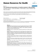
DNA and mRNA, using (RT)-PCR. In Figure 1, we demon-
strate that filtration, DNAse and RNAse treatment
increases the efficiency of amplification of Adenovirus
Type 17 DNA, whereas this combined treatment elimi-
nates amplification of genomic β-actin (upper band) and
β-actin cDNA (lower band). Although the addition of
RNAse alone has little effect on the β-actin mRNA, the
combination of DNAse and RNAse completely eliminates
β-actin mRNA. We postulate that this finding may be
related to the protection of RNA species by DNA:RNA
hybrids. Nevertheless, these data demonstrate that we are
able to remove host β-actin DNA and mRNA without
degrading Adenovirus Type 17 DNA.
5'-VVVVVVVVAA-3' Primers Enable Random Multiplex
Amplification of Plasmid DNA
We next designed a mixture of random primers that
would enable the priming and amplification of all DNA
or cDNA molecules, but that would be resistant to primer-
dimer amplification. We chose 10 base pairs as a compro-
mise length to simultaneously optimize hybridization
and possible binding sites. Taq polymerase requires that
the three 3' bases of a primer be 100% complementary in
order to enable efficient polymerization. We postulated
that "locking" the 3' end of the primers with 2 adenosines
and not incorporating any thymidines into the upstream
5' portion of the primers would prevent primer-dimer
amplification (i.e., 5'-VVVVVVVVAA-3'; where V = A, G or
C). This primer design will limit the frequency of binding
sites within a particular viral genome, but using the Edit-
Seq DNA Editing application (DNAStar), we observed
that primers within this mixture will bind every 200–500
bp in both orientations in the 19 viral genomes that were
examined, thus providing ample hybridization and
amplification potential.
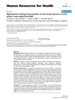
As a first test of these random primers we employed DNA
plasmids of known sequence. We found that this mixture
of primers enabled a continuum of discrete PCR products
to be amplified from 4 different circular DNA plasmids
(Figure 2A). We shot-gun cloned these PCR products and
confirmed that the amplified DNA had derived from the
source plasmids (data not shown). In contrast, primers con-
sisting of the sequence 5'-NNNNNNNNNN-3' (where N =
A, T, G, or C) did not enable the production of discrete or
clonable PCR amplicons (Figure 2A). We then examined
the ability of the V8AA primers to PCR amplify the plas-
mid, pBabe, and found that we could amplify as little as
100 fg (Figure 2B).
5'-VVVVVVVVAA-3' Primers Allow For the Random
Multiplex Amplification of DNA from Human Plasma
Inoculated with Adenovirus Type 17
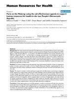
We inoculated 1 ml of human plasma with Adenovirus
Type 17. The plasma was then filtered (220 nm filter) and
incubated for 2 hours with nucleases. Remaining nucleic
acids were purified and PCR amplified using the 5'-
VVVVVVVVAA-3' primers. We found that this mixture of
primers enabled discrete PCR products to be amplified in
the human plasma that had been inoculated with Adeno-
virus Type 17 and treated with filtration, DNAse and
RNase treatment (Figure 3). Importantly, we observed
only minimal amplification products in the un-inocu-
lated plasma. We next shot-gun cloned the PCR ampli-
cons into the TOPO 2.1 cloning vector and PCR screened
colonies from E. coli that had been transformed with mix-
tures of recombined plasmids. In the un-inoculated
plasma samples, we were unable to clone and sequence
the amplicons (data not shown). In subsequent experi-
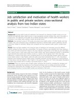
Filtration and Nuclease Treatment Improves Viral DNA Amplification by Removing Host DNA and mRNAFigure 1
Filtration and Nuclease Treatment Improves Viral
DNA Amplification by Removing Host DNA and
mRNA. Human plasma was inoculated with 1 µl of an Aden-
ovirus Type 17 suspension, filtered and incubated for 2 hrs
with either 10 u DNAse or 5 u RNAse as indicated. Remain-
ing nucleic acids were purified with the QiaAmp UltrasSens
Virus Kit and subjected to 1
st
strand cDNA synthesis and 50
cycles PCR using primers specific for either Adenovirus Type
17 or human β-Actin DNA (upper band; primers cross an
intron) or cDNA (lower band). Amplicons were then visual-
ized on an ethidium bromide impregnated agarose gel.
Adenovirus Type 17 β-Actin (genomic + cDNA)
+
+
+ + + +
+
+
Filter
DNAse
RNAse
+ + + +
+
+
+
Virology Journal 2007, 4:65 />Page 4 of 11
(page number not for citation purposes)
ments we have had variable success obtaining random
multiplex PCR amplification from human plasma and
have obtained only 7 sequences in total from un-inocu-
lated plasma. These sequences did not align with any
known sequences in NCBI Non-Redundant database
(Blastn searches were conducted.) However, when we
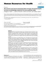
shot-gun cloned the DNA amplified from the Adenovirus
17-inoculated human plasma, we obtained 12 recom-
bined vector clones, and all clones matched to Adenovirus
Type 17 (see representative example, Figure 4). These data
demonstrate that the combination of filtration, DNAse/
RNAse treatment and V8AAmer Random Multiplex PCR
allows for the detection and identification of Adenovirus
Type 17 without using any virus-specific reagents such as
primers, antibodies or microarray chips.
Sensitivity of the Random Multiplex PCR Using 5'-
VVVVVVVVAA-3' Primers
The suspension of Adenovirus Type 17 received from the
ATCC had not been quantified and the sensitivity of the
random multiplex PCR method thus was uncertain. In
order to generate a standard curve, we conducted real-
time PCR of a known concentration of PCR-amplified
Adenovirus Type 17 genomic template using Adenovirus
Type 17-specific primers and found that we could detect
as little as 10 copies/ml of template (data not shown). We
then subjected the ATCC Adenovirus Type 17 suspension
from the ATCC to real-time PCR using the same Adenovi-
rus Type 17-specific primers and found that we could
readily detect the genomic DNA (data not shown). Based
on the real-time PCR generated from the known concen-
trations of Adenovirus Type 17 genomic template, we gen-
5'-VVVVVVVVAA-3' Primers Enable Random Multiplex Amplification of DNA from Human Plasma Inoculated with Adenovirus Type 17Figure 3
5'-VVVVVVVVAA-3' Primers Enable Random Multi-
plex Amplification of DNA from Human Plasma
Inoculated with Adenovirus Type 17. 1 ml of human
plasma was inoculated with 0–5 µl of suspended Adenovirus
Type 17 (ATCC), filtered and incubated for 2 hours with
nucleases. Remaining nucleic acids were purified with the
QiaAmp UltrasSens Virus Kit (Qiagen) and subjected to the
random multiplex PCR with 5'-VVVVVVVVAA-5' primers as
detailed in the Methods section. Amplicons were then visual-
ized on an ethidium bromide impregnated agarose gel.
+Adenovirus Type 17 (µ
µµ
µl)
MW 0 0.5 1 5 MW
2.0-
1.0-
0.6-
0.4-
0.2-
Kb
Random Multiplex PCR With 5'-VVVVVVVVAA-3' Primers Amplifies Circular Plasmid DNAFigure 2
Random Multiplex PCR With 5'-VVVVVVVVAA-3'
Primers Amplifies Circular Plasmid DNA. A. The indi-
cated plasmids were amplified using random multiplex PCR
with either 5'-VVVVVVVVAA-3' or 5'-NNNNNNNNNN-3'
primers as indicated in the Methods section. B. The pBabe
plasmid was diluted to the indicated amounts and amplified
using random multiplex PCR with 5'-VVVVVVVVAA-3' prim-
ers.
V
8
AA Primers
N10 Primers
H
2
O
G
F
P
p
S
u
p
e
r
P
e
t
3
0
b
p
B
a
b
e
H
2
O
G
F
P
p
S
u
p
e
r
P
e
t
3
0
b
p
B
a
b
e
1
0
0
f
g
1
0
f
g
1
0
0
p
g
H
2
O
p
S
u
p
e
r
P
e
t
3
0
b
1
0
n
g
1
n
g
1
0
p
g
1
p
g
pBabe
G
F
P
V
8
AA Primers
A.
B.
Virology Journal 2007, 4:65 />Page 5 of 11
(page number not for citation purposes)
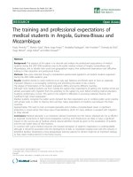
PCR Screening and Sequencing of Randomly Amplified Adenovirus Type 17 DNAFigure 4
PCR Screening and Sequencing of Randomly Amplified Adenovirus Type 17 DNA. Randomly amplified DNA from
Adenovirus 17-infected plasma was shot-gun ligated into pCR 2.1-TOPO and competent E. coli were transformed. Resultant
colonies were screened for the presence of recombinant plasmid DNA (A) and plasmid DNA from positive colonies was then
purified and sequenced (B). Sequence data from recombinant plasmid #9 (see *) was aligned to all sequence data in the Non-
Redundant NCBI Database using the NCBI Nucleotide-Nucleotide BLAST (blastn) Search Algorithm (version 2.2.8) (C).
TOPO 2.1
Multiple-
Cloning Site
Sequence of Randomly Amplified DNA
A.
B.
*
> gi|4416335|gb|AF108105.1|AF108105 Human adenovirus type 17 complete genome
Length = 35100
Score = 866 bits (437), Expect = 0.0
Identities = 443/445 (99%)
Strand = Plus / Minus
Query: 3 gcaaggaaggtaatacagcaccgctatgcacccacccaggaccacccctaaaatcaaata 62
||||||||||||||||||||||||||||||||||||||||||||||||||||||||||||
Sbjct: 26967 gcaaggaaggtaatacagcaccgctatgcacccacccaggaccacccctaaaatcaaata 26908
Query: 63 cccaaccgcttcaatatgtttacccccctctgttagagagggaacccagagctcacctcc 122
||||||||||||||||||||||||||||||||||||||||||||||||||||||||||||
Sbjct: 26907 cccaaccgcttcaatatgtttacccccctctgttagagagggaacccagagctcacctcc 26848
Query: 123 ggttttaggagtgttagtatcaaaaagaaggttaggtgtttctgtagcggctgtgctgct 182
||||||||||||||||||||||||||||||||||| ||||||||||||||||||||||||
Sbjct: 26847 ggttttaggagtgttagtatcaaaaagaaggttagatgtttctgtagcggctgtgctgct 26788
Query: 183 gtcggtaacgttcaccaaagtgaaagtgtggaagcaaggtccgctctggcagtggtaggt 242
||||||||||||||||||||||||||||||||||||||||||||||||||||||||||||
Sbjct: 26787 gtcggtaacgttcaccaaagtgaaagtgtggaagcaaggtccgctctggcagtggtaggt 26728
Query: 243 tccctctacaaaaggattgtagagtactagcttagtctttctggtagtataagttagtcc 302
||||||||||||||||||||||||||||||||||||||||||||||||||||||||||||
Sbjct: 26727 tccctctacaaaaggattgtagagtactagcttagtctttctggtagtataagttagtcc 26668
Query: 303 actggtaaggttgttgggtatttcaataccgtcgttggcgcaggcgttggagactgcgaa 362
||||||||||||||||||||||||||||||||||||||||||||||||||||||||||||
Sbjct: 26667 actggtaaggttgttgggtatttcaataccgtcgttggcgcaggcgttggagactgcgaa 26608
Query: 363 ggaagtgtttttaaaaagccagaggatgtatttgtcccccagtctgcagttgaattttac 422
|||||||||||||||||||||||||||||||||||||||| |||||||||||||||||||
Sbjct: 26607 ggaagtgtttttaaaaagccagaggatgtatttgtcccccggtctgcagttgaattttac 26548
Query: 423 ctcagtctggttggtgaggttgaag 447
|||||||||||||||||||||||||
Sbjct: 26547 ctcagtctggttggtgaggttgaag 26523
C.
Virology Journal 2007, 4:65 />Page 6 of 11
(page number not for citation purposes)
erated a standard curve plotting the concentration of
Adenovirus Type 17 genome equivalents versus the cycle
number when fluorescence passed the pre-set threshold
(Ct; Figure 4A). Based on this standard curve, we deter-
mined that the stock solution of Adenovirus Type 17 con-
tained 1 × 10
7
genome equivalents/ml. We then titrated
down the concentration of virus in human plasma and
conducted the combination of filtration, DNAse/RNAse
treatment and random multiplex PCR using the 5'-
VVVVVVVVAA-3' primers. We found that the random
multiplex (RT)-PCR method is sensitive to 1000 genome
equivalents/ml (Figure 4B).
Application of Random Multiplex PCR to RNA Viruses
We postulated that the random multiplex PCR method
would also detect RNA viruses if the viral nucleic acids
were first subjected to 1
st
strand cDNA synthesis. We inoc-
ulated human plasma with the RNA virus Coxsackievirus
A7 (CSV A7) and then filtered, treated with DNAse and
RNAse, purified the remaining nucleic acids and con-
ducted (RT)-PCR, using two commercially available
reverse transcriptases (Omniscript and SuperScript II) and
CSV A7 specific primers separated by either 200 bp or 1.4
Kb. We found that whereas Omniscript RT was more effi-
cient at amplifying 200 bp cDNA species, SuperScript II
RT was more efficient at amplifying 1.4 Kb fragments (Fig-
ure 5A). Next, we used the 5'-VVVVVVVVAA-3' primers to
random multiplex PCR amplify the cDNA and observed
distinct albeit faint amplicons (Figure 5B). We cloned the
amplification products and found that the amplified DNA
products matched the sequence of the cloned RNA
genomes as determined by BlastN alignment searches of
the NCBI Non-Redundant database (Figure 6). We were
surprised that the cloned DNA from Coxsackievirus A7
aligned with the known sequence for Coxsackievirus A16
until we learned that the Coxsackievirus A7 strain had not
been fully sequenced and is thus not in the NCBI Non-
Redundant Database (search terms Coxsackievirus AND
A7). Accordingly, we had amplified portions of the Cox-
sackievirus A7 genome that had not previously been
sequenced (Figure 7). We also inoculated human plasma
with Respiratory Syncytial Virus B and were able to
amplify and identify genomic RNA using the 5'-
VVVVVVVVAA-3' primers (data not shown). However, the
PCR amplification efficiency was poor in both RNA
viruses relative to the DNA virus, AV17, and we suspect
that this may be related to the efficiency of the reverse
transcriptases or the stability of the RNA.
Conclusion
In November 2002, an outbreak of atypical pneumonia
occurred in Guangdong Province, People's Republic of
China [13,14]. The disease did not respond to empirical
antibiotic treatment, and no known bacterial or viral
pathogens were identified using serological and (RT)-PCR
analyses. The disorder was called Severe Acute Respiratory
Syndrome (SARS), and by July 2003 it had caused 8439
reported cases and 812 fatalities [13,14]. In an effort to
identify the causative agent of SARS, the WHO SARS Aeti-
ology Study Group coordinated the distribution of infor-
mation and infected materials and the analyses of the data
Real-Time PCR Quantitation of Adenovirus Type 17 DNA TemplateFigure 5
Real-Time PCR Quantitation of Adenovirus Type 17
DNA Template. A. PCR-amplified genomic DNA from
Adenovirus Type 17 was quantitated by spectrophotometry
(OD 260 nm) and the genome equivalents/ml calculated
based on Avogadro's number (6.02205 × 10
23
/mol). Real-
time PCR was conducted with Adenovirus Type 17 specific
primers using 10
9
to 10
1
genome equivalents/ml as template
and then using 1 µl of the stock solution of Adenovirus Type
17 as template. A standard curve was produced based on the
threshold cycle number of the tested concentrations of Ade-
novirus Type 17 DNA template and plotted compared to the
threshold cycle number (C
T
) of the stock solution of Adeno-
virus Type 17. B. The indicated genome equivalents of Aden-
ovirus Type 17 were added to 1 ml of human plasma, treated
with 220 nm filtration and DNAse/RNAse digestion. Total
nucleic acids then were purified, amplified by random multi-
plex PCR (50 cycles) and sequenced as described in the
Methods section.
copies/ml
10
0
10
1
10
2
10
3
10
4
10
5
10
6
10
7
10
8
10
9
Cycle Number
10
15
20
25
30
r ² = 0.9600440817
Stock Virus (AV17) 17.3
10
8
10
7
10
6
10
4
10
5
10
9
10
3
10
1
10
2
0
A.
B.
Cycle Number (Ct)
Virology Journal 2007, 4:65 />Page 7 of 11
(page number not for citation purposes)
amongst eleven separate laboratories [14]. Researchers
cultured fetal rhesus kidney cells with infected nasopha-
ryngeal aspirates and then conducted random RT-PCR
using the primer, 5'-GCCGGAGCTCTGCAGAATTC-
NNNNNN-3' for reverse transcription, and 5'-GCCG-
GAGCTCTGCAGAATTC-3' for PCR. Unique PCR products
in the infected cell preparation were cloned and
sequenced, and 57% homology was found with bovine
Coronavirus[13]. RT-PCR of hundreds of infected clinical
specimens, using primers specific for the novel virus, con-
firmed the identification [13,14]. While this approach was
successful, it is worthwhile to note that it required 5
months and 11 laboratories to identify the virus. By con-
trast, the procedures described herein are – at least theo-
retically – capable of identifying an unknown virus
without the use of specially designed primers. Further-
more, is possible to at least partially identify an unknown
agent within 2–3 days in one laboratory without interme-
diate culture steps.
In the current study, we have detected and identified 3
separate viruses (i.e., Adenovirus Type 17, Coxsackievirus
A7, Respiratory Syncytial Virus B), using the novel
approach of random multiplex (RT)-PCR of purified viral
nucleic acids. The detection was accomplished in 7 hours
using only a micro-centrifuge and PCR machine and did
not require any virus-specific reagents. The sensitivity is
~1000 genome equivalents/ml for Adenovirus Type 17
which is not as sensitive as virus-specific PCR (<50
genome equivalents/ml) but should be adequate for the
detection of free virus in untreated individuals. Coupled
with the potential for direct sequence information, we are
confident that this approach may prove useful for our
defense against acts of bioterrorism, as well as for the
detection and characterization of novel viruses in blood
products and in patients displaying hallmarks of infection
(e.g., leukocytosis, fever).
Although we believe that the method is adequate in cur-
rent form to test clinical specimens in a randomized,
blinded diagnostic trial, we consider that certain alterna-
tive approaches may yield a method that has improved
sensitivity and specificity. In particular, we intend to
decrease the background amplification that has been var-
iably observed in order to prevent false positives and to
increase sensitivity by applying SYBR green fluorescence
for DNA detection. Finally, we will apply the optimized
approach to blood specimens from virally infected
patients in order to detect unsuspected variables and to
validate the method within the clinical environment.
Our long-term goal is to provide emergency room and pri-
mary care physicians with the ability to universally detect
viruses in human plasma. The rationale is, in part, that if
an emerging or recombined pathogenic virus were inten-
tionally released into a population, then the ability of
physicians to detect the pathogen will be crucial for con-
tainment. A second and related application of this tech-
nique involves identification of previously undescribed –
but naturally occurring – viruses in patients suffering with
idiopathic disease states of possible viral etiology. A third
application of these procedures may be in the screening of
blood bank blood just for the presence of virus. Although
the precise identification of the virus requires cloning and
sequencing, the amplification pattern will provide an
5'-VVVVVVVVAA-3' Primers Enable Random Multiplex Amplification of Viral RNA from Human Plasma Inoculated with Coxsackie Virus A7Figure 6
5'-VVVVVVVVAA-3' Primers Enable Random Multi-
plex Amplification of Viral RNA from Human Plasma
Inoculated with Coxsackie Virus A7. A. 1 ml of human
plasma was inoculated with 5 µl of suspended Coxsackie
Virus A7 (ATCC), filtered and incubated for 2 hours with
nucleases. Remaining nucleic acids were purified with the
QiaAmp UltrasSens Virus Kit (Qiagen) and subjected to first
strand cDNA synthesis using either Omniscript or Super-
script reverse transcriptase. Virus-specific primers were used
to amplify either a 200 bp or a 1.4 Kb portion of the viral
genome. B. The same samples were then subjected to ran-
dom multiplex PCR with 5'-VVVVVVVVAA-5' primers as
detailed in the Methods section. Amplicons were then visual-
ized on an ethidium bromide impregnated agarose gel.
Omniscript
SuperScript II
A
H
2
O
Plasma
CSV
+
+
+
+
+
+
+
+
+
+
+
+
+
+
+
200bp 1.4Kb
200bp 1.4Kb
+
Omniscript
SuperScript II
A
H
2
O
Plasma
CSV
+
+
+
+
+
+
+
+
+
+
+
+
+
+
+
200bp 1.4Kb
200bp 1.4Kb
+
B
Omniscript
SuperScript II
H
2
O
Plasma
CSV
+
+
+
+
V
8
AA Primers
+
++
+
Virology Journal 2007, 4:65 />Page 8 of 11
(page number not for citation purposes)
PCR Screening and Sequencing of Randomly Amplified Coxsackie Virus A7 cDNAFigure 7
PCR Screening and Sequencing of Randomly Amplified Coxsackie Virus A7 cDNA. Randomly amplified cDNA
from Coxsackie Virus A7-infected plasma was shot-gun ligated into pCR 2.1-TOPO and competent E. coli were transformed.
Resultant colonies were screened for the presence of recombinant plasmid DNA (A) and plasmid DNA from positive colonies
was then purified and sequenced (B). Sequence data from recombinant plasmid #7 (see *) was aligned to all sequence data in
the Non-Redundant NCBI Database using the NCBI Nucleotide-Nucleotide BLAST (blastn) Search Algorithm (version 2.2.8).
*
A.
B.
C.
TOPO 2.1
Multi-Cloning
Site
Sequence of Randomly Amplified DNA
Virology Journal 2007, 4:65 />Page 9 of 11
(page number not for citation purposes)
immediate signature indicating the presence of virus.
Most clinical laboratories that service emergency rooms
and primary care clinics possess the ability to conduct RT-
PCR for known viruses such as HIV, and the unmodified
random multiplex (RT)-PCR method does not require any
further technical expertise. In conclusion, the optimiza-
tion and testing of the random multiplex (RT)-PCR tech-
nology should allow for the development of a universal
virus detection assay that will vastly improve our collec-
tive defense against viral bioweapons and against emerg-
ing, heretofore unidentified natural viruses.
Methods
PCR of plasmids using 5'-VVVVVVVVAA-3' and 5'-
NNNNNNNNNN-3' primers
Plasmids (10 ng each) pEGFP (Clontech), pSuperNeo
(Oligoengine), Pet 30b (Calbiochem), and pBabe
(Addgene) were added to a PCR mix containing (per reac-
tion): 1 µl of 40 µM of one of the primers indicated (Oli-
gos Etc), 2.5 µl 10× PCR buffer (Promega), 5 µl (5 mM)
MgCl
2
, 1 µl (5 mM) dNTPs, 14 µl water, and 0.6 µl Taq
polymerase (Promega). PCR was performed according to
the following protocol on an Eppendorf Mastercycler Gra-
dient: 95°C-2 minutes and then 94°C-30 seconds, 33°C-
59 seconds, 34°C-6 seconds (increase in increments of
1°C for 6 seconds each up to 57°C for 6 seconds), 58°C-
2 minutes (+ .3°/s), repeat 50 times, 37°C-5 minutes,
65°C-5 minutes (+ .3°/s), and then hold at 4°C. Products
were run on a 1% agarose gel containing ethidium bro-
mide and images were captured on a UVP, Bio-Doc It, gel
documentation system.
Viral samples
Adenovirus Type 17 was purchased from ATCC (VR-
1094). This virus is a dsDNA virus. The source was con-
junctival scraping from a 1-year old female, Saudi Arabia,
1955. Coxsackievirus A7 W.P. Parker was purchased from
ATCC (VR-166). This virus is a ssRNA virus. The source
was stool of a 7-year old with fever, headache, sore throat,
stiff neck, and back pain, New York, 1949. Respiratory
syncytial virus was purchased from ATCC (VR-1580). This
virus is a ssRNA, linear virus. The source was the respira-
tory secretions from a child with acute respiratory disease.
Blood Plasma Isolation and Viral Separation
10 ml of heparinized whole blood was placed into a 15 ml
Falcon tube (BD Biosciences) and centrifuged at 1700 × g
for 10 minutes. 250 µl of plasma layer was added to 250
µl room temperature 1× PBS and placed in a SPIN-X .22
micron, 2 ml centrifuge tube (Costar). Plasma was then
inoculated with virus and the mixture was centrifuged at
2500 × g for 15 minutes. If any of the mixture remained
above the filter, centrifugation was continued at higher
speeds until all plasma had passed the filter into the col-
lection tube. 500 µl of PBS was added to the filtered mix-
ture and treated with 7 µl each of un-diluted Riboshredder
(Epicentre) and Omnicleave (Epicentre). Treatment of
samples with the RNAse and DNAse should cleave any
DNA or RNA not protected by viral capsid. All samples
were placed in a Thermomixer R (Eppendorf) at 37°C and
shaken for 15 seconds every 3 minutes for 2 hours. Plasma
was then ready for viral isolation.
Isolation of Virus from Plasma
1 ml of blood plasma ± virus was cooled to room temper-
ature and placed into a 2 ml tube. Viral isolation was car-
ried out according to the manufacturer's protocol
(Qiagen). Viral DNA or RNA was eluted off the column in
a final volume of 30 µl elution buffer. This product was
either used directly for random PCR or used in the reverse
transcription reaction.
Reverse Transcription of Viral RNA
Reverse transcription was carried out according to the
manufacturer's protocol either with the Omniscript RT
(Qiagen) or SuperScript II RT (Invitrogen). The maximum
amount of viral RNA was used in place of water for both
reactions.
Isolation and cloning of random PCR products
The PCR of viral DNA isolated from whole blood was
done according to the method described above utilizing
the 5'-VVVVVVVVAA-3' primers. Products were run on a
1% agarose gel containing ethidium bromide. The bands
which differed from the water control were excised and
the DNA was extracted using the Qiaquick gel extraction
kit (Qiagen). DNA was eluted off in a final volume of 20
µl elution buffer. The DNA was cloned into the TOPO
cloning vector (Invitrogen) following the manufacturer's
protocol and transformation was carried out in TOP 10
chemically competent E. coli. Resulting colonies were
picked randomly and mixed with 20 µl PBS. 1 µl of this
mixture was used in the PCR reaction using the universal
M13 forward and reverse (1 µl of 10 µM) primers and 2 ×
PCR master mix (Promega). The following parameters
were used for colony screening: 95°C-2 minutes, and then
36 cycles of 94°C-30 seconds, 55°C-30 seconds, 72°C-1
minute. PCR products were run out on a 1% agarose gel
containing ethidium bromide. Those colonies which
yielded products greater than 200 bp were grown in LB
broth overnight and the DNA was isolated according to
the manufacturer's protocol (Qiagen) eluting the DNA off
in a final volume of 25 µl. DNA was sequenced using the
universal M13 forward and reverse primers at the Univer-
sity of Louisville's core sequencing facility. Resulting
sequences were screened using the BLAST program on the
NCBI web site to determine sequence origin.
Virology Journal 2007, 4:65 />Page 10 of 11
(page number not for citation purposes)
PCR from Viral DNA or cDNA with Virus-specific Primers
PCR was carried out with either viral DNA or cDNA using
a 2× PCR Master Mix (Promega) and 1 µl of viral DNA. 1
µl of 10 µM primers was used for each reaction. The total
volume was equal to 25 µl per reaction. The following
parameters were used for viral-specific amplification:
94°C-5 minutes, 94°C-30 seconds, 55°C-30 seconds,
72°C-2 minutes, repeat 45 times, 72°C-7 minutes.
Virus-Specific Primers (standard PCR)
For generation of 200 bp fragment:
Coxsackievirus forward primer (923): 5'-TTATCAGAGAT-
GGCAGCACC-3'
Coxsackievirus reverse primer (1105C): 5'-CTTGTCCAC-
CGCTGTAGCCT-3'
For generation of 1.4 Kb fragment:
Coxsackievirus forward primer (923): 5'-TTATCAGAGAT-
GGCAGCACC-3'
Coxsackievirus reverse primer (2161C): 5'-CATTGCCT-
GCATTCTGTTGG-3'
Virus-Specific Primers (For Real Time PCR)
Coxsackievirus forward primer (937): 5'-AGCACCACT-
GCAATCACCGA-3'
Coxsackievirus reverse primer (1094C): 5'-CTGTAGCGT-
CAGTGTCAGGA-3'
Adenovirus forward primer (5770): 5'-TGTAGGTGTAG-
GCCACGTGA-3'
Adenovirus reverse primer (6084C): 5'-TCGCCAAGCT-
TCTCTCCAAC-3'
Template Production Primers:
Adenovirus forward primer (5492): 5'-CTCTTAC-
CTCGCGTCTCCAT-3'
Adenovirus reverse primer (6084C): 5'-TCGCCAAGCT-
TCTCTCCAAC-3'
Coxsackievirus forward primer (923): 5'-TTATCAGAGAT-
GGCAGCACC-3'
Coxsackievirus reverse primer (1105C): 5'-CTTGTCCAC-
CGCTGTAGCCT-3
Generating viral DNA templates for real time PCR
Viral DNA isolated from whole blood plasma was used to
generate a template for use in real time PCR. 2 µl of DNA
from the virus was used with Platinum PCR Master Mix
(Invitrogen) and 2 µl of a 10 µM solution of each of the
template primers outlined above. The total volume of the
reaction was 50 µl. The same PCR parameters were used as
were used for viral-specific amplification. The resulting
products were run on a 1% agarose gel containing ethid-
ium bromide. The bands were excised and DNA was
eluted according to manufacturer's instructions (Qiagen).
Real Time PCR
All real time PCR samples were run on a Cepheid Smart
Cycler. OmniMix Hs Reagent (Cephied, packged by
Takara) was re-suspended in 50 µl of water, which was
sufficient for two PCR reactions. 1 µl of the stock virus and
1 µl of each of the template primers (10 µM) were added
and mixed. Samples were run at 95°C for 30 sec, 54°C for
15 sec, and 72°C for 20 sec for a total of 40 cycles.
Determination of viral molecules in viral template
The molecular weight was determined for each of the
dsDNA templates generated by PCR. The OD of the result-
ant DNA was also recorded. The OD, in grams/µl was
divided by the molecular weight of the product and the
result was equal to moles/µl. This number was then mul-
tiplied by Avogadro's number (6.022 × 10
23
molecules/
mole) and the resulting number is equal to the number of
viral DNA molecules per microliter.
Competing interests
The author(s) declare that they have no competing inter-
ests.
Authors' contributions
ALC: Conducted multiplex (RT)-PCR, cloning, virus-spe-
cific PCR, template production and sequencing.
JS: Optimized the random multiplex (RT)-PCR of RNA
viruses.
ST: Phlebotomy, serum preparation and assisted with the
initial development of the 5-VVVVVVVVAA-3' primers.
JWE: Provided scientific input regarding the universal
application of this approach and assisted with the optimi-
zation of the random multiplex (RT)-PCR of RNA viruses.
JC: Conceived and directed the entire project. Conducted
initial multiplex PCR for AV17.
All authors read and approved the final manuscript.
Publish with Bio Med Central and every
scientist can read your work free of charge
"BioMed Central will be the most significant development for
disseminating the results of biomedical research in our lifetime."
Sir Paul Nurse, Cancer Research UK
Your research papers will be:
available free of charge to the entire biomedical community
peer reviewed and published immediately upon acceptance
cited in PubMed and archived on PubMed Central
yours — you keep the copyright
Submit your manuscript here:
/>BioMedcentral
Virology Journal 2007, 4:65 />Page 11 of 11
(page number not for citation purposes)
Acknowledgements
This study was funded by a grant from the Center for Genetics and Molec-
ular Medicine, University of Louisville.
References
1. Jackson RJ, Ramsay AJ, Christensen CD, Beaton S, Hall DF, Ramshaw
IA: Expression of mouse interleukin-4 by a recombinant
ectromelia virus suppresses cytolytic lymphocyte responses
and overcomes genetic resistance to mousepox. J Virol 2001,
75(3):1205-1210.
2. Espy MJ, Cockerill IF, Meyer RF, Bowen MD, Poland GA, Hadfield TL,
Smith TF: Detection of smallpox virus DNA by LightCycler
PCR. J Clin Microbiol 2002, 40(6):1985-1988.
3. Fatal yellow fever in a traveler returning from Amazonas,
Brazil, 2002. Jama 2002, 287(19):2499-2500.
4. Wilson WJ, Strout CL, DeSantis TZ, Stilwell JL, Carrano AV,
Andersen GL: Sequence-specific identification of 18 patho-
genic microorganisms using microarray technology. Mol Cell
Probes 2002, 16(2):119-127.
5. Seo TK, Thorne JL, Hasegawa M, Kishino H: Estimation of effec-
tive population size of HIV-1 within a host: a pseudomaxi-
mum-likelihood approach. Genetics 2002, 160(4):1283-1293.
6. Liang P, Pardee AB: Differential display of eukaryotic messen-
ger RNA by means of the polymerase chain reaction. Science
1992, 257(5072):967-971.
7. Nie X, Singh RP: A novel usage of random primers for multi-
plex RT-PCR detection of virus and viroid in aphids, leaves,
and tubers. J Virol Methods 2001, 91(1):37-49.
8. Ralph D, McClelland M, Welsh J: RNA fingerprinting using arbi-
trarily primed PCR identifies differentially regulated RNAs
in mink lung (Mv1Lu) cells growth arrested by transforming
growth factor beta 1. Proc Natl Acad Sci U S A 1993,
90(22):10710-10714.
9. van den Hoogen BG, de Jong JC, Groen J, Kuiken T, de Groot R,
Fouchier RA, Osterhaus AD: A newly discovered human pneu-
movirus isolated from young children with respiratory tract
disease. Nat Med 2001, 7(6):719-724.
10. Reyes GR, Kim JP: Sequence-independent, single-primer
amplification (SISPA) of complex DNA populations. Mol Cell
Probes 1991, 5(6):473-481.
11. Matsui SM, Kim JP, Greenberg HB, Su W, Sun Q, Johnson PC, DuPont
HL, Oshiro LS, Reyes GR: The isolation and characterization of
a Norwalk virus-specific cDNA. J Clin Invest 1991,
87(4):1456-1461.
12. Allander T, Emerson SU, Engle RE, Purcell RH, Bukh J: A virus dis-
covery method incorporating DNase treatment and its
application to the identification of two bovine parvovirus
species. Proc Natl Acad Sci U S A 2001, 98(20):11609-11614.
13. Kuiken T, Fouchier RA, Schutten M, Rimmelzwaan GF, van
Amerongen G, van Riel D, Laman JD, de Jong T, van Doornum G, Lim
W, Ling AE, Chan PK, Tam JS, Zambon MC, Gopal R, Drosten C, van
der Werf S, Escriou N, Manuguerra JC, Stohr K, Peiris JS, Osterhaus
AD: Newly discovered coronavirus as the primary cause of
severe acute respiratory syndrome. Lancet 2003,
362(9380):263-270.
14. Peiris JS, Lai ST, Poon LL, Guan Y, Yam LY, Lim W, Nicholls J, Yee
WK, Yan WW, Cheung MT, Cheng VC, Chan KH, Tsang DN, Yung
RW, Ng TK, Yuen KY: Coronavirus as a possible cause of
severe acute respiratory syndrome. Lancet 2003,
361(9366):1319-1325.