Báo cáo sinh học: " The complete genomes of three viruses assembled from shotgun libraries of marine RNA virus communities" ppt
Bạn đang xem bản rút gọn của tài liệu. Xem và tải ngay bản đầy đủ của tài liệu tại đây (997.7 KB, 9 trang )
BioMed Central
Page 1 of 9
(page number not for citation purposes)
Virology Journal
Open Access
Research
The complete genomes of three viruses assembled from shotgun
libraries of marine RNA virus communities
Alexander I Culley
1
, Andrew S Lang
2
and Curtis A Suttle*
1,3
Address:
1
University of British Columbia, Department of Botany, 3529-6270 University Blvd, Vancouver, B.C. V6T 1Z4, Canada,
2
Department of
Biology, Memorial University of Newfoundland, St. John's, NL A1B 3X9, Canada and
3
University of British Columbia, Department of Earth and
Ocean Sciences, Department of Microbiology and Immunology, 1461-6270 University Blvd, Vancouver, BC, V6T 1Z4, Canada
Email: Alexander I Culley - ; Andrew S Lang - ; Curtis A Suttle* -
* Corresponding author
Abstract
Background: RNA viruses have been isolated that infect marine organisms ranging from bacteria
to whales, but little is known about the composition and population structure of the in situ marine
RNA virus community. In a recent study, the majority of three genomes of previously unknown
positive-sense single-stranded (ss) RNA viruses were assembled from reverse-transcribed whole-
genome shotgun libraries. The present contribution comparatively analyzes these genomes with
respect to representative viruses from established viral taxa.
Results: Two of the genomes (JP-A and JP-B), appear to be polycistronic viruses in the proposed
order Picornavirales that fall into a well-supported clade of marine picorna-like viruses, the
characterized members of which all infect marine protists. A temporal and geographic survey
indicates that the JP genomes are persistent and widespread in British Columbia waters. The third
genome, SOG, encodes a putative RNA-dependent RNA polymerase (RdRp) that is related to the
RdRp of viruses in the family Tombusviridae, but the remaining SOG sequence has no significant
similarity to any sequences in the NCBI database.
Conclusion: The complete genomes of these viruses permitted analyses that resulted in a more
comprehensive comparison of these pathogens with established taxa. For example, in concordance
with phylogenies based on the RdRp, our results support a close homology between JP-A and JP-
B and RsRNAV. In contrast, although classification of the SOG genome based on the RdRp places
SOG within the Tombusviridae, SOG lacks a capsid and movement protein conserved within this
family and SOG is thus likely more distantly related to the Tombusivridae than the RdRp phylogeney
indicates.
Background
RNA viruses of every classification have been isolated
from the ocean; nevertheless, the marine RNA virus com-
munity remains largely uncharacterized. Although there
are several examples of RNA viruses that infect marine ani-
mals [1] these organisms represent a very small portion of
the organisms in the sea; therefore it is unlikely that
viruses infecting these organisms make up a significant
fraction of the natural RNA virioplankton. Marine RNA
phages appear to be rare [2] and thus it is more likely that
the dominant RNA viruses infect the diverse and abun-
dant marine protists. For example, RNA viruses have
Published: 6 July 2007
Virology Journal 2007, 4:69 doi:10.1186/1743-422X-4-69
Received: 10 May 2007
Accepted: 6 July 2007
This article is available from: />© 2007 Culley et al; licensee BioMed Central Ltd.
This is an Open Access article distributed under the terms of the Creative Commons Attribution License ( />),
which permits unrestricted use, distribution, and reproduction in any medium, provided the original work is properly cited.
Virology Journal 2007, 4:69 />Page 2 of 9
(page number not for citation purposes)
recently been isolated that infect a number of marine pro-
tists including a diatom [3], a dinoflagellate [4], a
raphidophyte [5], a prasinophyte [6] and a thrausto-
chytrid [7].
Picorna-like viruses are a "superfamily" of positive-sense
single-stranded RNA (ssRNA) viruses that have similar
genome features and several conserved protein domains
[8]. Previously, we investigated the diversity of marine
picorna-like viruses by analysis of RNA-dependent RNA
polymerase (RdRp) sequences amplified from marine
virus communities and demonstrated that picorna-like
viruses are present and persistent in a diversity of marine
environments [9]. Furthermore, phylogenetic analyses
showed that none of the environmental sequences fell
within established virus families.
In a recent study, reverse-transcribed whole-genome shot-
gun libraries were used to characterize two marine RNA
virus communities [10]. Positive-sense ssRNA viruses that
are distant relatives of known RNA viruses dominated the
libraries. One RNA virus library (JP) was characterized by
a diverse, monophyletic clade of picorna-like viruses, but
the second library (SOG) was dominated by viruses dis-
tantly related to members of the family Tombusviridae and
the genus Umbravirus. Moreover, in both libraries, a high
percentage of sequence fragments were part of only a few
contiguous segments of sequence (contigs). Specifically,
in the SOG sample 59% of the sequence fragments
formed a single contig. Similarly, 66% of JP sequence frag-
ments contributed to only four contigs that represented
two viral genomes. Using a RT-PCR-based approach to
increase the amount of sequence for each dominant con-
tig resulted in the assembly of three complete viral
genomes. This contribution analyzes these genomes from
three previously unknown marine RNA viruses and inves-
tigates their similarities and differences with respect to
representative genotypes from established viral taxa.
Results and Discussion
Jericho Pier site
The two assembled genomes (JP-A and JP-B) from the Jeri-
cho Pier sampling site (Figure 1) are single molecules of
linear ssRNA.
The JP-A genome is positive-sense, 9212 nt in length with
a 632 nt 5' untranslated region (UTR) followed by 2 pre-
dicted open reading frames (ORFs) of 5067 nt (ORF 1, nt
position 633 to 5699) and 3044 nt (ORF 2, nt position
5848 to 8799) separated by an intergenic region (IGR) of
149 nt (Figure 2A). ORF 2 is followed by a 3' UTR of 413
nt (nt position 8800 to 9212) and a polyadenylate [poly
(A)] tail. The base composition of JP-A is 27.1% A, 19.4%
C, 22.0% G, and 31.6% U; this results in a G+C of 41%, a
percentage similar to other polycistronic picorna-like
viruses (Table 1).
Comparison to known viral sequences shows that the pro-
tein sequence predicted to be encoded by ORF 1 of JP-A
contains conserved sequence motifs characteristic of a
type III viral Helicase (aa residues 430 to 545), a 3C-like
cysteine protease (aa residues 1077 to 1103) and a type I
RdRp (aa residues 1350 to 1591) [11] (Figure 1A).
BLASTp [12] searches of the NCBI database with the pre-
dicted ORF 1 protein sequence showed significant
sequence similarities (E value < 0.001) to nonstructural
protein motifs of several viruses, including members of
the families Dicistroviridae (Drosophila C virus), Marna-
viridae (HaRNAV), Comoviridae (Cowpea mosaic virus)
and the unassigned genus Iflavirus (Kakugo virus). The top
matches for ORF 1 were to RsRNAV [E value = 3 × 10
-119
,
identities = 302/908 (33%)], a newly sequenced, unclas-
sified positive-sense ssRNA virus that infects the widely
distributed diatom Rhizosolenia setigera [3], HaRNAV [E
value = 2 × 10
-32
, identities = 156/624 (25%)] and Dro-
sophila C virus [E value = 1 × 10
-29
, identities = 148/603
(24%)], a positive-sense ssRNA virus that infects fruit flies.
Comparison of the protein sequence predicted to be
encoded by ORF 2 of JP-A to known viral sequences shows
that it has significant similarities to the structural proteins
of viruses from the families Dicistroviridae (Drosophila C
virus), Marnaviridae (HaRNAV), and the genus Iflavirus
(Varroa destructor virus 1). The sequences that are most
similar to ORF 2 of JP-A were the structural protein
regions of RsRNAV [E value = 6 × 10
-78
, identities = 212/
632 (33%)], HaRNAV [E value = 6 × 10
-68
, identities =
187/607 (30%)] and SssRNAV [E value = 2 × 10
-49
, iden-
tities = 241/962 (25%)].
The JP-B RNA genome is also likely from a positive-sense
ssRNA virus. The 8839 nt genome consists of a 5' UTR of
774 nt followed by two predicted ORFs of 4842 nt (ORF
1, nt position 775 to 5616) and 2589 nt (ORF 2, nt posi-
tion 5914 to 8502) separated by an IGR of 298 nt (nt posi-
tion 5617 to 5913) (Figure 2B). The 3' UTR is 337 nt long
and followed by a poly (A) tail. The base composition of
the genome is A, 30.8%; C, 17.9%; G, 19.7%; U, 31.6%.
Like JP-A, this % G+C value of 38% is comparable to the
% G+C observed in other polycistronic picorna-like
viruses (Table 1).
The position of core sequence motifs conserved among
positive-sense ssRNA viruses and BLAST searches of the
NCBI database with the translated JP-B genome suggest
that nonstructural proteins are encoded by ORF1, and the
structural proteins are encoded by ORF2. We identified
conserved sequence motifs in ORF 1 characteristic of a
type III viral Helicase (aa residues 328 to 441), a 3C-like
cysteine protease (aa residues 882 to 909) and a type I
Virology Journal 2007, 4:69 />Page 3 of 9
(page number not for citation purposes)
RdRp (aa residues 1143 to 1408) [11] (Figure 2B).
BLASTp [12] searches of the GenBank database showed
that ORF 1 has significant similarities (E value < 0.001) to
nonstructural genes from positive-sense ssRNA viruses
from a variety of families, including the Comoviridae
(Peach rosette mosaic virus), Dicistroviridae (Taura syn-
drome virus), Marnaviridae (HaRNAV), Sequiviridae (Rice
tungro spherical virus) and Picornaviridae (Avian enceph-
alomyelitis virus). The top scoring sequences [E value = 2
× 10
-69
, identities = 232/854 (27%)] were to a RdRp
sequence from RsRNAV and a partial picorna-like virus
RdRp from an unidentified virus [E value = 2 × 10
-40
, iden-
tities = 85/150 (56%)] amplified from the same JP station
during an earlier study [9]. Significant similarities to ORF
2 include the structural genes of viruses from the families
Dicistroviridae (Rhopalosiphum padi virus), Marnaviridae
(HaRNAV) and Picornaviridae (Human parechovirus 2), as
well as the unclassified genus Iflavirus (Ectropis obliqua
picorna-like virus). The top scoring sequences were to the
capsid protein precursor regions of RsRNAV [E value = 9 ×
10
-88
, identities = 244/799 (30%)] and HaRNAV [E value
= 8 × 10
-60
, identities = 180/736(24%)] and SssRNAV [E
value = 1 × 10
-40
. identities = 156/588 (26%)].
The JP-A and JP-B genomes appear to have a polycistronic
genome organization similar to that found in viruses in
the family Dicistroviridae. Several of these viruses contain
internal ribosome entry sites (IRES) [13-16] that position
the ribosome on the genome, actuating translation initia-
tion even in the absence of known canonical initiation
factors [13]. For example, TSV, a marine dicistrovirus, has
an IRES located in the IGR that directs the synthesis of the
structural proteins [15]. Computational searches did not
identify the secondary structure elements characteristic of
dicistrovirus IGR-IRESs in the JP genomes [16,17], how-
ever, JP-A and JP-B genomes have extensive predicted sec-
ondary structure in the 5' UTRs and IGRs [18,19],
suggestive of an IRES function. Moreover, start codons in
a favorable Kozak context, i.e. conserved sequences
upstream of the start codon that are thought to play a role
in initiation of translation [20], were not found in the JP
genomes. However to unequivocally demonstrate IRES
elements in the JP genomes, they must be confirmed
experimentally in polycistronic constructs. Nevertheless,
it seems reasonable that JP-A and JP-B use similar mecha-
nisms to initiate translation of the ORF 2 genes as are
known to be employed by several dicistroviruses.
We used RT-PCR to assess the distribution and persistence
of the JP-A and JP-B viruses in situ. Amplification with spe-
cific primers that target each of these viruses occurred in
samples from throughout the Strait of Georgia, the West
coast of Vancouver Island, and in every season and tidal
state at Jericho pier (Figure 1, Table 2). These results sug-
gest that JP-A and JP-B are ubiquitous in the coastal waters
of British Columbia.
It has long been recognized that several other groups of
small, positive-sense ssRNA viruses share many character-
istics with viruses in the family Picornaviridae. Recently,
Christian et al. [8] proposed creating an order (the Picor-
navirales) of virus families (Picornaviridae, Dicistroviridae,
Marnaviridae, Sequiviridae and Comoviridae) and unas-
signed genera (Iflavirus, Cheravirus, and Sadwavirus) that
have picornavirus-like characteristics. Viruses in the pro-
posed order have genomes with a protein covalently
attached to the 5' end, a 3' poly (A) tail, a conserved order
of non-structural proteins (Helicase-VpG-Proteinase-
RdRp), regions of high sequence similarity in the helicase,
proteinase and RdRp, post translational protein process-
ing during replication, an icosahedral capsid with a
unique "pseudo-T3" symmetry, and only infect eukaryo-
tes.
Although the capsid morphology, presence of a 5' termi-
nal protein and replication strategy and hosts are
unknown, signature genomic features and phylogenetic
analyses suggest that the JP viruses fall within the pro-
posed order Picornavirales. Both JP genomes encode the
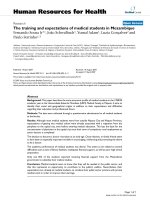
Map of southwestern British Columbia, Canada showing locations where samples were collectedFigure 1
Map of southwestern British Columbia, Canada
showing locations where samples were collected.Sites
in coastal BC waters where the JP-A and JP-B genomes were
detected are indicated and labelled. Both JP-A and JP-B were
detected in samples from 5 of the 9 stations that were
screened. The SOG station was not assayed for JP-A or JP-B.
See Table 2 for additional information about the stations.
Virology Journal 2007, 4:69 />Page 4 of 9
(page number not for citation purposes)
conserved core aa motifs and have the non-structural gene
order characteristic of viruses in the proposed Picornavi-
rales. Furthermore, both JP genomes have a poly (A) tail
and G+C content commensurate with these other viruses.
Bayesian trees [21] based on alignments of conserved
RdRp domains [11] (Figure 3), as well as concatenated
(putative) Hel+RdRp+VP3 capsid-like protein sequences
(Figure 4), of the JP genomes and representative members
of the proposed Picornavirales, resolves established taxa
according to previous taxonomic divisions. These analyses
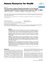
Analysis of genomes for putative open reading framesFigure 2
Analysis of genomes for putative open reading frames.
In the ORF maps created with DNA Strider [28], for each reading frame, potential start codons (AUG) are shown with a half-
height line and stop codons (UGA, UAA, and UAG) are shown by full-height lines. Recognizable conserved RNA virus protein
domains (Hel = helicase, Pro = Protease, RdRp = RNA-dependent RNA polymerase) and other genomic features (UTR =
untranslated region, IGR = intergenic region) are noted below each genome. See text for more detail. A. Map of the JP-A
genome. B. Map of the JP-B genome. C. Map of the SOG genome.
Virology Journal 2007, 4:69 />Page 5 of 9
(page number not for citation purposes)
also provide strong support for a clade comprised of
viruses (HaRNAV, RsRNAV and SssRNAV) that infect
marine protists and the JP-A and JP-B viruses. Within this
clade, RsRNAV, JP-A and JP-B have the most characteris-
tics in common. For example, they have the same order of
structural and non-structural genes, they are polycistronic
and the phylogenetic analyses indicate they are more
closely related (Figures 3 and 4). Whether JP-A and JP-B
infect host organisms related to Rhizosolenia setigera
remains unclear, although because of the inclusion of the
JP genomes within this clade and the fact that protists are
the most abundant eukaryotes in the sea, we suggest that
both JP viruses likely have a protist host.
Strait of Georgia site
The SOG genome was assembled from the Strait of Geor-
gia metagenomic library, and subsequently completed as
described in Methods. The genome has features character-
istic of a positive-sense ssRNA virus. The genome is 4449
nt long and comprised of a 5' UTR of 334 bp followed by
three putative ORFs (nt position 335–1228, nt position
1385–2860 and nt position 2903–4228) and is termi-
nated with a 3' UTR of 221 nt. A poly (A) tail was not
detected. Another putative ORF located at nt position 49
to 783 is in an alternative reading frame relative to the
ORFs discussed above (Figure 2C). The G+C content of
the SOG genome is 52%.
We identified the eight conserved motifs of the RdRp [11]
in the SOG genome (aa residues pos 451 to 700) (Figure
2C). tBLASTx [12] searches with the remainder of the
genome sequence showed no significant matches (E value
< 0.001) to sequences in the NCBI database (including the
five environmental metagenomes that have been depos-
ited). BLASTp searches with the putative RdRp sequence
resulted in significant similarities (E value < 0.001) to
RdRp sequences from positive-sense ssRNA viruses from
the family Tombusviridae and the unassigned genus
Umbravirus. The sequence with the most similarity to SOG
was from Olive latent virus 1 [E value = 3 × 10
-66
, identi-
ties = 180/508 (35%)]. This virus belongs to the genus
Necrovirus in the family Tombusviridae that has a host
range restricted to higher plants [22]. SOG is also signifi-
cantly similar to the Carrot mottle mimic virus sequence
[E value = 6 × 10
-66
, identities = 178/492 (36%)], a mem-
ber of the unclassified genus Umbravirus whose known
members infect only flowering plants [23].
Although the SOG putative RdRp sequence has similarity
to the RdRp of viruses from the family Tombusviridae and
genus Umbravirus, the remaining SOG sequence has no
detectable similarity to any other known sequence. A
Bayesian maximum likelihood tree based on alignments
of the SOG RdRp with the available Umbravirus sequences
and representative members of the Tombusviridae indi-
cates that the SOG genome forms a well supported clade
(Bayesian clade support value of 100) with the single
member of the genus Avenavirus, OCSV (Figure 5). Addi-
tionally, the presence of an amber stop codon (nt position
1230–1232) at the end of ORF 1 of the SOG genome (Fig-
ure 2C), resembles the in-frame termination codon char-
acteristic of the replicase gene of viruses in 7 of the 8
genera of the Tombusviridae [24]. This division of the rep-
licase of the Tombusviridae by a termination codon is
thought to be part of a translational read though gene
expression strategy [24]. Other similarities to the Tombus-
Table 1: Comparison of base composition between polycistronic picorna-like viruses
Genome* A C G U % G+C
JP-A 27.119.422.031.6 41
JP-B 30.817.919.731.6 38
ABPV 35.7 15.4 20.1 28.9 36
ALPV 31.3 19.4 19.2 30.2 39
BQCV 29.2 18.5 21.6 30.6 40
CrPV 32.6 18.4 20.9 28.1 39
DCV 29.9 16.3 20.4 33.4 37
HiPV 29.2 18.7 20.9 31.2 39
KBV 33.8 17.5 20.2 28.6 38
PSIV 31.3 17.0 19.4 32.3 36
RhPV 30.0 18.6 20.2 31.2 39
RsRNAV 31.2 16.7 19.5 32.5 36
SINV-1 32.9 18.3 20.5 28.2 39
SssRNAV 24.2 26.1 23.6 26.0 50
TSV 28.0 20.2 23.0 28.8 43
TrV 28.7 16.1 19.8 35.4 36
Average 30.4 18.4 20.7 30.5 39
* See Additional file 2 for the complete virus names
Virology Journal 2007, 4:69 />Page 6 of 9
(page number not for citation purposes)
viridae include a similar genome size, the absence of an
obvious helicase motif and the 5' proximal relative posi-
tion of the RdRp within the genome [22]. However,
unlike viruses in the Tombusviridae, there is no recogniza-
ble sequence for conserved movement or capsid proteins
in the SOG genome. The absence of a recognizable move-
ment protein could indicate the SOG virus does not infect
a higher plant. Our inability to identify structural genes
may indicate that, like the umbraviruses, the SOG virus
does not encode capsid proteins. However, it is also pos-
sible that movement or structural proteins encoded in the
SOG genome have no sequence similarity to those cur-
rently in the NCBI database.
Conclusion
Our analyses suggest that a persistent, widespread and
possibly dominant population of novel polycistronic
picorna-like viruses is an important component of the
RNA virioplankton in coastal waters. Nevertheless, as
exemplified by the SOG genome from the Strait of Geor-
gia site, other marine RNA virus assemblages appear to
contain viruses whose detectable sequence similarity with
established groups of viruses is limited to only the most
conserved genes (i.e. RdRp). The novelty of JP-A, JP-B and
SOG, as revealed by sequence analyses and genome char-
acterization, suggests that most of the diversity in the
marine RNA virus community remains uncharacterized.
Furthermore, these results raise the hypothesis that the
genomes of these marine RNA viruses that we propose to
infect single-celled eukaryotes may be more similar to the
ancestral RNA viruses that gave rise to those that infect
higher organisms.
Methods
Station descriptions
The shotgun libraries were constructed from seawater
samples collected from two stations, JP (Jericho Pier), a
site in English Bay adjacent to the city of Vancouver, Brit-
ish Columbia and SOG (Strait of Georgia), located in the
central Strait of Georgia next to Powell River, B.C. (Figure
1).
The locations of the stations where one or both of the JP
genomes were detected are shown in Figure 2. Details for
each station are listed in Table 2. In summary, samples
were collected from sites throughout the Strait of Georgia,
including repeated sampling from the JP site during differ-
ent seasons, and from the West coast of Vancouver Island
in Barkley Sound.
Virus concentration method
Concentrated virus communities were produced as
described by Suttle et al. [25]. Twenty to sixty litres of sea-
water from each station were filtered through glass fibre
(nominal pore size 1.2 μm) and then 0.45 μm pore-size
Durapore polyvinylidene fluoride (PVDF) membranes
Table 2: JP genome survey sample sites and results of assays
Station Name Station location
(B.C., Canada)
Date (mm/dd/yy) Location (Lat., Long.) Depth (m) Temp (°C) Salinity (ppt) JP-A PCR JP-B PCR
JP Jericho Pier 04/28/00 49.27, -123.20 S 9 26 ++
JP Jericho Pier 06/15/00 49.27, -123.20 S 14 12 ++
JP Jericho Pier 06/29/00 49.27, -123.20 S 17 12 ++
JP Jericho Pier 07/06/00 49.27, -123.20 S 16 13 ++
JP Jericho Pier 07/13/00 49.27, -123.20 S 18 8
JP Jericho Pier 07/27/00 49.27, -123.20 S 18 11 ++
JP Jericho Pier 08/17/00 49.27, -123.20 S 18 18 ++
JP Jericho Pier 09/14/00 49.27, -123.20 S 15 19 ++
JP Jericho Pier 09/21/00 49.27, -123.20 S 15 16 -+
JP Jericho Pier 09/28/00 49.27, -123.20 S 14 21 ++
JP Jericho Pier 11/23/00 49.27, -123.20 S 8 27 ++
JP Jericho Pier 02/15/01 49.27, -123.20 S 7 27 ++
JP Jericho Pier 06/14/01 49.27, -123.20 S 15 13 ++
SEC Sechelt Inlet 07/06/03 49.69, -123.84 4 13 26 -+
TEA Teakearne Inlet 07/07/03 50.19, -124.85 5 13 28 +-
QUA Quadra Island 07/07/03 50.19, -125.14 3 13 28
ARR Arrow Pass 07/09/03 50.72, -126.67 2 10 31 ++
IEC Imperial Eagle
Channel
06/20/99 48.87, -125.21 7 n.a. n.a. +-
TRE Trevor Channel 06/28/99 48.97, -125.16 S n.a. n.a. ++
BAM Bamfield Inlet 07/06/99 48.81, -125.16 S n.a. n.a. ++
NUM Numukamis Bay 07/12/99 48.90, -125.01 8 n.a. n.a. ++
A "+" indicates amplification and "-" indicates no amplification occurred. "n.a." indicates the data is not available and "S" means the sample was taken
from the surface.
Virology Journal 2007, 4:69 />Page 7 of 9
(page number not for citation purposes)
(Millipore, Cambridge, Canada), to remove particulates
larger than most viruses. This filtrate was subsequently
concentrated approximately 200 fold through a Tangen-
tial Flow Filter cartridge (Millipore) with a 30 kDa molec-
ular cut-off, essentially concentrating the 2 to 450 nm size
fraction of seawater. Remaining bacteria were removed by
filtering the concentrate two times through a 0.22 μm
Durapore PVDF membrane (Millipore). Virus-sized parti-
cles in each VC were pelleted via ultracentrifugation (5 h
at 113 000 × g at 4°C). Pellets were resuspended overnight
at 4°C in sterile 50 μM Tris chloride (pH 7.8).
Whole genome library construction
A detailed description of the whole genome shotgun
library construction protocol can be found in Culley et al.
[10]. Briefly, before extraction, concentrated viral lysates
were treated with RNase (Roche, Mississauga, Canada)
and then extracted with a QIAamp Minelute Virus Spin Kit
(Qiagen, Mississauga, Canada) according to the manufac-
turer's instructions. An aliquot of each extract was used in
a PCR reaction with universal 16S primers to ensure sam-
ples were free of bacteria. To isolate the RNA fraction,
samples were treated with DNase 1 (Invitrogen, Burling-
ton, Canada) and used as templates for reverse transcrip-
tion with random hexamer primers. Double-stranded (ds)
cDNA fragments were synthesized from single stranded
DNA with Superscript III reverse transcriptase (Invitro-
gen) using nick translational replacement of genomic
RNA [26]. After degradation of overhanging ends with T4
DNA polymerase (Invitrogen), adapters were attached to
the blunted products with T4 DNA ligase (Invitrogen).
Subsequently, excess reagents were removed and cDNA
products were separated by size with a Sephacryl column
(Invitrogen). To increase the amount of product for clon-
ing, size fractions greater than 600 bp were amplified with
primers targeting the adapters. Products from each PCR
reaction were purified and cloned with the TOPO TA
Cloning system (Invitrogen). Clones were screened for
inserts by PCR with vector-specific primers. Insert PCR
products greater than 600 bp were purified and sequenced
at the University of British Columbia's Nucleic Acid and
Protein Service Facility (Vancouver, Canada). Sequence
fragments were assembled into overlapping segments
using Sequencher v 4.5 (Gene Codes, Ann Arbor, U.S.A.)
based on a minimum match % of 98 and a minimum bp
overlap of 20. Sequences were compared against the NCBI
database with tBLASTx [12]. A sequence was considered
significantly similar if BLAST E values were < 0.001. The
details for viruses used in phylogenetic analyses are listed
in additional file 2. Virus protein sequences were aligned
using CLUSTAL X v 1.83 with the Gonnet series protein
matrix [27]. Alignments were transformed into likelihood
distances with Mr. Bayes v3.1.1 [21] and 250,000 genera-
tions. Neighbor-joining trees were constructed with PAUP
Bayesian maximum likelihood trees of aligned concatenated helicase, RdRp and VP3-like capsid amino acid sequences from the JP-A and JP-B genomes and other picorna-like virusesFigure 4
Bayesian maximum likelihood trees of aligned con-
catenated helicase, RdRp and VP3-like capsid amino
acid sequences from the JP-A and JP-B genomes and
other picorna-like viruses. Bayesian clade credibility val-
ues are shown for relevant nodes in boldface followed by
bootstrap values based on neighbour-joining analysis. The
Bayesian scale bar indicates a distance of 0.1. See Additional
file 2 for complete virus names and accession numbers.
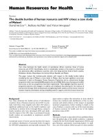
Bayesian maximum likelihood trees of aligned RdRp amino acid sequences from the JP-A and JP-B genomes and repre-sentative members of the proposed order PicornaviralesFigure 3
Bayesian maximum likelihood trees of aligned RdRp
amino acid sequences from the JP-A and JP-B
genomes and representative members of the pro-
posed order Picornavirales. Bayesian clade credibility val-
ues are shown for relevant nodes in boldface followed by
bootstrap values based on neighbour-joining analysis. The
Bayesian scale bar indicates a distance of 0.1. See Additional
file 2 for complete virus names and accession numbers.
Virology Journal 2007, 4:69 />Page 8 of 9
(page number not for citation purposes)
v4.0 [28], and bootstrap values calculated based on per-
centages of 10,000 replicates.
5' and 3' RACE
The 5' and 3' ends of the environmental viral genomes
were cloned using the 5' and 3' RACE systems (Invitrogen)
according to manufacturer's instructions. The 3' RACE
with the SOG genome required the addition of a poly (A)
tract with poly (A) polymerase (Invitrogen) according to
manufacturer directions before cDNA synthesis. cDNA
was synthesized directly from extracted viral RNA from
the appropriate library. Three clones of each 5' and 3' end
were sequenced.
PCR
Closing gaps in the assembly
PCR with primers targeting specific regions of the two JP
environmental genomes were used to verify the genome
assembly, increase sequencing coverage and reconfirm the
presence of notable genome features. The template for
these reactions was the amplified and purified PCR prod-
uct from the JP and SOG shotgun libraries. Additional file
1 lists the sequence and genome position of primers used.
The standard PCR conditions were reactions with 1 U of
Platinum Taq DNA polymerase (Invitrogen) in 1× Plati-
num Taq buffer, 1.5 mM MgCl
2
, 0.2 mM of each dNTP,
and 0.2 μM of each primer (see Additional file 1), in a
final volume of 50 μl. Thermocycler conditions were, acti-
vation of the enzyme at 94°C for 1 min 15 s, followed by
30 cycles of denaturation at 94°C for 45 s, annealing at
50°C for 45s and extension at 72°C for 1 minute. The
reaction was terminated after a final extension stage of 5
min at 72°C. PCR products were purified with a PCR Min-
elute cleanup kit (Qiagen) and sequenced directly with
both primers.
Environmental screening
To assess the temporal and geographic distribution of the
JP genomes, extracted RNA from viral concentrates were
screened with Superscript III One-step RT-PCR System
with Platinum Taq DNA Polymerase (Invitrogen) with
primers JP-A 5 and 6 and JP-B 6 and 7 (see Additional file
1). The template for the reactions was DNase 1 treated
viral RNA, extracted with a QIAamp Minelute Virus Spin
Kit (Qiagen) according to the manufacturer's instructions.
Each reaction consisted of RNA template, 1× reaction mix,
0.2 μM of each primer, 1 μl RT/Platinum Taq mix in a vol-
ume of 50 μl. Reactions were incubated 30 min at 50°C,
then immediately heated to 94°C for 45 s, followed by 35
cycles of denaturation at 94°C for 15 s, annealing at 50°C
for 30 s and extension at 68°C for 1 min. After a final
extension step at 68°C for 5 min, RT-PCR products were
analyzed by agarose gel electrophoresis. Products were
sequenced to verify the correct target had been amplified.
Competing interests
The author(s) declare that they have no competing inter-
ests.
Authors' contributions
AC contributed to the design of the study, performed the
lab work, analyzed the data and drafted the manuscript.
AL contributed to the design of the study, analyzed the
data and helped prepare the manuscript. CS was involved
in the conceptualization and design of the research and in
manuscript preparation. AC, AL and CS have read and
approved this manuscript.
Additional material
Additional file 1
PCR primers used to complete the three genome sequences. The table pro-
vides detailed information about the primers used to complete the three
viral genome sequences.
Click here for file
[ />422X-4-69-S1.doc]
Bayesian maximum likelihood trees of aligned RdRp amino acid sequences from the SOG genome and members of the family Tombusviridae and unassigned genus UmbravirusFigure 5
Bayesian maximum likelihood trees of aligned RdRp
amino acid sequences from the SOG genome and
members of the family Tombusviridae and unas-
signed genus Umbravirus. Bayesian clade credibility val-
ues are shown for relevant nodes in boldface followed by
bootstrap values based on neighbour-joining analysis. The
Bayesian scale bar indicates a distance of 0.1. See Additional
file 2 for complete virus names and accession numbers.
Publish with BioMed Central and every
scientist can read your work free of charge
"BioMed Central will be the most significant development for
disseminating the results of biomedical research in our lifetime."
Sir Paul Nurse, Cancer Research UK
Your research papers will be:
available free of charge to the entire biomedical community
peer reviewed and published immediately upon acceptance
cited in PubMed and archived on PubMed Central
yours — you keep the copyright
Submit your manuscript here:
/>BioMedcentral
Virology Journal 2007, 4:69 />Page 9 of 9
(page number not for citation purposes)
Acknowledgements
We would like to thank Professor Nakashima for evaluating the IGRs of the
JP genomes for the presence of dicistrovirus IRES elements and Debbie
Adams from the Nucleic Acid Protein Service Unit at the University of Brit-
ish Columbia for her generosity. Sequences have been deposited in Gen-
Bank with accession numbers EF198240
, EF198241 and EF198242. This
work was supported by grants from the Natural Science and Engineering
Research Council of Canada.
References
1. Smith A: Aquatic virus cycles. In Viral Ecology Edited by: Hurst C.
San Diego: Academic Press; 2000:447-491.
2. Weinbauer M: Ecology of prokaryotic viruses. FEMS Microbiol Rev
2004, 28:127-181.
3. Nagasaki K, Tomaru Y, Katanozaka N, Shirai Y, Nishida K, Itakura S,
Yamaguchi M: Isolation and characterization of a novel single-
stranded RNA virus infecting the bloom-forming diatom
Rhizosolenia setigera. Appl Environ Microbiol 2004, 70:704-711.
4. Tomaru Y, Katanozaka N, Nishida K, Shirai Y, Tarutani K, Yamaguchi
M, Nagasaki K: Isolation and characterization of two distinct
types of HcRNAV, a single-stranded RNA virus infecting the
bivalve-killing microalga Heterocapsa circularisquama. Aquat
Microb Ecol 2004, 34:207-218.
5. Tai V, Lawrence JE, Lang AS, Chan AM, Culley AI, Suttle CA: Char-
acterization of HaRNAV, a single-stranded RNA virus caus-
ing lysis of Hetersigma akashiwo (Raphidophyceae). J Phycol
2003, 39:343-352.
6. Brussaard CPD, Noordeloos AAM, Sandaa RA, Heldal M, Bratbak G:
Discovery of a dsRNA virus infecting the marine photosyn-
thetic protist Micromonas pusilla. Virol 2004, 319:280-291.
7. Takao Y, Nagasaki K, Mise K, Okuno T, Honda D: Isolation and
characterization of a novel single-stranded RNA virus infec-
tious to a marine fungoid protist, Schizochytrium sp. (Thraus-
tochytriaceae, labyrinthulea). Appl Environ Microbiol 2005,
71:4516-4522.
8. Christian P, Fauquet CM, Gorbalenya AE, King AMG, Knowles N,
LeGall O, Stanway G: A proposed Picornavirales order. In
Microbes in a Changing World Edited by: Fauquet CM. San Francisco:
International Unions of Microbiological Societies; 2005.
9. Culley AI, Lang AS, Suttle CA: High diversity of unknown
picorna-like viruses in the sea. Nature 2003, 424:1054-1057.
10. Culley AI, Lang AS, Suttle CA: Metagenomic analysis of coastal
RNA virus communities. Science
2006, 312:1795-1798.
11. Koonin EV, Dolja VV: Evolution and taxonomy of positive-
strand RNA viruses – implications of comparative-analysis of
amino-acid-sequences. Crit Rev Biochem Mol Biol 1993,
28:375-430.
12. Altschul SF, Madden TL, Schaffer AA, Zhang JH, Zhang Z, Miller W,
Lipman DJ: Gapped BLAST and PSI-BLAST: a new generation
of protein database search programs. Nucleic Acids Res 1997,
25:3389-3402.
13. Jan E, Sarnow P: Factorless ribosome assembly on the internal
ribosome entry site of cricket paralysis virus. J Mol Biol 2002,
324:889-902.
14. Nishiyama T, Yamamoto H, Shibuya N, Hatakeyama Y, Hachimori A,
Uchiumi T, Nakashima N: Structural elements in the internal
ribosome entry site of Plautia stali intestine virus responsible
for binding with ribosomes. Nucleic Acids Res 2003,
31:2434-2442.
15. Cevallos RC, Sarnow P: Factor-independent assembly of elon-
gation-competent ribosomes by an internal ribosome entry
site located in an RNA virus that infects penaeid shrimp. J
Virol 2005, 79:677-683.
16. Czibener C, Alvarez D, Scodeller E, Gamarnik AV: Characteriza-
tion of internal ribosomal entry sites of Triatoma virus. J Gen
Virol 2005, 86:2275-2280.
17. Hatakeyama Y, Shibuya N, Nishiyama T, Nakashima N: Structural
variant of the intergenic internal ribosome entry site ele-
ments in dicistroviruses and computational search for their
counterparts. RNA 2004, 10:779-786.
18. Mathews DH, Sabina J, Zuker M, Turner DH: Expanded sequence
dependence of thermodynamic parameters improves pre-
diction of RNA secondary structure. J Mol Biol 1999,
288:911-940.
19. Zuker M, Mathews DH, Turner DH: Algorithms and thermody-
namics for RNA secondary structure prediction: a practical
guide. In RNA Biochemistry and Biotechnology Edited by: Barciszewski
J, Clark BFC. Boston: Kluwer Academic Publishers; 1999:11-43.
20. Kozak M: Point mutations define a sequence flanking the
AUG initiator codon that modulates translation by eukaryo-
tic ribosomes. Cell 1986,
44:283-292.
21. Altekar G, Dwarkadas S, Huelsenbeck JP, Ronquist F: Parallel
metropolis coupled Markov chain Monte Carlo for Bayesian
phylogenetic inference. Bioinformatics 2004, 20:407-415.
22. Lommel SA, Martelli GP, Rubino L, Russo M: Tombusviridae. In
Virus Taxonomy Eight Report of the International Committee on Taxonomy
of Viruses Edited by: Fauquet CM, Mayo MA, Maniloff J, Desselberger
U, Ball LA. San Diego: Elsevier Academic Press; 2004:907-936.
23. Taliansky ME, Robinson DJ: Molecular biology of umbraviruses:
phantom warriors. J Gen Virol 2003, 84:1951-60.
24. White KA, Nagy PD: Advances in the molecular biology of tom-
busviruses: Gene expression, genome replication, and
recombination. Prog Nucleic Acid Res Mol Biol 2004, 78:187-226.
25. Suttle CA, Chan AM, Cottrell MT: Use of ultrafiltration to isolate
viruses from seawater which are pathogens of marine phyto-
plankton. Appl Environ Microbiol 1991, 57:721-726.
26. Okayama H, Berg P: High-efficiency cloning of full-length
cDNA. Mol Cell Biol 1982, 2:161-170.
27. Thompson JD, Gibson TJ, Plewniak F, Jeanmougin F, Higgins DG: The
CLUSTAL_X windows interface: flexible strategies for mul-
tiple sequence alignment aided by quality analysis tools.
Nucleic Acids Res 1997, 24:4876-4882.
28. Swofford DL: PAUP*. Phylogenetic analysis using parsimony
(*and other methods). Version 4. Sunderland, MA: Sinauer Asso-
ciates; 2003.
29. Marck C: "DNA Strider": a "C" program for the fast analysis
of DNA and protein sequences on the Apple Macintosh fam-
ily of computers. Nucleic Acids Res 1988, 16:1829-1836.
Additional file 2
Virus sequence details. Organized by taxonomic group, the table provides
the full name, acronym and NCBI accession number for the viruses used
in phylogenetic analyses.
Click here for file
[ />422X-4-69-S2.doc]