Báo cáo sinh học: " Evolution of naturally occurring 5''''non-coding region variants of Hepatitis C virus in human populations of the South American region" doc
Bạn đang xem bản rút gọn của tài liệu. Xem và tải ngay bản đầy đủ của tài liệu tại đây (706.52 KB, 12 trang )
BioMed Central
Page 1 of 12
(page number not for citation purposes)
Virology Journal
Open Access
Research
Evolution of naturally occurring 5'non-coding region variants of
Hepatitis C virus in human populations of the South American
region
Gonzalo Moratorio
1
, Mariela Martínez
1
, María F Gutiérrez
2
,
Katiuska González
3
, Rodney Colina
6
, Fernando López-Tort
1
, Lilia López
1
,
Ricardo Recarey
1
, Alejandro G Schijman
4,5
, María P Moreno
1
, Laura García-
Aguirre
1
, Aura R Manascero
2
and Juan Cristina*
1
Address:
1
Laboratorio de Virología Molecular. Centro de Investigaciones Nucleares. Facultad de Ciencias, Iguá 4225, 11400 Montevideo, Uruguay,
2
Laboratorio de Virología, Departamento de Microbiología, Facultad de Ciencias, Pontificia Universidad Javeriana, Cra 7 # 43-82 Ed 50 of 313,
Bogotá, Colombia,
3
Facultad de Ciencias Médicas y Bioquímicas, Universidad Mayor de San Andrés, Av. Villazón No. 1995 Monoblock Central,
La Paz, Bolivia,
4
Laboratorio de Biología Molecular, Grupo CentraLab, Buenos Aires, Argentina,
5
Instituto de Investigaciones en Ingeniería
Genética y Biología Molecular, Vuelta de Obligado 2490, Second Floor, 1428 Buenos Aires, Argentina and
6
Department of Biochemistry and
McGill Cancer Center, McGill University, Montreal, Quebec, Canada H3G 1Y6
Email: Gonzalo Moratorio - ; Mariela Martínez - ; María F Gutiérrez - ;
Katiuska González - ; Rodney Colina - ; Fernando López-Tort - ;
Lilia López - ; Ricardo Recarey - ; Alejandro G Schijman - ;
María P Moreno - ; Laura García-Aguirre - ; Aura R Manascero - ;
Juan Cristina* -
* Corresponding author
Abstract
Background: Hepatitis C virus (HCV) has been the subject of intense research and clinical investigation as its major
role in human disease has emerged. Previous and recent studies have suggested a diversification of type 1 HCV in the
South American region. The degree of genetic variation among HCV strains circulating in Bolivia and Colombia is
currently unknown. In order to get insight into these matters, we performed a phylogenetic analysis of HCV 5' non-
coding region (5'NCR) sequences from strains isolated in Bolivia, Colombia and Uruguay, as well as available comparable
sequences of HCV strains isolated in South America.
Methods: Phylogenetic tree analysis was performed using the neighbor-joining method under a matrix of genetic
distances established under the Kimura-two parameter model. Signature pattern analysis, which identifies particular sites
in nucleic acid alignments of variable sequences that are distinctly representative relative to a background set, was
performed using the method of Korber & Myers, as implemented in the VESPA program. Prediction of RNA secondary
structures was done by the method of Zuker & Turner, as implemented in the mfold program.
Results: Phylogenetic tree analysis of HCV strains isolated in the South American region revealed the presence of a
distinct genetic lineage inside genotype 1. Signature pattern analysis revealed that the presence of this lineage is consistent
with the presence of a sequence signature in the 5'NCR of HCV strains isolated in South America. Comparisons of these
results with the ones found for Europe or North America revealed that this sequence signature is characteristic of the
South American region.
Published: 2 August 2007
Virology Journal 2007, 4:79 doi:10.1186/1743-422X-4-79
Received: 3 May 2007
Accepted: 2 August 2007
This article is available from: />© 2007 Moratorio et al; licensee BioMed Central Ltd.
This is an Open Access article distributed under the terms of the Creative Commons Attribution License ( />),
which permits unrestricted use, distribution, and reproduction in any medium, provided the original work is properly cited.
Virology Journal 2007, 4:79 />Page 2 of 12
(page number not for citation purposes)
Conclusion: Phylogentic analysis revealed the presence of a sequence signature in the 5'NCR of type 1 HCV strains
isolated in South America. This signature is frequent enough in type 1 HCV populations circulating South America to be
detected in a phylogenetic tree analysis as a distinct type 1 sub-population. The coexistence of distinct type 1 HCV
subpopulations is consistent with quasispecies dynamics, and suggests that multiple coexisting subpopulations may allow
the virus to adapt to its human host populations.
Background
Hepatitis C virus (HCV) has infected an estimated 170
million people worldwide and therefore creates a huge
disease burden due to chronic, progressive liver disease
[1]. Infections with HCV have become a major cause of
liver cancer and one of the most common indications for
liver transplantation [2-4]. The virus has been classified in
the family Flaviviridae, although it differs from other
members of the family in many details of its genome
organization [2].
HCV is an enveloped virus with an RNA genome of
approximately 9400 bp in length. Most of the genome
forms a single open reading frame (ORF) that encodes
three structural (core, E1, E2) and seven non-structural
(p7, NS2-NS5B) proteins. Short untranslated regions at
each end of the genome (5'NCR and 3'NCR) are required
for replication of the genome. This process also requires a
cis-acting replication element in the coding sequence of
NS5B recently described [5]. Translation of the single ORF
is dependent on an internal ribosomal entry site (IRES) in
the 5'NCR, which interacts directly with the 40S ribos-
omal subunit during translation initiation [6].
Comparison of nucleotide sequences of variants recov-
ered from different individuals and geographical regions
has revealed the existence of six major genetic groups [1].
Each of the six major genetic groups of HCV contains a
series of more closely related sub-types.
Little is known about the earlier divergence of the six
major genotypes of HCV, the origins of infection in
humans and the underlying bases of the current geograph-
ical distribution of genotypes. Some genotypes, such as
1a, 1b or 3a have become widely distributed and now are
responsible for the vast majority of infections in Western
countries [2].
Genotype 1 is the most prevalent type in the Latin Ameri-
can region [7]. Previous and recent studies on genetic var-
iation of HCV revealed a diversification of type 1 HCV
strains circulating in that region [8-12]. There is no knowl-
edge about the degree of genetic variability of HCV strains
circulating in Bolivia and Colombia. This study aimed to
elucidate these matters by performing a phylogenetic
analysis of 5'NCR sequences from type 1 HCV strains
recently isolated in Bolivia, Colombia and Uruguay, as
well as available comparable sequences of HCV strains
isolated in other regions of South America. In order to
compare the results found for the South American region
with other regions of the world, the same approach was
used to perform a phylogenetic analysis of HCV strains
isolated in Europe and North America.
Results
Phylogenetic tree analysis of HCV strains isolated in the
South American region
To study the degree of genetic variation of HCV strains iso-
lated in Bolivia and Colombia, sequences from the 5'NCR
of Bolivian, Colombian and Uruguayan strains recently
isolated by us, as well as all available comparable
sequences (i.e. longer than 220 nucleotides) from HCV
strains isolated in the South American region were
aligned. Once aligned, phylogenetic trees were created by
the neighbor-joining method applied to a distance matrix
obtained under the Kimura two-parameter model [13]. As
a measure of the robustness of each node, we employed
the bootstrap method (1000 pseudo-replicas). The results
of these studies are shown in Fig. 1A.
All HCV strains included in this study are clustered
according to their genotype. Inside the main cluster of
type 1 strains, different genetic lineages can be observed.
One main line represents sub-type 1b strains (Fig. 1A,
upper part), another represents type 1a strains (Fig. 1A,
middle). Interestingly, type 1 HCV strains isolated in
Bolivia, Colombia and some of the Uruguayan strains do
not clustered together with major type 1 sub-types (1a and
1b). Instead, they are assigned to a different genetic line-
age together with strains [EMBL:DQ077818
],
[EMBL:AY376833
] and [EMBL:DQ313454], recently
reported by Gismondi et al.[8,9] and Schijman et al.
(EMBL database submissions) as a new type 1 genetic lin-
eage circulating in Argentina (see Fig. 1A, middle, cluster
in red).
To observe if similar results can be found in other geo-
graphic regions of the world, the same studies were carried
out for strains isolated in North America and Europe. The
results of these studies are shown in Figs. 1B and 1C,
respectively.
As it can be seen in the figures, while three different clus-
ters can be clearly identified in HCV type 1 strains isolated
Virology Journal 2007, 4:79 />Page 3 of 12
(page number not for citation purposes)
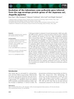
Phylogenetic analysis of 5'NCR sequences of HCV strainsFigure 1
Phylogenetic analysis of 5'NCR sequences of HCV strains. Strains in the trees are shown by their accession numbers
for strains previously described and their genotypes are indicated at the right side of the figure. Bolivian, Colombian and Uru-
guayan strains are shown by name. Number at the branches show bootstrap values obtained after 1000 replications of boot-
strap sampling. Bar at the bottom of the trees denotes distance. In (A) the phylogenetic tree for HCV strains isolated in South
America is shown. Strains assigned to a newly genetic lineage in HCV type 1 cluster are shown in red. Argentinean strains
[EMBL:DQ077818
] (Schijman et al., unpublished data), [EMBL:DQ313454] and [EMBL:AY376833] (Gismondi et al. [8, 9] previ-
ously reported as a new genetic lineage inside type 1 strains are shown in italics and an arrows denote its position in the figure.
Phylogeny for HCV strains isolated in North America and Europe are shown in (B), (C), respectively.
AY576550
AY576556
L27903
L27899
DQ06133
8
L27902
L38342
L38350
L27898
U51783
U51773
U51769
U5177
7
U5174
7
U51781
U51766
U51755
M74806
U5175
4
L27894
U51771
DQ319983
DQ06133
4
DQ319981
AY576577
DQ061340
M84842
DQ319985
L27904
DQ31997
8
L38318
M74809
U5175
8
L44599
U51761
M84838
AY576559
M84863
AF387732
U51752
L27896
AB154178
L27905
L27895
AB154179
AY576551
DQ061336
DQ061339
U5176
4
DQ061341
DQ319982
AJ238799
AJ132997
AB154180
D3172
4
DQ061335
DQ061331
L38351
DQ31998
4
M84841
L27901
AB154177
AY576558
DQ061333
AJ132996
DQ06133
7
DQ319980
AF387733
DQ061332
DQ319979
L27897
M84840
U51762
U51782
U51785
U51786
AY885238
U5178
8
U51753
U51748
M84839
M74811
D31722
AY576557
AY576576
L27871
AY725958
L27873
L27874
L27875
M74812
U51780
U51757
X84079
L27872
M84851
Z84280
DQ16474
8
DQ164751
DQ164752
DQ164753
Y13184
D31972
L38320
L38322
L38333
M84831
U51778
U51784
AB031663
L38319
L38321
L38334
L38335
L38336
L38337
M84833
U51759
U51775
Z84276
Z84279
U51779
Z84275
Z84277
Z84278
D31723
M84864
X76918
U51768
L12355
M84834
M84837
U51746
U51763
U51765
67
42
63
5
18
26
39
99
12
22
58
27
36
19
28
55
96
71
24
7
3
59
19
1
39
44
76
63
1
4
2
1
0.02
DQ061309
DQ061315
AY695436
DQ06132
4
L3438
4
DQ061316
U05029
DQ06131
4
L34386
M67463
M74808
DQ061320
AY446063
L34376
AY446046
DQ061323
AY446050
AY695437
DQ061312
DQ061310
AY446059
AY446061
DQ061325
AY446039
AY446065
AY446062
AY446043
AY446036
AF009606
AY446041
AF011752
AY446048
DQ06131
7
AY446042
DQ061318
AY446049
AY446064
AY446037
AF011753
AY446044
AY446045
AY446038
AY446057
AY446058
AF011751
AY446040
AY446047
AY446060
DQ010313
DQ061322
M84865
DQ061326
DQ061301
DQ061303
AY446051
AY446052
AY446053
AY446054
AY446055
AY446056
AY446066
AY446067
AY446068
DQ061296
DQ06129
7
DQ061299
L3437
7
L34385
L34388
M74813
M84830
M84857
U05028
L34387
U52810
U05026
U33430
U05023
U33432
AY434142
AY434155
AY434139
L34366
U05022
U05032
L34365
L34393
D14309
L34391
L34392
L34367
L34390
U05033
U05034
AY734478
AY434152
L3437
4
L34375
L34373
L34371
L34372
L34369
U05030
L34364
L34368
66
19
61
39
73
95
27
19
28
26
27
40
99
50
64
99
75
52
64
39
79
73
88
62
13
63
61
31
42
27
5
10
0.02
1a
5
4
BOL3
M84838
M84844
AJ291458
U05028
AF077232
M84863
AJ291457
AY576550
M84857
AJ238799
AJ132996
M84856
URU7B
URU23
L12354
AY576558
M84830
Z84287
DQ319979
Z84288
L34385
AJ438617
URU27
L34377
URU26
D31724
AJ132997
DQ319981
AY576553
DQ319980
L34388
AY576559
COL29
M84841
URU72
U45476
AB154177
AB154178
DQ319985
DQ319982
DQ31997
8
AB154180
L12353
AF077231
M84840
M84855
DQ319983
M84842
AY576552
AY576555
AB154179
AY576551
DQ31998
4
L34387
L34389
URU1
AF077236
URU51
M84839
L34386
M84865
L34376
URU20
X84079
AY576557
M67463
AJ438620
AF011751
DQ010313
URU41
URU60
AJ438619
AF011752
AF011753
AY576576
L34384
URU99
Z84280
URU64
URU7A
URUG8
M84851
URU8
BOL2
URU4
COL2
COL26
BOL6
BOL7
COL29
URU2
COL4
AY37683
3
COL20
BOL5
COL14
COL11
BOL1
DQ31345
4
URU14
COL18
URU6
URU7
BOL4
URU9
DQ07781
8
L34374
L34375
URUHCV20
L34373
L34368
L34369
L34371
L34372
M84860
M84852
M84858
U05026
M84862
M84832
L28058
COL5
COL25
Z84276
L34366
D31723
AF077233
M84864
URU17
X76918
L34365
URU66
L12355
M84834
D14309
U05032
M84837
L34367
D13448
L34390
URU18
AF077229
U05033
Z84279
URU29
L34392
L34391
L34393
AF077228
Z84275
Z84277
Z84278
62
34
64
77
94
47
46
17
16
48
45
13
11
1
4
0
0
24
1
0
33
17
57
25
41
43
96
27
62
100
91
91
95
65
38
1
0
0
83
83
71
43
72
63
36
0.02
4
6
A
1b
1a
2
3
B
C
1b
3i
3a
6
2
2b
1b
1a
5
2
3a
3
Virology Journal 2007, 4:79 />Page 4 of 12
(page number not for citation purposes)
in South America, this is not observed for type 1 strains
isolated in North America or Europe (compare Fig. 1A
with Figs. 1B and 1C).
Signature pattern analysis of type 1 HCV strains isolated in
South America
In order to test if the presence of the third phylogenetic
lineage in type 1 HCV strains isolated in South America
was due to a particular sequence signature, present exclu-
sively in HCV strains assigned to that lineage, a signature
pattern analysis was performed to assess viral sequence
relatedness. For that purpose, a query dataset of 19 type 1
HCV sequences belonging to this third cluster was ana-
lyzed using a background dataset of 19 type 1 HCV
sequences assigned to the two other clusters found in the
South American region (see Fig. 1A). The results of these
studies detected the presence of a sequence signature in
type 1 HCV strains assigned to the third genetic lineage in
the phylogenetic tree analysis (Fig. 2). Comparison of the
frequencies obtained for each particular nucleotide and
position in the signature gives statistical support to these
findings (Table 1). When similar studies were performed
using the same query dataset and background datasets of
sequences from strains isolated in Europe or North Amer-
ica, similar results were obtained (Table 1). These results
suggest that the sequence signature found in HCV type 1
strains isolated in South America may be characteristic of
this geographic region of the world. To observe if this
nucleotide sequence signature can be found indeed in
strains isolated outside the South American region, BLAST
studies were performed using sequences from strains bear-
ing the sequence signature as a query against all HCV
strains reported to HCV LANL Database [14]. Only strains
isolated in the South American region have 100% similar-
ity to the signature sequence strains (not shown).
Prediction of secondary structure of signature RNA
sequences
Biochemical and functional studies have revealed that the
5'NCR of HCV folds into a highly ordered complex struc-
ture with multiple stem-loops [15]. This complex RNA
structure contains four distinct domains, with domains II,
III and part of domain IV forming the IRES. These highly
folded secondary RNA elements function as cis-signals for
interaction with the 40S ribosome subunit and/or eukary-
otic translation initiation factors [6]. Signature mutations
map in IRES stem-loops II (G107A) and III (G243A,
C247U and U248C) relative to strain HCV1b [16] (see
Fig. 3).
To observe how these substitutions may affect IRES sec-
ondary RNA structure, predicted secondary structures of
HCV IRES domains II and III of consensus dataset
sequences of type 1 strains isolated in South America
(background dataset) and consensus signature sequence
dataset (query dataset) were compared. The results of
these studies are shown in Figs. 4 and 5, respectively.
As it can be seen in Fig. 4, the predicted secondary struc-
ture of domains II of background and signature consensus
sequences give similar structures. Nevertheless, mutation
A107 in the sequence signature might help to stabilize a
buckle in the structure by base pairing with U75 (compare
Figs. 4A and 4B).
In the case of IRES stem-loop III predicted secondary
structure, similar structures have also been obtained for
background and signature sequences (see Fig. 5). Never-
theless, mutations in stem-loop III does not seem to have
a particular effect in loop III folding (compare Figs. 5A
and 5B).
Discussion
Phylogenetic tree analysis of the 5'NCR from HCV strains
isolated in South America revealed that genotype 1 is the
most predominant in that region, in agreement with pre-
vious results [7]. There are no previous reports on the
genetic variation of HCV circulating in Bolivia. All Boliv-
ian strains enrolled in these studies have been clearly
assigned to genotype 1. Although more studies will be
needed in order to have a definitive picture on the degree
of genetic heterogeneity of HCV strains circulating in
Bolivia, the results of these studies suggests that genotype
1 might also be prevalent in that country (see Fig. 1A). In
the case of Colombia, previous studies suggested the pres-
ence of genotype 1 and 3 [17]. This is in agreement with
the results found in the present study. Interestingly, the
phylogenetic analysis revealed the presence of genotype 4
in Colombia for the first time (see Fig. 1A, bottom). This
genotype is prevalent in the Middle East [2] and not par-
ticularly in the South American region, although genotype
4 has been also found in Argentina [7]. More studies will
be needed to address the epidemiological situation of this
genotype in Colombia.
The phylogenetic analysis of HCV strains isolated in South
America also revealed the presence of a new genetic line-
age in HCV type 1 strains (Fig. 1A). These results are in
agreement with previous ones obtained for type 1 HCV
isolates circulating in Central and South America [8-12].
These previous data have suggested the presence of a dis-
tinct type 1 HCV sub-population in South America and a
diversification of HCV in that region. In this study, we
have analyzed more than 150 HCV strains isolated in
South America. The results of this work revealed that the
third type 1 sub-population observed in the phylogenetic
tree analysis of the HCV strains isolated in South America
is in fact due to the presence of a particular nucleotide sig-
nature sequence (Fig. 2 and Table 1). This sequence signa-
ture is frequent enough to be detected in a phylogenetic
Virology Journal 2007, 4:79 />Page 5 of 12
(page number not for citation purposes)
Signature pattern analysis of type 1 HCV strains isolated in South AmericaFigure 2
Signature pattern analysis of type 1 HCV strains isolated in South America. In (A) the consensus nucleotide
sequence in the background set of type 1 HCV strains isolated in South America is shown in black. The consensus nucleotide
sequence in the query (signature sequence) set is shown in red. Query sequence signature identified by VESPA is shown in
green. Numbers in the figure shows IRES nucleotide positions, relative to strain HCV1b [16]. In (B) an alignment of 5'NCR
sequences from strains belonging to the third cluster observed in type 1 HCV strains isolated in the South American region
with corresponding consensus sequences of type 1 HCV strains isolated in South America (Background1), Europe (Background
2) or North America (Background3) is shown. Strains are shown by accession number for strains previously described, or by
name at the left side of the figure. Identity to consensus sequences is indicated by a dash. Gaps introduced during alignment are
indicated by a dot.
A
63
TTCACGCAGAAAGCGTCTAGCCATGGCGTTAGTATGAGTGTCGTGCAGCCTCCAGGACCCCCCCTCCCGGGAGA
TTCACGCAGAAAGCGTCTAGCCATGGCGTTAGTATGAGTGTCGTACAGCCTCCAGGACCCCCCCTCCCGGGAGA
GCCATAGTGGTCTGCGGAACCGGTGAGTACACCGGAATTGCCAGGACGACCGGGTCCTTTCTTGGATCAACCCG
GCCATAGTGGTCTGCGGAACCGGTGAGTACACCGGAATTGCCAGGACGACCGGGTCCTTTCTTGGATCAACCCG
CTCAATGCCTGGAGATTTGGGCGTGCCCCCGCGAGACTGCTAGCCGAGTAGTGTTGGGTCGCGAAAGGCCTT
CTCAATGCCTGGAGATTTGGGCGTGCCCCCGCAAGATCGCTAGCCGAGTAGTGTTGGGTCGCGAAAGGCCTT
283
B
Background1 TTCACGCAGAAAGCGTCTAGCCATGGCGTTAGTATGAGTGTCGTGCAGCCTCCAGGACCCCCCCTCCCGGGAGAGCCATAGTGGTCTGCG
Background2
Background3
DQ313454 A
DQ077818 A
AY376833 A
Col20 A
Col26 A
Col18 A
Bol1 A
Bol2 A
Bol5 A
Uru8 A
Uru6 A
Uru2 A
Background1 GAACCGGTGAGTACACCGGAATTGCCAGGACGACCGGGTCCTTTCTTGGATCAACCCGCTCAATGCCTGGAGATTTGGGCGTGCCCCCGC
Background2
Background3
DQ313454
DQ077818
AY376833
Col20
Col26
Col18
Bol1
Bol2
Bol5
Uru8
Uru6
Uru2
Background1 GAGACTGCTAGCCGAGTAGTGTTGGGTCGCGAAAGGCCTT
Background2
Background3
DQ313454 A TC
DQ077818 A TC
AY376833 A TC
Col20 A TC
Col26 A TC
Col18 A TC
Bol1 A TC
Bol2 A TC
Bol5 A TC
Uru8 A TC
Uru6 A TC
Uru2 A TC
Virology Journal 2007, 4:79 />Page 6 of 12
(page number not for citation purposes)
tree analysis as a distinct type 1 sub-population (see Fig.
1A). Nevertheless, when the same analysis is carried out in
type 1 HCV strains isolated in Europe or North America,
only two genetic lineages are observed which correspond
to the major type 1 sub-types (see Fig. 1B and 1C).
Sequence signature pattern analysis has been useful for
epidemiological linkage, to corroborate transmission link
hypothesis or sequence relatedness studies [18-21]. The
identification of a sequence signature in the 5'NCR of type
1 HCV strains isolated in South America may permit a
more in-depth study on the molecular epidemiology of
HCV in this region.
Nevertheless, more studies will be needed to determine
the extent of distribution of this particular signature.
BLAST studies, on the other hand, have shown that only
type 1 HCV strains circulating in the South American
region have 100% similarity to the nucleotide sequence
signature found in that region.
HCV, as many other RNA viruses, replicates as complex
mutant distributions termed quasispecies [22-25]. Qua-
sispecies dynamics is characterized by continuous genera-
tion of variant viral genomes, competition among them,
and selection of the fittest mutant distributions in any
given environment [23]. The coexistence of distinct type 1
HCV subpopulations is consistent with quasispecies
dynamics, and suggests that multiple coexisting subpopu-
lations may occupy different regions on a fitness land-
scape to allow the virus to adapt rapidly to changes in the
landscape topology. This, in turn, may allow the virus to
adapt to its human host populations.
The 5'NCR, even though is one of the most conserved part
of the virus genome, shows a quasispecies distribution
with minor variants observed in the population [26] (Fig.
3). Since virus particles in serum are likely to be released
from the liver but also from compartments such as lym-
phocytes or dendritic cells, it has been suggested that the
sequence diversity found in the IRESs may reflect their
translational activity and tropism for these compartments
[27-29].
If all this is correct, the results of these studies may also be
related to these facts. Owing to the error-prone nature of
the HCV polymerase, mutations are expected to occur ran-
domly distributed over the 5'NCR. However, only muta-
tions compatible with replication and translation can be
propagated. Whether the stem-loop II and III mutations
observed confer a survival advantage or disadvantage in
vivo remains unknown. Nevertheless, the in silico pre-
dicted RNA secondary structures of IRES stem-loops sug-
gest that some mutations in the signature sequence might
have an effect in IRES structure. Further work with HCV
replicons containing the observed signature mutations
may help to clarify this point.
The unique structure of the HCV IRES makes it an attrac-
tive target for the development of antiviral agents directed
against this RNA element [30]. Mapping sequence signa-
tures in that region may help to understand their effects in
HCV IRES functions.
Conclusion
Phylogenetic analysis revealed the presence of a sequence
signature in the 5'NCR of type 1 HCV strains isolated in
South America. This signature is frequent enough in type
1 HCV populations circulating South America to be
detected in a phylogenetic tree analysis as a distinct type 1
sub-population. The coexistence of distinct type 1 HCV
subpopulations is consistent with quasispecies dynamics,
and suggests that multiple coexisting subpopulations may
allow the virus to adapt to its human host populations.
Methods
Serum samples
Serum samples were obtained from 7 volunteer blood
donors from Banco de Sangre de Referencia Departamen-
tal, La Paz, Bolivia, 14 volunteer blood donors from
Banco de Sangre de la Cruz Roja, Bogotá, Colombia and
26 HCV chronic patients from Servicio Nacional de San-
Table 1: Frequencies of signature nucleotides identified in the 5'NCR of type 1 HCV strains isolated in South America
a
Frequency of query nucleotides Frequency of background nucleotides
Position
b
: 107 243 247 248 107 243 247 248
Nucleotide: A A T C G G C T
Among query set: 0.947 0.947 0.947 0.947 0.053 0.053 0.053 0.053
Among background set 1: 0.000 0.421 0.000 0.000 1.000 0.579 1.000 1.000
Among background set 2: 0.000 0.316 0.105 0.105 1.000 0.684 0.895 0.895
Among background set 3: 0.158 0.368 0.000 0.000 0.842 0.632 1.000 1.000
a
Background sets 1, 2 and 3 are composed by type 1 HCV strains isolated in South America, Europe and North America, respectively.
b
Numbers refer to nucleotide sequence position relative to strain HCV1b sequence [14].
Virology Journal 2007, 4:79 />Page 7 of 12
(page number not for citation purposes)
gre, Montevideo, Uruguay. All patients tested positive in
an enzyme immunoassay from Abbott, used accordingly
to manufacturer's instructions. All patients were from La
Paz, Bogotá and Montevideo, respectively. For epidemio-
logical data of Bolivian, Colombian and Uruguayan
strains, see Table 2.
PCR amplification of 5'NCR of HCV strains
The 5'NCR of the HCV genome from samples that were
reactive in the enzyme immunoassay were amplified by
PCR, as previously described [31,32]. To avoid false posi-
tive results, the recommendations of Kwok and Higuchi
[33] were strictly adhered to. Amplicons were purified
using QIAquick PCR Purification Kit from QIAGEN,
according to instructions from the manufacturers.
Sequencing of PCR amplicons
The same primers used for amplification were used for
sequencing the PCR fragments, and the sequence reaction
was carried out using the Big Dye DNA sequencing kit
(Perkin-Elmer) on a 373 DNA sequencer apparatus (Per-
kin-Elmer). Both strands of the PCR product were
sequenced in order to avoid discrepancies. 5'NCR
sequences from position 62 through 285 (relative to the
genome of strain AF009606, sub-type 1A) were obtained.
For sequence accession numbers of Bolivian, Colombian
and Uruguayan HCV strains, see Table 2.
Phylogenetic tree analysis
5'NCR from HCV strains previously reported in South
America, Europe and North America were obtained from
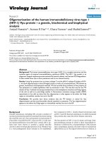
HCV IRES mutations found in sequence signature strains isolated in South AmericaFigure 3
HCV IRES mutations found in sequence signature strains isolated in South America. The 5'NCR sequences of
strain HCV1b [16] is shown. The locations of the nucleotide mutations found in the sequence signature are shown in bold and
a solid arrow indicates each particular substitution. Sequences previously identified to belong to a specific IRES domain [16] are
indicated by colours and domain number is indicated bellow the sequence. IRES nucleotide substitutions positions previously
reported in the literature [16] or in the HCV Database [14] are indicated in bold italics underlined. Each particular previously
reported substitution is indicated by a dotted arrow. Δ means deletion. Numbers in the figure denote nucleotide position in
HCV sequence according to strain HCV1b [16].
1 50
ʜ˨˨ ʜ
gccagcccccuguugggggcgacacuccaccauagaucacuccccugugaggaa
cuacugucuucacgcagaaag
domain I domain II
gg ¨
100 150
ʜʜ
cgucua
gccauggcguuaguaugagugucgugcagccuccaggaccccccccucccgggagagccauaguggucu
domain II
gu a g u u c
200
ʜ
gcggaaccggugaguacac
cggaauuccaggcagaccgguccuuucuuggaucaacccgcucaaugccuggagauu
domain IIIa domain IIIb
g u u g u
250
ʜ
uugggcgugcccccgcgagacugcuagccguaguguugggucgcgaaag
gccuugugguacugccugauagggu
domain IIIc domain IIId domain IIIe
a uc ¨ a a
Virology Journal 2007, 4:79 />Page 8 of 12
(page number not for citation purposes)
the LANL HCV Database [14]. Sequences were aligned
using the CLUSTAL W program [34]. Phylogenetic trees
were generated by the neighbor-joining method under a
matrix of genetic distances established under the Kimura-
two parameter model [13], using the MEGA3 program
[35]. The robustness of each node was assessed by boot-
strap resampling (1,000 pseudo-replicas).
Signature pattern analysis
Signature pattern analysis identifies particular sites in
amino acid or nucleic acid alignments of variable
sequences that are distinctly representative of a query set
relative to a background set. We employed the method
described by Korber & Myers [36] as implemented in the
VESPA program [37]. Sequences in the query and back-
ground datasets where aligned using the CLUSTAL W pro-
gram [34] and then transformed to the FASTA format
using the MEGA 3 program [35]. The query set was
formed by 19 type 1 HCV sequences isolated in South
America and representative of the third genetic lineage
identified in the phylogenetic tree analysis (see Fig. 1A).
The background set was formed by 19 type 1 HCV
sequences isolated in South America. The same studies
were performed using background sets of 19 type 1 HCV
strains isolated in Europe or North America. The thresh-
old was set to 0 (the program will use the majority consen-
Prediction of stem-loop II IRES RNA secondary structureFigure 4
Prediction of stem-loop II IRES RNA secondary structure. mfold results of IRES stem-loop II are shown. Numbers in
the figure denote nucleotide positions, ΔG obtained for the structures are shown on the bottom of the figure. In (A) mfold
results for consensus type 1 strains isolated in South America is shown. (B) shows mfold results for signature consensus
sequences.
73
83
B
63
93
A
73
83
63
93
53
53
113 113
103 103
Virology Journal 2007, 4:79 />Page 9 of 12
(page number not for citation purposes)
sus sequence in the query dataset for calculations) or 0.5
(the program will require that the signature nucleotides
be included at least in the 50% of the sequences in the
query set to be included for calculations). Both thresholds
gave the same results (not shown). For accession numbers
of strains included in query and background datasets see
Table 3.
Sequence similarity studies
Sequence similarity among query signature strain URU2
and all HCV strains of all types, isolated elsewhere, was
established using BLAST program [38], using the HCV
LANL Database [14].
Prediction of RNA secondary structure
Secondary structure prediction was done by the method
of Zuker & Turner [39], as implemented in the mfold pro-
gram (version 3.2) [40]. The core algorithm of this
method predicts a minimum free energy, ΔG, as well as
minimum free energies for foldings that must contain any
particular base pair. The folding temperature was set to
37°C. Ionic conditions was set to 1M NaCl, non divalent
ions. Base pairs that occur in all predicted folding struc-
tures are colored black. Otherwise, base pairs are assigned
in a multi-color mode that displays precisely what fold-
ings contain that base pair.
Competing interests
The author(s) declare that they have no competing inter-
ests.
Authors' contributions
JC and GM conceived and designed the study. MFG, KG,
ARM, and AGS contributed with HCV samples from
Colombia, Bolivia and Argentina, respectively, and to the
discussion of the results found in the study. GM, MM and
FL obtained PCR amplicons and sequences from Bolivian
and Colombian strains. MM contributed to the discussion
Prediction of stem-loop III IRES RNA secondary structureFigure 5
Prediction of stem-loop III IRES RNA secondary structure. Mfold results of IRES stem-loop III are shown. The rest
same as Fig. 4.
160 160
180
200
220
240
180
220
240
BA
200
Virology Journal 2007, 4:79 />Page 10 of 12
(page number not for citation purposes)
Table 2: Origins of Bolivian, Colombian and Uruguayan HCV strains
Name Accession Number Patient ID Age
a
Sex
Col2 [EMBL:AM269927] 451103850 39 Male
Col3 [EMBL:AM269928
] 451209881 54 Male
Col4 [EMBL:AM269929
] 451202563 20 Female
Col5 [EMBL:AM269926
] 451200819 30 Female
Col11 [EMBL:AM269930
] 451202594 49 Female
Col14 [EMBL:AM269931
] 451201641 23 Female
Col18 [EMBL:AM269932
] 451201714 24 Female
Col20 [EMBL:AM269936
] 451204950 29 Male
Col24 [EMBL:AM269937
] 451201157 28 Female
Col25 [EMBL:AM269925
] 451201208 31 Female
Col26 [EMBL:AM269933
] 451205577 25 Female
Col28 [EMBL:AM269934
] 451209889 21 Female
Col29 [EMBL:AM269935
] 451203054 25 Male
Bol1 [EMBL:AM400873
] 13183 46 Male
Bol2 [EMBL:AM400874
] 12713 48 Female
Bol3 [EMBL:AM400875
] 12577 42 Male
Bol4 [EMBL:AM400876
] 13410 42 Male
Bol5 [EMBL:AM400877
] 12573 43 Male
Bol6 [EMBL:AM400878
] 13177 42 Male
Bol7 [EMBL:AM400879
]13322 9 Male
Uru1 [AM709653
]H1 29 Male
Uru2 [AM709654
]H2 36 Male
Uru4 [AM709655
]H4 27 Male
Uru6 [AM709656
]H6 32 Male
Uru7 [AM709657
]H7 41 Male
Uru7A [AM709676
] HCV7A Adult Male
Uru7B [AM709671
] HCV7B Adult Male
Uru8 [AM709658
]H8 34 Male
UruG8 [AM709676
] HCVG8 Adult Female
Uru9 [AM709659
]H9 78 Male
Uru14 [AM709660
] H14 39 Male
Uru17 [AM709661
] H17 28 Male
Uru18 [AM709662
] H18 29 Male
Uru20 [AM709663
] H20 20 Male
Uru23 [AM709664
] H23 25 Male
Uru26 [AM709665
] H26 56 Male
Uru27 [AM709666
] H27 59 Male
Uru29 [AM709667
] H29 57 Male
UruHCV20 [AM709668
]HCV20 68 Female
Uru41 [AM709669
] HCV21 32 Male
Uru51 [AM709673
] HCV51 55 Male
Uru60 [AM709675
] HCV60 Adult Female
Uru64 [AM709672
] HCV64 Adult Female
Uru66 [AM709678
] HCV66 29 Male
Uru72 [AM709670
] HCV72 Adult Male
Uru99 [AM709474
] HCV99 Adult Male
a
Adult means older than 18.
Virology Journal 2007, 4:79 />Page 11 of 12
(page number not for citation purposes)
of the results found. RC, LL, RR, MPM and LG obtaining
PCR amplicons and sequences from Uruguayan strains. JC
wrote the paper. All authors have read and approved the
final document.
Acknowledgements
This work was supported by ICGEB, PAHO, and RELAB through Project
CRP.LA/URU03-032, and DINACYT, Uruguay, through Project No. 8006.
We thank Dr. Martín Abril, from Banco de Sangre de la Cruz Roja, Colom-
bia for invaluable help in HCV samples collection.
We thank Gustavo Saez (Grupo CentraLab, Argentina) for RT-PCR related
work with Argentinean HCV isolates.
References
1. Simmonds P, Bukh J, Combet C, Deleage G, Enomoto N, Feinstone S,
Halfon P, Inchauspe G, Kuiken C, Maertens G, Mizokami M, Murphy
DG, Okamoto H, Pawlotsky JM, Penin F, Sablon E, Shin-IT , Stuyver
LJ, Thiel HJ, Viazov S, Weiner AD, Widell A: Consensus proposals
for a unified system of nomenclature of hepatitis C virus gen-
otypes. Hematology 2005, 42:962-973.
2. Simmonds P: Genetic diversity and evolution of hepatitis C
virus 15 years on. J Gen Virol 2004, 85:3173-3188.
3. Hoofnagle JH: Course and outcome of hepatitis C. Hepatology
2002, 36:S21-S29.
4. Pawlotski JM: The nature of interferon-alfa resistance in hepa-
titis C virus infection. Curr Opin Infect Dis 2003, 16:587-592.
5. You S, Stump DD, Branch AD, Rice CM: A cis-acting replication
element in the sequence encoding the NS5B RNA-depend-
ent RNA polymerase is required for hepatitis C virus RNA
replication. J Virol 2004, 78:1352-1366.
6. Pestova TV, Shatsky IN, Fletcher SP, Jackson RJ, Hellen CUT: A
prokaryotic-like mode of cytoplasmic eukaryotic ribosome
binging to the initiation codon during internal translation ini-
tiation of hepatitis C and classical swine fever virus RNAs.
Genes Dev 1998, 12:6783.
7. Cristina J: Genetic diversity and evolution of hepatitis C virus
in the Latin American region. J Clin Virol 2005, 34:S1-S7.
8. Gismondi MI, Becker PD, Valva P, Guzman CA, Preciado MV: Phylo-
genetic analysis of previously nontypeable Hepatitis C virus
isolates from Argentina. J Cin Microbiol 2006, 44:2229-2232.
9. Gismondi MI, Staendner LH, Grinstein S, Guzman CA, Preciado MV:
Hepatitis C virus isolates from Argentina disclose a novel
genotype 1-associated restriction pattern. J Clin Microbiol 2004,
42:1298-12301.
10. San Roman M, Lezama L, Rojas E, Colina R, Garcia L, Carlos A, Khan
B, Cristina J: Analysis of genetic heterogeneity of hepatitis C
viruses in Central America reveals a novel genetic lineage.
Arch Virol 2002, 147:2239-2246.
11. Vega I, Colina R, García L, Uriarte R, Mogdasy C, Cristina J: Diversi-
fication of hepatitis C viruses in South America reveals a
novel genetic lineage. Arch Virol 2001, 146:1623-1629.
12. Colina R, Azambuja C, Uriarte R, Mogdasy C, Cristina J: Evidence of
increasing diversification of hepatitis C viruses. J Gen Virol
1999, 80:1377-1382.
13. Felsenstein J: Phylogeny interference package, version 3.5.
Department of Genetics, University of Washington, Seattle, U.S.A;
1993.
14. Kuiken C, Yusim K, Boykin L, Richardon R: The HCV Sequence
Database. Bioinformatics 2005, 21:379-384.
15. Rijnbrand RC, Lemon SM: Internal ribosome entry site-medi-
ated translation in hepatitis C replication. Curr Top Microbiol
Immunol 2000, 242:85-116.
16. van Leeuwen HC, Reusken CB, Roeten M, Dalebout TJ, Riezu-Boj JI,
Ruiz J, Spaan WJ: Evolution of naturally occurring 5'non-trans-
lated region variants of hepatitis C virus genotype 1b in
selectable replicons. J Gen Virol 2004, 85:1859-1866.
17. Yepes A, Alvarez C, Restrepo JC, Correa G, Zapata JC: [Viral gen-
otypes in patients with hepatitis C virus infection in Medel-
lin] [Article in Spanish]. Gastroenterol Hepatol 2002, 25:334-335.
18. Biswas S, Sanyal A, Hemadri D, Tosh C, Mohapatra JK, Manoj R, Ban-
dyopadhvav SK: Sequence analysis of non-structural 3A and 3C
protein-coding regions of foot-and-mouth disease virus sero-
type Asia 1 field isolates from an endemic country. Vet Micro-
biol 2006, 116:187-193.
19. Pistello M, Del Santo B, Butto S, Bargagna M, Domenici R, Bendinelli
M: Genetic and phylogenetic analyses of HIV-1 corroborate
the transmission link hypothesis. J Clin Virol 2004, 30:11-18.
20. Burke B, Derby NR, Kraft Z, Sauders CJ, Dai C, Llewellyn N, Zharkikh
I, Voitech L, Zhu T, Srivastava IK, Barnett SW, Stamatatos L: Viral
evolution in macaques coinfected with CCR5-and CXCR4-
tropic SHIVs in the presence or absence of vaccine-elicited
anti-CCR5 SHIV neutralizing antibodies. Virology 2006,
355:138-151.
21. Soares MA, De Oliveira T, Brindeiro RM, Diaz RS, Sabino EC, Brigido
L, Pires IL, Morgado MG, Dantas MC, Barreira D, Teixeira PR, Cassol
S, Tanuri A:
A specific subtype C of human immunodeficiency
virus type 1 circulates in Brazil. AIDS 2003, 17:11-21.
22. Chambers TJ, Fan X, Droll DA, Hembrador E, Slater T, Nickells MW,
Dustin LB, Dibisceglie AM: Quasispecies heterogeneity within
the E1/E2 region as a pretreatment variable during
pegylated interferon therapy of chronic hepatitis C virus
infection. J Virol 2005, 79:3071-3083.
23. Domingo E: Antiviral strategy on the horizon. Virus Res 2005,
107:115-116.
24. Feliu A, Gay E, Garcia-Retortillo M, Saiz JC, Foms X: Evolution of
hepatitis C virus quasispecies immediately following liver
transplantation. Liver Transpl 2004, 10:1131-1139.
25. Laskus T, Wilkinson J, Gallegos-Orozco JF, Radkowski M, Adair DM,
Nowicki M, Operskalski E, Buskell Z, Seeff LB, Vargas H, Rakela J:
Table 3: HCV strains included in query and background datasets for sequence signature studies
a
Dataset
b
Strains included
Query [EMBL:AM266927], URU1, URU2, URU4, URU6, URU8, URU9, URU14, [EMBL:AM269928], [EMBL:AM269929],
[EMBL:AM269930
], [EMBL:AM269931], [EMBL:AM269932], [EMBL:AM269933], [EMBL:AM269934], [EMBL:AM269935],
[EMBL:AM269936], [EMBL:DQ077818], [EMBL:DQ313454].
Background1 URUG7B, [EMBL:M84855
], [EMBL:M84856], URU11, [EMBL:AB154179], [EMBL:AY576553], [EMBL:AY576557],
[EMBL:DQ319979], [EMBL:M84838], [EMBL:M84839], [EMBL:M84841], [EMBL:AF077232], [EMBL:AF077236], [EMBL:AJ291457],
[EMBL:AJ438617
], [EMBL:AJ438619], [EMBL:AF011751], [EMBL:DQ010313], [EMBL:L34386].
Background2 [EMBL:AY576557
], [EMBL:AY576576], [EMBL:DQ319979], [EMBL:DQ313980], [EMBL:DQ319983], [EMBL:M84838],
[EMBL:M84840
], [EMBL:M84841], [EMBL:M84842], [EMBL:Z84279], [EMBL:Z84280], [EMBL:D31722], [EMBL:AB154177],
[EMBL:AB154178
], [EMBL:Z84284], [EMBL:AB154179], [EMBL:AB154180], [EMBL:D31723], [EMBL:D31724].
Background3 [EMBL:AF009606
], [EMBL:AY446036], [EMBL:AY446039], [EMBL:AY446043], [EMBL:AY446044], [EMBL:AY446049],
[EMBL:AY446050
], [EMBL:AY446051], [EMBL:AY446052], [EMBL:AY446053], [EMBL:AY446067], [EMBL:AY446068],
[EMBL:DQ061296
], [EMBL:DQ061297], [EMBL:DQ061299], [EMBL:L34377], [EMBL:L34385], [EMBL:L34388], [EMBL:L34389].
a
Strains previously reported are indicated by accession number, strains reported in this work are indicated by name.
b
Background datasets 1, 2 and 3, correspond to strains isolated in South America, Europe or North America, respectively.
Publish with BioMed Central and every
scientist can read your work free of charge
"BioMed Central will be the most significant development for
disseminating the results of biomedical researc h in our lifetime."
Sir Paul Nurse, Cancer Research UK
Your research papers will be:
available free of charge to the entire biomedical community
peer reviewed and published immediately upon acceptance
cited in PubMed and archived on PubMed Central
yours — you keep the copyright
Submit your manuscript here:
/>BioMedcentral
Virology Journal 2007, 4:79 />Page 12 of 12
(page number not for citation purposes)
Analysis of hepatitis C virus quasispecies transmission and
evolution in patients infected through blood transfusion. Gas-
troenterology 2004, 127:764-776.
26. Lu M, Kruppenbacher J, Roggendorf M: The importance of the
quasispecies nature of hepatitis C virus (HCV) for the evolu-
tion of HCV populations in patients: study on a single source
outbreak of HCV infection. Arch Virol 145:2201-2210.
27. Laporte J, Bain C, Maurel P, Inchauspe G, Agut H, Cahour A: Differ-
ential distribution and internal translation efficiency of hep-
atitis C virus quasispecies present in dendritic and livel cells.
Blood 2003, 101:52-57.
28. Laporte J, Malet I, Andrieu T: Comparative analysis of transla-
tion efficiencies of hepatitis C virus 5' untranslated regions
among intraindividual quasispecies present in chronic infec-
tion: opposite behaviours depending on cell type. J Virol 2000,
74:10827-10833.
29. Lerat H, Shimizu YK, Lemon SM: Cell type-specific enhancement
of hepatitis C virus internal ribosome entry site-directed
translation due to 5' nontranslated region substitutions
selected during passage of virus in lymphoblastoid cells. J Virol
2000, 74:7024-7031.
30. Kurreck J: Antisense technologies. Improvement through
novel chemical modifications. Eur J Biochem 2003,
270:1628-1644.
31. Chan SW, McOmish F, Holmes EC, Dow B, Peutherer JF, Follett E,
Yap PL, Simmonds P: Analysis of a new hepatitis C virus type
and its phylogenetic relationship to existing variants. J Gen
Virol 1991, 73:1131-1141.
32. Davidson F, Simmonds P, Ferguson JC, Jarvis LM, Dow BC, Follett EA,
Seed CR, Krusius T, Lin C, Madyuesi GA: Survey of major geno-
types and subtypes of hepatitis C virus using RFLP of
sequences amplified from the 5' non-coding regions. J Gen
Virol 1995, 76:1197-1204.
33. Kwok S, Higuchi R: Avoiding false positives with PCR. Nature
1989, 339:237-238.
34. Thompson JD, Higgins DG, Gibson TJ: CLUSTAL W: improv-
ingthe sensitivity of progressive multiple sequence align-
ment throughsequence weighting, position-specific gap
penalties and weight matrix choice. Nucleic Acid Res 1994,
22:
4673-4680.
35. Kumar S, Tamura K, Nei M: MEGA 3: Integrated software for
Molecular Evolutionary Genetics Analysis and sequence
alignment. Brief Bioinformatics 2004, 5:150-163.
36. Korber B, Myers G: Signature pattern analysis: a method for
assessing viral sequence relatedness. AIDS Res Hum Retroviruses
1992, 8:1549-1560.
37. [ />].
38. [ />].
39. Mathews DH, Sabina J, Zuker M, Turner DH: Expanded sequence
dependence of thermodynamic parameters improve predic-
tion of RNA secondary structure. J Mol Biol 1999, 288:911-940.
40. [ />].