Báo cáo sinh học: " Cytopathic Mechanisms of HIV-1" pot
Bạn đang xem bản rút gọn của tài liệu. Xem và tải ngay bản đầy đủ của tài liệu tại đây (805.64 KB, 22 trang )
BioMed Central
Page 1 of 22
(page number not for citation purposes)
Virology Journal
Open Access
Review
Cytopathic Mechanisms of HIV-1
Joshua M Costin
Address: Biotechnology Research Group, Department of Biology, Florida Gulf Coast University, 10501 FGCU Blvd. S., Fort Myers, Fl, 33965, USA
Email: Joshua M Costin -
Abstract
The human immunodeficiency virus type 1 (HIV-1) has been intensely investigated since its
discovery in 1983 as the cause of acquired immune deficiency syndrome (AIDS). With relatively
few proteins made by the virus, it is able to accomplish many tasks, with each protein serving
multiple functions. The Envelope glycoprotein, composed of the two noncovalently linked subunits,
SU (surface glycoprotein) and TM (transmembrane glycoprotein) is largely responsible for host cell
recognition and entry respectively. While the roles of the N-terminal residues of TM is well
established as a fusion pore and anchor for Env into cell membranes, the role of the C-terminus of
the protein is not well understood and is fiercely debated. This review gathers information on TM
in an attempt to shed some light on the functional regions of this protein.
Review
HIV discovery and clinical presentation
In 1981 the CDC (USA) began noting a group of homo-
sexual men presenting with symptoms of a rare opportun-
istic infections at a San Francisco clinic [1,2]. These
patients were later found to be suffering from severe
immune deficiency and their syndrome was dubbed
acquired immune deficiency syndrome (AIDS). In 1983,
two viruses were simultaneously isolated in the United
States and France thought to be the cause of these infec-
tions, named HTLV-III (Human T Lymphotropic Virus)
and LAV (Lymphadenopathy Associated Virus) respec-
tively [3-8]. HTLV-III and LAV, along with a third virus
isolated from AIDS patients in San Francisco, named ARV
for AIDS-associated Retrovirus [9] were later discovered to
be the same virus and renamed Human Immunodefi-
ciency Virus, or HIV [10].
Since its discovery it has been estimated that more than
64.9 million people have been infected with HIV world-
wide, with greater than 32 million AIDS-related deaths
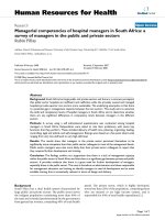
(refer to [222]. Infection with HIV is characterized by
three clinical stages – acute viremia, a latency phase of var-
iable duration, and a classification of clinical AIDS (Figure
1). Concurrent with initial infection, virus can be detected
in the blood of patients [11,12]. After the initial viremia
peaks, the level of virus in the blood falls off and a phase
of "latency" ensues. During the latency phase, HIV load is
generally very low to non-detectable, though there is a
high turnover of CD4
+
T cells and HIV virion production
[13-17]. Before the advent of highly active antiretroviral
therapy (haart), it was established that the levels of virus
in the blood at this stage are negatively correlated with
prognosis and time course of progression to AIDS [17-19].
It is during the latency phase that CD4
+
T cell counts also
begin to decline and an inversion of the CD4
+
/CD8
+
T cell
ratio occurs. A CD4
+
T cell count below 200 cells/mm
3
and infection with at least one opportunistic infection,
such as Pneumocystis Carinii defines clinical AIDS. It is at
this final stage where patients' immune systems are no
longer able to function properly and patients eventually
succumb to their secondary infections, to otherwise rare
cancers (such as Kaposi's sarcoma) or to other manifesta-
tions of HIV infection (such as neuropathy).
Published: 18 October 2007
Virology Journal 2007, 4:100 doi:10.1186/1743-422X-4-100
Received: 4 September 2007
Accepted: 18 October 2007
This article is available from: />© 2007 Costin; licensee BioMed Central Ltd.
This is an Open Access article distributed under the terms of the Creative Commons Attribution License ( />),
which permits unrestricted use, distribution, and reproduction in any medium, provided the original work is properly cited.
Virology Journal 2007, 4:100 />Page 2 of 22
(page number not for citation purposes)
HIV classification, structure, genome, and replication cycle
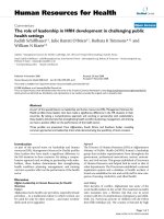
HIV is enveloped, contains reverse transcriptase and 2
identical copies of a positive sense, linear RNA genome
(Figure 2). HIV is classified in a subgroup of retroviruses
called the lentiviridae based on these "morphological,
genetic, and biological properties" [10,20]. HIV is a slow
virus – the clinical "latency" phase can last more than 20
years. During this time, HIV can have widespread effects
on immunological and neurological systems. Lentiviruses
are known for their cytolytic and immunosuppressive
properties and include viruses such as simian immunode-
ficiency virus (SIV), feline immunodeficiency virus (FIV),
caprine arthritis-encephalitis virus (CAEV), and equine
infectious anemia virus (EIAV).
As with all lentiviruses, HIV possesses a complex genome
(in this case, 9.8 kb) containing accessory and regulatory
genes (Figure 3). An additional, novel open reading
frame, vpu separates the pol and env regions [10,21]. In
total 9 genes are present that can be classified into 3 func-
tional groups. Gag, Pol, and Env are structural genes; Tat
and Rev are regulatory genes; Vpu, Vpr, Vif, and Nef are
accessory genes. A general overview of the replication
cycle in a single cell is presented in Figure 4. After direct
fusion of the virion and cellular lipid membranes, the
viral core is released into the cytoplasm where it uncoats
and releases the RNA genome. The viral genome is then
reverse transcribed and transported to the nucleus where
it integrates as a provirus. The early gene products, tat, rev,
and nef are first transcribed, followed later by the rest of
the HIV genome. Assembly and budding of progeny viri-
ons takes place at the plasma membrane.
Gag codes for the capsid protein which recruits two copies
of the RNA genome, the pol gene products (reverse tran-
scriptase, protease, and integrase), and other viral and cel-
lular gene products to the plasma membrane for budding
of the virus. Env encodes the Envelope protein, or Env,
which is synthesized as a single polyprotein in the endo-
plasmic reticulum. After synthesis, Env (gp160) is heavily
glycosylated in the Golgi complex before a cellular pro-
tease cleaves it into the noncovalently associated proteins,
surface glycoprotein (SU, or gp120) and transmembrane
glycoprotein (TM, or gp41).
SU is an extracellular protein which primarily functions to
recognize HIV's primary and secondary cellular receptors,
CD4 and CCR5/CXCR4 respectively on target cells [22].
Analysis of the expression of these receptors in immune
cells is sufficient to explain the tropism of HIV, primarily
macrophages and T lymphocytes. TM on the other hand
appears to function in membrane interactions. It is an
integral membrane protein which contains a transmem-
brane anchor domain that anchors Env into the lipid
membrane [10]. TM is responsible for fusion of the viral
and cellular membranes via its fusion peptide located in
TM's extracellular, N-terminal domain. The fusion pep-
tide of HIV-1 has shown some structural and functional
similarities to the hydrophobic internal region of bovine
prion protein (BPrP
tm
) [23]. Both of these peptides are
notable for their ability to interact with, and insert into
membranes. After the addition of calcium, there is a shift
in conformation from α-helix to β-sheet which accompa-
nies membrane fusion. The C-terminal, cytoplasmic tail
of TM is known to help direct the assembly of virions at
the cell surface [24], among other functions (see below).
The regulatory proteins, Tat and Rev are both RNA bind-
ing proteins. Tat is an RNA binding protein and transcrip-
tional activator that works to ensure full length HIV
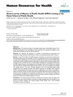
The HIV-1 virionFigure 2
The HIV-1 virion. Graphical depiction of the HIV-1 virion.
Vpu is not thought to be present in the virion in any appreci-
able amount.
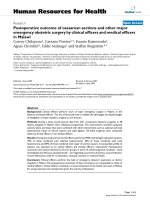
Time course of HIV infectionFigure 1
Time course of HIV infection. Time course of HIV infec-
tion showing correlation of viral load, CD4
+
T cell, and CD8
+
T cell counts.
Virology Journal 2007, 4:100 />Page 3 of 22
(page number not for citation purposes)
genomes are produced [25]. Tat is also known to activate
cellular genes such as TNF-β and TGF-β as well as down-
regulate the expression of other cellular genes such as bcl-
2 and MIP1-α. HIV's other regulatory protein, Rev, is an
RNA binding protein that is required for the transition of
HIV gene expression from the early phase to the late phase
[26]. Rev accomplishes this through binding of unspliced
or incompletely spliced viral RNA's in the nucleus and
nucleolus and then transporting them into the cytoplasm,
leaving fewer viral RNA's to be completely spliced.
The accessory proteins coded in the HIV genome are
known to be multifunctional. Nef, or negative factor, has
been shown to downregulate existing CD4 and MHC I
expression at the cell surface via degradation in lysosomes
[27,28]. Nef can perturb T cell activation (up- or down-
regulate) and stimulate HIV virion infectivity. Nef shows
sequence and structural features of scorpion peptides
known to interact with K
+
channels. When Nef is added to
chick dorsal root ganglion an increase in K
+
current is
observed [29]. Vpr allows HIV to infect nondividing cells
by acting as a nucleocytoplasmic transport factor [30]. Vpr
has reported cation-selective ion channel activity in planar
lipid bilayers [31]. Vpr "pores" may be active in both
nuclear and mitochondrial membranes [32-34]. In the
nuclear membrane, Vpr may facilitate the translocation of
the HIV-1 preintegration complex from the cytoplasm to
the nucleus. In mitochondrial membranes, Vpr binds to
the adenine nucleotide translocase (ANT), part of the
mitochondrial permeability transition pore (MPTP).
Binding of Vpr to ANT can convert it to a pro-apoptotic
pore, leading to uncoupling of mitochondrial respiration,
loss of transmembrane potential, swelling of the matrix,
and release of intermembrane proteins. Additionally, Vpr
acts to arrest the cell cycle in the G2 phase, preventing
entry into mitosis [35]. The internal membrane localized
Vpu functions to downmodulate CD4 expression via
ubiquitin-mediated degradation and to enhance virion
release through the formation of an ion channel which
collapses membrane potential and may promote virion
release (discussed in greater depth below) [27]. Finally,
Vif is essential for the replication of HIV in PBMC's, lym-
phocytes, macrophages, and certain cell lines suggesting
that it may act through interaction with a cellular factor
that is host species specific [26].
HIV cytopathology and induced ion modifications
Selective depletion of CD4
+
T cells is a hallmark of HIV
infection and is accomplished, at least in part, due to
direct cytopathic effects (CPE) of the virus [36]. The HIV
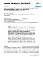
HIV genome and replication cycleFigure 3
HIV genome and replication cycle. Depiction of the ~10 Kb HIV-1 genome showing the organization of genes and their
transcriptional splicing (dashed lines). Relevant TM domains are highlighted.
Virology Journal 2007, 4:100 />Page 4 of 22
(page number not for citation purposes)
replication cycle is complex and not completely under-
stood. It is increasingly thought to begin via interaction
with dendritic cells during transmission [37]. A protein
present on dendritic cells, DC-SIGN, reversibly binds HIV,
with or without internalizing it, and shuttles it to a
regional lymph node, thought to be the primary site for
replication and spread of HIV. When the virus encounters
a macrophage or T cell with its primary CD4 receptor and
a coreceptor, either CXCR4 or CCR5, conformational
changes caused by the binding of SU expose the fusion
peptide of TM triggering direct fusion of the HIV and host
cell membranes. CD4 is expressed on many cells in the
body, but is found in highest levels on T lymphocytes,
macrophages, and in the brain, primarily astrocytes [38].
The specificity for the coreceptor is determined by the V3
loop region of SU and explains the tropism of the virus for
specific cell types [39]. CCR5-utilizing HIV (macrophage
tropic, non-syncytium inducing) strains are preferentially
transmitted over CXCR4-utilizing (T cell tropic, syncy-
tium inducing) strains for reasons that are not completely
understood [40,41]. A naturally occurring ∆CCR5 muta-
tion in humans correlates with resistance to infection by
HIV [42]. The emergence of CXCR4 strains during the
course of an infection is correlated with increased CD4
+
T
cell depletion and accelerated progression towards AIDS
[43]. This increase in T cell depletion can at least be par-
tially explained by the fact that a higher percentage of T
cells express CXCR4 (90–100%) than express CCR5 (10–
30%) [44,45] and suggests a role for direct cytopathic
effect by HIV.
The ability to directly lyse CD4
+
T cells have been postu-
lated to at least partially cause the reduction of these
immune effecter cells which leads to the clinical condition
of AIDS. Three additional mechanisms have been postu-
lated for CD4
+
T cell depletion including immune destruc-
tion of infected cells, apoptosis, and impaired lymphocyte
regeneration. These alternative mechanisms for in vivo
CD
+
T cell depletion are reviewed in McMichael et al.,
2000, Alimonti et al., 2003, and Douek et al., 2003 respec-
tively [46-48]. The relative contribution of each of these
mechanisms, if any, is still not clear. However, there is
strong evidence that direct cytopathic effects of the virus
play a large role in its pathogenicity.
Only cells expressing CD4 along with the proper corecep-
tor are infected by HIV [38,49]. HIV kills cells in cell cul-
ture as well as in vivo. Through the course of natural
disease, the virus switches use of coreceptors from the less
cytopathic CCR5 (R5), non-syncytium inducing (NSI)
variants to the more cytopathic CXCR4 (X4), syncytium
inducing (SI) variants [41]. The emergence of X4 variants
during an infection is associated with an accelerated pro-
gression towards AIDS [43]. After the development of
Highly Active Anti-Retroviral Therapy (HAART), it
became clear that HIV-1 infection was a highly dynamic
process involving massive covert replication of HIV-1 in
lymphoid tissues at all stages of an infection with contin-
ual destruction and regeneration of CD4
+
lymphocytes
[50]. It is estimated that HIV-infected cells and plasma vir-
ions have drastically shortened average life spans in vivo
– 2.2 and 0.3 days respectively [14-16,51]. Uninfected T
lymphocytes can survive >80 days by comparison [51]. If
the estimates of total HIV virion production of 10.3 × 10
9
virions a day are correct, then statistically there are enough
virions present in an in vivo infection to cause massive
direct CPE [52,53].
In vitro, HIV causes two types of CPE – syncytia and single
cell lysis. Syncytia are formed when Env expressed on an
infected cell late in infection interacts with CD4 of a
neighboring cell, triggering the fusion peptide of TM to
fuse the two membranes. Repeated occurrences of this
event allows for the formation of giant, multinucleated
cells. This type of CPE is thought rarely, if ever to occur in
vivo, and in fact rarely occurs during infection of human
Overview of the replication cycle of HIV-1Figure 4
Overview of the replication cycle of HIV-1. Overview
of some of the basic steps of HIV infection of a cell.
Virology Journal 2007, 4:100 />Page 5 of 22
(page number not for citation purposes)
PBL's in vitro, with the possible notable exception of the
brain [54-56]. HIV patients with AIDS Dementia Complex
(ADC) are found to have many giant, multinucleated cells
in the brain upon autopsy, mostly consisting of glial cells
known to express CD4. In addition to multinucleated syn-
cytial cells, single cells infected with HIV undergo a proc-
ess termed balloon degeneration whereby cells swell up
beyond the limits of their membrane integrity and lyse.
This is by far the most common type of CPE observed in
vitro [10,20,36,57]. Cell swelling in this case appears to be
irreversible in most cells, though it has been hypothesized
that those cells which can overcome these alterations in
cell volume may survive to become a population of chron-
ically infected cells [20]. One factor that both of these
types of CPE have in common is increases in cell volume.
Though syncytia do not generally lyse, they do show
increases in cell volume.
Experimenting with Sendai virus, Micklem and Pasternak,
1977 observed that alterations in the plasma membrane
of infected cells occurred within minutes of adsorption of
the virus [58]. These alterations included: changes in
intracellular ion concentrations, osmotically driven water
entry, and an increase in cell volume [59,60]. Basford et
al., 1984 hypothesized that after direct fusion of Sendai
virus lipid membrane with the host cell, the viral lipids
and proteins introduced into the host cell were able to
perturb the membrane in a manner reminiscent of the bee
venom melittin [61]. In the case of HIV, Grewe et al., 1990
noted that early interactions of HIV with host cell mem-
branes were similar to those observed with Sendai virus
[62]. Further evidence provided by Rasheed et al., 1986
showed that HIV was able to cause CPE as an early event.
UV-irradiated HIV, lacking the ability to replicate but still
able to infect cells by direct fusion of its lipid membrane
to the host cell still caused single cell balloon degenera-
tion of the RH9 T lymphoblastoid cell line. Cloyd and
Lynn, 1991 further proved that the permeability of the
host plasma membrane was enhanced early (12–24
hours) post infection to small molecules such as Ca
2+
and
sucrose, with greater permeability seen later (24–72
hours) post infection [54].
Viral ion channels, or viroporins, are present in many lytic
animal viruses. The cellular plasma membrane maintains
cellular materials and ionic gradients necessary for the
proper functioning of the cell. The ability to alter intracel-
lular ion concentrations is necessary for many of these
animal viruses in their life cycles and is a common theme
of cytolytic viruses [63,64].
HIV infection causes increases in intracellular monovalent
cations during infection analogous to what has been
observed for other animal cytolytic viruses, such as polio-
virus and sindbis virus. Acute infection of RH9 cells, a T-
lymphoblastoid cell line, with HIV-1
HXB2
, a lab adapted
strain, increases intracellular Na
+
and K
+
concentrations as
measured by ion sensitive dyes [65,66]. The flow of the
osmotically active monovalent cations, K
+
and Na
+
into
infected cells correlates with CPE. Increased intracellular
ion content is expected to be associated with increased
water influx into the cell to balance osmolarity, thereby
expanding the total volume of both single and syncytial
cells. Furthermore, strains of HIV known to be more cyto-
pathic, the syncytium inducing (SI) strains, induced
greater increases in [Na
+
]
i
and [K
+
]
i
than did non-syncy-
tium inducing (NSI) strains of HIV [66]. This correlation
remained when primary isolates of HIV were used in place
of the lab adapted strain, and when primary human
PBMC's were used in place of immortalized RH9 cells
[66].
Addition of loop diuretics such as bumetanide and furo-
samide, specific inhibitors of the Na
+
/K
+
/2Cl
-
cotrans-
porter, at least partially blocked increases in [Na
+
]
i
and
[K
+
]
i
levels, suggesting that HIV alters this transporter's
normal function in cell volume control [67]. Makutonina
et al., 1996 observed a concomitant decrease in pH, from
pH 7.2 in uninfected cells, to pH 6.7 in HIV-infected RH9
cells using a pH sensitive dye [68]. Use of the Na
+
/H
+
ant-
iporter inhibitor amiloride did not further decrease HIV
infected cell pH
i
, but did decrease control cells. This
implies that HIV may be inhibiting the Na
+
/H
+
antiporter
in some manner. The authors further suggest that the
increases in [Na
+
]
i
observed during infection may itself
lead to this shutoff as it would be unfavorable to exchange
an extracellular Na
+
for an intracellular H
+
when the [Na
+
]
i
is already high.
Some viruses alter intracellular ion concentrations in
order to get their mRNA's preferentially translated. Cellu-
lar mRNA's are only functional within a narrow range of
intracellular ion concentrations, while viral RNA's have
been shown to be more resistant [69-73]. Previous studies
involving animal cytolytic viruses have shown that alter-
ing the external ion concentration can affect internal ion
concentrations and pathogenesis of the virus. Altering the
external concentration of K
+
in the medium of HIV-
infected RH9 cells alters the cytopathicity of HIV [65].
Decreasing [K
+
]
e
to zero abrogates visible CPE in cell cul-
ture and lowers HIV protein translation by 40–50%. Alter-
natively, increasing [K
+
]
e
from 5 mM (normal) up to as
much as 75 mM increases visible CPE and increases HIV
protein translation as much as three fold. Altering [K
+
]
e
with primary human PBMC's has an even greater effect on
CPE and protein translation than it had with cell culture.
Alteration of the external Na
+
concentration did not affect
CPE or HIV protein translation [65]. For comparison,
increased [K
+
]
e
does not increase poliovirus or Sindbis
virus production or CPE [69,70].
Virology Journal 2007, 4:100 />Page 6 of 22
(page number not for citation purposes)
Selected Literature review of viral membrane permeability
altering proteins
Increased membrane permeability caused by viroporins,
glycoproteins, and proteases is a typical feature of animal
virus infections [63]. Viroporins are virally encoded, small
(generally ≤120 amino acid residues) membrane proteins
that form selective channels in lipid membranes. These
channels are less discriminating than the highly selective
ion channels of bacteria and eukarya and have been
hypothesized to be a family of primordial proteins which
predate the latter [27]. Features common to viroporins
include: promoting the release of virus, altering cellular
vesicular and glycoprotein trafficking, and increasing
membrane permeability. Amphipathic α-helical domains
of viroporins generally oligomerize to form the channel
by inserting into lipid membranes with the hydrophobic
residues oriented towards the lipid bilayer and the
hydrophilic residues facing in towards the lumen of the
channel. Though viroporins are not essential for virus rep-
lication, they may be necessary for full pathogenesis in
vivo as they are known to enhance virion production and
release [64,74,75]. Many lytic viruses employ altered
[ion]
i
(intracellular ion concentrations) in various stages
of their replication cycles. This can include steps such as
uncoating, host cell translation shutoff, and release of vir-
ions from infected cells. Viroporins are not the only strat-
egy viruses employ to alter [ion]
i
– other strategies include
generalized membrane destabilization and alteration of
existing ion channel and pump functions or expression
[20,63,64].
Influenza virus
The prototype viroporin, M2 protein, was first isolated
from the influenza A virus. M2 protein is one of three pro-
teins found in the virion envelope and is present in less
abundance than either of the other two envelope proteins,
hemagglutinin (HA) and neuraminidase (N) [76]. Early
studies to block influenza A virus infection showed that
the virus was sensitive to the compound amantadine at
two stages of its replication cycle [77,78]. The first block
occurs early in infection after attachment, but before
uncoating. As a consequence of this block, a buildup of
nondissociated matrix (M1) and ribonucleoprotein
(RNP) occurs in endosomal compartments [79,80]. The
second block occurs late in infection and inhibits the
release of virions [81]. At this late stage of infection,
amandatine causes a buildup of HA protein during trans-
port through the trans Golgi network that has undergone
the acid-induced conformational changes normally
observed with viral entry.
Sequencing of viruses with amantadine resistance
mapped the mutations responsible for resistance to the
transmembrane domain of the M2 protein, a highly con-
served protein, even across human, swine, equine, and
avian strains of influenza A virus [78,82]. The transmem-
brane domain of the M2 protein models to form amphip-
athic α-helices that associate minimally as homotetramers
in membranes, forming an ion channel [81,83]. Expres-
sion of M2 RNA in Xenopus oocytes and analysis of whole
cell currents showed a channel selective for monovalent
cations that was activated by low pH [84], though later
experiments showed the channel to be ~1.5 – 2.0 × 10
6
more selective for H
+
than Na
+
[85]. Mutations in the
membrane spanning domain of M2 that conferred aman-
tadine resistance also decreased the conductance of these
variant M2 proteins when expressed in Xenopus oocytes.
Purified M2 protein, as well as peptides corresponding to
the TM region of M2, produced an increased conductance
of planar lipid bilayers at low pH that was able to be
blocked by the addition of amantadine [86,87]. It was
then theorized that the M2 protein acts after receptor
mediated endocytosis to acidify the interior of the virion
and dissociate the matrix protein from the ribonucleopro-
tein (the first block seen with amantadine), allowing the
ribonucleoprotein (RNP) to enter the cytoplasm. The M2
protein was also theorized to work late in infection to pre-
vent Golgi vesicle acidification. This prohibits a prema-
ture change in conformation of the HA protein (the
second block seen with amantadine), which would halt
the assembly of virions. It is important to note that viruses
deficient in M2, while severely delayed in growth kinetics
are able to undergo multiple rounds of replication in cul-
tured cells. Thus the M2 protein is not essential for influ-
enza A virus replication, but does enhance viral
productivity [64,82,88].
A protein analogous to the M2 protein of influenza A virus
was discovered in the influenza B virus genome. The NB
protein (a.k.a. – BM2) shares many characteristics with
the M2 protein. Peptides corresponding to the predicted
transmembrane region form α-helices and increase the
conductance of lipid bilayers [89,90]. This conductance is
inhibited by amantadine, though at a higher concentra-
tion than is necessary for the M2 protein of influenza A
virus [91]. Purified whole NB protein also increases the
conductance of lipid bilayers in a fashion similar to the
TM region peptides [92]. NB protein expressed in either
Xenopus oocytes or mammalian cells form a proton selec-
tive channel that is presumably used in a manner analo-
gous to the M2 protein of influenza A virus; for
acidification of the virion during uncoating in the endo-
somal compartment and to equilibrate Golgi vesicles to
prohibit premature acid-induced conformational changes
in the HA protein of influenza B virus [93]. Single amino
acid mutations in the transmembrane region of the NB
protein abrogate proton selectivity of the channel, further
supporting an analogous role for NB in influenza B virus
infections [94].
Virology Journal 2007, 4:100 />Page 7 of 22
(page number not for citation purposes)
Early evidence suggests that influenza C virus also encodes
an ion channel (CM2) that is a minor virion component
[95]. CM2 protein has been shown to possess an α-helical
transmembrane domain similar to both the M2 and NB
proteins discussed above [96]. However, expression of
CM2 protein in Xenopus oocytes shows a voltage-acti-
vated, Cl
-
-selective ion channel that was not activated by
low pH, nor was it inhibited by even high (1 mM) concen-
trations of amantadine [97]. Studies involving influenza
C virus uncoating do not show a dependence on low pH
to dissociate the matrix and ribonucleoproteins. At the
present time it remains unclear how CM2 protein func-
tions during influenza C virus infection.
HIV
Viral protein U (Vpu) of HIV-1 (and SIV
cpz
) is an integral
membrane protein found predominantly in the endoplas-
mic reticulum (ER) and Golgi. It is possibly found to a
lesser extent the plasma membrane, but does not seem to
be present in the virion [98,99]. Vpu is expressed late in
infection as a bicistronic RNA that also codes for the Env
protein, which is differentially spliced to produce each
protein (see Figure 3). HIV-1 virions deficient in Vpu are
impaired in their ability for correct assembly and release.
A large proportion of these mutant virus particles display-
ing altered size and shape from wild type virions remain
attached to the cell surface [75]. Vpu possesses two func-
tional domains known to enhance the release of virions
from infected cells. The C-terminal cytoplasmic tail of Vpu
functions to enhance the degradation of CD4 in the ER
[100]. Vpu does not accomplish this task directly, but
instead binds CD4 and β-transducin repeats-containing
protein (β-TrCP), forming a ternary complex. Formation
of this complex requires two phosphorylated serine resi-
dues (52 and 56) of the Vpu cytoplasmic tail and targets
CD4 for proteolysis using the ubiquitin-dependent prote-
osome pathway [27,101,102]. It is thought that decreas-
ing the level of expression of CD4 decreases the formation
of CD4:Env complexes in the ER, allowing for increased
levels of Env expression on the plasma membrane.
Increased levels of Env expression at the plasma mem-
brane in turn increases the frequency of virion budding.
The N-terminus of Vpu contains a string of hydrophobic
amino acid residues that are predicted to form an α-heli-
cal secondary structure and span the ER membrane [90].
This predicted structure is supported by experimental evi-
dence employing solution and solid-state NMR spectros-
copy, as well as CD spectroscopy [102-104]. The presence
of a functional transmembrane domain of Vpu is corre-
lated with an enhanced rate of release of virus [105].
When Vpu is expressed in E. coli, Xenopus oocytes, or
incorporated into lipid bilayers an increased conductance
across each of these membranes is observed
[102,105,106]. Analyses of conductances observed in the
presence of altered extracellular cation concentrations in
these studies suggest that Vpu is selective for monovalent
cations. Expression of Vpu with a scrambled transmem-
brane sequence ablated the increased membrane conduct-
ance of lipid bilayers and Xenopus oocytes [105]. Just how
altering the intracellular ion concentration ([ion]
i
) of the
ER and/or Golgi enhances the release of virus particles is
still unclear. It has been hypothesized that a collapse of
the membrane potential at various points (ER, mitochon-
drial, and/or plasma membranes) could help to promote
virion fusion and release [27], though how this works
mechanistically has yet to be worked out.
Incorporation of only the transmembrane domain of Vpu
was sufficient to increase planar lipid membrane conduct-
ance, whereas expression of the C-terminal intracellular
domain did not [102,105]. However, addition of the two
amphipathic α-helices just C-terminal to the transmem-
brane domain, and surrounding the two serine residues
necessary for the CD4 degradation function of Vpu seems
to promote the oligomerization of Vpu in membranes as
well as stabilize the conductive state of the channel [102].
Vpu oligomerizes minimally as a four-helix bundle, but
most likely as a five helix bundle [107,108]. Tryptophan
residues at position 22 are thought to situate their head-
groups into the lumen of the channel, creating a narrow
constriction or gate in the closed form of the channel.
Rotation of the hydrophobic tryptophan residues around
the helical axis is thought to create a more open structure
and expose polar serine residues at position 23 in the
open state of the channel, allowing monovalent cations to
selectively flow through the channel.
Alternatively, Vpu could be interacting with an endog-
enous ion channel to alter its normal function and modify
membrane conductance. Coady et al., 1998 report that
expression of Vpu in Xenopus oocytes decreases membrane
conductance by decreasing expression of an unidentified
endogenous membrane channel via degradation in the ER
[109]. Furthermore, these authors purport that the
increased membrane conductance observed in previous
studies was an artifact of the injection of large amounts of
RNA and that randomization of the TM sequence also
served to ablate its ability to interact with the endogenous
ion channel. Expression of exogenous proteins in Xenopus
oocytes has been shown to sometimes induce non-spe-
cific conductances [110]. In support of this theory, Hsu et
al., 2004 show that Vpu can physically interact with and
inhibit TASK-1, an endogenous mammalian K
+
channel
[111]. Though the results using planar lipid bilayers in the
absence of all proteins except Vpu argues against the con-
clusion that Vpu conductance is solely caused by interac-
tion with endogenous channels, it does not eliminate this
Virology Journal 2007, 4:100 />Page 8 of 22
(page number not for citation purposes)
possibility as Vpu's primary mode of action or that Vpu
may employ both modes of action.
Sindbis virus
Sindbis virus, a member of the family Togaviridae is an
enveloped and positive sense RNA cytolytic virus of ani-
mals. Sindbis virus is known to increase and decrease the
intracellular concentration of Na
+
and K
+
respectively
[112,113]. Late in Sindbis virus infection there is a mas-
sive shut-off of host protein translation. An increase in
[Na
+
]
i
correlates with the shutoff of host protein synthesis,
though Sindbis virus protein synthesis continues and
appears to favor these intracellular ionic conditions to
force its proteins to be preferentially expressed over host
cell proteins.
The cause of the observed increase in membrane permea-
bility appears to be an accessory protein named 6K pro-
tein. 6K protein has many similarities to Vpu of HIV-1 –
they are small (~60 amino acid residues) hydrophobic, α-
helical proteins that associate with membranes [114].
Viruses deficient in 6K protein are replication competent,
but are deficient in virion budding [74,115]. 6K protein is
produced in the ER and is post-translationally cleaved
from the virion glycoproteins E1 and E2. All three pro-
teins are then transported via the Golgi to the cell surface,
but 6K protein is not incorporated into virions. 6K-defi-
cient sindbis virus mutants are at least partly restored by
the expression of Vpu in trans [116].
6K protein increases membrane permeability to the trans-
lation inhibitor Hygromycin B in eukaryotic cells [115].
Inducibly expressed in E. coli, 6K protein induces leakage
in the bacterial cell membrane and cell death [117]. Incor-
porated into planar lipid bilayers, 6K proteins (produced
in E. coli or synthetically derived) increase membrane
conductance and form cation selective ion channels that
are reversibly inhibited by polyclonal antibodies [118].
Wengler et al., 2003 reported the identification of another
possible pore that Sindbis virus uses during uncoating
that resides in the virion [119]. Sindbis virus enters the
cell by way of binding to a cellular receptor to induce
uptake into endosomal compartments [63]. Upon acidifi-
cation of these compartments, the E1 glycoprotein under-
goes conformational changes that allow for the formation
of a proposed "fusion pore". This pore is of sufficient size
to allow the capsid to enter the cytoplasm to begin
uncoating, a process that is facilitated in Sindbis virus by
a more acidic pH during interaction of the core with the
60S ribosome [120,121]. Therefore it is proposed that the
already formed fusion pore also allows H
+
ions to exit the
endosome, creating a localized area of lower pH that facil-
itates disassembly of the core while not creating globally
acidic conditions in the cytoplasm that would destabilize
capsids assembled late in the viral life cycle for budding of
progeny virus [119].
In support of this idea, Nieva et al., 2004 recently reported
the identification of a membrane permeabilizing region
of E1 protein of Simliki Forest virus (a related alphavirus)
capable of permeabilizing E. coli as efficiently as 6K pro-
tein [122]. The authors suggest that this E1 domain may
additionally act as a backup membrane permeabilizing
protein to allow budding at the cell surface.
Hepatitis C virus
Hepatitis C virus (HCV), a hepacivirus of the family Flavi-
viridae, encodes a 63 amino acid non-structural protein,
P7, that is required for the formation of infectious parti-
cles and resembles the 6K protein of sindbis virus
[123,124]. When peptides corresponding to the P7 pro-
tein are mixed with planar lipid bilayers, ion channels of
variable conductance were detected [125,126]. These
channels were discovered to be selective for Ca
2+
over Na
+
and K
+
. Amantadine, a known inhibitor of the influenza
virus M2 ion channel, as well as hexamethylene amilo-
ride, a known inhibitor of the HIV-encoded Vpu, both
inhibited P7 in planar lipid bilayers [125,126]. In fact,
amantadine has shown some efficacy in clinical trials
when given in conjunction with the current treatment reg-
imen of IFN-α and ribavirin.
Sequence analysis shows that P7 contains two domains
separated by a hydrophilic stretch of amino acid residues
which are expected to span the membrane as an α-helix in
an "α-loop-α" motif [123]. Expression of P7 in HepG2
cells followed by crosslinking and analysis via Western
blot shows the formation of hexameric complexes. In
good agreement, transmission electron microscopy of
negatively stained E. coli expressing P7 shows ring struc-
tures with a diameter consistent with a hexameric arrange-
ment of proteins [126].
Though it has been observed as being present in small
amounts in the plasma membrane, P7 protein is mostly
localized to the ER, where it presumably would function
to release intracellular calcium stores. P7 from bovine
viral diarrheal virus (BVDV; a related pestivirus) is known
to facilitate virion release from the plasma membrane.
BVDV lacking P7 still replicates, but does not produce
infectious virions [124]. When P7 protein is added back in
trans, infectious virions are detected. It has been suggested
that P7 from HCV may serve a similar function, though
the mechanism of the release of calcium from intracellular
stores is as of yet unclear. These studies are complicated to
perform directly in HCV due to the inherent difficulty of
culturing HCV in vitro.
Virology Journal 2007, 4:100 />Page 9 of 22
(page number not for citation purposes)
Poliovirus
Poliovirus, a non-enveloped virus and a member of the
family of Picornaviridae alters intracellular monovalent
ion concentrations during infection. An increased total
cell volume correlating with increased [Na
+
]
i
and
decreased [K
+
]
i
is detected after a couple hours post infec-
tion with poliovirus, and has the effect of decreasing the
overall rate of protein synthesis of infected cells
[112,113,120]. Mammalian cells are known to be sensi-
tive to changes in intracellular monovalent ion concentra-
tions during translation of cellular mRNA [69,127].
Certain viral mRNA, including poliovirus mRNA, has
been shown to be less sensitive to altered intracellular cat-
ion concentrations. This presents a mechanism by which
viruses coax the host cell to preferentially translate viral
RNA over most cellular RNA. A decrease in the [NaCl] or
an increase in the [KCl] in the medium of infected cells is
able to compensate for the induced alterations of intracel-
lular monovalent cation concentrations and allow
infected cells to resume normal protein synthesis [128].
The first evidence for a particular poliovirus protein
responsible for altering intracellular ion concentrations
came from the study of a replication competent poliovirus
possessing a mutation in its 2A protease [128]. Expression
of individual poliovirus proteins using vaccinia virus in
HeLa cells identified the 2B protein (just downstream of
the 2A protein) as being responsible for actually increas-
ing membrane permeability [129]. Expression of 2B and
2BC (a precursor protein that stably exists in poliovirus
infected cells, some of which is cleaved to produce 2B and
2C), but not any other poliovirus proteins increased HeLa
cell permeability to hygromycin B. Mutations in the 2C
region of 2BC did not seem to affect its ability to increase
plasma membrane permeability suggesting that the 2B
region is primarily responsible for this task.
Analysis of the overall hydrophobic 100 amino acid resi-
dues present in the sequence of 2B reveals that it contains
two predicted α-helical regions separated by a stretch of
hydrophilic amino acids [130]. Most of the N-terminal α-
helix is amphipathic, while the C-terminal α-helix con-
tains hydrophobic residues are expected to form a trans-
membrane domain. 2B induces leakage of large
unilamellar vesicles (LUV's) composed of phosphatidyli-
nositol in an ANTS/DPX assay [130]. Mutation of various
positively charged amino acids in the amphipathic α-helix
domain known to decrease membrane permeability to
hygromycin B during infection decrease the amount of
observed leakage in ANTS/DPX assays. 2B pores allow free
diffusion of compounds up to approximately 1000 Da
into or out of LUV's. Fluorescence resonance energy trans-
fer (FRET) microscopy shows multimerization in the pres-
ence of phosphatidylinositol. Western blot analysis
showed these multimers to be SDS-resistant tetramers.
Yeast 2-hybrid assays, GST pulldown assays, and FRET
microscopy in single living cells have all been in agree-
ment that 2B oligomerizes to form a pore [131-133].
Nieva et al., 2003 modeled the 2B protein to oligomerize
in such as way as to form a "barrel-stave"-like pore, where
the four amphipathic domains have the hydrophilic resi-
dues facing the lumen of the pore and the transmembrane
domains surrounding these domains to form a transmem-
brane anchor.
There have been conflicting reports on the intracellular
location of 2B – it has been reported to reside in the ER
and Golgi, as well as the plasma membrane [64,134-138].
Concurrent with increased monovalent ion concentration
during poliovirus infection, there is a profound rearrange-
ment of the ER and Golgi to the point where the Golgi
becomes unrecognizable and numerous membrane vesi-
cles fill most of the cytoplasm late in infection. Whether
the 2B protein resides in the plasma membrane to indi-
rectly affect the ER and Golgi, or resides in the ER and
Golgi having an indirect affect on the plasma membrane,
or is present in all three to produce its effects is unclear.
More research needs to be done in this area to distinguish
between these three possibilities.
It has been speculated that the capsid of poliovirus is able
to form a pore through which the virus is able to enter
cells for infection. 160S particles (intact infectious polio-
virus) possess a capsid comprised of four proteins – VP1–
VP4. VP1–VP3 make up the outer shell of the icosohe-
drally shaped capsid with their N-termini situated on the
inner surface where the entire VP4 protein resides
[139,140]. After poliovirus interacts with the Poliovirus
Receptor (PVR), there is a rearrangement of capsid pro-
teins such that the N-terminus of VP1 relocates to the
outer surface and VP4 is lost from the virion, creating
135S particles. The N-terminus of VP1, which models to
form amphipathic α-helices, and VP4, which localizes to
the cellular membrane after attachment primarily through
a myristolated amino acid residue, are then thought to
form a pore or ion channel. Increased conductances across
model lipid membranes after addition of 135S particles
were measured by Tosteson et al., 1997 and proved to be
consistent with this hypothesis [140-142].
Rotavirus
Rotavirus (RV) encodes two suspected ion pores that act
in different stages of its replication cycle. RV infects the
gastrointestinal tract and is a significant cause of diarrheal
disease in infants, but does not cause diarrhea in infected
adults. A single protein, nonstructural protein 4 (NSP4)
has been identified as causing diarrhea and was the first
virally encoded enterotoxin identified [143-145]. A pep-
tide corresponding to NSP4 amino acid residues 114–135
is also capable of evoking diarrhea in mice, albeit to a
Virology Journal 2007, 4:100 />Page 10 of 22
(page number not for citation purposes)
lesser extent than the full protein. Circular dichroism
shows this peptide to form α-helices and partition into
model lipid membranes and is thought to be the lipid
binding domain of NSP4 [146]. Addition of NSP4 protein
to gastrointestinal epithelial cells evokes intracellular cal-
cium mobilization most likely from the endoplasmic
reticulum (ER). This in turn triggers halide movement
across the plasma membrane in what is thought to be the
age-dependent step. Finally, transepithelial movement of
Cl
-
, followed by Na
+
and water into the lumen occur
[145]. This secretory diarrhea occurs independent of cyclic
nucleotides and the CFTR and in the absence of inflam-
mation. The crystal structure for NSP4 has been solved
and predicts that NSP4 could form a homotetrameric pore
to potentially span the ER and act as a calcium channel
[147]. While this theory has yet to be directly tested, there
are a couple facts which suggest this possibility: 1) the pre-
dicted hydrophobic interior of the NSP4 pore contains a
calcium-binding domain and 2) NSP4 does not alter
plasma membrane calcium permeability, but does alter
calcium release when expressed within cells.
Rotavirus is nonenveloped and is thought to enter cells
through direct penetration. VP4, a structural protein
present on the surface of the virion, is cleaved into VP5
and VP8 after treatment with trypsin or after uptake into
early endocytic vesicles. Golantsova et al., 2004 report
that VP5 has two discrete domains used to penetrate into
the cytoplasm. The first domain directs peripheral mem-
brane association, while the second permeabilizes, but
does not lyse membranes [148]. VP5 is thought to form
transient and size-selective lipidic pores ("ion flicker
pores") which allow small molecules to pass. The pres-
ence of this pore in an early endocytic vesicle containing a
rotavirus virion could allow the [Ca
2+
] in the vesicle to
drop – the first step needed for uncoating of the virion and
eventual penetration of the virion into the cytoplasm of
the cell.
Existing evidence that the LLP domains of TM HIV may
constitute a viroporin
Though it is thought that HIV contains at least one virop-
orin in Vpu, there is evidence that it codes for more than
one. First, Vpu is not present in virions, but membrane
perturbations leading to increased intracellular ion con-
centrations may be an early event in HIV infection
[36,54]. Addition of UV-inactivated virus to RH9 T-lym-
phoblastoid cells, which can attach to and enter cells, but
cannot replicate, causes syncytium formation and single
cell balloon degeneration. These cytopathic effects – syn-
cytial cell formation, balloon degeneration and cell death
– are all observed in the absence of reverse transcription
and provirus formation [36].
Ultrastructural analysis of RH9 cells infected with intact
HIV virions illustrates partial separation of lipid bilayers,
formation of distinct "pores", perturbations, or mem-
brane thickenings within one hour of exposure [149].
Concurrent with these plasma membrane observations
were observations of extensive cytoplasmic vacuolization
of the endoplasmic reticulum (ER). Vacuolization was
most prominent in cells with the highest numbers of
bound virions. The authors hypothesize that the cell may
be pumping excess ions into the ER, which is followed by
water to osmotically balance the ER lumen. This could
direct the maintenance of total cell volume in the early
stages of infection after disruption of the plasma mem-
brane and resulting ion influxes. Both of these studies
indicate the involvement of an actual virion component
in CPE.
Analysis of the Env protein, the major protein present in
the virion envelope, led to the discovery of 2 domains in
the extreme C-terminus of the long (~150 amino acid)
cytoplasmic tail of TM that have a high hydrophobic
moment [150]. These domains were identified on the
basis of their structural motifs and similarities to several
natural cytolytic peptides, such as magainin-2 and were
given the names LLP-1 and LLP-2 [150,151]. A third
domain located between the first two, LLP-3, was discov-
ered later [152]. Magainins are hemolytic, but at concen-
trations 1–3 orders of magnitude higher than is needed
for bactericidal activity [153]. Analysis using the patch
clamp technique identified magainin-2 as a voltage-
dependent ion channel [154]. Biochemical analyses
yielded insights into the mechanism of action of
magainin-2. This peptide is cationic, amphipathic, and
adopts an α-helical secondary structure in the presence of
lipid [153,155]. Molecular modeling studies supported by
experimental evidence suggested that the activity of
magainin-2 is tied to its ability to form a multimeric struc-
ture after insertion into lipid membranes [156,157]. Sim-
ilar structure-function relationships have been discovered
for other natural lytic peptides, such as the cecropins of
the North American silk moth, Hyalophora cecropia, and
melittin from the venom of the honey bee, Apis mellifera
[157,158].
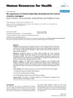
Figure 5A contains helical wheel diagrams of each LLP
domain from the HXB2 strain of HIV-1, as well as their
primary amino acid sequence (Figure 5B). When plotted
as α-helices, it is apparent that all three domains are
amphipathic, generally with hydrophilic residues
(colored blue) clustered on one face of the α-helix and
hydrophobic residues (colored red) clustered on the
opposite face. LLP-3 differs from LLP-1 and -2 in that it
lacks the positively charged residues on its hydrophilic
face. This secondary structure is conserved across HIV-1
clades, though primary amino acid identity is not [151].
Virology Journal 2007, 4:100 />Page 11 of 22
(page number not for citation purposes)
As discussed in Figure 5, helical wheel diagrams suggest
these regions have a high propensity to form amphipathic
α-helices [150]. Not only are the structure of these LLP-1
and -2 domains highly conserved across all strains of HIV-
1, they are conserved across other lytic lentiviruses such as
HIV-2, SIV, and equine infectious anemia virus (EIAV) as
well, though primary amino acid sequence does vary and
as such is not conserved [151]. Closely related nonlytic
oncoviruses, including murine leukemia virus lack these
conserved amphipathic α-helical structural motifs,
though they contain similarly long cytoplasmic tails. The
structure of LLP-1 and -2 domains resemble proteins of
ion-selective channels, such as the S4 domain of K
+
chan-
nels, as do the natural cytolytic peptides of the honey bee
(melittin) and amphibians (magainins)
[150,151,158,159]. Based on analogy to other lytic pep-
tides with similar secondary structure such as magainin-2,
and on the observation that when in an anti-parallel
arrangement, LLP-1 and -2 exhibit charge complementa-
rity, it has been hypothesized that LLP-1 and LLP-2 could
aggregate in lipid membranes with their charged (mostly
arginine) residues facing towards each other and their
hydrophobic residues facing out toward the lipid bilayer
to form an ion channel or a pore [160]. A third domain
dubbed LLP-3, located between the LLP-1 and -2 domains
shows a propensity to form an amphipathic α-helix, but
lacks the charged residues on one face, instead possessing
a leucine zipper-like sequence [152]. LLP-3 could, in the
context of the full protein, span the lipid bilayer to help
form the channel. It is also possible that LLP-3 could inter-
act with LLP-1 and 2 in a manner that aids in their aggre-
gation, an assertion the Kliger et al., 1997 based solely on
analogy to other leucine zipper-containing proteins.
Studies utilizing synthetic peptides of identical amino
acid sequence to the LLP-1 domain have supported the
hypothesis that LLP-1 can oligomerize, insert into mem-
branes and form pores. LLP-1 peptides of HIV-1 and SIV
were bactericidal to both gram(-) and gram(+) bacteria at
micromolar concentrations within a few minutes
The LLP domainsFigure 5
The LLP domains. (A) Helical wheel diagrams showing the amphipathic nature of each LLP domain. The coloring scheme is
from Benner et al. and graphs were generated using a java applet available at the PredictProtein server [221]. (B) Primary
amino acid sequence of LLP peptides which correspond to the LLP-1, -2, and -3 domains of the TM protein are given from the
helical wheels in (A).
Virology Journal 2007, 4:100 />Page 12 of 22
(page number not for citation purposes)
[151,161-163]. These peptides were also capable of lysing
red blood cells (RBC's) and RH9 T-lymphoblastoid cells
in similar concentration ranges. LLP-1 peptides were more
effective than the natural amphibian cytolytic magainin
peptides in these assays. Mutation of 2 – 3 of 7 positively
charged arginine residues present in LLP-1 to neutral
glutamate residues resulted in an almost complete loss of
activity against both prokaryotic and eukaryotic cells in
lysis assays [151,161]. Glutamate was chosen to preserve
the overall hydrophobic moment of LLP-1 as well as to
preserve its secondary structure. Thus, the overall positive
charge provided by its arginine residues is most likely nec-
essary for its function.
Experimental evidence supports the theoretical models of
the LLP domains' secondary structure and function. Circu-
lar dichroism studies show that synthetic peptides corre-
sponding to these regions have little secondary structure
in water, but adopt an α-helical secondary structure in the
presence of a lipid environment [152,162,164-166].
Transmission electron microscopy studies confirm that,
similar to data gathered using magainin-2, LLP-1 interacts
with both the inner and outer leaflets of the cytoplasmic
membrane of the bacteria Serratia marcescens [167]. Bacte-
ria exposed to LLP-1 displayed a decreased cytoplasmic
density from negative controls indicating that the mem-
brane had been compromised. Furthermore, addition of
membrane impermeable ONPG to LLP-1-incubated, but
not control cultures, led to its hydrolysis over time, indi-
cating that it was able to gain access to the β-galactosidase
enzyme located in the cytoplasm of bacteria [162]. Both
LLP-1 and LLP-2 were able to cause time- and dosage-
dependent release carboxyfluorescein entrapped egg PC
vesicles at micromolar concentrations. When added to
LUV's of various lipid compositions, 15-mer peptides
spanning all three LLP regions were capable of causing
leakage, phospholipid mixing, and fusion to differing
extents [168]. The presence and amount of sphengomye-
lin as well as cholesterol correlated positively with these
peptides' functions in these assays, though similar trends
were observed between strains of HIV.
In a subsequent attempt to define the size of the pore cre-
ated by LLP-1, Miller et al., 1993 measured the amounts
of
45
Ca,
14
C-sucrose, and
14
C-inulin that were able to enter
LLP-1 treated CEM cell cultures [169].
45
Ca (M.W. = 45
Da) and
14
C-sucrose (M.W. = 342.3 Da), but not
14
C-inu-
lin (M.W. ~5000 Da) were able to pass through LLP-1
treated membranes, suggesting that LLP-1 could form a
pore of a definable size and did not simply destabilize or
disintegrate the membrane. In good agreement, mem-
brane perturbation studies utilizing whole virus show that
hygromycin b (MW 527) was able to enter cells after infec-
tion with HIV-1, while G418 (MW 693) was not able to
enter [170]. This suggests that the pore created by the LLP
domains has a cutoff between MW 527 and 693.
Topological analysis of full length TM of HIV-1 using
sequence specific antibodies showed that not only did TM
contain an N-terminal transmembrane anchoring
domain, but it also formed secondary associations at the
C-terminal end which blocked antibody binding [171].
Furthermore, the association of the cytoplasmic tail with
microsomal membranes made from canine pancreas con-
ferred resistance to extraction via carbonate treatment,
suggesting that the association observed earlier was not
merely an artifact of having a conformational dependent
antibody. The association of the cytoplasmic tail with
lipid membranes was also resistant to high salt extraction
and proteolysis [172]. Expression of just the cytoplasmic
tail of TM associated with lipid membranes and was suffi-
cient to get cell surface expression of the tail fragment
[173]. Sucrose gradient centrifugation, chemical cross-
linking, and gel filtration analysis of an MBP-cytoplasmic
tail fusion protein proved the formation of a higher
ordered, multimerized structure, dominantly a hexamer.
Analysis of the same fusion protein in a mammalian 2-
hybrid assay and in a GST pull-down assay complemented
these studies in eukaryotic cells [174]. In fact, the authors
concluded that the cytoplasmic tail itself is sufficient to
oliogomerize Env.
LLP-1 causes increased the conductance of various mem-
branes when added exogenously. LLP-1 bound preferen-
tially to planar lipid bilayers composed of negatively
charged phosphatidylserine (PS) over neutral diphytanoyl
phosphatidylcholine (DPC) bilayers and as such, all
experiments were performed using PS bilayers [175]. At
micromolar concentrations there was an overall increased
conductance at negative and positive voltages. A prefer-
ence for cations over anions was observed, with no prefer-
ence of Na
+
or K
+
. The effect of exogenous LLP-1 peptides
on whole-cell conductance of (Sf9) insect cells was meas-
ured in the same study. Nanomolar LLP-1 concentrations
increased mean membrane conductance at positive and
negative voltages by approximately 10 fold using the
patch clamp technique. Nanomolar concentrations of
exogenous LLP-1 were also able to increase the whole cell
conductance of Xenopus laevis oocytes [176]. Much
Smaller conductances were induced by equal concentra-
tions of the lytic peptide melittin. Up to four times the
concentration of HIV-1 Nef accessory protein did not
increase oocyte membrane conductance over control,
untreated oocytes.
Ultrastructural analysis of eukaryotic cells incubated with
LLP-1 peptides uncovered features of necrosis as the main
cause of death of these cells [177]. The most striking fea-
tures visible under electron microscopy are extensive vac-
Virology Journal 2007, 4:100 />Page 13 of 22
(page number not for citation purposes)
uolization of the endoplasmic reticulum and
mitochondria. The authors attribute this to a concentra-
tion of ions and water into these cell organelles attempt-
ing to compensate for increased intracellular levels of ions
and water caused by LLP-1 peptides. Plymale et al., 1996
also observed a small increase in the levels of apoptosis in
these cells [178]. The increase in the numbers of cells
undergoing apoptosis under these conditions was very
small compared to the increase in cells undergoing necro-
sis. The magnitude of the increases was influenced by the
cell type and the concentration of LLP-1. Lower "sub-lytic"
concentrations of LLP-1 tended to cause more apoptosis
than higher "lytic" LLP-1 concentrations. Thus apoptosis,
like syncytia formation, appears to be a mechanism which
HIV can utilize to cause cytopathology, but is not likely
the dominant mechanism utilized in vivo.
By virtue of its amphipathic α-helical secondary structure,
LLP-1 has homology to calmodulin binding proteins and
has been proposed as a mechanism behind HIV's ability
to cause apoptosis in cell culture. HIV virions which pos-
sess full-length TM cytoplasmic tails, but not virions with
truncated tails lacking LLP domains, are able to bind cal-
modulin [179]. Peptides corresponding to both the LLP-1
and -2 domains bind calmodulin with high affinity, irre-
spective of natural sequence variation present in different
clades of HIV [161,180]. When added exogenously to T
cells in vitro, both LLP-1 and -2 inhibited T cell signal
transduction through the NF-AT complex via sequestra-
tion and titration of available calmodulin (Figure 6). LLP-
1 and -2 were as effective at inhibiting calmodulin as the
known calmodulin inhibitor, W-7 [181]. The end result of
calmodulin inhibition observed in these studies was a
decrease in IL-2 production leading to T cell anergy and
increased levels of apoptosis. It has long been observed
that immune cells from HIV infected patients are less
responsive than HIV seronegative persons though it
remains to be seen whether significantly increased HIV-
induced apoptosis occurs in vivo at all [182]. However, cell
culture experiments tend to suggest that apoptosis is not a
major contributor to cell death as only marginal increases
in apoptotic cells are observed throughout the course of
HIV infected cell cultures compared to necrotic cell death
[177].
TM has been implicated in altering [ion]
i
thought to be
responsible for the impairment of brain function known
as AIDS Dementia Complex (ADC). Upon necropsy, neu-
rohistopathology of the CNS shows morphological
abnormalities and death of neurons, astrogliosis, micro-
glial nodules, and multi-nucleated giant cells consisting of
monocyte-macrophages and microglial cells [55]. Patients
with ADC have increased levels of glutamate
+
in their CSF
[183]. Glutamate
+
is an excitatory amino acid (EAA)
whose levels are closely regulated by glial cells in the brain
because excess glutamate
+
in neuronal synapses is toxic.
Though neuronal infection is generally non-productive in
vitro, an increase in extracellular [EAA] is observed after
acute infection of neurons by HIV. Analysis of virion pro-
teins proved that TM was sufficient to cause the increased
[EAA]
e
. Addition of exogenous peptides with sequences
corresponding to the LLP-1 domain of TM had the same
effect as the expressed TM protein. It was postulated by
Kart et al., 1998 that LLP-1 had its action through ablation
of the Na
+
gradient, a postulated action of the LLP
domains (discussed more in depth below)[183]. Without
the Na
+
gradient to drive the Na
+
-glutamate
+
cotrans-
porter-mediated electrogenic uptake of glutamate
+
against
its large concentration gradient across the plasma mem-
brane, [EAA]
e
and [cation]
e
increase. Bubien et al., 1995
report that SU added exogenously to cultures of rat or
human astrocytes stimulates the Na
+
/H
+
exchanger to
alkalinize the cytoplasm [184]. This in turn inhibits the
Na
+
-dependent uptake of glutamate
+
against its concen-
tration gradient and activates pH-sensitive K
+
channels to
release intracellular K
+
. It is thought that impairment of
the astrocytes' ability to maintain the proper [EAA]
e
and
[ion]
e
leads to improper firing of neuron potentials and
neuronal cell death. It is unknown at this time what the
relative contributions of each of these pathways may be in
vivo to developing ADC.
Previous work on TM and its ability to perturb mem-
branes focused on truncations in the context of whole vir-
ions [185-189]. These studies produced conflicting
reports of the function and necessity of the C-terminus of
TM during infection. Results gained from these trunca-
Calmodulin binding activity of the LLP domainsFigure 6
Calmodulin binding activity of the LLP domains. Pro-
posed action of LLP-1 and -2 binding of calmodulin in T cell
anergy. The LLP domains are thought to disrupt the signaling
cascade through titration of calmodulin. Figure was produced
based on data from Beary et. al, 1998 [181].
Virology Journal 2007, 4:100 />Page 14 of 22
(page number not for citation purposes)
tions vary between producing no effect at all to one or
more of decreases in viral entry, infectivity, cytopathic
effect, and envelope production, processing, stability, cell
surface expression, and virion incorporation. Two points
that these studies generally agree on is that HIV virions
with truncated TM cytoplasmic tails are replication com-
petent and that most effects observed in these mutant
viruses are cell type dependent. Discrepancies between
studies likely involve the disruption of multiple domains
contained within the cytoplasmic tail of TM, depending
upon the extent of each truncation. Several studies have
indicated that there may be domain(s) in the C-terminus
of TM that interact with HIV Gag proteins to assemble vir-
ions [24,190]. In addition to interacting with Gag, it has
been hypothesized, though not proven, that the cytoplas-
mic tail of TM may interact with cellular factors, and that
this may be the source of the cell-type dependent effect
[191-193].
Studies involving site-directed mutagenesis of specific
domains within the cytoplasmic tail may help to clear up
some of the confusion caused by the truncation studies.
Kalia et al., 2003 engineered infectious virions with muta-
tions of the LLP-1 and -2 domains [194]. Three mutant
viruses were produced, one with 2 arginine to glutamate
mutations in LLP-1, another with 2 arginine to glutamate
mutations in LLP-2, and a third incorporating the same
mutations in LLP-1 and -2, but in the same virus. Syn-
thetic peptides corresponding to these domains which
include these mutations were previously found to lose
their lytic properties, as well as their calmodulin binding
activity [169,180]. The LLP-1 mutant virions displayed an
approximate 85% decrease in TM incorporation, though
Env expression and processing were unaffected. LLP-2
mutant viruses were unaffected in Env expression,
processing, oligomerization, and incorporation. Both
LLP-1 and -2 mutant viruses were decreased in their capac-
ity to cause syncytia by 70 and 90% respectively. Double
mutant viruses were similar to LLP-1 mutant viruses in
envelope expression, processing, and incorporation, but
failed to cause syncytia to the same extent as LLP-2 mutant
viruses [194].
Syncytia formation and single cell balloon degeneration
have traditionally been thought to be two distinct cyto-
pathic phenotypes caused by two distinct regions of Env –
the extracellular fusion peptide and the intracellular LLP
domains respectively. However, these two processes may
be more closely linked than previously thought. Giant,
multinucleated syncytial cells undergo increases in total
cell volume, owing to the LLP domains perturbing the
plasma membrane just as in the case of single cells. On the
other hand, Kalia et al., 2003 report that infectious clones
containing 2 each of arginine to glutamate site directed
mutations in the LLP-1 and/or LLP-2 domains exhibit a
decrease in their ability to cause cell:cell fusion [194]. It is
possible, though it remains to be proven, that the mem-
brane perturbation properties of the LLP domains could
synergize with the fusion peptide to increase the efficiency
of cell:cell fusion in a manner analogous to its function in
virion budding. Increases in cell volume due to osmotic
balancing after ion influx could disrupt the cell cytoskele-
ton allowing for greater ease of membrane fusion.
The case is more clear-cut for truncations of the cytoplas-
mic tail of the Env protein of SIV. Env truncations are doc-
umented to arise during culturing of SIV in human cell
lines [195]. Mutants revert back if cultured again in simian
cells [195,196]. While these SIV TM truncation mutants
are replication competent in vitro as well as in vivo, they
lack full pathogenicity in vivo and tend to revert back over
time [197]. Shacklett et al., 2000 showed that the cyto-
plasmic tail of SIV Env is necessary for persistence of
viremia and pathogenesis of the virus in rhesus macaques
[198]. Their group engineered 3 stop codons, a +1
frameshift mutation, and 3 arginine to glutamate site
directed mutations in the LLP-1 domain of mac239 viri-
ons, eliminating both LLP-1 and -2 regions. The resulting
mutant virions cause an initial viremia, but levels fall off
and become non-detectable over time. All viruses recov-
ered from these macaques maintained their mutations
without exception and none of the infected macaques
developed SAIDS. Juvenile macaques infected i.v. with
this mutant virus maintained low level viremia, but also
never progressed to SAIDS. 100% of macaques infected
with wild type mac239 develop SAIDS over the same time
course.
LLP viroporin models and discussion
If the LLP domains form a viroporin and allow ions to
enter the cell more freely down its concentration gradient,
then water would follow in attempt to osmotically bal-
ance those ion influxes. This would represent a mecha-
nism by which HIV-1-infected cells undergo the process of
balloon degeneration.
This hypothesis allows for a mechanism by which large
versus moderate ion influxes could direct differential out-
comes for infected cells [199]. Those cells that can over-
come the osmotic imbalance may then live to become
chronically infected. Those that can't overcome this
osmotic imbalance undergo balloon degeneration. In
support of this theory, the concentration of LLP peptides
exogenously added to mammalian cell culture were previ-
ously determined to correlate with a differential outcome
on cell death [178]. The levels of apoptosis versus necrosis
of these cells – higher concentrations (>100 nM) were
shown to result in more necrosis, while lower concentra-
tions (~20 nM) resulted in more apoptosis with exoge-
nous LLP-1.
Virology Journal 2007, 4:100 />Page 15 of 22
(page number not for citation purposes)
It is unknown to what extent single cell killing versus syn-
cytial cell death occurs in vivo. The central nervous system
is the only tissue where syncytial cells have been observed
in vivo at autopsy [55,200]. In the absence of observed
syncytia, it is assumed that single cell death occurs in the
rest of the body, possibly due to a lack of opportunity for
infected cells to be in close enough proximity to allow
syncytial formation. As an estimate for the amount of sin-
gle cell death that can be caused by Env in the absence of
syncytia formation, 43% of RH9 T-lymphoblastoid cells
died by single cell death after inducibly expressing Env in
the presence of soluble CD4 to prevent syncytia formation
[199].
There are three general mechanisms by which the LLP
peptides and expressed Env may act to alter oocyte mem-
brane permeability. They may be acting to modify endog-
enous ion channel function, altering its activity and
thereby increasing whole-cell conductance. This could
occur through direct contact or by shifting the intracellu-
lar ionic environment. For example, LLP-1 is known to
have the ability to bind calmodulin leading to an increase
in intracellular free calcium [161,179,180]. Calcium lev-
els have a large effect on protein activation in the cell, thus
the LLP domains could be working through this mecha-
nism exclusively, or in addition to forming a membrane
pore. Second, the LLP domains could act to nonspecifi-
cally disrupt the membrane, or specific regions of the
membrane in a detergent-like effect. Lastly, the LLP
domains may serve as viroporins i.e., insert into the mem-
brane to form pores. Mounting evidence suggests that the
last possibility is likely; however definitive differentiation
of these mechanisms would require the discovery of spe-
cific inhibitors, such as amantadine for the M2 channel of
influenza.
When partitioning into a lipid environment, LLP-1, -2,
and -3 form highly ordered α-helices that were able to dis-
rupt a variety of model lipid bilayers in the absence of all
other protein (discussed above). Consequently these
domains are capable of disrupting membranes and are
not solely dependent upon alteration of endogenous ion
channel function to alter the intracellular ionic environ-
ment. In the context of a living membrane, LLP-1 and -2
have been shown to lyse bacteria, fungus, red blood cells,
and various cultured eukaryotic cells [151,162,169,178-
180,201,202]. When added exogenously, LLP-1 can
increase the conductance of Xenopus oocytes, presumably
caused by the formation of transmembrane pores which
increase the membrane permeability of electrogenically
active ions [176]. It has thus been postulated that LLP-1,
and possibly LLP-2 peptides, oligomerize to form a "bar-
rel-stave"-like pore, which are conducting pores (barrels)
in membranes formed by the self-assembly of a variable
number of alpha-helical rods (staves). It is the formation
of these pores which then allow ions to be redistributed
across the membrane to cause osmolysis.
Recently it has been proposed that HIV, along with several
animal viruses including influenza, may utilize lipid rafts
to enter and exit cells. Lipid rafts are lipid microdomains
enriched for sphingomyelin and cholesterol that are
thought to function by aggregating proteins that require
close proximity for proper function, such as in the case of
the T cell synapse [203]. It has been proposed by several
groups that HIV-1 exploits lipid rafts during both the
entry and budding stages of its replication cycle. During
entry, lipid rafts may serve to concentrate cellular recep-
tors (CD4, CXCR4, and CCR5) for ease of entry by HIV
[204]. This could potentially be important for HIV since
entry has been shown to be dependent on the density of
receptors on the cell surface [205].
There is ample evidence that HIV-1 buds from lipid rafts
[28,206]. The composition of the HIV envelope is prima-
rily sphingolipid and cholesterol, resembling the compo-
sition of rafts. The HIV lipid envelope is furthermore
enriched for proteins known to partition into lipid rafts
and excludes proteins known to not associate with rafts
(such as CD45). HIV proteins extract with lipid raft
domains during nonionic detergent extraction at 4°C.
Lastly, the cytoplasmic tail of Env has two palmitoylated
sites known for targeting proteins to lipid rafts in a man-
ner analogous to cellular proteins' targeting to lipid rafts
[207]. One result of entry and budding through lipid rafts
is that Env proteins would be concentrated in distinct
areas on the cell surface, possibly allowing interaction of
LLP domains to form pores on the cell surface. These
pores in turn could weaken the normally stable raft area
through altered intracellular ion milieu, thereby creating
a more favorable environment for replication and bud-
ding of the virus. Hence, as infection progresses and
increasing amounts of Env are deposited on the cell sur-
face in preparation for budding, there would be a con-
comitant increase in cytopathic effects, such as
intracellular ion imbalance, cell volume dysregulation,
and balloon degeneration.
Prior observations that LLP-1 can bind to intracellular sig-
naling molecules, such as calmodulin to ultimately
induce apoptosis and/or necrosis [161,178-180] suggest
that the LLP domains may be configured in certain situa-
tions as a pore passing through the membrane and part of
their time associated with the inner leaflet of the lipid
membrane where they are able to interact with these intra-
cellular molecules. Flip-flopping between lipid bilayers of
amphipathic pore forming peptides has been docu-
mented with melittin [208,209]. Based on reported simi-
larities between melittin and LLP peptides, it is reasonable
to hypothesize that the LLP domains may be flip-flopping
Virology Journal 2007, 4:100 />Page 16 of 22
(page number not for citation purposes)
between a transmembrane state and parallel association
with the inner leaflet of the lipid bilayer. On the other
hand, the LLP domains may possess different activities in
the different cell types that it infects, or there may be some
as of yet undefined temporal control that allows these two
alternate functions to take place at appropriate times dur-
ing infection.
Since the LLP domains are also present in the context of
the virion, it is possible that they would have an effect at
this stage of the HIV replication cycle. There is at least one
report of an increase in natural endogenous reverse tran-
scription (NERT) cause by the LLP domains increasing the
virion envelope permeability to dNTP's [210].
That HIV may code a viroporin in its major surface glyco-
protein would ensure that the membrane perturbation,
ion fluxes, volume changes, and resulting "loosening" of
the plasma membrane and cytoskeleton always occur
when and where it is needed for budding, syncytial forma-
tion, and/or single cell balloon degeneration. Concentrat-
ing HIV glycoproteins in lipid rafts could allow for
localized unstable membrane regions at the exact points
where it is needed by HIV. While it seems possible that
Vpu could also act at these stages to accomplish the same
goals, it is more difficult to envision how it could accom-
plish the task as Vpu has been shown to be excluded from
the plasma membrane and HIV virions [75,99].
Since SIV does not contain an equivalent of Vpu in its
genome (except SIV
cpz
), there is some conjecture in the lit-
erature that it utilizes its Env protein and specifically the
LLP domains of Env to take the place of Vpu in enhance-
ment of virion budding [211]. In this situation it makes
sense that deleting the LLP domains in SIV makes a more
clear cut phenotype in vivo than HIV-1. SIV minus the LLP
regions is still able to replicate, but does not cause SAIDS
[198]. It may be that the LLP domains of SIV are func-
tional equivalents of Vpu in the case of SIV. By extension,
it is possible that these two proteins represent backup sys-
tems in HIV-1 for budding, or maybe that Vpu and LLP
have specialized such that Vpu helps Env traffic through
the ER and Golgi while the LLP's increase budding more
specifically through cytoskeletal disruptions at the cell
surface. Though admittedly it is not understood to what
extent deletion of LLP domains in HIV-1 would do in vivo
since that type of controlled experiment cannot be done
ethically – truncated mutants have been found in produc-
tive human infections which cause AIDS and make it dif-
ficult to dissect the roles of these two proteins in infection
[212,213].
In addition to the LLP's involvement as a backup system
for cell volume regulation and cytoskeletal disruption,
they may produce secondary effects, such as AIDS-related
dementia complex and bystander cell death. LLP domains
could be cleaved by cellular proteases from the C-termini
of TM proteins and act as exogenous peptides for all
intents and purposes in vivo. In this way they could pro-
duce the effects generated by LLP in cell culture thought to
cause AIDS-related dementia (described above). An anal-
ogous role could be played in the death of bystander cells
– a population of cells that die in HIV-infected individu-
als, but are not productively infected [214,215].
The Lentiviral Lytic Peptide motif may not be unique to
lentiviruses, as their name would imply. Recently it was
discovered that the carboxyl termini of pestivirus enve-
lope glycoproteins contain a sequence predicted to have a
high propensity to form an amphipathic α-helix [216].
This sequence is conserved among pestiviruses. It remains
to be proven that this region of pestiviruses employ any
functional characteristics of LLP's, however it could be
hypothesized that a functional motif is beginning to show
itself. Functional substitutions of 6K protein of sindbis
virus and Vpu of HIV-1 have been proven [116]. Thus it
begs the question, could the 6K protein and LLP domains
of the E1 surface glycoprotein of sindbis virus be func-
tional equivalents of the Vpu protein and the LLP
domains of the Env surface glycoprotein of HIV-1? Func-
tional equivalents may exist in other viruses, such as the
P7 protein and LLP domains of BVDV (also a pestivirus),
as well as the VP1 and 2B proteins of poliovirus, a picor-
navirus [64,217,218].
In 2004 alone it was estimated that there were approxi-
mately 39.4 million people living with HIV/AIDS, with
around 3.1 million AIDS related deaths, and 13,500 new
infections each day [222]. Even with the advent of Highly
Active Anti-Retroviral Therapy (HAART), which combines
the use of protease inhibitors and reverse transcriptase
inhibitors, and use of the newer fusion inhibitors such as
T20, HIV continues to be a serious threat to world health
[219,220]. A lack of resources for most infected persons to
purchase the drugs, the intensive treatment regimen, the
toxicity of drug regiments, and emerging drug resistance
all contribute to a lack of general efficacy of the current
treatment regimen and highlight the necessity for more
basic research with the ultimate goal of development of
new treatments. The LLP domains may represent a new
target for HIV drugs to inhibit HIV infection. Otherwise
the development of eLLP's as a new class of antibacterial
drugs could be used to help resolve AIDS-related infec-
tions, as well as serve as a new class of antibiotics – virally
derived antibiotic peptides.
In order to develop the LLP domains as an attractive target
for the development of novel anti-HIV therapies, it will
likely be necessary to achieve a better understanding of
the mechanism of action of this domain. The use of bio-
Virology Journal 2007, 4:100 />Page 17 of 22
(page number not for citation purposes)
chemical techniques such as oriented circular dichroism
(OCD) spectroscopy could be used to determine the ori-
entation of LLP peptides in lipid membranes, either span-
ning the membrane, lying on the surface, or somewhere in
between. Expanding on the experiments of Comardelle et
al., 1997 and continuing to define the effects of LLP pep-
tides on Xenopus oocyte membranes could also be helpful
as this system was helpful in defining the activity of the
M2 protein of influenza. Ion-selective electrodes could be
employed to characterize the ion selectivity of LLP peptide
domains. Specific LLP domain mutations could be made
to dissect each LLP domains' role in the proposed virop-
orin. Specific inhibitors of the LLP domains could be
screened for, which could help prove specific activity of
the LLP domains as a viroporin and could themselves be
a candidate for a novel class of anti-HIV drugs.
Summary of Results
Mechanisms by which HIV-1 mediates reductions in
CD4
+
cell levels in infected persons are intensely investi-
gated, and have broad implications for AIDS drug and
vaccine development. Virally induced changes isn mem-
brane ionic permeability contribute to cytopathogenesis
induced by lytic viruses of many families. HIV-1 induces
disturbances in plasma membrane ion transport. The car-
boxyl terminus of TM contains amphipathic α-helical
motifs identified because of their structural similarities to
melittin, a naturally occurring cytolytic peptide, and were
dubbed lentiviral lytic peptides (LLP) -1, -2, and -3. Pep-
tides corresponding to these domains from HIV-1
HXB2
(a
clade B laboratory adapted virus) partition into lipid
membranes as α-helices and disrupt model lipid mem-
branes. A peptide corresponding to the LLP-1 domain of a
clade D HIV-1 virus, dubbed LLP-1D displayed similar
activity to the LLP-1 domain of the clade B virus in all
assays, despite a lack of amino acid sequence identity.
When individual peptides are incubated exogenously with
Xenopus oocytes, LLP-1 and -2, but not LLP-3 increased the
whole cell conductance across the plasma membrane. The
increased conductance observed with LLP-1 and -2
appears to be at least in part due to an increased permea-
bility of Na
+
ions. A peptide corresponding to the LLP-1
domain of a clade D HIV-1 virus, dubbed LLP-1D, dis-
played similar activity to the LLP-1 domain of the clade B
virus in all assays, despite a lack of amino acid sequence
identity.
Combinations of LLP peptides appear to act cooperatively
to increase the whole cell conductance of Xenopus oocyte
plasma membranes. Taken together, these results suggest
that the C-terminal domains of HIV-1 Env proteins may
form an ion channel, or viroporin, that is capable of con-
ducting Na
+
ions. Alternatively, HIV-1 Env protein may
activate a silent Xenopus oocyte Na
+
-conductive ion chan-
nel. Increased understanding of the function of LLP
domains and their role in the viral replication cycle could
allow for the development of novel HIV drugs.
Biography
Joshua Costin was born in Colorado Springs, CO on July
14, 1976. He grew up in the tropical paradise that is
Naples, Florida and graduated 7th in his class at Naples
High School. He went on to graduate magna cum laude
from Florida State University, with honors, as well as hon-
ors in the major, receiving a B.S. in Biology, a B.S. in psy-
chology, and a minor in chemistry. Joshua enrolled in the
Department of Microbiology and Immunology at Tulane
University in the fall semester of 1998 and began what
was to become his dissertation work in November of that
year under the guidance of Dr. Robert F. Garry and com-
pleted his dissertation work in the summer of 2005. Along
the way Joshua received several awards, including a
Tulane University Cancer Center Grant in 1999, the
Center for Infectious Diseases Award for Basic Research in
Infectious Diseases 2001, and a Graduate School Disserta-
tion Year Fellowship for Sciences and Engineering for
2004–2005.
Competing interests
The author(s) declare that they have no competing inter-
ests.
Authors' contributions
JC researched and wrote the review article.
Acknowledgements
The author would like to give thanks to the members of his committee, Dr.
Robert F. Garry, Dr. Nazih Nakhoul, Dr. William Wimley, Dr. Laura Levy,
and Dr. Cindy Morris for their guidance, time, and encouragement through-
out my dissertation. I am extremely grateful to Dr. Nazih Nakhoul and Dr.
William Wimley for their time and willingness to teach me the techniques
of their respective labs in pursuit of my dissertation.
References
1. Gottlieb MS, Schroff R, Schanker HM, Weisman JD, Fan PT, Wolf RA,
Saxon A: Pneumocystis carinii pneumonia and mucosal candi-
diasis in previously healthy homosexual men: evidence of a
new acquired cellular immunodeficiency. N Engl J Med 1981,
305:1425-1431.
2. Masur H, Michelis MA, Greene JB, Onorato I, Stouwe RA, Holzman
RS, Wormser G, Brettman L, Lange M, Murray HW, Cunningham-
Rundles S: An outbreak of community-acquired Pneumocystis
carinii pneumonia: initial manifestation of cellular immune
dysfunction. N Engl J Med 1981, 305:1431-1438.
3. Gallo RC, Montagnier L: Historical essay. Prospects for the
future. Science 2002, 298:1730-1731.
4. Barre-Sinoussi F, Chermann JC, Rey F, Nugeyre MT, Chamaret S,
Gruest J, Dauguet C, Axler-Blin C, Vezinet-Brun F, Rouzioux C, et al.:
Isolation of a T-lymphotropic retrovirus from a patient at
risk for acquired immune deficiency syndrome (AIDS). Sci-
ence 1983, 220:868-871.
5. Gallo RC, Salahuddin SZ, Popovic M, Shearer GM, Kaplan M, Haynes
BF, Palker TJ, Redfield R, Oleske J, Safai B, et al.: Frequent detection
and isolation of cytopathic retroviruses (HTLV-III) from
patients with AIDS and at risk for AIDS. Science 1984,
224:500-503.
6. Gallo RC, Montagnier L: The discovery of HIV as the cause of
AIDS. N Engl J Med 2003, 349:2283-2285.
Virology Journal 2007, 4:100 />Page 18 of 22
(page number not for citation purposes)
7. Montagnier L: Historical essay. A history of HIV discovery. Sci-
ence 2002, 298:1727-1728.
8. Gallo RC: Historical essay. The early years of HIV/AIDS. Sci-
ence 2002, 298:1728-1730.
9. Levy JA, Hoffman AD, Kramer SM, Landis JA, Shimabukuro JM, Oshiro
LS: Isolation of lymphocytopathic retroviruses from San
Francisco patients with AIDS. Science 1984, 225:840-842.
10. Levy JA: Pathogenesis of human immunodeficiency virus
infection. Microbiol Rev 1993, 57:183-289.
11. Daar ES, Moudgil T, Meyer RD, Ho DD: Transient high levels of
viremia in patients with primary human immunodeficiency
virus type 1 infection. N Engl J Med 1991, 324:961-964.
12. Zhu T, Mo H, Wang N, Nam DS, Cao Y, Koup RA, Ho DD: Geno-
typic and phenotypic characterization of HIV-1 patients with
primary infection. Science 1993, 261:1179-1181.
13. Coffin JM: HIV population dynamics in vivo: implications for
genetic variation, pathogenesis, and therapy. Science 1995,
267:483-489.
14. Ho DD, Neumann AU, Perelson AS, Chen W, Leonard JM, Markowitz
M: Rapid turnover of plasma virions and CD4 lymphocytes in
HIV-1 infection. Nature 1995, 373:123-126.
15. Wei X, Ghosh SK, Taylor ME, Johnson VA, Emini EA, Deutsch P, Lif-
son JD, Bonhoeffer S, Nowak MA, Hahn BH, et al.: Viral dynamics
in human immunodeficiency virus type 1 infection. Nature
1995, 373:117-122.
16. Perelson AS, Neumann AU, Markowitz M, Leonard JM, Ho DD: HIV-
1 dynamics in vivo: virion clearance rate, infected cell life-
span, and viral generation time. Science 1996, 271:1582-1586.
17. Piatak M Jr, Saag MS, Yang LC, Clark SJ, Kappes JC, Luk KC, Hahn BH,
Shaw GM, Lifson JD: High levels of HIV-1 in plasma during all
stages of infection determined by competitive PCR. Science
1993, 259:1749-1754.
18. Fahey JL, Taylor JM, Manna B, Nishanian P, Aziz N, Giorgi JV, Detels
R: Prognostic significance of plasma markers of immune acti-
vation, HIV viral load and CD4 T-cell measurements. Aids
1998, 12:1581-1590.
19. Arnaout RA, Lloyd AL, O'Brien TR, Goedert JJ, Leonard JM, Nowak
MA: A simple relationship between viral load and survival
time in HIV-1 infection. Proc Natl Acad Sci USA 1999,
96:11549-11553.
20. Garry RF: Potential mechanisms for the cytopathic properties
of HIV. Aids 1989, 3:683-694.
21. Strebel K, Klimkait T, Martin MA: A novel gene of HIV-1, vpu, and
its 16-kilodalton product. Science 1988, 241:1221-1223.
22. Deng H, Liu R, Ellmeier W, Choe S, Unutmaz D, Burkhart M, Di Mar-
zio P, Marmon S, Sutton RE, Hill CM, et al.: Identification of a
major co-receptor for primary isolates of HIV-1. Nature 1996,
381:661-666.
23. Saez-Cirion A, Nieva JL, Gallaher WR: The hydrophobic internal
region of bovine prion protein shares structural and func-
tional properties with HIV type 1 fusion peptide. AIDS Res
Hum Retroviruses 2003, 19:969-978.
24. Wyma DJ, Kotov A, Aiken C: Evidence for a stable interaction
of gp41 with Pr55(Gag) in immature human immunodefi-
ciency virus type 1 particles. J Virol 2000, 74:9381-9387.
25. Pugliese A, Vidotto V, Beltramo T, Petrini S, Torre D: A review of
HIV-1 Tat protein biological effects. Cell Biochem Funct :2004.
26. Strebel K: Virus-host interactions: role of HIV proteins Vif,
Tat, and Rev. Aids 2003, 17(Suppl 4):S25-34.
27. Montal M: Structure-function correlates of Vpu, a membrane
protein of HIV-1. FEBS Lett 2003, 552:47-53.
28. Doms RW, Trono D: The plasma membrane as a combat zone
in the HIV battlefield. Genes Dev 2000, 14:2677-2688.
29. Werner T, Ferroni S, Saermark T, Brack-Werner R, Banati RB, Mager
R, Steinaa L, Kreutzberg GW, Erfle V: HIV-1 Nef protein exhibits
structural and functional similarity to scorpion peptides
interacting with K+ channels. Aids 1991, 5:1301-1308.
30. Sherman MP, de Noronha CM, Heusch MI, Greene S, Greene WC:
Nucleocytoplasmic shuttling by human immunodeficiency
virus type 1 Vpr. J Virol 2001, 75:1522-1532.
31. Piller SC, Ewart GD, Premkumar A, Cox GB, Gage PW: Vpr protein
of human immunodeficiency virus type 1 forms cation-selec-
tive channels in planar lipid bilayers. Proc Natl Acad Sci USA 1996,
93:111-115.
32. Boya P, Pauleau AL, Poncet D, Gonzalez-Polo RA, Zamzami N, Kro-
emer G: Viral proteins targeting mitochondria: controlling
cell death. Biochim Biophys Acta 2004, 1659:178-189.
33. Basanez G, Zimmerberg J: HIV and apoptosis death and the
mitochondrion. J Exp Med 2001, 193:F11-14.
34. Halestrap AP, Brennerb C: The adenine nucleotide translocase:
a central component of the mitochondrial permeability
transition pore and key player in cell death. Curr Med Chem
2003, 10:1507-1525.
35. Sherman MP, De Noronha CM, Williams SA, Greene WC: Insights
into the biology of HIV-1 viral protein R. DNA Cell Biol 2002,
21:679-688.
36. Rasheed S, Gottlieb AA, Garry RF: Cell killing by ultraviolet-inac-
tivated human immunodeficiency virus. Virology 1986,
154:395-400.
37. Geijtenbeek TB, Kwon DS, Torensma R, van Vliet SJ, van Duijnhoven
GC, Middel J, Cornelissen IL, Nottet HS, KewalRamani VN, Littman
DR, et al.: DC-SIGN, a dendritic cell-specific HIV-1-binding
protein that enhances trans-infection of T cells. Cell 2000,
100:587-597.
38. Maddon PJ, Dalgleish AG, McDougal JS, Clapham PR, Weiss RA, Axel
R: The T4 gene encodes the AIDS virus receptor and is
expressed in the immune system and the brain. Cell 1986,
47:333-348.
39. Cocchi F, DeVico AL, Garzino-Demo A, Cara A, Gallo RC, Lusso P:
The V3 domain of the HIV-1 gp120 envelope glycoprotein is
critical for chemokine-mediated blockade of infection. Nat
Med 1996, 2:1244-1247.
40. Wahl SM, Greenwell-Wild T, Peng G, Ma G, Orenstein JM, Vazquez
N: Viral and host cofactors facilitate HIV-1 replication in
macrophages. J Leukoc Biol 2003, 74:726-735.
41. Philpott SM: HIV-1 coreceptor usage, transmission, and dis-
ease progression. Curr HIV Res 2003, 1:217-227.
42. Liu R, Paxton WA, Choe S, Ceradini D, Martin SR, Horuk R, MacDon-
ald ME, Stuhlmann H, Koup RA, Landau NR: Homozygous defect
in HIV-1 coreceptor accounts for resistance of some multi-
ply-exposed individuals to HIV-1 infection. Cell 1996,
86:367-377.
43. Miedema F, Meyaard L, Koot M, Klein MR, Roos MT, Groenink M,
Fouchier RA, Van't Wout AB, Tersmette M, Schellekens PT, et al.:
Changing virus-host interactions in the course of HIV-1
infection. Immunol Rev 1994, 140:35-72.
44. Holm GH, Zhang C, Gorry PR, Peden K, Schols D, De Clercq E,
Gabuzda D: Apoptosis of bystander T cells induced by human
immunodeficiency virus type 1 with increased envelope/
receptor affinity and coreceptor binding site exposure. J Virol
2004, 78:4541-4551.
45. Bleul CC, Wu L, Hoxie JA, Springer TA, Mackay CR: The HIV core-
ceptors CXCR4 and CCR5 are differentially expressed and
regulated on human T lymphocytes. Proc Natl Acad Sci USA
1997, 94:1925-1930.
46. Alimonti JB, Ball TB, Fowke KR: Mechanisms of CD4+ T lym-
phocyte cell death in human immunodeficiency virus infec-
tion and AIDS. J Gen Virol 2003, 84:1649-1661.
47. Douek DC: Disrupting T-cell homeostasis: how HIV-1 infec-
tion causes disease. AIDS Rev 2003, 5:172-177.
48. McMichael AJ, Ogg G, Wilson J, Callan M, Hambleton S, Appay V, Kel-
leher T, Rowland-Jones S: Memory CD8+ T cells in HIV infec-
tion. Philos Trans R Soc Lond B Biol Sci 2000, 355:363-367.
49. Dalgleish AG, Beverley PC, Clapham PR, Crawford DH, Greaves MF,
Weiss RA: The CD4 (T4) antigen is an essential component of
the receptor for the AIDS retrovirus. Nature 1984,
312:763-767.
50. Simon V, Ho DD: HIV-1 dynamics in vivo: implications for ther-
apy. Nat Rev Microbiol 2003, 1:181-190.
51. McLean AR, Michie CA: In vivo estimates of division and death
rates of human T lymphocytes. Proc Natl Acad Sci USA 1995,
92:3707-3711.
52. Lawn SD, Butera ST, Folks TM: Contribution of immune activa-
tion to the pathogenesis and transmission of human immun-
odeficiency virus type 1 infection. Clin Microbiol Rev 2001,
14:753-777. table of contents.
53. Mohri H, Bonhoeffer S, Monard S, Perelson AS, Ho DD: Rapid turn-
over of T lymphocytes in SIV-infected rhesus macaques. Sci-
ence 1998, 279:1223-1227.
Virology Journal 2007, 4:100 />Page 19 of 22
(page number not for citation purposes)
54. Cloyd MW, Lynn WS: Perturbation of host-cell membrane is a
primary mechanism of HIV cytopathology. Virology 1991,
181:500-511.
55. Takeya M, Naito M, Eto K, Takahashi K: Immunohistochemical
characterization of multinucleated giant cells in the brain of
a Japanese AIDS patient. Acta Pathol Jpn 1991, 41:212-216.
56. Vago L, Castagna A, Lazzarin A, Trabattoni G, Cinque P, Costanzi G:
Reduced frequency of HIV-induced brain lesions in AIDS
patients treated with zidovudine. J Acquir Immune Defic Syndr
1993, 6:42-45.
57. Somasundaran M, Robinson HL: A major mechanism of human
immunodeficiency virus-induced cell killing does not involve
cell fusion. J Virol 1987, 61:3114-3119.
58. Micklem KJ, Pasternak CA: Surface components involved in
virally mediated membrane changes. Biochem J 1977,
162:405-410.
59. Impraim CC, Foster KA, Micklem KJ, Pasternak CA: Nature of
virally mediated changes in membrane permeability to small
molecules. Biochem J 1980, 186:847-860.
60. Foster KA, Gill K, Micklem KJ, Pasternak CA: Survey of virally
mediated permeability changes. Biochem J 1980, 190:639-646.
61. Bashford CL, Alder GM, Patel K, Pasternak CA: Common action of
certain viruses, toxins, and activated complement: pore for-
mation and its prevention by extracellular Ca2+. Biosci Rep
1984, 4:797-805.
62. Grewe C, Beck A, Gelderblom HR: HIV: early virus-cell interac-
tions. J Acquir Immune Defic Syndr 1990, 3:965-974.
63. Carrasco L: Modification of membrane permeability by ani-
mal viruses. Adv Virus Res 1995, 45:61-112.
64. Gonzalez ME, Carrasco L: Viroporins. FEBS Lett 2003, 552:28-34.
65. Choi B, Gatti PJ, Haislip AM, Fermin CD, Garry RF: Role of potas-
sium in human immunodeficiency virus production and cyto-
pathic effects. Virology 1998, 247:189-199.
66. Voss TG, Fermin CD, Levy JA, Vigh S, Choi B, Garry RF: Alteration
of intracellular potassium and sodium concentrations corre-
lates with induction of cytopathic effects by human immun-
odeficiency virus. J Virol 1996, 70:5447-5454.
67. Voss TG, Gatti PJ, Fermin CD, Garry RF: Reduction of human
immunodeficiency virus production and cytopathic effects
by inhibitors of the Na+/K+/2Cl- cotransporter. Virology 1996,
219:291-294.
68. Makutonina A, Voss TG, Plymale DR, Fermin CD, Norris CH, Vigh S,
Garry RF: Human immunodeficiency virus infection of T-lym-
phoblastoid cells reduces intracellular pH. J Virol 1996,
70:7049-7055.
69. Garry RF, Bishop JM, Parker S, Westbrook K, Lewis G, Waite MR:
Na+ and K+ concentrations and the regulation of protein
synthesis in Sindbis virus-infected chick cells. Virology 1979,
96:108-120.
70. Garry RF: Sindbis virus-induced inhibition of protein synthesis
is partially reversed by medium containing an elevated
potassium concentration. J Gen Virol 1994, 75(Pt 2):411-415.
71. Garry RF, Gottlieb AA, Zuckerman KP, Pace JR, Frank TW, Bostick
DA: Cell surface effects of human immunodeficiency virus.
Biosci Rep 1988, 8:35-48.
72. Garry RF, Westbrook K, Waite MR: Differential effects of oua-
bain on host- and sindbis virus-specified protein synthesis.
Virology 1979, 99:179-182.
73. Castrillo JL, Lopez-Rivas A, Carrasco L: Effects of extracellular
cations on translation in poliovirus-infected cells. J Gen Virol
1987, 68(Pt 2):325-333.
74. Loewy A, Smyth J, von Bonsdorff CH, Liljestrom P, Schlesinger MJ:
The 6-kilodalton membrane protein of Semliki Forest virus
is involved in the budding process. J Virol 1995, 69:469-475.
75. Klimkait T, Strebel K, Hoggan MD, Martin MA, Orenstein JM: The
human immunodeficiency virus type 1-specific protein vpu is
required for efficient virus maturation and release. J Virol
1990, 64:621-629.
76. Zebedee SL, Lamb RA: Influenza A virus M2 protein: mono-
clonal antibody restriction of virus growth and detection of
M2 in virions. J Virol 1988, 62:2762-2772.
77. Hay AJ, Wolstenholme AJ, Skehel JJ, Smith MH: The molecular
basis of the specific anti-influenza action of amantadine.
Embo J 1985, 4:3021-3024.
78. Wang C, Lamb RA, Pinto LH: Direct measurement of the influ-
enza A virus M2 protein ion channel activity in mammalian
cells. Virology 1994, 205:133-140.
79. Helenius A: Unpacking the incoming influenza virus. Cell 1992,
69:577-578.
80. Bui M, Whittaker G, Helenius A: Effect of M1 protein and low pH
on nuclear transport of influenza virus ribonucleoproteins. J
Virol 1996, 70:8391-8401.
81. Sugrue RJ, Hay AJ: Structural characteristics of the M2 protein
of influenza A viruses: evidence that it forms a tetrameric
channel. Virology 1991, 180:617-624.
82. Takeda M, Pekosz A, Shuck K, Pinto LH, Lamb RA: Influenza a virus
M2 ion channel activity is essential for efficient replication in
tissue culture. J Virol 2002, 76:1391-1399.
83. Holsinger LJ, Nichani D, Pinto LH, Lamb RA: Influenza A virus M2
ion channel protein: a structure-function analysis. J Virol 1994,
68:1551-1563.
84. Pinto LH, Holsinger LJ, Lamb RA: Influenza virus M2 protein has
ion channel activity. Cell 1992, 69:517-528.
85. Mould JA, Drury JE, Frings SM, Kaupp UB, Pekosz A, Lamb RA, Pinto
LH: Permeation and activation of the M2 ion channel of influ-
enza A virus. J Biol Chem 2000, 275:31038-31050.
86. Duff KC, Ashley RH: The transmembrane domain of influenza
A M2 protein forms amantadine-sensitive proton channels in
planar lipid bilayers. Virology 1992, 190:485-489.
87. Tosteson MT, Pinto LH, Holsinger LJ, Lamb RA: Reconstitution of
the influenza virus M2 ion channel in lipid bilayers. J Membr
Biol 1994, 142:117-126.
88. Watanabe T, Watanabe S, Neumann G, Kida H, Kawaoka Y: Immu-
nogenicity and protective efficacy of replication-incompe-
tent influenza virus-like particles. J Virol 2002, 76:767-773.
89. Fischer WB, Pitkeathly M, Wallace BA, Forrest LR, Smith GR, Sansom
MS: Transmembrane peptide NB of influenza B: a simulation,
structure, and conductance study. Biochemistry 2000,
39:12708-12716.
90. Fischer WB, Forrest LR, Smith GR, Sansom MS: Transmembrane
domains of viral ion channel proteins: a molecular dynamics
simulation study. Biopolymers 2000, 53:529-538.
91. Fischer WB, Pitkeathly M, Sansom MS: Amantadine blocks chan-
nel activity of the transmembrane segment of the NB pro-
tein from influenza B. Eur Biophys J 2001, 30:416-420.
92. Sunstrom NA, Premkumar LS, Premkumar A, Ewart G, Cox GB, Gage
PW: Ion channels formed by NB, an influenza B virus protein.
J Membr Biol 1996, 150:127-132.
93. Mould JA, Paterson RG, Takeda M, Ohigashi Y, Venkataraman P, Lamb
RA, Pinto LH: Influenza B virus BM2 protein has ion channel
activity that conducts protons across membranes. Dev Cell
2003, 5:175-184.
94. Premkumar A, Ewart GD, Cox GB, Gage PW: An amino-acid sub-
stitution in the influenza-B NB protein affects ion-channel
gating. J Membr Biol 2004, 197:135-143.
95. Hongo S, Sugawara K, Muraki Y, Kitame F, Nakamura K: Character-
ization of a second protein (CM2) encoded by RNA segment
6 of influenza C virus. J Virol 1997, 71:2786-2792.
96. Kukol A, Arkin IT: Structure of the influenza C virus CM2 pro-
tein transmembrane domain obtained by site-specific infra-
red dichroism and global molecular dynamics searching. J
Biol Chem 2000, 275:4225-4229.
97. Hongo S, Ishii K, Mori K, Takashita E, Muraki Y, Matsuzaki Y, Suga-
wara K: Detection of ion channel activity in Xenopus laevis
oocytes expressing Influenza C virus CM2 protein. Arch Virol
2004, 149:35-50.
98. Maldarelli F, Chen MY, Willey RL, Strebel K: Human immunodefi-
ciency virus type 1 Vpu protein is an oligomeric type I inte-
gral membrane protein. J Virol 1993, 67:5056-5061.
99. Strebel K, Klimkait T, Maldarelli F, Martin MA: Molecular and bio-
chemical analyses of human immunodeficiency virus type 1
vpu protein. J Virol 1989, 63:3784-3791.
100. Willey RL, Maldarelli F, Martin MA, Strebel K: Human immunode-
ficiency virus type 1 Vpu protein induces rapid degradation
of CD4. J Virol 1992, 66:7193-7200.
101. Margottin F, Bour SP, Durand H, Selig L, Benichou S, Richard V, Tho-
mas D, Strebel K, Benarous R: A novel human WD protein, h-
beta TrCp, that interacts with HIV-1 Vpu connects CD4 to
the ER degradation pathway through an F-box motif. Mol Cell
1998, 1:565-574.
Virology Journal 2007, 4:100 />Page 20 of 22
(page number not for citation purposes)
102. Ma C, Marassi FM, Jones DH, Straus SK, Bour S, Strebel K, Schubert
U, Oblatt-Montal M, Montal M, Opella SJ: Expression, purification,
and activities of full-length and truncated versions of the
integral membrane protein Vpu from HIV-1. Protein Sci 2002,
11:546-557.
103. Wray V, Federau T, Henklein P, Klabunde S, Kunert O, Schomburg D,
Schubert U: Solution structure of the hydrophilic region of
HIV-1 encoded virus protein U (Vpu) by CD and 1H NMR
spectroscopy. Int J Pept Protein Res 1995, 45:35-43.
104. Fischer WB, Sansom MS: Viral ion channels: structure and func-
tion. Biochim Biophys Acta 2002, 1561:27-45.
105. Schubert U, Bour S, Ferrer-Montiel AV, Montal M, Maldarell F, Strebel
K: The two biological activities of human immunodeficiency
virus type 1 Vpu protein involve two separable structural
domains. J Virol 1996, 70:809-819.
106. Ewart GD, Sutherland T, Gage PW, Cox GB: The Vpu protein of
human immunodeficiency virus type 1 forms cation-selec-
tive ion channels. J Virol 1996, 70:7108-7115.
107. Park SH, Mrse AA, Nevzorov AA, Mesleh MF, Oblatt-Montal M, Mon-
tal M, Opella SJ: Three-dimensional structure of the channel-
forming trans-membrane domain of virus protein "u" (Vpu)
from HIV-1. J Mol Biol 2003, 333:409-424.
108. Becker CF, Oblatt-Montal M, Kochendoerfer GG, Montal M: Chem-
ical synthesis and single channel properties of tetrameric
and pentameric TASPs (template-assembled synthetic pro-
teins) derived from the transmembrane domain of HIV virus
protein u (Vpu). J Biol Chem 2004, 279:17483-17489.
109. Coady MJ, Daniel NG, Tiganos E, Allain B, Friborg J, Lapointe JY,
Cohen EA: Effects of Vpu expression on Xenopus oocyte
membrane conductance. Virology 1998, 244:39-49.
110. Tzounopoulos T, Maylie J, Adelman JP: Induction of endogenous
channels by high levels of heterologous membrane proteins
in Xenopus oocytes. Biophys J 1995, 69:904-908.
111. Hsu K, Seharaseyon J, Dong P, Bour S, Marban E: Mutual functional
destruction of HIV-1 Vpu and host TASK-1 channel. Mol Cell
2004, 14:259-267.
112. Ulug ET, Garry RF, Bose HR Jr: The role of monovalent cation
transport in Sindbis virus maturation and release. Virology
1989, 172:42-50.
113. Ulug ET, Garry RF, Bose HR Jr: Inhibition of Na+K+ATPase
activity in membranes of Sindbis virus-infected chick cells.
Virology 1996, 216:299-308.
114. Welch WJ, Sefton BM: Characterization of a small, nonstruc-
tural viral polypeptide present late during infection of BHK
cells by Semliki Forest virus. J Virol 1980, 33:230-237.
115. Sanz MA, Madan V, Carrasco L, Nieva JL: Interfacial domains in
Sindbis virus 6 K protein. Detection and functional charac-
terization. J Biol Chem 2003, 278:2051-2057.
116. Gonzalez ME, Carrasco L: Human immunodeficiency virus type
1 VPU protein affects Sindbis virus glycoprotein processing
and enhances membrane permeabilization. Virology 2001,
279:201-209.
117. Sanz MA, Perez L, Carrasco L: Semliki Forest virus 6 K protein
modifies membrane permeability after inducible expression
in Escherichia coli cells. J Biol Chem 1994, 269:12106-12110.
118. Melton JV, Ewart GD, Weir RC, Board PG, Lee E, Gage PW:
Alphavirus 6 K proteins form ion channels. J Biol Chem 2002,
277:46923-46931.
119. Wengler G, Koschinski A, Dreyer F: Entry of alphaviruses at the
plasma membrane converts the viral surface proteins into
an ion-permeable pore that can be detected by electrophys-
iological analyses of whole-cell membrane currents. J Gen
Virol 2003, 84:173-181.
120. Moore LL, Bostick DA, Garry RF: Sindbis virus infection
decreases intracellular pH: alkaline medium inhibits
processing of Sindbis virus polyproteins. Virology 1988, 166:1-9.
121. Wengler G: In vitro analysis of factors involved in the disas-
sembly of Sindbis virus cores by 60S ribosomal subunits iden-
tifies a possible role of low pH. J Gen Virol 2002, 83:2417-2426.
122. Nieva JL, Sanz MA, Carrasco L: Membrane-permeabilizing motif
in Semliki forest virus E1 glycoprotein. FEBS Lett 2004,
576:417-422.
123. Carrere-Kremer S, Montpellier-Pala C, Cocquerel L, Wychowski C,
Penin F, Dubuisson J: Subcellular localization and topology of
the p7 polypeptide of hepatitis C virus. J Virol 2002,
76:3720-3730.
124. Harada T, Tautz N, Thiel HJ: E2-p7 region of the bovine viral
diarrhea virus polyprotein: processing and functional studies.
J Virol 2000, 74:9498-9506.
125. Premkumar A, Wilson L, Ewart GD, Gage PW: Cation-selective
ion channels formed by p7 of hepatitis C virus are blocked by
hexamethylene amiloride. FEBS Lett 2004, 557:99-103.
126. Griffin SD, Beales LP, Clarke DS, Worsfold O, Evans SD, Jaeger J, Har-
ris MP, Rowlands DJ: The p7 protein of hepatitis C virus forms
an ion channel that is blocked by the antiviral drug, Amanta-
dine. FEBS Lett 2003, 535:34-38.
127. Garry RF, Ulug ET, Bose HR Jr: Membrane-mediated alterations
of intracellular Na+ and K+ in lytic-virus-infected and retro-
virus-transformed cells. Biosci Rep 1982, 2:617-623.
128. Garry RF: Alteration of intracellular monovalent cation con-
centrations by a poliovirus mutant which encodes a defective
2A protease. Virus Res 1989, 13:129-141.
129. Aldabe R, Barco A, Carrasco L: Membrane permeabilization by
poliovirus proteins 2B and 2BC. J Biol Chem 1996,
271:23134-23137.
130. Agirre A, Barco A, Carrasco L, Nieva JL: Viroporin-mediated
membrane permeabilization. Pore formation by nonstruc-
tural poliovirus 2B protein. J Biol Chem 2002, 277:40434-40441.
131. Cuconati A, Xiang W, Lahser F, Pfister T, Wimmer E: A protein
linkage map of the P2 nonstructural proteins of poliovirus. J
Virol 1998, 72:1297-1307.
132. van Kuppeveld FJ, Melchers WJ, Willems PH, Gadella TW Jr: Homo-
multimerization of the coxsackievirus 2B protein in living
cells visualized by fluorescence resonance energy transfer
microscopy. J Virol 2002, 76:9446-9456.
133. Nieva JL, Agirre A, Nir S, Carrasco L: Mechanisms of membrane
permeabilization by picornavirus 2B viroporin. FEBS Lett 2003,
552:68-73.
134. de Jong AS, Melchers WJ, Glaudemans DH, Willems PH, van Kuppe-
veld FJ: Mutational analysis of different regions in the coxsack-
ievirus 2B protein: requirements for homo-multimerization,
membrane permeabilization, subcellular localization, and
virus replication. J Biol Chem 2004, 279:19924-19935.
135. de Jong AS, Wessels E, Dijkman HB, Galama JM, Melchers WJ, Wil-
lems PH, van Kuppeveld FJ: Determinants for membrane associ-
ation and permeabilization of the coxsackievirus 2B protein
and the identification of the Golgi complex as the target
organelle. J Biol Chem 2003, 278:1012-1021.
136. Sandoval IV, Carrasco L: Poliovirus infection and expression of
the poliovirus protein 2B provoke the disassembly of the
Golgi complex, the organelle target for the antipoliovirus
drug Ro-090179. J Virol 1997, 71:4679-4693.
137. Suhy DA, Giddings TH Jr, Kirkegaard K: Remodeling the endo-
plasmic reticulum by poliovirus infection and by individual
viral proteins: an autophagy-like origin for virus-induced ves-
icles. J Virol 2000, 74:8953-8965.
138. Van kuppeveld FJ, Melchers WJ, Kirkegaard K, Doedens JR: Struc-
ture-function analysis of coxsackie B3 virus protein 2B. Virol-
ogy 1997, 227:111-118.
139. Huang Y, Hogle JM, Chow M: Is the 135S poliovirus particle an
intermediate during cell entry? J Virol 2000, 74:8757-8761.
140. Tosteson MT, Chow M: Characterization of the ion channels
formed by poliovirus in planar lipid membranes. J Virol 1997,
71:507-511.
141. Tosteson MT, Wang H, Naumov A, Chow M: Poliovirus binding to
its receptor in lipid bilayers results in particle-specific, tem-
perature-sensitive channels. J Gen Virol 2004, 85:1581-1589.
142. Danthi P, Tosteson M, Li QH, Chow M: Genome delivery and ion
channel properties are altered in VP4 mutants of poliovirus.
J Virol 2003, 77:5266-5274.
143. Estes MK, Morris AP: A viral enterotoxin. A new mechanism of
virus-induced pathogenesis. Adv Exp Med Biol 1999, 473:73-82.
144. Morris AP, Scott JK, Ball JM, Zeng CQ, O'Neal WK, Estes MK: NSP4
elicits age-dependent diarrhea and Ca(2+)mediated I(-)
influx into intestinal crypts of CF mice. Am J Physiol 1999,
277:G431-444.
145. Morris AP, Estes MK: Microbes and microbial toxins: para-
digms for microbial-mucosal interactions. VIII. Pathological
consequences of rotavirus infection and its enterotoxin. Am
J Physiol Gastrointest Liver Physiol 2001, 281:G303-310.
146. Huang H, Schroeder F, Zeng C, Estes MK, Schoer JK, Ball JM: Mem-
brane interactions of a novel viral enterotoxin: rotavirus
Virology Journal 2007, 4:100 />Page 21 of 22
(page number not for citation purposes)
nonstructural glycoprotein NSP4. Biochemistry 2001,
40:4169-4180.
147. Bowman GD, Nodelman IM, Levy O, Lin SL, Tian P, Zamb TJ, Udem
SA, Venkataraghavan B, Schutt CE: Crystal structure of the oli-
gomerization domain of NSP4 from rotavirus reveals a core
metal-binding site. J Mol Biol 2000, 304:861-871.
148. Golantsova NE, Gorbunova EE, Mackow ER: Discrete domains
within the rotavirus VP5* direct peripheral membrane asso-
ciation and membrane permeability. J Virol 2004,
78:2037-2044.
149. Fermin CD, Garry RF: Membrane alterations linked to early
interactions of HIV with the cell surface. Virology 1992,
191:941-946.
150. Eisenberg D, Wesson M: The most highly amphiphilic alpha-hel-
ices include two amino acid segments in human immunode-
ficiency virus glycoprotein 41. Biopolymers 1990, 29:171-177.
151. Miller MA, Garry RF, Jaynes JM, Montelaro RC: A structural corre-
lation between lentivirus transmembrane proteins and nat-
ural cytolytic peptides. AIDS Res Hum Retroviruses 1991,
7:511-519.
152. Kliger Y, Shai Y: A leucine zipper-like sequence from the cyto-
plasmic tail of the HIV-1 envelope glycoprotein binds and
perturbs lipid bilayers. Biochemistry 1997, 36:5157-5169.
153. Chen HC, Brown JH, Morell JL, Huang CM: Synthetic magainin
analogues with improved antimicrobial activity. FEBS Lett
1988, 236:462-466.
154. Cruciani RA, Barker JL, Durell SR, Raghunathan G, Guy HR, Zasloff
M, Stanley EF: Magainin 2, a natural antibiotic from frog skin,
forms ion channels in lipid bilayer membranes. Eur J Pharmacol
1992, 226:287-296.
155. Marion D, Zasloff M, Bax A: A two-dimensional NMR study of
the antimicrobial peptide magainin 2. FEBS Lett 1988,
227:21-26.
156. Murzyn K, Pasenkiewicz-Gierula M: Construction of a toroidal
model for the magainin pore. J Mol Model (Online) 2003,
9:217-224.
157. Yang L, Harroun TA, Weiss TM, Ding L, Huang HW: Barrel-stave
model or toroidal model? A case study on melittin pores. Bio-
phys J 2001, 81:1475-1485.
158. Andreu D, Merrifield RB, Steiner H, Boman HG: N-terminal ana-
logues of cecropin A: synthesis, antibacterial activity, and
conformational properties. Biochemistry 1985, 24:1683-1688.
159. Berkowitz BA, Bevins CL, Zasloff MA: Magainins: a new family of
membrane-active host defense peptides. Biochem Pharmacol
1990, 39:625-629.
160. Venable RM, Pastor RW, Brooks BR, Carson FW: Theoretically
determined three-dimensional structures for amphipathic
segments of the HIV-1 gp41 envelope protein. AIDS Res Hum
Retroviruses 1989, 5:7-22.
161. Tencza SB, Mietzner TA, Montelaro RC: Calmodulin-binding
function of LLP segments from the HIV type 1 transmem-
brane protein is conserved among natural sequence vari-
ants. AIDS Res Hum Retroviruses 1997, 13:263-269.
162. Srinivas SK, Srinivas RV, Anantharamaiah GM, Segrest JP, Compans
RW: Membrane interactions of synthetic peptides corre-
sponding to amphipathic helical segments of the human
immunodeficiency virus type-1 envelope glycoprotein. J Biol
Chem 1992, 267:7121-7127.
163. Phadke SM, Islam K, Deslouches B, Kapoor SA, Beer Stolz D, Watkins
SC, Montelaro RC, Pilewski JM, Mietzner TA: Selective toxicity of
engineered lentivirus lytic peptides in a CF airway cell
model. Peptides 2003, 24:1099-1107.
164. Gawrisch K, Han KH, Yang JS, Bergelson LD, Ferretti JA: Interaction
of peptide fragment 828–848 of the envelope glycoprotein of
human immunodeficiency virus type I with lipid bilayers. Bio-
chemistry 1993, 32:3112-3118.
165. Koenig BW, Bergelson LD, Gawrisch K, Ward J, Ferretti JA: Effect
of the conformation of a peptide from gp41 on binding and
domain formation in model membranes. Mol Membr Biol 1995,
12:77-82.
166. Fujii G, Horvath S, Woodward S, Eiserling F, Eisenberg D: A molec-
ular model for membrane fusion based on solution studies of
an amphiphilic peptide from HIV gp41. Protein Sci 1992,
1:1454-1464.
167. Phadke SM, Lazarevic V, Bahr CC, Islam K, Stolz DB, Watkins S,
Tencza SB, Vogel HJ, Montelaro RC, Mietzner TA: Lentivirus lytic
peptide 1 perturbs both outer and inner membranes of Ser-
ratia marcescens. Antimicrob Agents Chemother 2002,
46:2041-2045.
168. Moreno MR, Giudici M, Villalain J: The membranotropic regions
of the endo and ecto domains of HIV gp41 envelope glyco-
protein. Biochim Biophys Acta 2006, 1758:111-123.
169. Miller MA, Cloyd MW, Liebmann J, Rinaldo CR Jr, Islam KR, Wang SZ,
Mietzner TA, Montelaro RC: Alterations in cell membrane per-
meability by the lentivirus lytic peptide (LLP-1) of HIV-1
transmembrane protein. Virology 1993, 196:89-100.
170. Gatti PJ, Choi B, Haislip AM, Fermin CD, Garry RF: Inhibition of
HIV type 1 production by hygromycin B. AIDS Res Hum Retro-
viruses 1998, 14:885-892.
171. Haffar OK, Dowbenko DJ, Berman PW: Topogenic analysis of the
human immunodeficiency virus type 1 envelope glycopro-
tein, gp160, in microsomal membranes. J Cell Biol 1988,
107:1677-1687.
172. Chen SS, Lee SF, Wang CT: Cellular membrane-binding ability
of the C-terminal cytoplasmic domain of human immunode-
ficiency virus type 1 envelope transmembrane protein gp41.
J Virol 2001, 75:9925-9938.
173. Haffar OK, Dowbenko DJ, Berman PW: The cytoplasmic tail of
HIV-1 gp160 contains regions that associate with cellular
membranes. Virology 1991, 180:439-441.
174. Lee SF, Wang CT, Liang JY, Hong SL, Huang CC, Chen SS: Multimer-
ization potential of the cytoplasmic domain of the human
immunodeficiency virus type 1 transmembrane glycoprotein
gp41. J Biol Chem 2000, 275:15809-15819.
175. Chernomordik L, Chanturiya AN, Suss-Toby E, Nora E, Zimmerberg
J: An amphipathic peptide from the C-terminal region of the
human immunodeficiency virus envelope glycoprotein
causes pore formation in membranes. J Virol 1994,
68:7115-7123.
176. Comardelle AM, Norris CH, Plymale DR, Gatti PJ, Choi B, Fermin
CD, Haislip AM, Tencza SB, Mietzner TA, Montelaro RC, Garry RF:
A synthetic peptide corresponding to the carboxy terminus
of human immunodeficiency virus type 1 transmembrane
glycoprotein induces alterations in the ionic permeability of
Xenopus laevis oocytes. AIDS Res Hum Retroviruses 1997,
13:1525-1532.
177. Plymale DR, Tang DS, Comardelle AM, Fermin CD, Lewis DE, Garry
RF: Both necrosis and apoptosis contribute to HIV-1-induced
killing of CD4 cells. Aids 1999, 13:1827-1839.
178. Plymale DR, Comardelle AM, Fermi CD, Martin DS, Costin JM, Nor-
ris CH, Tencza SB, Mietzner TA, Montelaro RC, Garry RF: Concen-
tration-dependent differential induction of necrosis or
apoptosis by HIV-1 lytic peptide 1. Peptides 1999, 20:1275-1283.
179. Miller MA, Mietzner TA, Cloyd MW, Robey WG, Montelaro RC:
Identification of a calmodulin-binding and inhibitory peptide
domain in the HIV-1 transmembrane glycoprotein. AIDS Res
Hum Retroviruses 1993, 9:1057-1066.
180. Tencza SB, Miller MA, Islam K, Mietzner TA, Montelaro RC: Effect
of amino acid substitutions on calmodulin binding and cyto-
lytic properties of the LLP-1 peptide segment of human
immunodeficiency virus type 1 transmembrane protein. J
Virol 1995, 69:5199-5202.
181. Beary TP, Tencza SB, Mietzner TA, Montelaro RC: Interruption of
T-cell signal transduction by lentivirus lytic peptides from
HIV-1 transmembrane protein. J Pept Res 1998, 51:75-79.
182. Nokta MA, Hassan MI, Morgan JA, Loesch KA, Pollard RB: Protein
kinase C and intracellular free Ca++: relationship to human
immunodeficiency virus (HIV)-induced cellular hyporespon-
siveness. Proc Soc Exp Biol Med 1994, 207:284-291.
183. Kort JJ: Impairment of excitatory amino acid transport in
astroglial cells infected with the human immunodeficiency
virus type 1. AIDS Res Hum Retroviruses 1998, 14:1329-1339.
184. Bubien JK, Benveniste EN, Benos DJ: HIV-gp120 activates large-
conductance apamin-sensitive potassium channels in rat
astrocytes. Am J Physiol 1995, 268:C1440-1449.
185. Lee SJ, Hu W, Fisher AG, Looney DJ, Kao VF, Mitsuya H, Ratner L,
Wong-Staal F: Role of the carboxy-terminal portion of the
HIV-1 transmembrane protein in viral transmission and
cytopathogenicity. AIDS Res Hum Retroviruses 1989, 5:441-449.
186. Fisher AG, Ratner L, Mitsuya H, Marselle LM, Harper ME, Broder S,
Gallo RC, Wong-Staal F: Infectious mutants of HTLV-III with
Publish with BioMed Central and every
scientist can read your work free of charge
"BioMed Central will be the most significant development for
disseminating the results of biomedical research in our lifetime."
Sir Paul Nurse, Cancer Research UK
Your research papers will be:
available free of charge to the entire biomedical community
peer reviewed and published immediately upon acceptance
cited in PubMed and archived on PubMed Central
yours — you keep the copyright
Submit your manuscript here:
/>BioMedcentral
Virology Journal 2007, 4:100 />Page 22 of 22
(page number not for citation purposes)
changes in the 3' region and markedly reduced cytopathic
effects. Science 1986, 233:655-659.
187. Wilk T, Pfeiffer T, Bosch V: Retained in vitro infectivity and
cytopathogenicity of HIV-1 despite truncation of the C-ter-
minal tail of the env gene product. Virology 1992, 189:167-177.
188. Gabuzda DH, Lever A, Terwilliger E, Sodroski J: Effects of deletions
in the cytoplasmic domain on biological functions of human
immunodeficiency virus type 1 envelope glycoproteins. J Virol
1992, 66:3306-3315.
189. Yu X, Yuan X, McLane MF, Lee TH, Essex M: Mutations in the
cytoplasmic domain of human immunodeficiency virus type
1 transmembrane protein impair the incorporation of Env
proteins into mature virions. J Virol 1993, 67:213-221.
190. Freed EO, Martin MA: Virion incorporation of envelope glyco-
proteins with long but not short cytoplasmic tails is blocked
by specific, single amino acid substitutions in the human
immunodeficiency virus type 1 matrix. J Virol 1995,
69:1984-1989.
191. Iwatani Y, Ueno T, Nishimura A, Zhang X, Hattori T, Ishimoto A, Ito
M, Sakai H: Modification of virus infectivity by cytoplasmic tail
of HIV-1 TM protein. Virus Res 2001, 74:75-87.
192. Kim EM, Lee KH, Kim JW: The cytoplasmic domain of HIV-1
gp41 interacts with the carboxyl-terminal region of alpha-
catenin. Mol Cells 1999, 9:281-285.
193. Kim JT, Kim EM, Lee KH, Choi JE, Jhun BH, Kim JW: Leucine zipper
domain of HIV-1 gp41 interacted specifically with alpha-cat-
enin. Biochem Biophys Res Commun 2002, 291:1239-1244.
194. Kalia V, Sarkar S, Gupta P, Montelaro RC: Rational site-directed
mutations of the LLP-1 and LLP-2 lentivirus lytic peptide
domains in the intracytoplasmic tail of human immunodefi-
ciency virus type 1 gp41 indicate common functions in cell-
cell fusion but distinct roles in virion envelope incorporation.
J Virol 2003, 77:3634-3646.
195. Hirsch VM, Edmondson P, Murphey-Corb M, Arbeille B, Johnson PR,
Mullins JI: SIV adaptation to human cells. Nature 1989,
341:573-574.
196. Kodama T, Wooley DP, Naidu YM, Kestler HW 3rd, Daniel MD, Li
Y, Desrosiers RC: Significance of premature stop codons in
env of simian immunodeficiency virus. J Virol 1989,
63:4709-4714.
197. Luciw PA, Shaw KE, Shacklett BL, Marthas ML: Importance of the
intracytoplasmic domain of the simian immunodeficiency
virus (SIV) envelope glycoprotein for pathogenesis. Virology
1998, 252:9-16.
198. Shacklett BL, Weber CJ, Shaw KE, Keddie EM, Gardner MB, Sonigo P,
Luciw PA: The intracytoplasmic domain of the Env trans-
membrane protein is a locus for attenuation of simian
immunodeficiency virus SIVmac in rhesus macaques. J Virol
2000, 74:5836-5844.
199. Gatti PJ: Role of Membrane Alterations in HIV-1 Infection. In
PhD Tulane University, Department of Microbiology and Immunology;
1998.
200. Maier H, Budka H, Lassmann H, Pohl P: Vacuolar myelopathy with
multinucleated giant cells in the acquired immune deficiency
syndrome (AIDS). Light and electron microscopic distribu-
tion of human immunodeficiency virus (HIV) antigens. Acta
Neuropathol (Berl) 1989, 78:497-503.
201. Sung JH, Shin SA, Park HK, Montelaro RC, Chong YH: Protective
effect of glutathione in HIV-1 lytic peptide 1-induced cell
death in human neuronal cells. J Neurovirol 2001, 7:454-465.
202. Tencza SB, Douglass JP, Creighton DJ Jr, Montelaro RC, Mietzner TA:
Novel antimicrobial peptides derived from human immuno-
deficiency virus type 1 and other lentivirus transmembrane
proteins. Antimicrob Agents Chemother 1997, 41:2394-2398.
203. Miceli MC, Moran M, Chung CD, Patel VP, Low T, Zinnanti W: Co-
stimulation and counter-stimulation: lipid raft clustering
controls TCR signaling and functional outcomes. Semin Immu-
nol 2001, 13:115-128.
204. Popik W, Alce TM, Au WC: Human immunodeficiency virus
type 1 uses lipid raft-colocalized CD4 and chemokine recep-
tors for productive entry into CD4(+) T cells. J Virol 2002,
76:4709-4722.
205. Bannert N, Schenten D, Craig S, Sodroski J: The level of CD4
expression limits infection of primary rhesus monkey mac-
rophages by a T-tropic simian immunodeficiency virus and
macrophagetropic human immunodeficiency viruses. J Virol
2000, 74:10984-10993.
206. Ono A, Freed EO: Plasma membrane rafts play a critical role
in HIV-1 assembly and release. Proc Natl Acad Sci USA 2001,
98:13925-13930.
207. Rousso I, Mixon MB, Chen BK, Kim PS: Palmitoylation of the HIV-
1 envelope glycoprotein is critical for viral infectivity. Proc
Natl Acad Sci USA 2000, 97:13523-13525.
208. Fattal E, Nir S, Parente RA, Szoka FC Jr: Pore-forming peptides
induce rapid phospholipid flip-flop in membranes. Biochemistry
1994, 33:6721-6731.
209. Wimley WC, White SH: Determining the membrane topology
of peptides by fluorescence quenching. Biochemistry 2000,
39:161-170.
210. Zhang H, Dornadula G, Pomerantz RJ: Endogenous reverse tran-
scription of human immunodeficiency virus type 1 in physio-
logical microenviroments: an important stage for viral
infection of nondividing cells. J Virol 1996, 70:2809-2824.
211. Bour S, Strebel K: The HIV-1 Vpu protein: a multifunctional
enhancer of viral particle release. Microbes Infect 2003,
5:1029-1039.
212. Shimizu H, Hasebe F, Tsuchie H, Morikawa S, Ushijima H, Kitamura
T: Analysis of a human immunodeficiency virus type 1 isolate
carrying a truncated transmembrane glycoprotein. Virology
1992, 189:534-546.
213. Shimizu H, Morikawa S, Yamaguchi K, Tsuchie H, Hachimori K, Ush-
ijima H, Kitamura T: Shorter size of transmembrane glycopro-
tein of an HIV-1 isolate. Aids 1990, 4:575-576.
214. Finkel TH, Banda NK: Indirect mechanisms of HIV pathogene-
sis: how does HIV kill T cells? Curr Opin Immunol 1994, 6:605-615.
215. Carbonari M, Pesce AM, Cibati M, Modica A, Dell'Anna L, D'Offizi G,
Angelici A, Uccini S, Modesti A, Fiorilli M: Death of bystander cells
by a novel pathway involving early mitochondrial damage in
human immunodeficiency virus-related lymphadenopathy.
Blood 1997, 90:209-216.
216. Garry RF: Unexpected similarity between the carboxyl ter-
mini of lentivirus and pestivirus envelope glycoproteins. Aids
2003, 17:276-277.
217. Ciampor F: The ion channels coded by viruses. Acta Microbiol
Immunol Hung 2003, 50:433-442.
218. Griffin SD, Harvey R, Clarke DS, Barclay WS, Harris M, Rowlands DJ:
A conserved basic loop in hepatitis C virus p7 protein is
required for amantadine-sensitive ion channel activity in
mammalian cells but is dispensable for localization to mito-
chondria. J Gen Virol 2004, 85:451-461.
219. Finzi D, Hermankova M, Pierson T, Carruth LM, Buck C, Chaisson RE,
Quinn TC, Chadwick K, Margolick J, Brookmeyer R, et al.: Identifi-
cation of a reservoir for HIV-1 in patients on highly active
antiretroviral therapy. Science 1997, 278:1295-1300.
220. Kilby JM, Hopkins S, Venetta TM, DiMassimo B, Cloud GA, Lee JY,
Alldredge L, Hunter E, Lambert D, Bolognesi D, et al.: Potent sup-
pression of HIV-1 replication in humans by T-20, a peptide
inhibitor of gp41-mediated virus entry. Nat Med 1998,
4:1302-1307.
221. The PredictProtein server [ />predictprotein/tools/helicalWheel/]
222. [ />faq.html].