Báo cáo sinh học: " The E5 protein of the human papillomavirus type 16 down-regulates HLA-I surface expression in calnexin-expressing but not in calnexin-deficient c" pptx
Bạn đang xem bản rút gọn của tài liệu. Xem và tải ngay bản đầy đủ của tài liệu tại đây (1.2 MB, 15 trang )
BioMed Central
Page 1 of 15
(page number not for citation purposes)
Virology Journal
Open Access
Research
The E5 protein of the human papillomavirus type 16
down-regulates HLA-I surface expression in calnexin-expressing
but not in calnexin-deficient cells
Myriam Gruener
1
, Ignacio G Bravo*
2,5
, Frank Momburg
3
, Angel Alonso
1
and
Pascal Tomakidi
4
Address:
1
Division of Cell Differentiation, German Cancer Research Center, Heidelberg, Germany,
2
Division of Genome Modifications and
Carcinogenesis, German Cancer Research Center, Heidelberg, Germany,
3
Division of Molecular Immunology, German Cancer Research Center,
Heidelberg, Germany,
4
Department of Dental Medicine, University of Heidelberg, Heidelberg, Germany; Germany and
5
Experimental Molecular
Evolution. Institute for Evolution and Biodiversity, University of Muenster, Muenster, Germany
Email: Myriam Gruener - ; Ignacio G Bravo* - ; Frank Momburg - ;
Angel Alonso - ; Pascal Tomakidi -
* Corresponding author
Abstract
The human papillomavirus type 16 E5 protein (HPV16 E5) down-regulates surface expression of
HLA-I molecules. The molecular mechanisms underlying this effect are so far unknown. Here we
show that HPV16 E5 down-regulates HLA-I surface expression in calnexin-containing but not in
calnexin-deficient cells. Immunoprecipitation experiments reveal that calnexin and HPV16E5 can be
co-precipitated and that this association depends on the presence of a wild-type first hydrophobic
region of E5. When an E5 mutant (M1) in which the first putative transmembrane helix had been
disrupted was used for the transfections calnexin-E5 co-precipitation was strongly impaired. In
addition, we show that the M1 mutant is only able to marginally down-regulate HLA-I surface
expression compared to the wild-type protein. Besides, we demonstrate that E5 forms a ternary
complex with calnexin and the heavy chain of HLA-I, which is mediated by the first hydrophobic
region of the E5 protein. On the basis of our results we conclude that formation of this complex
is responsible for retention of HLA-I molecules in the ER of the cells.
Introduction
Epidemiological analyses have demonstrated a close asso-
ciation between infection of certain human papillomavi-
rus (HPV) species within the Alphapapillomavirus genus
and malignant growth of the human cervix epithelium [1-
3], as HPV sequences have been found in virtually all cer-
vical cancers [4]. HPV types associated to cervical cancer
are phenomenologically named as "high-risk HPVes", and
about 70 % of the HPV sequences isolated from cervical
lesions have been identified as being HPV type 16 or 18
[5,6]. High-risk HPV infection of the stratified epithelium
occurs first in the basal cell layer, where transcription of
the early genes E5, E6 and E7 takes place [7,8]. Upon
upwards migration towards more superficial layers and
concomitant differentiation of the infected keratinocyte,
the late genes of the virus are expressed leading to the for-
mation of viral particles and their release upon cell death.
During evolution the arms race between papillomaviruses
(PVes) and their hosts has resulted in parallel selection of
Published: 30 October 2007
Virology Journal 2007, 4:116 doi:10.1186/1743-422X-4-116
Received: 7 September 2007
Accepted: 30 October 2007
This article is available from: />© 2007 Gruener et al; licensee BioMed Central Ltd.
This is an Open Access article distributed under the terms of the Creative Commons Attribution License ( />),
which permits unrestricted use, distribution, and reproduction in any medium, provided the original work is properly cited.
Virology Journal 2007, 4:116 />Page 2 of 15
(page number not for citation purposes)
cellular mechanisms aiming to clear viral infection, such
as inhibition of cellular apoptosis or uncoupling of the
normal proliferation/differentiation program of the epi-
thelium on the one hand, and in selection of viral mech-
anisms aiming to hamper cellular reaction directed to
clear infection on the other. In this context, several molec-
ular interactions between the oncogenes HPV16 E5, E6
and E7 and different apoptotic pathways have already
been identified [9]. E6 and E7 modulate apoptosis by
binding and inactivating p53 and the product of tumour
suppressor gene Rb1 respectively [10,11], thereby deregu-
lating the cell cycle. E5 impairs ligand-mediated apoptosis
by reducing the amount of surface CD95 proteins or
inhibiting the formation of the DISC complex [12], and
affects the normal functioning of a number of membrane
associated proteins, probably by modifying the composi-
tion and the interactions in the cell membranes [13].
Another mechanism evolved in certain PVes proceeds
through down-modulation of the host adaptive immu-
noresponse. In this context it should be mentioned that
whereas antibodies against E6 and against E7 have been
found in blood of infected patients [14,15], no antibodies
against E5 have been so far detected [16-18].
Using cellular systems it has been shown that HPV16 E5
expression results in down-regulation of cell surface
expression of HLA-I and HLA-II molecules [19-22]. This
down-regulation might result in diminished antigen-pres-
entation and decreased adaptive immunoresponse of the
host. Interestingly, a reduced expression of HLA-I mole-
cules has also been detected in squamous cell carcinomas
of the cervix compared to uninfected epithelium [23]. The
decrease in HLA-I surface expression seems to be medi-
ated by a failure in the HLA-complex transport systems to
the cell membrane, which accumulate instead in the
endoplasmic reticulum [22,24]. The molecular mecha-
nisms that lead to this impaired intracellular trafficking
are unknown. Recently it has been shown that HPV16 E5
may co-precipitate with the heavy chain of HLA-I in cells
over-expressing the E5 protein [21]. Nevertheless, no bio-
logical evidence has been presented demonstrating that
this association is responsible for the down-regulation of
HLA-I surface expression. Thus, the intimate mechanisms
responsible for the reduced amount of HLA-I molecules at
the cell surface remain still elusive.
Calnexin is a chaperone that plays a major role in HLA-I
maturation and surface transport [25-27]. Based on the
observation that in cervical cancer lesions the expression
of calnexin is deregulated [28], we hypothesyse that this
chaperone is involved in the E5-mediated down-regula-
tion of HLA-I surface expression. In this communication
we present experimental evidence showing that HPV16 E5
down-regulates cell surface expression of HLA-I in cal-
nexin-expressing but not in calnexin-deficient cells. We
further show that E5 associates and co-localizes with cal-
nexin and forms a ternary complex with the heavy chain
of HLA-I molecules. Further, we show that E5 mutants
unable to bind calnexin fail to down-regulate cell surface
expression of HLA-I molecules.
Methods
Cells and recombinants
HaCaT, Hela and HEK-293T cells were grown in DMEM
(Gibco) supplemented with 10% heat-inactivated fetal
calf serum (FCS) and 1% penicillin/streptomycin. The
two subclones of a human T cell leukaemia cell line CEM-
C7 [29] and the calnexin-deficient CEM-NKR [30,31]
were grown in RPMI 1640 (Gibco) with 10% heat-inacti-
vated FCS and supplements. The coding region of HPV16
E5, an E5 alpha type protein [32], containing a HA-tag at
the 5-end terminus and was cloned into the pCI vector
(Promega) devoid of the starting methionine. Further, an
AU1-tagged version of the E5 gene with codon usage
adapted to the human relative synonymous codon usage
preferences (Accession Number EF463082) was cloned
into the pCDNA 3.1(+) vector (Invitrogen). A GFP-E5
fusion recombinant was synthesized by ligating the E5
wild-type coding region to the C-terminal end of the green
fluorescence protein gene of the pEGFP vector [33].
Mutant recombinants were prepared by changing amino
acids (QuickChange
®
Site-Directed Mutagenesis Kit of
Stratagene) in order to disrupt the putative transmem-
brane helix of each of the three domains of the E5 protein
[34-36] without altering the length of the protein. All
PCR-generated recombinants were confirmed by sequenc-
ing. Putative transmembrane domains of the E5 protein
and the mutants were analysed using the TMHMM server
version 2.0 [37,38].
Transfections and confocal microscopy
Cells were transfected with Lipofectamine (HaCaT cells)
or using the calcium phosphate method (Hela, HEK-
293T). CEM-C7 and CEM-NKR cell lines were electropo-
rated using 1×10⁷ cells in 200 µl PBS, 10 µg DNA and set-
ting the pulser to 220 Volt and 960 µFarad (Bio-Rad Gene-
Pulser). Transfected CEM-C7 and CEM-NKR clones were
selected with 0.8 mg/ml G418. For microscopy, trans-
fected HaCaT cells were grown for 24 hours after transfec-
tion and then fixed with 4 % paraformaldehyde.
Permeabilized, fixed cells were incubated with anti-AU1
(1:1000, Covance) or anti-calnexin (1:100, Santa Cruz),
thoroughly washed and incubated with a secondary anti-
body labelled either with AlexaFluor
®
488 or AlexaFluor
®
594 (Molecular Probes). A LEICA laser scanning micro-
scope (LEICA TCS SP) was used in all experiments.
Virology Journal 2007, 4:116 />Page 3 of 15
(page number not for citation purposes)
Immunoprecipitation
CEM-NKR and CEM-C7 transfectants were lysed with a
modified RIPA buffer (150 mM NaCl, 1% NP-40, 0,5%
sodium deoxycholate, 0,1% SDS, 1 mM EDTA, 1 mM
EGTA, 50 mM Tris-HCl pH 8.0) supplemented with pro-
tease inhibitors. HEK-293T and Hela cells were trans-
fected with the corresponding recombinants or with the
empty vector. At 20–24 hours post transfection, the cells
were lysed with a CHAPS buffer (0.2 M NaCl, 50 mM
HEPES pH 7.5, 2% CHAPS) containing phosphatase- and
proteinase-inhibitors for 20 min at 4°C. From the cell
extracts 0.5 up to 1.5 mg proteins were immunoprecipi-
tated with 2 µg of anti-AU1, anti-HA, anti-GFP or anti-cal-
nexin. Immunoprecipitates were collected with protein G-
sepharose, separated on acrylamide gels, blotted onto
PVDF membranes and incubated with the appropriate
antibodies. Reacting bands were revealed with the West-
ern Lightning™ Chemiluminescence Reagent Plus (Perkin
Elmer).
Peptide translocation-assay
This assay was performed essentially as described [39]
using the glycosylable peptide TNKTRIDGQY labeled
with 125I by chloramine-T-catalyzed iodination. Cells
were permeabilized with Streptolysin-O (Murex Diagnos-
tics, Dartford, UK). 2 × 10⁶ CEM-C7 or CEM-NKR cells
were incubated with peptide and 10 mM ATP in 0.1 ml
translocation buffer (130 mM KCl, 10 mM NaCl, 1 mM
CaCl2, 2 mM EGTA 2 mM MgCl2, 5 mM HEPES pH 7.3)
for 20 min at 37°C. Following lysis in 1% NP-40 (Sigma-
Aldrich, Taufkirchen, Germany) the glycosylated peptide
fraction was isolated with 30 µl concanavalin A-Sepharose
slurry (Amersham-Pharmacia, Freiburg, Germany) and
quantified by γ-counting. For control 5.0 mM EDTA was
added instead of ATP.
Flow cytometry and antibodies
HEK-293T cells were trypsinised 20 h post-transfection
and incubated for 1 h in 37°C CO2-incubator to recover
molecules expressed on the surface. CEM-NKR and CEM-
C7 transfectants were stained with the HLA-A, B, C-reac-
tive mAbs B9.12 [40]. Secondary antibodies were FITC-
conjugated goat anti-mouse IgG (Dianova, 1:100) or PE-
conjugated donkey anti-mouse IgG (Jackson ImmunoRe-
search Laboratories, 1:200). Incubations were performed
in Eppendorf tubes for 45 min on ice in the dark, followed
by two washes with ice-cold PBS/BSA. Cells were resus-
pended in 300 µl PBS/BSA and filtered in round-bottom
polystyrene tubes (Greiner bio-one). Flow cytometry was
performed with a FACSsort (Becton Dickinson).
Statistical analysis
Analysis of FACS data and Kolmogorov-Smirnov statistics
were performed with CellQuest™ software (BD Bio-
science). Paired data were analysed with both the Wil-
coxon Matched-Pairs Signed-Ranks Test -more
conservative- and with the paired Student's t-test -less con-
servative. Inter-group comparisons were performed with
both a Kruskal-Wallis test -more conservative- and with a
one-way Analysis Of Variance (ANOVA) -less conserva-
tive. Differences below p value of 0.05 were considered
significant.
Results
HPV16 E5 decreases surface expression of HLA-I molecules
Experimental results have shown that BPV E5 as well as
HPV16 E5 and HPV2 E5 proteins down-regulate surface
expression of HLA-I molecules [22,24,41,42]. To evaluate
this effect under our experimental conditions, we trans-
fected pEGFP-HPV16-E5 or pCI-HPV16-E5-HA into HEK-
293T cells and analysed cell surface expression of HLA-I
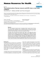
by flow cytometry. Both constructs lead to a significant
down-regulation of HLA-I surface expression (p ≤ 0.001,
Kolmogorov-Smirnov test, Fig. 1). For the pEGFP-HPV16-
E5 and pEGFP constructs, the intracellular GFP-depend-
ent fluorescence allowed us to gate GFP-expressing trans-
fected cells making it possible to compare GFP-E5 with
GFP positive populations in respect to their HLA-I signals
(Fig. 1A). Further, in our hands the anti-HA antibody did
not render sharp results differentiating transfected from
untransfected cells. For this reason, the effects for the pCI-
HPV16-E5-HA and pCI constructs were assessed by com-
paring total living cell populations (Fig. 1B). Since trans-
fection efficiency never reached 100 %, reduction in
relative values of the HLA-I surface expression tended to
be more discrete in HPV16E5-HA than in pEGFP-HPV16-
E5 transfected cells, leading to clearly significant though
smaller values in the statistical analyses (Fig. 1A and 1B).
These results therefore demonstrate that HPV16 E5 can
down-regulate cell surface expression under our experi-
mental conditions. Further, they also show that neither
the small HA (10 amino acids) nor the large EGFP (239
amino acids) used for tagging the viral protein impairs the
ability of HPV16 E5 to down-regulate HLA-I cell surface
expression.
HPV16 E5 expression reduces cell surface expression of
HLA-I molecules in calnexin-expressing but not in
calnexin-deficient cells
Since calnexin plays an important role in maturation of
the HLA-I complex, we decided to analyze whether E5
affects HLA-I surface expression by a mechanism involv-
ing calnexin. We transfected CEM-NKR and CEM-C7 cells
with pCI-HPV16-E5HA or empty pCI vector and selected
clones stably expressing E5. CEM-NKR [31] is a variant of
the leukaemia cell line CEM [43] known to be deficient in
calnexin expression (Fig. 2A) [30]. First, we checked
whether the permanent transfectants expressed E5 at sim-
ilar amounts. Pooled clones of both CEM-NKR and CEM-
C7 cells were analysed by immunoblotting for E5 expres-
Virology Journal 2007, 4:116 />Page 4 of 15
(page number not for citation purposes)
sion. As shown in Fig. 2B no major difference in the
expression level was found between both cells types. We
then analysed surface expression of HLA-I molecules by
flow cytometry, using two different anti-HLA-I antibodies.
Whereas calnexin-expressing CEM-C7 transfected with the
E5 protein contained clearly reduced amounts of surface
HLA-I molecules (Fig. 2C, left panels, KS-test p ≤ 0.001),
the calnexin-defficient CEM-NKR transfectants showed no
differences in HLA-I surface expression between E5-
expressing cells and controls (Fig. 2C, right panels, KS-test
p ≥ 0.100).
To test whether this effect simply reflected the presence of
different total amounts of HLA-I proteins in the cells, we
analysed the total amount of HLA-I molecules in CEM-
NKR and CEM-C7 cells by immunoblotting. As shown in
Fig. 2D, no major differences in the HLA-I content
between CEM-NKR and CEM-C7 cells were found when
using total cellular protein extracts from both cell lines (N
= 5, pKW = 0.87, Kruskal-Wallis test, pA = 0.77, ANOVA).
The E5-mediated reduction in the HLA-I amount at the
cell surface was thus not mediated by a lower total cellular
content of HLA-I proteins in the CEM-C7 transfectants.
These results therefore strongly suggest that E5 affects sur-
face HLA-I expression by a mechanism that involves cal-
nexin.
HPV16 E5 does not influence the transport activity of TAP
Experimental evidence has been published showing that
certain viruses target the TAP peptide transport as an effec-
tive strategy to reduce the availability of HLA-I-peptide
complexes at the cell surface, thereby reducing the cellular
HPV16 E5 expression down-regulates HLA-I surface moleculesFigure 1
HPV16 E5 expression down-regulates HLA-I surface molecules. HEK-293T cells were transfected either with (A) pEGFP-
HPV16-E5 or empty pEGFP vector, (B) pCI-HPV16-E5-HA or empty pCI vector. HLA-I molecules were then detected by
immunostaining and flow cytometry using mouse monoclonal anti-HLA-A, B, C (mAb B9.12). Differences between the HLA-I
surface expression levels were assessed by Kolmogorov-Smirnov test. This statistic defines the maximum vertical deviation
between the two curves (pEGFP-E5 and GFP, pCI-E5-HA and pCI) as the statistic D. The p value of each single experiment was
in all cases ≤ 0.001.
10
0
10
1
10
2
10
3
10
4
B9.12-PE
grey line: pEGFP
black line: pEGFP-E5
HLA class I (B9.12-PE)
D
1
=0.25 p
1
0.001
D
2
=0.28 p
2
0.001
D
3
=0.15 p
3
0.001
10
0
10
1
10
2
10
3
10
4
Channels
10
0
10
1
10
2
10
3
10
4
B9.12-PE
10
0
10
1
10
2
10
3
10
4
Channels
HLA class I (B9.12-PE)
grey line: pCI
black line: pCI-E5HA
D
1
=0.11 p
1
0.001
D
2
=0.09 p
2
0.001
D
3
=0.11 p
3
0.001
A
Kolmogorov-Smirnov test
Kolmogorov-Smirnov test
B
Virology Journal 2007, 4:116 />Page 5 of 15
(page number not for citation purposes)
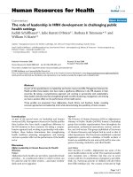
HPV16 E5 decreases HLA-I surface expression in calnexin-containing but not in calnexin-deficient cellsFigure 2
HPV16 E5 decreases HLA-I surface expression in calnexin-containing but not in calnexin-deficient cells. CEM-C7 (calnexin) and
CEM-NKR (no calnexin) cells were stably transfected with pCI-HPV16-E5-HA or pCI empty vector. A) Calnexin is only
expressed in CEM-C7 cells but not in CEM-NKR cells. B) E5-HA expression was analysed in each stable polyclone by immuno-
precipitation and -blot using mouse monoclonal anti-HA Ab and 500 µg RIPA cell lysate. C) FACS analysis of CEM-NKR and
CEM-C7 cells transfected with either the empty vector pCI or with pCI-E5-HA were stained with anti-HLA-A, B, C mAbs
B9.12. E5 expression results in diminished HLA-I surface staining in cells expressing calnexin, but not in calnexin deficient cells.
D) The upper part of the blot shown in A was incubated with anti-HC-10 antibodies (anti HLA-B, C). Incubation with anti-actin
antibodies was performed as loading control. Columns represent average values (N = 5) and the error bars comprise the cor-
responding standard deviations. There were no differences between the total amounts of cellular HLA (N = 5; pKW = 0.87,
Kruskal-Wallis test, and pA = 0.77, ANOVA). Molecular-mass markers (in kDa) are indicated in the left of the blots.
10
0
10
1
10
2
10
3
10
4
B9.12-MHCI-FITC
10
0
10
1
10
2
10
3
10
4
B9.12-MHCI-FITC
A
anti-calnexin
anti-E5-tag (HA)
100
10
B
D
anti-actin
anti-HC10
37
50
IP: a nti-E5-tag (HA)
pCI pCIE5 E5
NKR C7
C
N=5
pCI pCIE5 E5
NKR C7
0,0
0,2
0,4
0,6
0,8
1,0
1,2
1,4
p
KW
=0.87
pCI pCIE5 E5
NKR C7
CEM-C7
(Calnexin)
HLA-I (B9.12-FITC)
HLA-I (B9.12-FITC)
CEM-NKR
(no Calnexin)
KS-test:
p0.001
KS-test:
p 0.100
10
0
10
1
10
2
10
3
10
4
Channels
10
0
10
1
10
2
10
3
10
4
Channels
pCI
pCI-HPV16-
E5-HA
pCI
pCI-HPV16-
E5-HA
p
A
=0.77
Virology Journal 2007, 4:116 />Page 6 of 15
(page number not for citation purposes)
susceptibility to CTL control and eventual lysis [44,45]. To
determine whether HPV16 E5 interferes with the peptide
transport activity of TAP in CEM cells, leading to the
observed decrease in HLA-I surface expression, we applied
a peptide translocation/glycosylation assay previously
described [39]. As shown in Fig. 3, no differences in trans-
port rates between E5 expressing and control cells were
found, demonstrating that the transporter activity of TAP
is not affected by HPV16 E5 expression in CEM-C7 and
CEM-CEM-NKR transfectants.
HPV16 E5 and calnexin can be co-immunoprecipitated
from cellular extracts
To examine whether there is a physical interaction
between E5 and calnexin, we transfected cells with HPV16
E5 and analysed whether calnexin and E5 could be co-
immunoprecipitated. Since protein expression of the viral
E5 gene is very weak in transfected cells, we prepared a
codon-adapted version of the E5 sequence fitting to the
codon usage preferences in humans, a procedure known
to allow for increased protein expression of the protein in
eukaryotic cells [46-48]. HEK-293T cells were transfected
with the codon-adapted E5-coding DNA and protein
expression levels were tested by Western blot. As shown in
Fig. 4A (left) the codon-optimised E5 gene is well
expressed in HEK-293T cells, some orders of magnitude
above the expression achieved for the wild-type E5 gene
(Fig. 4A, right). Cellular proteins were immunoprecipi-
tated with antibodies against the AU1-tagged E5 protein,
separated on SDS-PAGE, blotted, and the membrane was
subsequently incubated with antibodies against calnexin.
A band of 90 kDa apparent molecular mass correspond-
ing to calnexin was identified in the immunoprecipitates,
demonstrating that HPV16 E5 and calnexin could be co-
immunoprecipitated in extracts of transfected cells (Fig.
4B). To further substantiate these results we performed
the reverse experiment immunoprecipitating the extracts
from transfected cells first with calnexin antibodies and
then incubating the separated immunoprecipitates on the
membrane with anti-E5-tag antibodies (anti-AU1). As
shown in Fig. 4C, a reacting band of about 10 kDa was
observed. This is the molecular mass found for HPV16 E5
when total cellular protein extracts were used for the
immunoblots. These results demonstrate that HPV16 E5
and calnexin either directly interact in vitro. This interac-
tion could also be reproduced when non-optimised viral
E5-coding DNA (pCI-HPV16-E5-HA) was used for trans-
fection (Fig. 4D and 4E), indicating that the effects did not
arise from the higher amount of protein expressed from
the codon-adapted version (Fig. 4A).
To further corroborate this finding at the intracellular
level we next sought to demonstrate co-localization of
both proteins in human keratinocytes expressing the E5
protein. HaCaT cells were transiently transfected with
AU1-tagged codon-adapted E5 and co-localization with
calnexin was analysed by laser confocal double immun-
ofluorescence microscopy. As shown in Fig. 5A we
observed a sharp colocalization of both proteins, confirm-
ing already published results for retroviral transduced
keratinocytes [48]. Similar results were obtained when the
GFP fusion protein was expressed instead of the AU1-
tagged codon-optimised E5 protein (Fig. 5B), indicating
that the subcellular localization of the E5 protein does not
depend on the nature of the tag used to label E5.
An intact hydrophobic region of HPV16 E5 is necessary for
binding to calnexin
To analyze the characteristics of the E5-calnexin binding
in more detail, we prepared a series of point mutants -M1,
M2 and M3- in which we modified the E5 protein
sequence, altering the hydrophobic profile and the local
propensity to form helical structures. Leucine and/or iso-
leucine residues were mutated to proline, aspartate or
arginines and then the resulting hydrophobic profile, pro-
pensity to helical structure and potential for stably span-
ning the cellular membrane were analysed and compared
with those of the wild-type E5 protein (Fig. 6A, 6B). The
point mutations were chosen so that they resulted respe-
tively in the disruption of each of the three putative trans-
membrane helix within each of the three hydrophobic
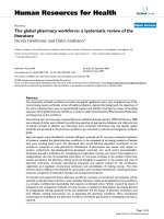
Transporter activity of TAP is not influenced by HPV16 E5Figure 3
Transporter activity of TAP is not influenced by HPV16 E5.
Streptolysin Opermeabilized calnexin-proficient CEM-C7 and
calnexin-deficient CEM-NKR cells (38) were analysed in a
peptide translocation/glyosylation assay using the indicated
input quantities of the radioiodinated reporter peptide TNK-
TRIDGQY (glycosylation consensus site underlined) in the
presence or absence of ATP. The glycosylated fraction, indic-
ative of TAP-mediated ER transport, is isolated by concanav-
alin A Sepharose and quantitated by γ-counting. No
significant differences could be detected between HPV16 E5-
expressing cells and the control cells irrespective from the
presence (CEM-C7) or absence (CEM-NKR) of calnexin.
Virology Journal 2007, 4:116 />Page 7 of 15
(page number not for citation purposes)
domains of the E5 protein, without changing the total
protein length. All three mutants were based on the
codon-optimised version of E5.
To test whether the mutants M1, M2 and M3 were
expressed at similar levels, HEK-293T cells were trans-
fected with the original codon-optimised E5 sequences or
with each of the mutants, and the protein content was
analysed by immunoblotting. As shown in Fig. 7A, all
recombinants showed similar levels of expression, being
differences in SDS-PAGE migration attributable to the dif-
ferent hydrophobicity of the proteins.
To analyze the differential involvement of the each of the
three E5 transmembrane domains in the interaction
between E5 and calnexin, we performed immunoprecipi-
tation experiments with the three mutants M1, M2 and
M3 as described above. Protein extracts from transfected
cells were immunoprecipitated with antibodies against
the AU1 epitope, and the precipitates were analysed for
calnexin content by immunoblotting. As shown in Fig.
7B, the original codon-optimized E5 protein and the
mutants M2 and M3 co-precipitated calnexin to similar
extents, whereas mutant M1 precipitated clearly reduced
amounts of calnexin. To discard artefacts due to different
inputs of antibody, protein G-sepharose or protein, the
experiments were repeated six times. As shown in Fig. 7C
mutant M1 co-precipitated calnexin to only 50 % of the
levels precipitated by the wild-type and mutants M2 and
M3. These results could be reproduced when non-opti-
Calnexin interacts with the HPV16 E5 protein in cellular extractsFigure 4
Calnexin interacts with the HPV16 E5 protein in cellular extracts. HEK-293T cells were transfected with AU1-tagged codon-
optimised HPV16 E5, pCI-HPV16-E5-HA or corresponding empty vectors and lysed at 24 h posttransfection with CHAPS lysis
buffer. A) Immunoblot showing the expression levels of the codon-optimised E5 gene (left panel) and of the viral E5 gene (right
panel). Note the differences in the immunoreactivity signals despite the higher amount of total protein loaded in the non-opti-
mised gene (100 µg vs 30 µg). B) Immunoprecipitations were performed using monoclonal anti-E5-tag (AU1) antibodies and
proteins in the immune complexes were probed using anti-AU1 and anti-calnexin antibodies. C) Immunoprecipitations were
performed using monoclonal anti-calnexin antibodies and proteins in the immune complexes were probed using anti-calnexin
and anti-E5-tag (AU1) anti-bodies. D) Immunoprecipitations were performed using monoclonal anti-E5-tag (HA) antibodies and
proteins in the immune complexes were probed using anti-HA and anti-calnexin antibodies. E) 2 Immunoprecipitations were
performed using monoclonal anti-calnexin antibodies and proteins in the immune complexes were probed using anti-calnexin
and anti-E5-tag (HA) antibodies. Molecular-mass markers in kDa are indicated at the left of the blots.
10
anti-E5-tag (AU1)
100
IP: anti-calnexin
anti-calnexin
E5pCDNA
10
anti-E5-tag (HA)
100
IP: anti-calnexin
anti-calnexin
E5pCI
10
anti-E5-tag (HA)
100
IP: anti-E5-tag (HA)
anti-calnexin
E5pCI
D
EC
anti-E5-tag
(AU1 left, HA right)
E5
30µg
pCIpCDNA
100µg
E5
10
10
anti-E5-tag (AU1)
100
IP: anti-E5-tag (AU1)
anti-calnexin
E5pCDNA
B
A
Virology Journal 2007, 4:116 />Page 8 of 15
(page number not for citation purposes)
mised viral E5-coding DNA (pEGFP-HPV16-E5 and
pEGFP-M1) was used for transfection instead of the
codon-adapted E5-coding DNA (Fig. 7D and 7E). Taken
together, these results strongly suggest that the first hydro-
phobic region of E5, i.e. the first putative transmembrane
domain of the protein, is involved in the interaction with
calnexin.
Co-localization of HPV16 E5 and calnexin is dependent on
the presence of the first hydrophobic domain of E5
The experiments described above indicate that the intera-
cion between E5 and calnexin relies on the presence of an
intact first hydrophobic region, and that this binding may
be responsible for down-regulation of HLA-I expression.
Should this be true, a reduction in co-localization
between calnexin and mutant M1 would be expected in
immunofluorescence experiments. In order to address
this point, HaCaT cells were transfected with the three
mutants M1, M2, and M3 and double immunofluores-
cence with anti-calnexin and anti tag antibodies was per-
formed.
As shown in Fig. 8, calnexin colocalized with the E5 pro-
tein expressed from the codonoptimized gene (Fig. 8A), as
well as with the M2 and M3 mutants (Fig. 8C and 8D). In
contrast, the disruption of the first helix in mutant M1
results in a change in the subcellular localisation of the
protein, yielding a disperse and punctuate subcellular dis-
tribution, where only a partial co-localization with cal-
nexin (Fig. 8B). These results are consistent with those
found in the immunoprecipitation experiments and fur-
ther confirm that the interaction of HPV16 E5 and cal-
nexin requires a native, non-modified first
transmembrane domain of the viral protein.
Calnexin, HPV16 E5 and HLA form a trimeric complex
Recent results have shown that HPV16 E5 may co-precip-
itate with the heavy chain of HLA-I [21]. In the light of our
results presented above, and together with the fact that
HLA-I and calnexin associate during HLA maturation, we
hypothesized that the formation of a trimeric complex
between HLA-I heavy chain, calnexin and E5 might be
involved in the retention of HLA-I in the ER/Golgi appa-
ratus of the cells expressing E5. To address this question,
HeLa cells were transfected with AU1-tagged codon-opti-
mised E5 or with mutant M1, and protein extracts were
immunoprecipitated with anti-AU1. Immunoprecipitates
separated in SDS-PAGE, were blotted onto PVDF mem-
Co-localization of HPV16 E5 with calnexinFigure 5
Co-localization of HPV16 E5 with calnexin. HaCaT cells were transfected with AU1- tagged codon-optimised E5 or pEGFP-E5
and analysed after 24 h by confocal laser scanning microscopy using a monoclonal anti-AU1 and/or polyclonal anti-calnexin Abs.
A
B
mergecalnexin
E5-AU1
pEGFP-E5
10m
Virology Journal 2007, 4:116 />Page 9 of 15
(page number not for citation purposes)
brane and probed either with anti-HC10, recognizing
HLA-B, C heavy chains [49], or with anti-calnexin anti-
bodies. As shown in Fig. 9, both HLA-I heavy chain and
calnexin could be co-immunoprecipitated with anti-AU1
antibodies, which target E5. More important, the E5
mutant M1 previously shown to be deficient in immuno-
precipitation of calnexin, also failed to co-precipitate the
HLA-I heavy chain. These results demonstrate that HPV16
E5 forms a complex with calnexin and HLA-I heavy chain
and that this complex depends on the interaction of the
first hydrophobic region of E5 with calnexin.
Mutant M1 is not able to down-regulate HLA-I cell surface
expression in the same extent that wild type HPV16 E5
does
Since the experiments shown above demonstrate that
mutation of the first putative transmembrane helix of E5
results in the loss of binding to calnexin, we addressed the
question whether this loss correlates with the failure to
down-regulate HLA-I surface expression. HEK-293T cells
were transfected with the wild-type pEGFP-E5, mutant
pEGFP-M1 or pEGFP empty vector and the amount of
HLA-I expression at the cell surface was determined by
Transmembrane Hidden Markov Model posterior probabilities for the sequences of E5 and the mutants M1, M2 and M3Figure 6
Transmembrane Hidden Markov Model posterior probabilities for the sequences of E5 and the mutants M1, M2 and M3. A)
Amino acid sequence of the wild-type E5 protein and corresponding mutants. Aminoacids of the AU1-tag are underlined.
Arrows show the position of exchanged amino acids. B) Analysis of the wild-type E5 and mutants using the TMHMM 2.0 algo-
rithm (36, 37), showing the three hydrophobic regions predicted to be transmembrane domains, and the corresponding dis-
ruptions in the three mutants.
A
B
MDTYRYIT NLDTASTTLLACFLLCFCVLLCVCLLIRPLLLSVSTYTSLIILVLLLWITAASAFRCFIVYIIFVYI PLFLIHTHARFLIT
wt
M1
M2
M3
MDTYRYIT NLDTASTTLpACFLdCFCV rLCVCLLIRPLLLSVSTYTSLIILVLLLWITAASAFRCFI VYIIFVYI PLFLIHTHARFLIT
MDTYRYI
TN LDTASTTLLACFLLCFCVLLCVCLLIRPLLLSVSTYTSpIIdV LLrWITAASAFRCFI VYIIFVYI PLFLIHTHARFLIT
MDTYRYI
TN LDTASTTLLACFLLCFCVLLCVCLLIRPLLLSVSTYTSLIILVLLLWITAASAFRCFpVYIdFVYIPrFLIHTHARFLIT
M3
M2
M1
wt
inside
outside
transmembrane
posterior probability
Virology Journal 2007, 4:116 />Page 10 of 15
(page number not for citation purposes)
FACS analysis. While wild-type E5 expression resulted in
HLA-I down-regulation at the plasma membrane (Figs. 1
and 2C, Fig. 10), this effect was not observed when the
cells expressed the E5 mutant M1 (Fig. 10). To substanti-
ate this result, we did the experiment six times and ana-
lysed the median values of HLA-I surface expression in the
transfected cells (for statistical analysis, see Table 1).
Whereas the wild type E5 protein was able to down-regu-
late HLA-I surface expression down to 65% (median of six
experiments), the median HLA-I staining of HEK-293T
transfected with the E5 mutant M1 was 82% (median of
six experiments) as compared with HEK-293T control
transfectants (N = 6, pW = 0.0313, Wilcoxon matched-
Mutant M1 binds less calnexin than wild-type E5 proteinFigure 7
Mutant M1 binds less calnexin than wild-type E5 protein. HEK-293T cells were transfected with either (A-C) AU1-tagged
codon-optimised HPV16 E5, the mutants M1, M2 and M3 or pcDNA 3.1 empty vector as control, (D and E) pEGFP-tagged
HPV16 E5, mutant pEGFPM1, mock-control or pEGFP empty vector and lysed at 24 h posttransfection with CHAPS lysis
buffer. A) Similar expression levels of all HPV16 E5 and the mutants M1, M2 and M3. B) Immunoprecipitations were performed
using monoclonal anti-AU1, and proteins in the immune complex were detected using anti-AU1 and anti-calnexin. C) Quantifi-
cation of co-precipitated calnexin for wild-type HPV16E5 protein, the mutants M1, M2, M3 and the vector control. The wild-
type expression level was set to 100%. Data shown represent six independent experiments 2 plus standard errors of the mean.
P values were calculated with paired two-tailed Student's t-test. D) Similar expression levels of pEGFP-HPV16-E5 and the
mutant pEGFP-M1. E) Immunoprecipitations were performed using monoclonal anti-GFP, and proteins in the immune complex
were detected using anti-GFP and anti-calnexin. Molecular-mass markers in kDa are indicated at the left of the blots.
100
anti-calnexin
anti-actin
anti-E5-tag (AU1)
10
37
A
10
anti-E5-tag (AU1)
IP: anti-E5-tag (AU1)
pCDNA
pCDNA
E5 M2M1 M3
E5 M2M1 M3
pcDNA E5 M1 M2 M3
0
20
40
60
80
100
120
140
**
p<0.001
N=6
anti-actin
anti-E5-tag (GFP)
25
37
D
GFP E5 M1 mock
100
E
25
anti-E5-tag (GFP)
IP: anti-E5-tag (GFP)
anti-calnexin
B
C
GFP E5 M1 mock
percent
p=0.909
p=0.250
Virology Journal 2007, 4:116 />Page 11 of 15
(page number not for citation purposes)
HPV16 E5, M2 and M3 mutants but not M1 mutant strongly co-localize with calnexinFigure 8
HPV16 E5, M2 and M3 mutants but not M1 mutant strongly co-localize with calnexin. HaCaT cells were transfected with A)
AU1-tagged codon-optimised E5 or AU1- tagged codonoptimised E5 mutants M1 B), M2 C), and M3 D) and analysed after 24 h
by confocal laser scanning microscopy using a monoclonal anti-AU1 and polyclonal anticalnexin antibodies.
M1-AU1
M2-AU1
M3-AU1
10m
A
mergecalnexin
E5-AU1
B
D
C
Virology Journal 2007, 4:116 />Page 12 of 15
(page number not for citation purposes)
pairs signed ranks test, pST = 0.009, paired Student's t-
test).
Taken together, our results strongly indicate i) that E5-
mediated down-regulation of HLA-I surface expression
proceeds through the formation of a ternary complex
between E5, calnexin and the heavy chain of HLA-I; ii)
that the disruption of the first transmembrane domain of
HPV16 E5 modifies the subcellular distribution of the
protein; and iii) that the disruption of the first transmem-
brane domain of HPV16 E5 prevents the interaction, colo-
calisation and immunoprecipitation of the viral protein
with calnexin, and also of that with the heavy chain of
HLA-I.
Discussion
Eukaryotic cells respond to viral infection by activating
mechanisms aiming to abortion of the infection through
hindering of viral protein expression, virus maturation or
virus release, while viruses have developed during evolu-
tion molecular countermeasures to escape from these cel-
lular controls. One of these viral strategies leads to a
reduction in the adaptive immunoresponses of the host
by reducing the exposure of the infected cells to immune
surveillance. Reduced surface expression of HLA-I has
been described upon expression of HPV16 E5 or HPV2 E5
proteins [22,42], but the molecular mechanisms respon-
sible for the decrease of HLA-I on the cell surface have not
yet been elucidated. In this report we present experimen-
tal evidence demonstrating that HPV16 E5 down-regu-
lates HLA-I surface expression by a calnexin-mediated
mechanism. Using transient and stably transfected cells,
we have shown that HPV16 E5 is able to reduce HLA-I sur-
face expression in calnexin-containing cells, but not in a
calnexin-deficient cell line. Published reports have
described that the heavy chain of HLA-I molecules and
HPV16 E5 could be co-precipitated [21], suggesting that
this binding might be involved in HLA-I down-regulation.
Nevertheless, our results point to the binding of E5 to cal-
nexin as the critical molecular event directly involved in
HLA down-regulation. Expression of E5 in CEM-C7 cells,
which constitutively express calnexin, results in a
decreased amount of HLA-I at the cell surface, but no
down-regulation was observed in CEM-NKR cells devoid
of calnexin (see Fig. 2C). Since both cell types CEM-C7
and CEM-NKR contain similar amounts of HLA-I mole-
cules (Fig. 2D and see [30]) it seems unlikely that a puta-
tive binding of HPV16 E5 to the HLA-I heavy chain alone
could be solely responsible for the decreased surface
expression of HLA-I proteins in CEM-C7 cells.
Regarding other viruses, such as herpes simplex virus and
cytomegalovirus, it has been shown that they target the
transporter associated with antigen processing (TAP) in
order to down-regulate HLA-I surface expression [50,51].
In PVes it has been demonstrated that purified HPV11 E7
protein is able to inhibit ATP-dependent peptide transport
into the lumen of the ER in vitro [52]. In this context, our
peptide translocation-assay results show that HPV16 E5
does not influence the transport of antigen peptides from
the cytosol to the ER. Thus, the data here presented sug-
E5 mutant M1 down-regulates surface expression of HLA-I to a lesser extent than the E5 protein doesFigure 10
E5 mutant M1 down-regulates surface expression of HLA-I
to a lesser extent than the E5 protein does. HEK-293T cells
were transfected either with pEGFP-HPV16- E5, -M1, or
empty pEGFP vector. HLA-I molecules were detected by
immunostaining and flow cytometry using mouse monoclonal
anti-B9.12 Ab. Results for one representative experiment out
of six are shown. Statistic analysis is shown in Table 1.
grey line: pEGFP
dark grey line: pEGFP-M1
black line: pEGFP-E5
10
0
10
1
10
2
10
3
10
4
B9.12-PE
HLA-I (B9.12-PE)
HPV16 E5 forms a ternary complex with calnexin and the HLA-I heavy chainFigure 9
HPV16 E5 forms a ternary complex with calnexin and the
HLA-I heavy chain. HeLa cells were transiently transfected
with AU1-tagged codon-optimised HPV16 E5, M1, or empty
vector. 24 h later CHAPS lysates were immunoprecipitated
with antibodies against the E5-tag (anti-AU1). Precipitated
immune complexes were separated by SDS-PAGE and West-
ern blotted using anti-calnexin and anti-HLA-B, -C mAb
(HC10), respectively (band marked with *). Molecular-mass
markers in kDa are indicated at the left of the blots.
anti-HC10
anti-calnexin
37
100
*
pCDNA E5 M1 IP: anti-E5-tag (AU1)
Virology Journal 2007, 4:116 />Page 13 of 15
(page number not for citation purposes)
gest that HPV16 E5 does not target the TAP transporter
activity to control surface expression of HLA-I molecules.
Our co-immunoprecipitation experiments using either
antibodies against different tagged versions of the E5 pro-
tein or against calnexin demonstrate that HPV16 E5 asso-
ciates with calnexin in vitro. The biological significance of
this interaction is further supported by the previously
described intracellular co-localization of calnexin and
HPV16 E5 [48], that we confirmed in this report.
Upon interaction between the first and the third hydro-
phobic segments [53], HPV16 E5 could be organized as a
transmembrane protein with three putative transmem-
brane helices [54]. In the present work we have intro-
duced specific point mutations in this E5 gene, selectively
targeting local hydrophobicity and propensity towards
helix conformation in each of the three predicted trans-
membrane helices of the HPV16 E5 protein [32]. These
point mutations result in the selective and individual dis-
ruption of each helix without altering the overall length of
the protein. Our results reveal that the first hydrophobic
helix is mainly responsible for HPV16 E5 subcellular
localisation and concomitantly for colocalisation
between HPV16 E5 and calnexin. Mutant M1 -with the
first putative transmembrane helix being disrupted- was
able to bind reduced amounts of calnexin in immunopre-
cipitation assays, while co-localizing only weakly with cal-
nexin in transfected cells. In addition, M1 transfectants
did not down-regulate surface expression of HLA-I in the
same extent than wild-type E5. Together both results sug-
gest that i) the first putative transmembrane domain of
HPV16 E5 is responsible for the HPV16 E5 localisation; ii)
the interaction of HPV16 E5 and calnexin depends on the
integrity of the first putative transmembrane domain; iii)
the effect of HPV16 E5 on HLA-I surface expression
strongly depends on the integrity of the first putative
transmembrane domain and on the subsequent interac-
tion between HPV16 E5 and calnexin.
The definitive finding presented here is the existence of a
ternary protein complex of HPV16 E5, calnexin, and the
heavy chain of HLA-I molecules. The formation of this
complex depends on the presence of the first predicted
transmembrane domain of HPV16 E5. Since the dimer
calnexin-HLA is a natural step in the antigen processing
route, it can be hypothesized that HPV16 E5 binds to the
calnexin-HLA-I complex and that this binding blocks fur-
ther trafficking of the HLA-I complex to the plasma mem-
brane, leading instead to its accumulation in the ER/Golgi
of the infected cell. A direct binding of E5 to the heavy
chain of HLA-I seems under the light of our results
improbable. This is further supported by our findings
using calnexin-deficient cells lines. Although both cell
types, calnexin-containing and calnexin-deficient, express
similar amounts of heavy chain HLA-I, the E5-mediated
reduction of surface HLA-I becomes evident exclusively in
calnexin-containing cells.
The interaction between E5 and calnexin could be demon-
strated in cells transfected with the codon-adapted version
of the gene, and also in cells transfected with the wild-type
gene. This association is therefore independent from the
effective amount of E5 protein expressed, and cannot be
due to a very large overexpression from the optimised ver-
sion of the gene. This is not a trivial result, as it has been
shown that codon usage optimization can lead to changes
in the phenotype associated with protein expression
[55,56].
Table 1: E5 mutant M1 down-regulates surface expression of HLA-I to a lesser extent than the E5 protein does.
E5-GFP p- value
c
M1-GFP E5-GFP p- value
c
M1-GFP
HLA- 57.77% t- test 69.78% KS-D
b
0.29 t- test 0.20
surface 67.92% 0.009 75.67% 0.17 0.005 0.13
expression
a
75.67% 94.75% 0.15 0.04
69.16% Wilcoxon 82.04% 0.20 Wilcoxon 0.11
59.35% 0.0313 82.78% 0.25 0.0313 0.10
62.08% 100.90% 0.23 0.02
Median 65.00% 82.41% Median 0.215 0.105
Range 58%-76% 70%-100% Range 0.17–0.29 0.02–0.2
a
Values of pEGFP-E5 and pEGFP-M1 were normalized to the values of the pEGFP-control (E5-GFP, M1-GFP), which was set as 100% expression of
HLA-I surface expression.
b
Kolmogorov-Smirnov (KS) test was performed with CellQuest™ software (BD Bioscience). This statistic defines the maximum vertical deviation
between the two curves (pEGFP-E5 and pEGFP-control or pEGFP-M1 and pEGFP-control) as the statistic D. The p value of each single experiment
was ≤ 0.001.
c
Paired two tailed paired student's t-test and Wilcoxon matched-pairs signed-ranks test values for the raw percentages of immunorreactive cells
(left column) and for the D statistic (right column). The E5 mutant M1 does not affect HLA-I expression in the same extent than the original E5
protein does (p < 0.05 in all cases).
Virology Journal 2007, 4:116 />Page 14 of 15
(page number not for citation purposes)
Conclusion
In summary, our results support a model for the E5-medi-
ated HLA-I surface downregulation in which the viral pro-
tein interacts with calnexin, finally leading to an E5-
calnexin-HLA-I heavy chain ternary complex unable to be
further transported to the cell surface.
Authors' contributions
MG performed molecular biology, cell biology, confocal
microscopy and flow citometry experiments, and drafted
the manuscript. IGB participated in the design of the
research concept and in mutant design, performed statis-
tical analyses and drafted the manuscript. FM performed
the peptide translocation assay. AA conceived and super-
vised the study and drafted the manuscript. PT collabo-
rated in the supervision of the study and helped draft the
manuscript. All authors have read and approved the final
manuscript.
Acknowledgements
IGB is the recipient of a grant from the Volkswagen Stiftung under the The-
matic Impetus "Evolutionary Biology".
References
1. DiMaio D, Liao JB: Human papillomaviruses and cervical can-
cer. Adv Virus Res 2006, 66:125-159.
2. Munoz N, Bosch FX, de Sanjose S, Herrero R, Castellsague X, Shah
KV, Snijders PJ, Meijer CJ: Epidemiologic classification of human
papillomavirus types associated with cervical cancer. N Engl
J Med 2003, 348:518-527.
3. Franco EL, Schlecht NF, Saslow D: The epidemiology of cervical
cancer. Cancer J 2003, 9:348-359.
4. Walboomers JM, Jacobs MV, Manos MM, Bosch FX, Kummer JA, Shah
KV, Snijders PJ, Peto J, Meijer CJ, Munoz N: Human papillomavirus
is a necessary cause of invasive cervical cancer worldwide. J
Pathol 1999, 189:12-19.
5. Clifford GM, Smith JS, Aguado T, Franceschi S: Comparison of HPV
type distribution in high-grade cervical lesions and cervical
cancer: a meta-analysis. Br J Cancer 2003, 89:101-105.
6. Bosch FX, Lorincz A, Munoz N, Meijer CJ, Shah KV: The causal
relation between human papillomavirus and cervical cancer.
J Clin Pathol 2002, 55:244-265.
7. Stoler MH, Broker TR: In situ hybridization detection of human
papillomavirus DNAs and messenger RNAs in genital condy-
lomas and a cervical carcinoma. Hum Pathol 1986,
17:1250-1258.
8. Crum CP, Symbula M, Ward BE: Topography of early HPV 16
transcription in high-grade genital precancers. Am J Pathol
1989, 134:1183-1188.
9. Garnett TO, Duerksen-Hughes PJ: Modulation of apoptosis by
human papillomavirus (HPV) oncoproteins. Arch Virol 2006,
151:2321-2335.
10. Werness BA, Levine AJ, Howley PM: Association of human papil-
lomavirus types 16 and 18 E6 proteins with p53. Science 1990,
248:76-79.
11. Dyson N, Howley PM, Munger K, Harlow E: The human papilloma
virus-16 E7 oncoprotein is able to bind to the retinoblastoma
gene product. Science 1989, 243:934-937.
12. Kabsch K, Alonso A: The human papillomavirus type 16 E5 pro-
tein impairs TRAIL- and FasL-mediated apoptosis in HaCaT
cells by different mechanisms. J Virol 2002, 76:12162-12172.
13. Bravo IG, Crusius K, Alonso A: The E5 protein of human papil-
lomavirus type 16 modulates composition and dynamics of
membrane lipids in keratinocyte membranes. Arch Virol 2005,
150:231-246.
14. Sasagawa T, Inoue M, Tanizawa O, Yutsudo M, Hakura A: Identifica-
tion of antibodies against human papillomavirus type 16 E6
and E7 proteins in sera of patients with cervical neoplasias.
Jpn J Cancer Res 1992, 83:705-713.
15. Muller M, Viscidi RP, Sun Y, Guerrero E, Hill PM, Shah F, Bosch FX,
Munoz N, Gissmann L, Shah KV: Antibodies to HPV-16 E6 and E7
proteins as markers for HPV-16-associated invasive cervical
cancer. Virology 1992, 187:508-514.
16. Dillner J: Mapping of linear epitopes of human papillomavirus
type 16: the E1, E2, E4, E5, E6 and E7 open reading frames.
Int J Cancer 1990, 46:703-711.
17. Kochel HG, Sievert K, Monazahian M, Mittelstadt-Deterding A, Teich-
mann A, Thomssen R: Antibodies to human papillomavirus
type-16 in human sera as revealed by the use of prokaryoti-
cally expressed viral gene products. Virology 1991, 182:644-654.
18. Auvinen E, Crusius K, Steuer B, Alonso A: Human papillomavirus
type 16 E5 protein (Review). Int J Oncol 1997, 11:1297-1304.
19. O'Brien PM, Saveria Campo M: Evasion of host immunity
directed by papillomavirus-encoded proteins. Virus Res 2002,
88:103-117.
20. Zhang B, Li P, Wang E, Brahmi Z, Dunn KW, Blum JS, Roman A: The
E5 protein of human papillomavirus type 16 perturbs MHC
class II antigen maturation in human foreskin keratinocytes
treated with interferon-gamma. Virology 2003, 310:100-108.
21. Ashrafi GH, Haghshenas M, Marchetti B, Campo MS: E5 protein of
human papillomavirus 16 downregulates HLA class I and
interacts with the heavy chain via its first hydrophobic
domain. Int J Cancer 2006, 119:2105-2112.
22. Ashrafi GH, Haghshenas MR, Marchetti B, O'Brien PM, Campo MS:
E5 protein of human papillomavirus type 16 selectively
downregulates surface HLA class I. Int J Cancer 2005,
113:276-283.
23. Cromme FV, Snijders PJ, van den Brule AJ, Kenemans P, Meijer CJ,
Walboomers JM: MHC class I expression in HPV 16 positive
cervical carcinomas is post-transcriptionally controlled and
independent from c-myc overexpression. Oncogene 1993,
8:2969-2975.
24. Marchetti B, Ashrafi GH, Tsirimonaki E, O'Brien PM, Campo MS: The
bovine papillomavirus oncoprotein E5 retains MHC class I
molecules in the Golgi apparatus and prevents their trans-
port to the cell surface. Oncogene 2002, 21:7808-7816.
25. Ortmann B, Androlewicz MJ, Cresswell P: MHC class I/beta 2-
microglobulin complexes associate with TAP transporters
before peptide binding. Nature 1994, 368:864-867.
26. Ahluwalia N, Bergeron JJ, Wada I, Degen E, Williams DB: The p88
molecular chaperone is identical to the endoplasmic reticu-
lum membrane protein, calnexin. J Biol Chem 1992,
267:10914-10918.
27. Degen E, Cohen-Doyle MF, Williams DB: Efficient dissociation of
the p88 chaperone from major histocompatibility complex
class I molecules requires both beta 2-microglobulin and
peptide. J Exp Med 1992, 175:1653-1661.
28. Ritz U, Momburg F, Pilch H, Huber C, Maeurer MJ, Seliger B: Defi-
cient expression of components of the MHC class I antigen
processing machinery in human cervical carcinoma. Int J
Oncol 2001, 19:1211-1220.
29. Ayala-Torres S, Moller PC, Johnson BH, Thompson EB: Character-
istics of 25-hydroxycholesterol-induced apoptosis in the
human leukemic cell line CEM. Exp Cell Res 1997, 235:35-47.
30. Scott JE, Dawson JR: MHC class I expression and transport in a
calnexin-deficient cell line. J Immunol 1995, 155:143-148.
31. Howell DN, Andreotti PE, Dawson JR, Cresswell P: Natural killing
target antigens as inducers of interferon: studies with an
immunoselected, natural killing-resistant human T lym-
phoblastoid cell line. J Immunol 1985, 134:971-976.
32. Bravo IG, Alonso A: Mucosal human papillomaviruses encode
four different E5 proteins whose chemistry and phylogeny
correlate with malignant or benign growth. J Virol 2004,
78:13613-13626.
33. Oetke C, Auvinen E, Pawlita M, Alonso A: Human papillomavirus
type 16 E5 protein localizes to the Golgi apparatus but does
not grossly affect cellular glycosylation. Arch Virol 2000,
145:2183-2191.
34. Gieswein CE, Sharom FJ, Wildeman AG: Oligomerization of the
E5 protein of human papillomavirus type 16 occurs through
multiple hydrophobic regions. Virology 2003, 313:415-426.
35. Rodriguez MI, Finbow ME, Alonso A: Binding of human papillo-
mavirus 16 E5 to the 16 kDa subunit c (proteolipid) of the
Publish with BioMed Central and every
scientist can read your work free of charge
"BioMed Central will be the most significant development for
disseminating the results of biomedical research in our lifetime."
Sir Paul Nurse, Cancer Research UK
Your research papers will be:
available free of charge to the entire biomedical community
peer reviewed and published immediately upon acceptance
cited in PubMed and archived on PubMed Central
yours — you keep the copyright
Submit your manuscript here:
/>BioMedcentral
Virology Journal 2007, 4:116 />Page 15 of 15
(page number not for citation purposes)
vacuolar H+-ATPase can be dissociated from the E5-medi-
ated epidermal growth factor receptor overactivation. Onco-
gene 2000, 19:3727-3732.
36. Yang DH, Wildeman AG, Sharom FJ: Overexpression, purifica-
tion, and structural analysis of the hydrophobic E5 protein
from human papillomavirus type 16. Protein Expr Purif 2003,
30:1-10.
37. Sonnhammer EL, von Heijne G, Krogh A: A hidden Markov model
for predicting transmembrane helices in protein sequences.
Proc Int Conf Intell Syst Mol Biol 1998, 6:175-182.
38. Moller S, Croning MD, Apweiler R: Evaluation of methods for the
prediction of membrane spanning regions. Bioinformatics 2001,
17:646-653.
39. Neefjes JJ, Momburg F, Hammerling GJ: Selective and ATP-
dependent translocation of peptides by the MHC-encoded
transporter. Science 1993, 261:769-771.
40. Rebai N, Malissen B: Structural and genetic analyses of HLA
class I molecules using monoclonal xenoantibodies. Tissue
Antigens 1983, 22:107-117.
41. Ashrafi GH, Tsirimonaki E, Marchetti B, O'Brien PM, Sibbet GJ,
Andrew L, Campo MS: Down-regulation of MHC class I by
bovine papillomavirus E5 oncoproteins. Oncogene 2002,
21:248-259.
42. Cartin W, Alonso A: The human papillomavirus HPV2a E5 pro-
tein localizes to the Golgi apparatus and modulates signal
transduction. Virology 2003, 314:572-579.
43. Foley GE, Lazarus H, Farber S, Uzman BG, Boone BA, McCarthy RE:
Continuous Culture of Human Lymphoblasts from Periph-
eral Blood of a Child with Acute Leukemia. Cancer 1965,
18:522-529.
44. Loch S, Tampe R: Viral evasion of the MHC class I antigen-
processing machinery. Pflugers Arch 2005, 451:409-417.
45. Vossen MT, Westerhout EM, Soderberg-Naucler C, Wiertz EJ: Viral
immune evasion: a masterpiece of evolution. Immunogenetics
2002, 54:527-542.
46. Zhou J, Liu WJ, Peng SW, Sun XY, Frazer I: Papillomavirus capsid
protein expression level depends on the match between
codon usage and tRNA availability. J Virol 1999, 73:4972-4982.
47. Leder C, Kleinschmidt JA, Wiethe C, Muller M: Enhancement of
capsid gene expression: preparing the human papillomavirus
type 16 major structural gene L1 for DNA vaccination pur-
poses. J Virol 2001, 75:9201-9209.
48. Disbrow GL, Sunitha I, Baker CC, Hanover J, Schlegel R: Codon
optimization of the HPV-16 E5 gene enhances protein
expression. Virology 2003, 311:105-114.
49. Stam NJ, Spits H, Ploegh HL: Monoclonal antibodies raised
against denatured HLA-B locus heavy chains permit bio-
chemical characterization of certain HLA-C locus products.
J Immunol 1986, 137:2299-2306.
50. Seliger B, Ritz U, Ferrone S: Molecular mechanisms of HLA class
I antigen abnormalities following viral infection and transfor-
mation. Int J Cancer 2006, 118:129-138.
51. Yewdell JW, Bennink JR: Mechanisms of viral interference with
MHC class I antigen processing and presentation. Annu Rev
Cell Dev Biol 1999, 15:579-606.
52. Vambutas A, DeVoti J, Pinn W, Steinberg BM, Bonagura VR: Interac-
tion of human papillomavirus type 11 E7 protein with TAP-
1 results in the reduction of ATP-dependent peptide trans-
port. Clin Immunol 2001, 101:94-99.
53. Alonso A, Reed J: Modelling of the human papillomavirus type
16 E5 protein. Biochim Biophys Acta 2002, 1601:9-18.
54. Bravo I, Alonso A, Auvinen E: Human papillomavirus type 16 E5
protein. Papillomavirus Report 2004, 15:1-6.
55. Cid-Arregui A, Juarez V, zur Hausen H: A synthetic E7 gene of
human papillomavirus type 16 that yields enhanced expres-
sion of the protein in mammalian cells and is useful for DNA
immunization studies. J Virol 2003, 77:4928-4937.
56. Samorski R, Gissmann L, Osen W: Codon optimized expression
of HPV 16 E6 renders target cells susceptible to E6-specific
CTL recognition. Immunol Lett 2006, 107:41-49.