Báo cáo sinh học: " Roles of adjuvant and route of vaccination in antibody response and protection engendered by a synthetic matrix protein 2-based influenza A virus vaccine in the mouse" pdf
Bạn đang xem bản rút gọn của tài liệu. Xem và tải ngay bản đầy đủ của tài liệu tại đây (819.64 KB, 14 trang )
BioMed Central
Page 1 of 14
(page number not for citation purposes)
Virology Journal
Open Access
Research
Roles of adjuvant and route of vaccination in antibody response and
protection engendered by a synthetic matrix protein 2-based
influenza A virus vaccine in the mouse
Krystyna Mozdzanowska
1
, Darya Zharikova
1,2
, Mare Cudic
1,3
, Laszlo Otvos
1,4
and Walter Gerhard*
1
Address:
1
Immunology Program, The Wistar Institute, Philadelphia, USA,
2
Department of Pathology and Laboratory Medicine, University of
Wisconsin Hospital and Clinics, Madison, USA,
3
Department of Chemistry and Biochemistry, Florida Atlantic University, Boca Raton, USA and
4
Temple University, Sbarro Institute, Philadelphia, USA
Email: Krystyna Mozdzanowska - ; Darya Zharikova - ; Mare Cudic - ;
Laszlo Otvos - ; Walter Gerhard* -
* Corresponding author
Abstract
Background: The M2 ectodomain (M2e) of influenza A virus (IAV) strains that have circulated in humans during
the past 90 years shows remarkably little structural diversity. Since M2e-specific antibodies (Abs) are capable of
restricting IAV replication in vivo but are present only at minimal concentration in human sera, efforts are being
made to develop a M2e-specific vaccine. We are exploring a synthetic multiple antigenic peptide (MAP) vaccine
and here report on the role of adjuvants (cholera toxin and immunostimulatory oligodeoxynucleotide) and route
of immunization on Ab response and strength of protection.
Results: Independent of adjuvants and immunization route, on average 87% of the M2e-MAP-induced Abs were
specific for M2e peptide and a variable fraction of these M2e(pep)-specific Abs (average 15%) cross-reacted with
presumably native M2e expressed by M2-transfected cells. The titer of these cross-reactive M2e(pep-nat)-specific
Abs in sera of parenterally immunized mice displayed a sigmoidal relation to level of protection, with EC
50
of ~20
μg Ab/ml serum, though experiments with passive M2e(pep-nat) Abs indicated that serum Abs did not fully
account for protection in parenterally vaccinated mice, particularly in upper airways. Intranasal vaccination
engendered stronger protection and a higher proportion of G2a Abs than parenteral vaccination, and the strength
of protection failed to correlate with M2e(pep-nat)-specific serum Ab titers, suggesting a role of airway-associated
immunity in protection of intranasally vaccinated mice. Intranasal administration of M2e-MAP without adjuvant
engendered no response but coadministration with infectious IAV slightly enhanced the M2e(pep-nat) Ab
response and protection compared to vaccination with IAV or adjuvanted M2e-MAP alone.
Conclusion: M2e-MAP is an effective immunogen as ~15% of the total M2e-MAP-induced Ab response is of
desired specificity. While M2e(pep-nat)-specific serum Abs have an important role in restricting virus replication
in trachea and lung, M2e-specific T cells and/or locally produced Abs contribute to protection in upper airways.
Intranasal vaccination is preferable to parenteral vaccination, presumably because of induction of local protective
immunity by the former route. Intranasal coadministration of M2e-MAP with infectious IAV merits further
investigation in view of its potential applicability to human vaccination with live attenuated IAV.
Published: 31 October 2007
Virology Journal 2007, 4:118 doi:10.1186/1743-422X-4-118
Received: 6 September 2007
Accepted: 31 October 2007
This article is available from: />© 2007 Mozdzanowska et al; licensee BioMed Central Ltd.
This is an Open Access article distributed under the terms of the Creative Commons Attribution License ( />),
which permits unrestricted use, distribution, and reproduction in any medium, provided the original work is properly cited.
Virology Journal 2007, 4:118 />Page 2 of 14
(page number not for citation purposes)
Background
Two types of influenza A virus (IAV) vaccines are currently
used: 1) non-infectious preparations of detergent-dis-
rupted virus particles or purified viral glycoproteins,
hemagglutinin (HA) and neuraminidase (NA), which are
licensed for all ages ≥0.5 y and 2) live attenuated, temper-
ature sensitive and cold-adapted IAV, which are currently
licensed for vaccination of 5 to 49 y old subjects [1]. Both
vaccines attempt to engender strong Ab responses to HA
and NA, and can be 70–90% effective in preventing IAV-
induced illness [1]. Still, current vaccines have shortcom-
ings: First, the viral glycoproteins are highly variable tar-
gets and change from year to year. Thus, the efficacy of
current vaccines depends greatly on how well the glyco-
proteins of the vaccine strains, which must be selected 8–
9 months prior to the influenza season, match those of
the actual circulating epidemic strain. A mismatch is likely
to cause a decrease in protective efficacy. Second, the pres-
ently licensed inactivated vaccines have relatively low
(≤50%), if any [2], protective efficacy in the elderly (≥60
y). This is a problem because elderly people are at high
risk for severe disease, and 90% of influenza-associated
mortality in the U.S. (on average ~30,000/year) occurs in
this segment of the population [1]. Third, newborns (≤0.5
y), who also are at high risk for severe disease and are usu-
ally protected by passively acquired maternal Abs [3], may
be with no or low protection in case of a major mismatch
between vaccine and circulating IAV strains. These short-
comings of current vaccines could be lessened by a vaccine
or vaccine adjunct that engendered protective Abs against
viral structures of low or no variability, and thereby pro-
vided a constant level of long lasting resistance against
IAV infection, independent of the glycoprotein makeup of
circulating IAV strains.
The ectodomain of matrix protein 2 (M2e) is a promising
candidate for a broadly protective IAV vaccine as M2e
underwent remarkably little sequence variation amongst
human IAV strains isolated between 1918 to 2005, and
M2e-specific Abs have been shown to display significant
protective activity in animal models [4-11]. Most impor-
tantly, however, M2e-specific Ab titers are very low or
undetectable in human sera, suggesting that current vac-
cines or recovery from natural infection fail to induce sig-
nificant M2e-specific Ab responses [12-14]. Thus, humans
are currently without significant M2e-specific Ab-medi-
ated protection. Based on these premises, various M2e-
specific vaccine constructs have been explored in recent
years and tested for immunogenicity and protective activ-
ity in preclinical models [4-6,8,9,15-18]. In view of the
relatively small size of M2e (23aa), we chose to develop a
synthetic multiple antigenic peptide (MAP) vaccine. The
latter consists of four M2e and two helper T cell peptides
linked to a linear scaffold peptide [17]. In a previous
study, we showed that immunization of mice with M2e-
MAP plus cholera toxin (CT) and immunostimulatory oli-
godeoxynucleotide (ODN) by the i.n. route induced sig-
nificant M2e-specific Ab responses and protection [17].
Here, we report studies in which we investigated the roles
of adjuvant and route of vaccine administration on titer
and composition of the M2e-specific Ab response and
strength of protection.
Results
Specificity of the M2e-MAP-induced Ab response
M2e-MAP consists of a scaffold peptide to which M2e-
and Th determinant peptides are covalently attached (Fig
1). Each of these peptides or combinations thereof may
serve as target for MAP-induced Abs. We were interested in
learning what fraction of the total M2e-MAP-induced Ab
response was specific for M2e peptide and what fraction
of the M2e-peptide-specific Abs was capable of binding to
native tetrameric M2e. The latter was of particular interest
because only Abs capable of binding to native tetrameric
M2e would be expected to display protective activity. To
measure the total M2e-MAP-specific response, we tested
sera of M2e-MAP-immunized mice by ELISA against wells
coated with the M2e-MAP used for immunization as spe-
cific and uncoated (BSA-blocked) wells as non-specific
(background) immunosorbents. M2e-peptide (pep)-spe-
cific Ab titers were measured by using Cys-M2e coated
wells as specific and Cys-bb-coated wells as non-specific
immunosorbents. Abs specific for cell-expressed, presum-
ably native, tetrameric M2e were measured by using HeLa-
M2 cells as specific and HeLa-C10 cells as non-specific
immunosorbents. Since the latter Abs are a fraction of the
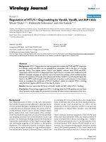
Composition of MAPsFigure 1
Composition of MAPs. The amino acid (aa) composition of the scaffolds of
G39d and G40d is shown in single letter code. The triple dash in the scaffolds
denotes the disulfide bond between adjacent cysteins. S1 and S2 are helper T
cell peptides and M2e the 24 N-terminal aa of M2, linked through their C-ter-
minal aa to the indicated lysines of the scaffold peptides.
Virology Journal 2007, 4:118 />Page 3 of 14
(page number not for citation purposes)
M2e(pep)-specific Abs, they will be referred to as
M2e(pep-nat)-specific to distinguish them from Abs that
react with native cell-expressed M2e but not with M2e
peptide, an Ab population detected in mice that have
recovered from repetitive IAV infections [13]. The M2e-
specific MAb 14C2-S1-4, which binds with comparable
efficacy to all three specific immunosorbents under the
present assay conditions (Fig 2A), was used as a standard
to quantify the ELISA data.
Fig 2B shows results from an experiment in which four
groups of mice were immunized three times by i.n. or s.c.
routes with the M2e-MAP G40d together with the immu-
nostimulatory oligodeoxynucleotide 1826 (ODN) or
ODN and cholera toxin (CT). Ab titers were measured in
pooled plasma samples (5 mice/group) collected three
weeks after secondary and tertiary immunization. It is evi-
dent that M2e(pep)-specific Abs accounted for the major-
ity (79% ± 18%, SD) of the total G40d-specific response
(defined in each sample as 100%). M2e(pep-nat)-specific
Abs made up a smaller and more variable fraction (10% ±
8%, SD) of the total G40d-specific response. In most
experiments, only M2e(pep)- and M2e(pep-nat)-specific
Ab titers were determined. Taking 27 distinct vaccination
groups into account, M2e(pep-nat)-specific Ab titers
ranged from ~1% to essentially 100% of the M2e(pep)-
specific Ab titers and accounted on average for 14.5%
(geometric mean, GM) of the M2e(pep)-specific response
(Fig 2C). The various immunization protocols employed
here had no significant effect on the size of the M2e(pep-
nat)-specific Ab fraction (Fig 2C).
Taken together, the results indicated that the majority of
the M2e-MAP-induced Abs were M2e(pep)-specific, and
that a variable fraction of these Abs crossreacted with M2
expressed by HeLa-M2 cells, i.e. displayed M2e(pep-nat)-
specificity.
Roles of adjuvant and immunization route on Ab response
and protection
In our previous study [17], we had shown that mice vacci-
nated with M2e-MAP, ODN and CT by the i.n. route
exhibited significant resistance to total respiratory tract
infection with IAV. Here, we wanted to determine whether
route of vaccination and use of CT as adjuvant made a sig-
nificant contribution to protection. To this end, mice were
immunized three times at 4–5 week intervals with M2e-
MAP plus ODN with or without CT by i.n. or s.c. (tail
base) routes. M2e-specific Ab titers in plasma (pools of 4–
5 mice per group) collected three weeks after the third
immunization were determined and mice challenged 7–
10 days later by nasal infection with X31. Results from
four independent repeat vaccination and challenge exper-
iments are compiled in Fig 3.
As shown in Fig 3A, M2e(pep)- and M2e(pep-nat)-specific
Ab titers were slightly higher in mice vaccinated with
ODN and CT by the s.c. route than in the other vaccina-
Fine specificity of the M2e-MAP induced Ab responseFigure 2
Fine specificity of the M2e-MAP induced Ab response. A. MAb 14C2-
S1-4, which was used in all assays for quantification of serum Ab titers, was
tested in ELISA against M2e-MAP Gd40 (squares), Cys-M2e (triangles) and
HeLa-M2 (circles) as described in the method section, using the same reagents
and incubation times for each assay. The mean OD (± SEM) above background
of six replicates at each Ab dilution are shown. The three sigmoidal titration
curves have similar EC50 values (-9.3 vs G40d, -9.5 vs Cys-M2e, -9.2 vs HeLa-
M2). To further demonstrate the similarity between the three titration curves,
OD values measured against HeLa-M2 were multiplied by 1.65 to generated the
stipulated curve. A representative assay is shown. B. Pooled plasma samples (5
mice/group), obtained 3 wks after second (left column) and third (right column)
immunization, were tested by ELISA for M2e-MAP- (squares), M2e(pep)- (trian-
gles) and M2e(pep-nat)-specific (circles) Ab titers as described in the method
section. The mice had been immunized with 3 μg M2e-MAP G40d and adjuvants
by i.n. or s.c. routes as indicated below the x axis. Each symbol shows the mean
serum Ab concentration determined in each sample by 2–3 independent assays.
Data from a single vaccination experiment are shown. C. The fraction of
M2e(pep-nat)-specific Abs is expressed as percent of the M2e(pep)-specific Ab
concentration within each sample. Each dot indicates the % of anti-M2e(pep-
nat) per group of 3–5 mice immunized by one of the protocols indicated below
the x axis. In most groups, samples from secondary and tertiary responses
were tested, and the mean % of these is shown. Horizontal bars indicate the
geometric means within a vaccination protocol. Data from 12 independent vac-
cination experiments are shown. Groups immunized by different protocols did
not differ significantly (ANOVA) with regards to percentage of anti-M2e(pep-
nat)-specific Abs.
Virology Journal 2007, 4:118 />Page 4 of 14
(page number not for citation purposes)
tion groups. Although this difference was not significant
(by ANOVA) in the four experiments shown in Fig 3A, it
was significant when Ab titers after the second immuniza-
tion were analyzed and additional vaccination experi-
Effect of immunization protocol on size and G2a content of the M2e(pep-nat)-specific Ab responseFigure 4
Effect of immunization protocol on size and G2a content of the
M2e(pep-nat)-specific Ab response. A. M2e(pep-nat)-specific Ab titers in
pooled plasma samples collected three weeks after second immunization from
mice vaccinated with M2e-MAP according to the protocol indicated below the
x axis. Each dot shows the titer of pooled plasma from 3–5 mice. Horizontal
bars indicate the GMTs of groups within a given vaccination protocol. Data
were analyzed by ANOVA and Tukey's Multiple Comparison post test. Statisti-
cally significant differences between group are indicated by asterisks above two-
sided arrows: p < 0.05 (*), p < 0.01 (**). B. Pooled plasma from 4–5 mice/group
collected three weeks after second and third immunization were tested for
concentration of Cκ- (total) and γ 2a-expressing M2e(pep-nat)-specific Ab tit-
ers and the latter were expressed as percentage of the former. In groups that
were immunized three times, the mean percentage of G2a after 2
nd
and 3
rd
immunization is shown. Groups with low M2e(pep-nat)-specific Ab titers that
did not permit detection of G2a at ≤5% were excluded from the analysis. Hori-
zontal bars show GMTs within distinct immunization protocols. Data were ana-
lyzed by ANOVA and Tukey's Multiple Comparison post test and marked as in
A.
Ab response and protection after various modes of vaccina-tionFigure 3
Ab response and protection after various modes of vaccination. A.
BALB/c mice were vaccinated three times at 4–5 week intervals with 3 μg M2e-
MAP (two experiments G39d, two G40d) and the indicated adjuvants (see bot-
tom of figure) by i.n. or s.c. route. Mice were bled 3 weeks after the third
immunization. Pooled plasma samples (3–5 mice/pool) were tested by ELISA for
M2e(pep)- (dots) and M2e(pep-nat)-specific (circles) Ab titers. Horizontal bars
indicate GMTs within each set. Data from four independent vaccination experi-
ments are shown. B, C, D. 7–10 days after the third vaccination, mice were
challenged by i.n. inoculation of 5 μl X31 (1000 TCID
50
/mouse). Five days later,
nose, trachea and lung were tested for virus titer. Each symbol indicates the
virus titer of an individual mouse. Horizontal bars indicate the GMT within each
vaccination set. Dashed (top) and stipulated (bottom) horizontal lines indicate
the mean virus titer of control mice and threshold of virus detection, respec-
tively. Tissues with undetectable virus were assumed to be virus free. Data
were analyzed by non-parametric ANOVA and Dunn's Multiple Comparison
post test. M2e-MAP vaccination groups with statistically significant reduction in
virus titer compared to the control group are indicated by asterisks right above
the group and statistical differences between M2e-MAP vaccination groups by
asterisks above two-sided arrows: p < 0.05 (*), p < 0.01 (**), p < 0.001 (***).
Virology Journal 2007, 4:118 />Page 5 of 14
(page number not for citation purposes)
ments taken into account (Fig 4A). Thus, in the presence
of ODN, CT strongly enhanced the Ab response upon
parenteral though not i.n. vaccination.
The strength of protection was assessed by i.n. inoculation
of mice with 5 μl (1000 TCID
50
) of X31 virus. This chal-
lenge induces an infection that is initially confined to the
nasal epithelium and from there spreads in non-immune
mice within a few days into the lower respiratory tract.
Five days after challenge, mice were euthanized and virus
titers determined in nose, trachea (together with extrapul-
monary bronchi) and lung. As shown in Fig 3B–D, the
infection had spread by this time in all control mice
(immunized with adjuvant only) into trachea and lung.
Compared to the control group, all M2e-MAP vaccination
groups showed significant restriction of similar strength
against virus growth in the nose (Fig 3B). The groups dif-
fered, however, with regards to resistance against descend-
ing infection. The least resistance was seen in mice
vaccinated with M2e-MAP and ODN by the s.c. route and
in fact did not differ significantly from the control group.
The strongest and most significant resistance was seen in
mice vaccinated with ODN and CT by the i.n. route. The
other two vaccination groups (i.n. with ODN but without
CT and s.c. with ODN and CT) displayed intermediate
and similar levels of protection.
Taken together, the results indicated that CT significantly
enhanced the systemic Ab response when administered
together with ODN by a parenteral route and strength-
ened protection both upon parenteral and i.n. vaccina-
tion. Furthermore, independent of the adjuvants used, the
i.n. route of vaccination engendered stronger protection
than parenteral vaccination. However, the relationship
between strength of protection and M2e-specific serum
Ab titer was not clear. For instance, mice vaccinated with
M2e-MAP and ODN by s.c. route displayed significantly
weaker resistance against descending infection than mice
immunized with M2e-MAP, ODN and CT by i.n. route, in
spite of similar serum Ab titers. This was unexpected in
view of previous findings showing that protection could
be transferred with serum from M2e-immune to naive
mice [4-6,8,9].
Relation between M2e-specific serum Ab titers and
protection
The absence of a clear relation between serum Ab titer and
strength of protection suggested that the concentration of
Cκ-positive M2e-specific Abs in serum (biotinylated anti-
Cκ was used for measurement of Ab titers) was not the
sole determinant of protection. Although λ light chains
are expressed only by ~5% of Abs in the BALB/c mouse,
they may be expressed at higher frequency in responses of
some specificities. We therefore tested selected serum
samples for λ-positive M2e-specific Abs but found no evi-
dence for the substantial use of λ light chains in the M2e-
specific Ab response (data not shown). Thus, differences
in the fine specificity, avidity or heavy chain isotype of
M2e-specific serum Abs or of immune phenomena that
are mostly confined to the respiratory tract and poorly
reflected in serum could make significant contributions to
protection. To further explore these possibilities, we ana-
lyzed the relation between M2e-specific serum Ab titers
and strength of protection in the above and additional
groups of mice that had been vaccinated with M2e-MAP,
challenged by localized nasal infection with the same
dose of X31 virus and analyzed for virus titer five days
later. To detect potential contributions of respiratory tract-
associated immune phenomena, which may be induced
preferentially by i.n. immunization, groups vaccinated by
i.n. and parenteral routes were analyzed separately. The
reduction in virus titer (on log
10
basis) in M2e-MAP
immunized groups compared to the control group (adju-
vant only) of the given immunization experiment was
used as measure of strength of protection. Tissues with
undetectable virus (threshold of 10 EID
50
for nose and tra-
chea and 10
1.3
for lung) were assumed to be virus-free.
Table 1: Correlation between M2e-specific serum Ab titer and reduction of virus titer in various sites of the respiratory tract after
parenteral and i.n. immunization.
Specificity/Isotype
of anti-M2e Abs
Spearman correlation coefficient r (p)
parenteral vaccination i.n. vaccination
Nose Trachea Lung Nose Trachea Lung
M2e(pep) 0.07 -0.03 0.18 0.2 0.24 0.02
M2e(pep-nat) 0.96(***) 0.82 (**) 0.8 (**) -0.38 -0.27 -0.31
M2e(pep-nat) G2a 0.56 0.51 0.75 (*) -0.43 -0.30 -0.35
Mean (4–5 mice/group) M2e-specific serum Ab titers 7–10 days before challenge with X31 virus (5 μl, 10
3
TCID
50
) were analyzed for correlation
with mean reduction (compared to control group) of virus titers in nose, trachea and lung. Included in the analysis are 9 groups of mice immunized
with M2e-MAP by a parenteral route (s.c., i.m., i.p.) and 14 groups immunized by the i.n. route. Statistical significance of correlation (Spearman r,
non parametric) is indicated by asterisk: (*): p < 0.05, (**): p = 0.01, (***): p = 0.002.
Virology Journal 2007, 4:118 />Page 6 of 14
(page number not for citation purposes)
As shown in Table 1, Cκ-positive M2e(pep)-specific
serum Ab titers showed no significant correlation with
strength of protection, both after i.n. and parenteral
immunization. By contrast, highly significant correlations
were seen between M2e(pep-nat)-specific Ab titers and
protection after parenteral though not i.n. immunization.
These findings indicated, firstly, that only Abs capable of
reacting with native cell-expressed M2e played a role in
protection. Since M2e(pep-nat)-specific Abs are a subpop-
ulation of the M2e(pep) specific response, the absence of
correlation between M2e(pep)-specific Ab titers and pro-
tection is apparently a consequence of the substantial var-
iation between groups in the proportion of M2e(pep-nat)-
specific Abs within the total M2e(pep)-specific response
(Fig 2C). Second, the absence of correlation between
M2e(pep-nat)-specific serum Ab titers and protection in
mice immunized by the i.n. route indicated that M2e(pep-
nat)-specific serum Abs were not the sole effectors of pro-
tection; conceivably, M2e-specific Abs produced in airway
tissues, whose titers are inadequately reflected in serum,
or M2e-specific T cells may contribute to protection.
Abs of G2a isotype have often been found to display
higher activity in vivo than Abs of other IgG isotypes. This
has been attributed to the ability IgG2a to interact with all
three activating IgG Fc receptors, FcγRI, FcγRIII and most
notably FcγRIV, for which G2a is the preferred iso-
type[19,20]. In agreement with this, naive mice, passively
protected with the G2a isotype switch variant of mAb
14C2, showed significantly less weight loss (p < 0.05) and
less mortality (p = 0.08) than mice passively protected
with the same dose of mAb 14C2 of G1 or G2b isotype
(Fig 5). Therefore, we determined also titers of M2e(pep-
nat)-specific G2a in sera, hoping Abs of this isotype may
show an improved correlation with protection. However,
the contrary was the case, possibly because positive effects
on correlation due to the increased protective activity of
G2a were outweighed by negative effects on correlation
due to the variability in the proportion of G2a within the
total M2e(pep-nat) response (Fig 4B). It is possible also
that the G2a isotype provides a smaller advantage over
other isotypes in inhibition of a descending infection by
X31 virus – the endpoint used for the data in table 1 –
than in reduction of morbidity and mortality after total
respiratory tract challenge with PR8 – the endpoint used
in the comparison of the isotype switch variants (Fig 5).
An interesting observation resulting from this analysis was
that i.n. vaccination engendered an Ab response with a
significantly larger proportion of G2a (GM: 45%) than
parenteral immunization (GM: 8%), independent of the
adjuvants used (Fig 4B).
In view of the significant correlation between total
M2e(pep-nat)-specific serum Ab titer and protection after
parenteral immunization, we subjected the data to linear
and non-linear regression analysis. Linear regression anal-
ysis showed a poor fit between Ab titer and protection,
with R
2
values of 0.45, 0.36 and 0.37 for protection in
nose, trachea and lung, respectively, though elimination
of one outlier group with the highest serum Ab titer
yielded linear regressions with R
2
and (p) values of 0.94
(<0.0001), 0.66 (0.014) and 0.78 (0.0039) for protection
in nose, trachea and lung, respectively. However, without
exclusion of any data, the relations between Ab titers and
protection could be described by sigmoidal curves that
exhibited R
2
values of 0.79 for nose and lung and 0.65 for
trachea (Fig 6A). They indicated that M2e-specific protec-
tion after parenteral immunization exhibited an upper
boundary and half-maximm protection was achieved in
each site of the respiratory tract at the serum Ab concen-
tration of ~20 μg/ml. By contrast, in i.n. vaccinated mice,
there was no obvious relation between serum Ab titer and
protection. Indeed, significant protection was seen in
many mice with serum Ab titers that were completely
non-protective in parenterally vaccinated mice. This is
demonstrated in Fig. 6B and 6C, which display the data
from individual i.n. (filled symbols) and parenterally vac-
cinated (open symbols and deduced sigmoidal curve)
groups for protection in the nose and lung, respectively.
Apparently, vaccination by the i.n. route was capable of
inducing potent protective activities other than those
mediated by M2e-specific serum Abs.
Ab response and protection after i.n. administration of
M2e-MAP together with infectious virus
Recovery from respiratory tract infection has been shown
to result in optimal protection [21]. This is generally
Role of heavy chain isotype in protectionFigure 5
Role of heavy chain isotype in protection. Naive BALB/c mice were
injected i.p. with 10 μg mAb 14C2 of G1 (triangles pointing down), G2b (dia-
monds) or G2a (triangles pointing up) isotype. The control group (open
squares) received PBS i.p. One day later, mice were exposed to a total respira-
tory tract challenge with PR8 (4 LD
50
in 50 μl) and monitored for weight loss.
Pooled data from two independent experiments are shown, each performed
with 4–5 mice/group. A. Symbols show mean % body weight and SEM (relative
to day 0) of 9–10 mice/group. Differences between treatment groups were
tested for statistical significance at individual days. Mice treated with G2a
showed significantly (p < 0,05, ANOVA) less weight loss than those treated
with G1 or G2b at days 6 to 13 p.i. B. Survival. Death was defined as >30%
weight loss, at which stage mice were euthanized. Differences between survival
curves were tested for statistical significance by log rank test.
Virology Journal 2007, 4:118 />Page 7 of 14
(page number not for citation purposes)
attributed to the combined effects of strong local and sys-
temic T and B cell responses against several viral proteins.
Since infection induces only a poor M2e-specific Ab
response [13], we wondered whether infection-induced
protection could be further strengthened by concomitant
immunization with M2e-MAP. This was tested by i.n.
administration of a sublethal dose of PR8, either alone or
together with 3 μg M2e-MAP. Both groups of mice devel-
oped a primary infection from which they recovered. Four
weeks later, the mice were inoculated i.n. with PR8-Seq14
(200 TCID
50
), again with or without M2e-MAP. Other
groups of mice were inoculated twice by the i.n. route
with A) ODN and CT (negative control), B) M2e-MAP (3
μg/dose) in PBS without adjuvant, C) M2e-MAP with
ODN and CT (positive control) or D) 5 μg purified uv-
inactivated PR8 virus. We decided on this dosage of inac-
tivated virus, which contains ~10
6
times the amount of
virus present in 200 TCID
50
, to compensate for the lack of
replication in vivo. Plasma samples were collected three
weeks after the boost, pooled within each group, and
tested by ELISA against M2e peptide and HeLa-M2.
Fig 7A shows Ab titers from three independent vaccina-
tion experiments. No M2e-specific Abs were detected in
sera of mice immunized with M2e-MAP without adjuvant
or with inactivated virus. The former shows that M2e-MAP
is not immunogenic in the absence of adjuvant and the
latter that M2 – a minor viral structural protein that makes
up only ~0.2% of the total protein mass of virus particles
[22] – is not immunogenic in the context of a large dose
of mature virus particles. Note, however, that mice immu-
nized twice with inactivated virus made a strong HA-spe-
cific Ab response (data not shown). M2e-specific Ab
responses were seen in all other groups. However, they
differed in titer and fine specificity in that mice immu-
nized with M2e-MAP plus adjuvant displayed higher Ab
titers against M2e peptide than HeLa-M2 while, as shown
previously [13], the reverse was the case for mice immu-
nized by infection. Importantly, the highest Ab titers
against HeLa-M2 were seen in mice immunized concomi-
tantly with infectious virus and M2e-MAP, and the major-
ity of these Abs appeared to display M2e(pep-nat)-
specificity.
Seven to fourteen days after the second immunization,
mice from two vaccination experiments were challenged
by i.n. instillation of 50 μl of X31. This mode of challenge
initiates an infection throughout the respiratory tract
(nose, trachea, pulmonary airways) and was chosen in
preference of the localized nasal infection because the lat-
ter did not descend within five days into the lower respi-
ratory tract in infection-immunized mice (our
unpublished observation) and therefore was unsuitable
for revealing differences between the groups immunized
by infection with/without M2e-MAP. Three days after
total respiratory tract infection, mice were euthanized and
virus titers in nose, trachea and lung determined. Com-
pared to control mice (adjuvant alone), significant reduc-
tions in virus titers were seen in mice immunized with
Relation between M2e(pep-nat)-specific Ab titer and protec-tion against virus challengeFigure 6
Relation between M2e(pep-nat)-specific Ab titer and protection
against virus challenge. Cκ-positive M2e(pep-nat)-specific Ab titers were
determined in pooled plasma (3–5 mice/group) collected 7–10 days before chal-
lenge of mice by localized nasal infection (5 μl X31, 1000 TCID
50
). Five days
after challenge, virus titers were determined in nose, trachea and lung of indi-
vidual mice and the group average was determined. The average reduction in
virus titer on log
10
basis compared to the control group (immunized with adju-
vant alone) was taken as measure of strength of protection (y axis). A. Protec-
tion in nose (squares), trachea (triangles) and lung (circles) from nine groups of
mice immunized by parenteral route is plotted against the M2e(pep-nat)-spe-
cific serum Ab titer (x axis). Non-linear regression analysis yielded sigmoidal
regression curves with R
2
of 0.79 for nose (stipulated) and lung (continuous)
and of 0.65 for trachea (dashed). B. Serum Ab titers and protection in nose
observed in mice immunized by the i.n. route (filled squares) are plotted
together with the regression line and corresponding data points (open squares)
from mice immunized by parenteral route (as in A). C. Serum Ab titers and
protection in lung of mice immunized by i.n. route (filled circles) are plotted
together with the regression line and corresponding data points (open circles)
from mice after parenteral immunization (as in A).
Virology Journal 2007, 4:118 />Page 8 of 14
(page number not for citation purposes)
M2e-MAP plus adjuvant, M2e-MAP plus infectious virus
or infectious virus alone, but the two infection-immu-
nized groups showed stronger protection in the lung than
the M2e-MAP/adjuvant-immunized group. Comparison
between the infection-immunized groups indicated a
slight increase in resistance against virus replication in the
nose and trachea in mice that had been co-immunized
with M2e-MAP, although the difference did not reach sta-
tistical significance with the few mice used in these exper-
iments.
Taken together, the results indicated that combined vacci-
nation with M2e-MAP and infectious virus may improve
the induction of HeLa-M2-reactive Abs and slightly
enhance protection in nose and trachea compared to vac-
cination with infectious virus or M2e-MAP alone.
Discussion
Relation between Ab specificity, titer and protection
We found that the concentration of M2e(pep-nat)-specific
Abs in sera of parenterally vaccinated mice correlated with
strength of protection (Table 1). This is consistent with
previous studies showing that protection can be trans-
ferred to naive mice by passive M2e-specific Abs
[10,11,17] and antisera [4-6,8,9]. It is consistent also with
the generally held view that M2e-specific Abs mediate pro-
tection by reaction with M2e expressed in the plasma
membrane of infected host cells. By contrast, M2e(pep)-
specific Ab titers showed no correlation with protection,
in spite of the fact that the M2e(pep-nat)-specific Abs are
a fraction of the larger M2e(pep)-specific Ab response
(Table 1). This lack of correlation between M2e(pep) Ab
titer and protection appears to be a consequence of the
large variability of the M2e(pep-nat)-fraction within the
M2e(pep) response (Fig 2C). The reason for this variabil-
ity is not known. It could be due to a low frequency of
M2e(pep)- and M2e(pep-nat)-specific B cells within the
naive B cell repertoire, which may result, for stochastic
reasons, in large differences in the composition of this
response between individuals and even pooled sera from
3–5 animals, as tested here. A low precursor B cell fre-
quency is consistent with our previous finding that seven
M2e(pep-nat)-specific hybridomas isolated from three
mice all expressed a highly restricted heavy chain variable
region (formed by recombination of the same V
H
, D and
J gene segments) in association with only two distinct V
L
genes [23]. The M2e(pep)-specific response has not been
analyzed at the clonal level but may be equally restricted.
Because of this variability, the protective M2e(pep-nat)-
specific Ab titers cannot be extrapolated from M2e(pep)-
specific Ab titers and must be measured specifically.
Given the importance of M2e(pep-nat)-specific Abs in
protection, the selective promotion of this Ab population
by a M2e vaccine would be advantageous. This may be
Immunization with the combination of infectious virus and M2e-MAPFigure 7
Immunization with the combination of infectious virus and M2e-
MAP. BALB/c mice were immunized twice (4 week interval) by the i.n. route
with the components listed at the bottom of the figure. Dosage/injection (50
μl): M2e-MAP G39d (3 μg), ODN (3 μg), CT (0.5 μg), Vir (150–200 TCID
50
of
PR8 for primary and of Seq14 for secondary immunization), Vir(uv) (5 μg of
purified uv-inactivated PR8, <1 TCID
50
). Plasma was collected three weeks
after second immunization and pooled within groups. A. Ab titer measured by
ELISA against M2e peptide (closed circles) and HeLa-M2 (open circles) in
pooled plasma samples of groups of 3–4 mice from three independent vaccina-
tion experiments. Bars indicate the GMTs. The stipulated horizontal line indi-
cates the threshold of detection of Ab titers against HeLa-M2. B, C, D. Four
weeks after the second immunization, mice from two vaccination experiments
were challenged by i.n. inoculation of 50 μl X31, which initiates an infection
throughout the entire respiratory tract. Virus titers in nose, trachea and lung
were determined three days later. Each symbol indicates the total virus titer
(TCID
50
) from an individual mouse in the nose (B), trachea (C) and lung (D).
Bars indicate GMTs. The data were analyzed by non-parametric ANOVA and
Dunn's Multiple Comparison Test. Statistical significance between experimental
and control groups and between experimental groups is indicated by asterisks
above each column and above two-sided arrows, respectively: p < 0.05 (*); p <
0.01 (**).
Virology Journal 2007, 4:118 />Page 9 of 14
(page number not for citation purposes)
achieved by development of a more effective vaccine con-
struct and/or vaccine administration. Of note in the latter
context is the present finding that concomitant adminis-
tration of M2e-MAP and a sublethal dose of infectious
virus by the i.n. route not only enhanced the M2e(pep-
nat)-specific serum Ab titer compared to vaccination with
infectious virus or M2e-MAP (plus adjuvants) alone, but
affected also the specificity of the response in that essen-
tially all M2e-specific Abs generated in these co-immu-
nized mice displayed M2e(pep-nat)-specificity (Fig 7A
and data not shown). The advantage of co-administration
of infectious virus and M2e-MAP with regard to strength
of protection against heterosubtypic IAV challenge (as
used in the present study) merit further investigation, par-
ticularly since this protocol may be adaptable to humans
in the form of i.n. vaccination with a combination of live
attenuated IAV and a M2e-vaccine.
The relation between M2e(pep-nat)-specific Ab titers in
sera of parenterally vaccinated mice and strength of pro-
tection followed sigmoidal curves (Fig 6A), which sug-
gested that M2e(pep-nat)-specific serum Abs were equally
protective in nose, trachea and lung (EC
50
~20 μg/ml).
This was unexpected in view of previous studies showing
that systemically administered passive anti-viral Abs of
IgG isotypes were significantly less protective in upper
than lower airways [24-26]. The reason for this appears to
be the lower rate of transudation of serum IgG through
the pseudostratified columnar epithelium of upper air-
ways than the thinner epithelium of respiratory airways
and alveoli [27]. To confirm that this differential effective-
ness applies also to M2e(pep-nat)-specific Abs, we
injected fifteen naive BALB/c mice with three different
purified mAbs (5 mice/Ab) to achieve a passive serum Ab
concentration of ~20 μg/ml and then challenged the mice
by i.n. inoculation of 5 μl X31. Determination of virus tit-
ers in lung, trachea and nose five days later confirmed the
decreasing protective activity of serum Ab from lower to
upper airways, in that mAb-treated mice exhibited, on
average, a 100 fold reduction in virus titer in the lung, 30
fold in the trachea and no reduction at all in the nose
compared to control mice treated with PBS (data not
shown). Accordingly, M2e(pep-nat)-specific serum Ab tit-
ers in mice that had been immunized by a parenteral
route appeared to account reasonably well for the protec-
tion in lung and trachea but not in the nose.
One possible explanation for this difference in protection
between actively and passively immunized mice was that
active immunization induced substantial levels of
M2e(pep-nat)-specific IgA. When dimerized with J chain,
IgA is actively transported by the polymeric Ig receptor
(pIgR) system through the columnar epithelium of con-
ducting airways and is therefore more abundant than IgG
in secretions of upper than lower airways [27,28]. Accord-
ingly, secretory IgA with virus-neutralizing activity has
been shown to be responsible for much of the protection
against IAV replication in the nasal cavity of mice, while
IgG is more important for protection of respiratory air-
ways [29-31]. However, we could not detect significant
levels of M2e(pep-nat)-specific IgA in sera of parenterally
vaccinated mice (data not shown), making this explana-
tion untenable. Another possibility, which is discussed in
more detail below, is that parenteral vaccination with
M2e-MAP induced significant airway-associated immu-
nity. Although induction of strong local airway-associated
immunity is generally thought to require administration
of antigen into the airways [31-33], there is evidence indi-
cating that parenteral immunization with CT may result
in the migration of dendritic cells to mucosa-associated
lymphoid tissues and thereby promote some level of
mucosa-associated immunity [34,35]. In this study, CT
significantly enhanced the systemic Ab response upon
parenteral vaccination but we do not know whether it also
resulted in the induction of nasal mucosa-associated
immunity that may have restricted virus replication in
nasal tissue, independent of serum Ab titer. Finally and
probably most likely, immunization with M2e-MAP may
have induced not only M2e-specific Abs but also T cells
that contributed to protection. This possibility is sup-
ported by previous studies showing that vaccination of
BALB/c mice with M2e-MAP [17] or M2-DNA and M2-
recombinant adenovirus [9] induced M2e-specific T cell
responses, most likely of CD4 phenotype, and that virus-
specific CD4 memory T cells could significantly restrict
virus replication in the nose but not the lung [36]. Accord-
ingly, M2e-specific CD4 T cells may have inhibited virus
replication in the nose and M2e-specific serum Abs in the
lung. This proposition does not conflict with the conclu-
sion of Jegerlehner et al. [6] that M2e-specific T cells
played no role in protection of mice against a lethal total
respiratory virus challenge, as the lethality of the infection
is determined by the level of virus replication in the lung
but not the nose. The contrasting finding by Tompkins et
al. [9] that T cells contributed to protection against lethal
IAV challenge in mice immunized by M2-DNA and M2-
recombinant adenovirus may be explained by induction
of M2-specific CD8 T cells in these mice. It is well estab-
lished that virus-specific memory CD8 T cells can contrib-
ute to resistance against a lethal IAV challenge.
Route of vaccination and strength of protection
I.n. vaccination resulted in stronger protection against
descending infection than parenteral vaccination (Fig
3C,D). Most remarkably, however, the strength of protec-
tion in i.n. vaccinated mice showed no correlation with
M2e(pep-nat)-specific serum Ab titers (Table 1). Indeed,
several groups of i.n. vaccinated mice with serum Ab titers
that were completely non-protective in parenterally vacci-
Virology Journal 2007, 4:118 />Page 10 of 14
(page number not for citation purposes)
nated mice showed nevertheless strong protection (Fig
6B,C). Several explanations can be considered.
First, i.n. administration of adjuvant alone has been
shown to result in a temporary increase in resistance
against virus replication in the respiratory tract [37-40].
However, such a non-specific enhancement of resistance
is unlikely to have affected the results of this study, since
M2e-MAP-vaccinated mice were always compared to con-
trol mice that had been vaccinated by the i.n. route with
adjuvant alone, thus canceling out adjuvant-induced non-
specific effects.
Second, i.n. vaccination may have induced local, airway-
associated immunity that was not adequately reflected by
serum Ab titers. To affect virus replication, Abs must be
present in airway secretions. Abs in this location may have
two distinct provenances [41]: 1) They may be serum Abs
that transudated into extravascular spaces of airway tis-
sues and, in the case of IgG, transudated further into the
airway lumen or, in the case of IgA and IgM, became trans-
ported through the epithelial cell layer by pIgR. 2) They
may have been secreted by B cells located in the lamina
propria of airways. As such locally produced Abs, particu-
larly J-chain associated IgA and IgM, can be expected to be
delivered more effectively into the airway lumen than into
the intravascular compartment, serum Ab titers do not
provide a reliable measure of the locally produced frac-
tion of Abs. The importance of nasal administration of
vaccine for promotion of local immunity has been docu-
mented both in animal models [31,42-45] and humans
[46-49]. Once induced, antigen-specific B and T cells may
persist in airway tissues for an extended period of time
and provide the host with long lasting enhanced protec-
tion [50-54]. Accordingly, local M2e(pep-nat)-specific B
and possibly also T cells may have provided strong protec-
tion in some i.n. vaccinated mice in the absence of protec-
tive serum Ab titers (Fig 6B,C).
Third, i.n. vaccination may have induced a qualitatively
different and more protective immune response than
parenteral vaccination. It is well established, for instance,
that i.n. vaccination typically promotes a stronger IgA
response than parenteral vaccination. The fact that we
could not detect significant M2e(pep-nat)-specific IgA in
pooled sera of i.n. vaccinated mice (data not shown) does
not exclude the possibility that M2e(pep-nat)-specific IgA
was produced locally and efficiently transported into air-
way secretions. In contrast to IgG, locally produced IgA
may interact intracellularly with M2e during its pIgR-
mediated transport through infected epithelial cells and
thereby restrict virus replication [55]. The substantial effi-
cacy of this mechanism in vivo has been demonstrated by
passive IgA mAb-mediated clearance of Rotavirus from
intestinal epithelium of mice with severe combined
immunodeficiency [56]. After its release into airway secre-
tions, secretory M2e(pep-nat)-specific IgA may have lesser
protective power than IgG, both in terms of activation of
FcR-expressing effector cells and complement. Neverthe-
less, cell-bound secretory IgA, while incapable of activat-
ing effector cells through one of the widely expressed
activating FcγRs, may still be able to activate effector cells
through interaction with the recently identified FcαμR in
mice [57] or CD86 in humans. In addition, while incapa-
ble of activating complement through the classic pathway,
IgA may still activate it through the alternative [58] and
lectin [59] pathways if complement activation were
involved in M2e-Ab-mediated protection. Another poten-
tially important qualitative change observed here after i.n.
administration of vaccine was the significant increase in
the proportion of M2e(pep-nat)-specific Abs of G2a iso-
type (Fig 4B). Firstly, IgG2a was the most protective IgG
isotype in passive transfer experiments (Fig 5). In addi-
tion, if T cells contributed to protection, the prevalence of
IgG2a may indicate a general bias of the response towards
type 1, which is typically associated with optimal T cell-
mediated protection in viral and bacterial infections.
Additional studies are needed to sort out the relative
importance of local immunity and quality of the response
in the improved protection after i.n. vaccination.
The enhanced protection seen here after i.n. vaccination
must be viewed in the context of the challenge used here.
It consisted of an infection that was initially confined to
the nasal epithelium and allowed to descend from there
into the lower respiratory tract over the course of five days.
In this scenario, strong immunity in the upper respiratory
tract would be expected to have a substantial impact on
the progress of the infection. By contrast, the more fre-
quently used challenge with an inoculum of 30–50 μl in
anesthetized mice initiates an infection in both upper and
lower respiratory tact, and virus titer in lung or survival
would hardly if at all be affected by immunity in the upper
respiratory tract. We believe this nasal challenge provides
a relevant model for the IAV infection in humans.
Conclusion
M2e-MAP is an effective immunogen as roughly 80% of
the total M2e-MAP-specific Ab response displayed
M2e(pep) specificity. A variable fraction (on average
15%) of these M2e(pep)-specific Abs cross-reacted with
presumably native tetrameric M2e expressed by M2-trans-
fected HeLa cells, and the concentration of these
M2e(pep-nat)-specific Abs in sera of parenterally immu-
nized mice showed a good correlation with protection
against virus challenge. However, M2e(pep-nat)-specific
serum Abs did not appear to fully account for protection,
particularly in the nose, of M2e-MAP-vaccinated mice,
suggesting the contribution of additional protective activ-
ities, possibly M2e-specific T cells and/or local airway-
Virology Journal 2007, 4:118 />Page 11 of 14
(page number not for citation purposes)
associated Ab responses. The latter was supported also by
the observation that immunization by the i.n. route
resulted in stronger protection than immunization by a
parenteral route and that the strength of protection in i.n.
vaccinated mice showed no correlation with M2e(pep-
nat)-specific serum Ab titers. Concomitant i.n. adminis-
tration of M2e-MAP with infectious virus enhanced the
M2e(pep-nat)-specific Ab response and protection com-
pared to i.n. vaccination with M2e-MAP plus adjuvant or
infectious virus alone. Concomitant i.n. administration of
M2e-MAP and attenuated cold-adapted live virus may be
applicable to human vaccination and merits further inves-
tigation.
Methods
Mice
Female BALB/c mice (5–6 week old) were purchased from
Harlan [60] and maintained in the Institute's Animal
Facility in microisolator cages under specific pathogen-
free conditions. Mice were rested for ≥2 weeks before use
in experiments. All procedures performed on animals
were approved by the Institutional Animal Care and Use
Committee.
Media, solutions and reagents
ISC-CM is Iscove's Dulbecco medium (Invitrogen) sup-
plemented with 0.05 mM 2-mercaptoethanol, 0.005 mg/
ml transferrin (Sigma), 2 mM L-glutamine (Mediatech
Inc) and 0.05 mg/ml gentamicin (Mediatech Inc). ISC-
CM was further supplemented, as indicated, with fetal
bovine serum (FBS, Gemini Bio-products) or bovine
serum albumin (BSA, Sigma). PBSN is phosphate buffered
saline (pH7.2) supplemented with 3 mM NaN3. Immu-
nostimulatory phosphorothionated oligodeoxynucle-
otide (ODN) 1826 [61] and cholera toxin (CT) were
purchased from Sigma. Multiple antigenic peptides
(MAPs) were synthesized in house [62] and have been
described previously [17]. The MAPs used here are: G39d
(dimer of disulfide-linked MAPs, each containing two
M2e(2–25)-peptides and two helper T cell peptides, one
(S1) presented by A
d
and the other (S2) by E
d
[63], linked
to the Lys of a Cys-(Gly-Lys)
4
-Ala scaffold peptide); G40d
(as G39d but with only one helper T cell peptide linked to
a Cys-(Gly-Lys)
3
-Ala scaffold peptide); Cys-M2e (two M2e
peptides linked to Cys-(Gly-Lys)
3
-Ala) and Cys-bb (Cys-
(Gly-Lys)
3
-Ala. G39d was used for all but two immuniza-
tions. Fig 1 shows the composition and sequence of the
MAPs.
Monoclonal Abs
The M2e-specific hybridoma 14C2 (IgG1) was originally
obtained from Zebedee and Lamb [22]. The 14C2 switch
variant of G2b isotype was selected by staining 20 million
parental (IgG1) hybridoma cells with rat-anti-mouse-G2b
mAb (R1.3-20), sorting by flow cytometry for the 1%
most intensively stained cells, culturing the sorted cells by
limiting dilution and testing growing cultures for secre-
tion of IgG1 and IgG2b. A G2a switch variant (14C2-S1-
4) was similarly selected from the G2b switch variant
(14C2-S1) by using the rat-anti-mouse-G2a mAb (G2a-3-
6.8) for staining of 14C2-S1 cells. MAbs were purified
from protein-free hybridoma medium PFHM-II (Gibco),
in which hybridoma cells, initially grown up in ISC-CM
5%FBS, had been cultured to exhaustion. Purified 14C2-
S1-4 mAb was used as standard for determination of M2e-
specific Ab concentration by ELISA.
Viruses
PR/8/34(H1N1)-Mt.Sinai (PR8) is a highly pathogenic
mouse-adapted IAV. 500 TCID
50
(50% tissue culture
infectious dose) correspond to ~1 LD
50
(50% lethal dose)
when administered in 50 μl into the respiratory tract of
anesthetized mice. PR8-Seq14 is an escape mutant derived
from PR8 through fourteen sequential selection steps,
each performed in the presence of a distinct PR8-specific
mAb that was capable of neutralizing the penultimate
escape mutant. PR8-Seq14 differs from PR8 by 15 amino
acid substitutions in HA, is not measurably inhibited in
hemagglutination inhibition assay by PR8-specific mouse
sera and retains the high pathogenicity of PR8 (observa-
tions to be published). X31 is a reassortant between PR8
and A/Aichi/68(H3N2). It contains all PR8-derived genes
except those encoding H3 and N2 and is of low patho-
genicity.
Immunization, infection and analysis of mice
For i.n. immunization, 50 μl of vaccine preparation (PBS
containing 3 μg M2e-MAP, 3 μg ODN 1826 and 0.5 μg
CT) was applied to the nares of mice anesthetized by
intraperitoneal (i.p.) injection of 0.2 ml ketamine-xyla-
zine (70/7 mg/kg); this results in the aspiration of the vac-
cine into upper and lower airways. For subcutaneous
(s.c.), intramuscular (i.m.) and i.p. immunization, 50 μl
of vaccine as above was injected into the tail base, quadri-
ceps or peritoneal cavity, respectively, of non-anesthetized
mice. Vaccinations were repeated once or twice, typically
in four week intervals. Three weeks after primary, second-
ary or tertiary vaccination, 0.1–0.2 ml blood was collected
by puncture of the retro-orbital plexus, and plasma sam-
ples were pooled within each immunization group. Four
weeks after the last vaccination, anesthetized mice were
challenged by administration of 5 μl PBS (containing
2000 TCID
50
of X31) to the nares, dispensing ~half of the
inoculum per nare. This challenge results in a sublethal
infection that is initially confined to the nasal epithelium
and then descends in naive mice over the next five days
into trachea and lung. Five days after infection, mice were
euthanized by exsanguination under ketamine/xylazine
anesthesia. Nose, trachea with attached extrapulmonary
bronchi and lungs were individually dissected and stored
Virology Journal 2007, 4:118 />Page 12 of 14
(page number not for citation purposes)
frozen for subsequent determination of infectious virus
titer by MDCK assay as described [36]. Tissue homoge-
nates that scored negative in the MDCK assay (sensitivity
threshold: 10
1.8
TCID
50
per total nasal and tracheal extract
and 10
2.1
TCID
50
per lung extract) were tested by inocula-
tion of 50 μl undiluted tissue extracts into the allantoic
cavity of two embryonated chicken eggs (sensitivity
threshold: 10 EID
50
for nose and trachea and 20 EID
50
for
lung). In some experiments, anesthetized mice were chal-
lenged by i.n. administration of PR8 in 50 μl PBS; this pro-
cedure initiates an infection of upper and lower airways
and, depending on virus dose, may be lethal. Infected
mice were euthanized three days later for determination
of virus titers in airway tissues, or were observed for
weight loss.
Determination of M2e-specific Ab concentration by ELISA
For measurement of Abs specific for cell-expressed (pre-
sumably native tetrameric) M2e, we used HeLa cells stably
transfected with M2-expressing (HeLa-M2) or empty con-
trol plasmid (HeLa-C10) as specific and non-specific
(background) immunosorbent, respectively [13]. The spe-
cific and non-specific immunosorbents used for measure-
ment of M2e-peptide-specific Ab concentration were Cys-
M2e and Cys-bb (see above), respectively [17]. Cys-M2e
and Cys-bb were used at equimolar concentration (85 nM
in 0.02 M NaCl) to coat wells of polyvinyl plastic plates
(25 μl/well, overnight at room temperature, under cover
to prevent evaporation). For measurement of total M2e-
MAP-specific Ab concentration, we used wells coated as
above with M2e-MAP as specific and uncoated wells (only
blocked with BSA) as non-specific immunosorbent.
Immunosorbent-bound Abs were detected with bioti-
nylated mAb 187 (ATCC HB58, rat-anti-mouse κ chain)
or G2a-3-6.8 (rat-anti-mouse-G2a) for measurement of
Cκ- or G2a-expressing Abs, respectively. The difference in
OD (ΔOD) between specific and non-specific immuno-
sorbent was used for quantification of Ab concentration
by comparison to ODs observed with known concentra-
tions of purified M2e-specific mAb 14C2-S1-4 (G2a/Cκ)
bound to the same immunosorbents. ELISA data were col-
lected with the e-max ELISA reader and analyzed with
Softmax Pro software (both Molecular Devices, Sunny-
vale, CA).
Statistical analyses
Prism 4 software [64] was used for plotting and statistical
analysis of data as indicated in figure legends.
Competing interests
The author(s) declare that they have no competing inter-
ests.
Authors' contributions
KM collected blood samples from vaccinated mice, and
performed ELISAs and virus titrations. DZ performed the
passive protection studies. GK and LO synthesized the
M2e-MAPs. WG designed the studies, immunized mice,
analyzed data and wrote the manuscript. All authors have
read and approved the manuscript.
Acknowledgements
This study was supported by NIAID grant AI46457 (WG) and the Com-
monwealth Universal Research Enhancement Program, Pennsylvania
Department of Health. The help of Marion Sacks in the preparation of the
manuscript and of Soheila Nikpour in the preparation of the figures is grate-
fully acknowledged.
References
1. Smith NM, Bresee JS, Shay DK, Uyeki TM, Cox N, Strikas RA, Centers
for Disease Control and Prevention (U.S.), Practices USACoI: Preven-
tion and control of influenza : recommendations of the Advisory Committee
on Immunization Practices (ACIP) Atlanta, GA: U.S. Dept. of Health &
Human Services Centers for Disease Control and Prevention; 2006.
2. Simonsen L, Taylor RJ, Viboud C, Miller MA, Jackson LA: Mortality
benefits of influenza vaccination in elderly people: an ongo-
ing controversy. Lancet Infect Dis 2007, 7:658-666.
3. Puck JM, Glezen WP, Frank AL, Six HR: Protection of infants from
infection with influenza A virus by transplacentally acquired
antibody. J Infect Dis 1980, 142:844-849.
4. Ernst WA, Kim HJ, Tumpey TM, Jansen AD, Tai W, Cramer DV,
Adler-Moore JP, Fujii G: Protection against H1, H5, H6 and H9
influenza A infection with liposomal matrix 2 epitope vac-
cines. Vaccine 2006, 24:5158-5168.
5. Fan J, Liang X, Horton MS, Perry HC, Citron MP, Heidecker GJ, Fu
TM, Joyce J, Przysiecki CT, Keller PM, et al.: Preclinical study of
influenza virus A M2 peptide conjugate vaccines in mice, fer-
rets, and rhesus monkeys. Vaccine 2004, 22:2993-3003.
6. Jegerlehner A, Schmitz N, Storni T, Bachmann MF: Influenza A vac-
cine based on the extracellular domain of M2: weak protec-
tion mediated via antibody-dependent NK cell activity. J
Immunol 2004, 172:5598-5605.
7. Mozdzanowska K, Maiese K, Furchner M, Gerhard W: Treatment
of influenza virus-infected SCID mice with nonneutralizing
antibodies specific for the transmembrane proteins matrix 2
and neuraminidase reduces the pulmonary virus titer but
fails to clear the infection. Virology 1999, 254:138-146.
8. Neirynck S, Deroo T, Saelens X, Vanlandschoot P, Jou WM, Fiers W:
A universal influenza A vaccine based on the extracellular
domain of the M2 protein. Nat Med 1999, 5:1157-1163.
9. Tompkins SM, Zhao ZS, Lo CY, Misplon JA, Liu T, Ye Z, Hogan RJ,
Wu Z, Benton KA, Tumpey TM, Epstein SL: Matrix protein 2 vac-
cination and protection against influenza viruses, including
subtype H5N1. Emerg Infect Dis 2007, 13:426-435.
10. Treanor JJ, Tierney EL, Zebedee SL, Lamb RA, Murphy BR: Passively
transferred monoclonal antibody to the M2 protein inhibits
influenza A virus replication in mice. J Virol 1990, 64:1375-1377.
11. Zharikova D, Mozdzanowska K, Feng J, Zhang M, Gerhard W: Influ-
enza type A virus escape mutants emerge in vivo in the pres-
ence of antibodies to the ectodomain of matrix protein 2. J
Virol 2005, 79:6644-6654.
12. Black RA, Rota PA, Gorodkova N, Klenk HD, Kendal AP: Antibody
response to the M2 protein of influenza A virus expressed in
insect cells. J Gen Virol 1993, 74(Pt 1):143-146.
13. Feng J, Zhang M, Mozdzanowska K, Zharikova D, Hoff H, Wunner W,
Couch RB, Gerhard W: Influenza A virus infection engenders a
poor antibody response against the ectodomain of matrix
protein 2. Virol J 2006, 3:102.
14. Liu W, Li H, Chen YH: N-terminus of M2 protein could induce
antibodies with inhibitory activity against influenza virus rep-
lication. FEMS Immunol Med Microbiol 2003, 35:141-146.
15. Frace AM, Klimov AI, Rowe T, Black RA, Katz JM: Modified M2 pro-
teins produce heterotypic immunity against influenza A
virus. Vaccine 1999, 17:2237-2244.
Virology Journal 2007, 4:118 />Page 13 of 14
(page number not for citation purposes)
16. Liu W, Peng Z, Liu Z, Lu Y, Ding J, Chen YH: High epitope density
in a single recombinant protein molecule of the extracellular
domain of influenza A virus M2 protein significantly
enhances protective immunity. Vaccine 2004, 23:366-371.
17. Mozdzanowska K, Feng J, Eid M, Kragol G, Cudic M, Otvos L Jr, Ger-
hard W: Induction of influenza type A virus-specific resistance
by immunization of mice with a synthetic multiple antigenic
peptide vaccine that contains ectodomains of matrix protein
2. Vaccine 2003, 21:2616-2626.
18. Slepushkin VA, Katz JM, Black RA, Gamble WC, Rota PA, Cox NJ:
Protection of mice against influenza A virus challenge by
vaccination with baculovirus-expressed M2 protein. Vaccine
1995, 13:1399-1402.
19. Nimmerjahn F, Bruhns P, Horiuchi K, Ravetch JV: FcgammaRIV: a
novel FcR with distinct IgG subclass specificity. Immunity 2005,
23:41-51.
20. Nimmerjahn F, Ravetch JV: Divergent immunoglobulin g sub-
class activity through selective Fc receptor binding. Science
2005, 310:1510-1512.
21. Ben Ahmeida ET, Gregoriadis G, Potter CW, Jennings R: Immuno-
potentiation of local and systemic humoral immune
responses by ISCOMs, liposomes and FCA: role in protec-
tion against influenza A in mice. Vaccine 1993, 11:1302-1309.
22. Zebedee SL, Lamb RA: Influenza A virus M2 protein: mono-
clonal antibody restriction of virus growth and detection of
M2 in virions. J Virol 1988, 62:2762-2772.
23. Zhang M, Zharikova D, Mozdzanowska K, Otvos L, Gerhard W: Fine
specificity and sequence of antibodies directed against the
ectodomain of matrix protein 2 of influenza A virus. Mol
Immunol 2006, 43:2195-2206.
24. Gerhard W, Mozdzanowska K, Furchner M, Washko G, Maiese K:
Role of the B-cell response in recovery of mice from primary
influenza virus infection. Immunol Rev 1997, 159:95-103.
25. Prince GA, Horswood RL, Chanock RM:
Quantitative aspects of
passive immunity to respiratory syncytial virus infection in
infant cotton rats. J Virol 1985, 55:517-520.
26. Ramphal R, Cogliano RC, Shands JW Jr, Small PA Jr: Serum anti-
body prevents lethal murine influenza pneumonitis but not
tracheitis. Infect Immun 1979, 25:992-997.
27. Ito R, Ozaki YA, Yoshikawa T, Hasegawa H, Sato Y, Suzuki Y, Inoue
R, Morishima T, Kondo N, Sata T, et al.: Roles of anti-hemaggluti-
nin IgA and IgG antibodies in different sites of the respira-
tory tract of vaccinated mice in preventing lethal influenza
pneumonia. Vaccine 2003, 21:2362-2371.
28. Kaltreider HB: Expression of immune mechanisms in the lung.
Am Rev Respir Dis 1976, 113:347-379.
29. Renegar KB, Small PA Jr: Immunoglobulin A mediation of
murine nasal anti-influenza virus immunity. J Virol 1991,
65:2146-2148.
30. Tamura S, Funato H, Hirabayashi Y, Kikuta K, Suzuki Y, Nagamine T,
Aizawa C, Nakagawa M, Kurata T: Functional role of respiratory
tract haemagglutinin-specific IgA antibodies in protection
against influenza. Vaccine 1990, 8:479-485.
31. Tamura SI, Asanuma H, Ito Y, Hirabayashi Y, Suzuki Y, Nagamine T,
Aizawa C, Kurata T, Oya A: Superior cross-protective effect of
nasal vaccination to subcutaneous inoculation with influenza
hemagglutinin vaccine. Eur J Immunol 1992, 22:477-481.
32. Asanuma H, Aizawa C, Kurata T, Tamura S: IgA antibody-forming
cell responses in the nasal-associated lymphoid tissue of
mice vaccinated by intranasal, intravenous and/or subcuta-
neous administration. Vaccine 1998, 16:1257-1262.
33. Gallichan WS, Rosenthal KL: Long-lived cytotoxic T lymphocyte
memory in mucosal tissues after mucosal but not systemic
immunization. J Exp Med 1996, 184:1879-1890.
34. Enioutina EY, Visic D, Daynes RA: The induction of systemic and
mucosal immune responses to antigen-adjuvant composi-
tions administered into the skin: alterations in the migratory
properties of dendritic cells appears to be important for
stimulating mucosal immunity. Vaccine 2000, 18:2753-2767.
35. de Haan L, Verweij WR, Holtrop M, Brands R, van Scharrenburg GJ,
Palache AM, Agsteribbe E, Wilschut J: Nasal or intramuscular
immunization of mice with influenza subunit antigen and the
B subunit of Escherichia coli heat-labile toxin induces IgA- or
IgG-mediated protective mucosal immunity. Vaccine 2001,
19:2898-2907.
36. Liang S, Mozdzanowska K, Palladino G, Gerhard W: Heterosub-
typic immunity to influenza type A virus in mice. Effector
mechanisms and their longevity. J Immunol 1994,
152:1653-1661.
37. De Filette M, Ramne A, Birkett A, Lycke N, Lowenadler B, Min Jou
W, Saelens X, Fiers W: The universal influenza vaccine M2e-
HBc administered intranasally in combination with the adju-
vant CTA1-DD provides complete protection. Vaccine 2006,
24:544-551.
38. Matsuo K, Yoshikawa T, Asanuma H, Iwasaki T, Hagiwara Y, Chen Z,
Kadowaki SE, Tsujimoto H, Kurata T, Tamura SI: Induction of
innate immunity by nasal influenza vaccine administered in
combination with an adjuvant (cholera toxin). Vaccine 2000,
18:2713-2722.
39. Rudent A, Zalisz R, Quero AM, Smets P: Enhanced resistance of
mice against influenza virus infection after local administra-
tion of glycoprotein extracts from Klebsiella pneumoniae.
Int J Immunopharmacol 1985, 7:525-531.
40. Spencer JC, Ganguly R, Waldman RH: Nonspecific protection of
mice against influenza virus infection by local or systemic
immunization with Bacille Calmette-Guerin. J Infect Dis 1977,
136:171-175.
41. Wagner DK, Clements ML, Reimer CB, Snyder M, Nelson DL, Mur-
phy BR: Analysis of immunoglobulin G antibody responses
after administration of live and inactivated influenza A vac-
cine indicates that nasal wash immunoglobulin G is a transu-
date from serum. J Clin Microbiol 1987, 25:559-562.
42. Fazekas de St Groth S, Donnelley M: Studies in experimental
immunology of influenza. IV. The protective value of active
immunization. Aust J Exp Biol Med Sci 1950, 28:61-75.
43. Fazekas de St Groth S, Donnelley M: Studies in experimental
immunology of influenza. III. The antibody response. Aust J
Exp Biol Med Sci 1950, 28:45-60.
44. Liew FY, Russell SM, Appleyard G, Brand CM, Beale J: Cross-protec-
tion in mice infected with influenza A virus by the respira-
tory route is correlated with local IgA antibody rather than
serum antibody or cytotoxic T cell reactivity. Eur J Immunol
1984, 14:350-356.
45. Tumpey TM, Renshaw M, Clements JD, Katz JM: Mucosal delivery
of inactivated influenza vaccine induces B-cell-dependent
heterosubtypic cross-protection against lethal influenza A
H5N1 virus infection. J Virol 2001, 75:5141-5150.
46. Clements ML, Betts RF, Murphy BR: Advantage of live attenuated
cold-adapted influenza A virus over inactivated vaccine for
A/Washington/80 (H3N2) wild-type virus infection. Lancet
1984, 1:705-708.
47. Clements ML, Betts RF, Tierney EL, Murphy BR: Serum and nasal
wash antibodies associated with resistance to experimental
challenge with influenza A wild-type virus. J Clin Microbiol 1986,
24:157-160.
48. Johnson PR Jr, Feldman S, Thompson JM, Mahoney JD, Wright PF:
Comparison of long-term systemic and secretory antibody
responses in children given live, attenuated, or inactivated
influenza A vaccine. J Med Virol 1985, 17:325-335.
49. Waldman RH, Mann JJ, Kasel JA: Influenza virus neutralizing anti-
body in human respiratory secretions. J Immunol 1968,
100:80-85.
50. Brokstad KA, Cox RJ, Eriksson JC, Olofsson J, Jonsson R, Davidsson
A: High prevalence of influenza specific antibody secreting
cells in nasal mucosa. Scand J Immunol 2001, 54:243-247.
51. Jones PD, Ada GL: Persistence of influenza virus-specific anti-
body-secreting cells and B-cell memory after primary
murine influenza virus infection. Cell Immunol 1987, 109:53-64.
52. Liang B, Hyland L, Hou S: Nasal-associated lymphoid tissue is a
site of long-term virus-specific antibody production following
respiratory virus infection of mice. J Virol 2001, 75:5416-5420.
53. Moyron-Quiroz JE, Rangel-Moreno J, Hartson L, Kusser K, Tighe MP,
Klonowski KD, Lefrancois L, Cauley LS, Harmsen AG, Lund FE, Ran-
dall TD: Persistence and responsiveness of immunologic
memory in the absence of secondary lymphoid organs. Immu-
nity 2006, 25:643-654.
54. van Panhuys N, Perret R, Prout M, Ronchese F, Le Gros G: Effector
lymphoid tissue and its crucial role in protective immunity.
Trends Immunol 2005, 26:242-247.
Publish with BioMed Central and every
scientist can read your work free of charge
"BioMed Central will be the most significant development for
disseminating the results of biomedical research in our lifetime."
Sir Paul Nurse, Cancer Research UK
Your research papers will be:
available free of charge to the entire biomedical community
peer reviewed and published immediately upon acceptance
cited in PubMed and archived on PubMed Central
yours — you keep the copyright
Submit your manuscript here:
/>BioMedcentral
Virology Journal 2007, 4:118 />Page 14 of 14
(page number not for citation purposes)
55. Mazanec MB, Coudret CL, Fletcher DR: Intracellular neutraliza-
tion of influenza virus by immunoglobulin A anti-hemagglu-
tinin monoclonal antibodies. J Virol 1995, 69:1339-1343.
56. Burns JW, Siadat-Pajouh M, Krishnaney AA, Greenberg HB: Protec-
tive effect of rotavirus VP6-specific IgA monoclonal antibod-
ies that lack neutralizing activity. Science 1996, 272:104-107.
57. Shibuya A, Honda S: Molecular and functional characteristics of
the Fcalpha/muR, a novel Fc receptor for IgM and IgA.
Springer Semin Immunopathol 2006, 28:377-382.
58. Janoff EN, Fasching C, Orenstein JM, Rubins JB, Opstad NL, Dalmasso
AP: Killing of Streptococcus pneumoniae by capsular polysac-
charide-specific polymeric IgA, complement, and phago-
cytes. J Clin Invest 1999, 104:1139-1147.
59. Roos A, Bouwman LH, van Gijlswijk-Janssen DJ, Faber-Krol MC, Stahl
GL, Daha MR: Human IgA activates the complement system
via the mannan-binding lectin pathway. J Immunol 2001,
167:2861-2868.
60. Harlan Sprague Dawley, Inc: [
].
61. Hartmann G, Weeratna RD, Ballas ZK, Payette P, Blackwell S, Suparto
I, Rasmussen WL, Waldschmidt M, Sajuthi D, Purcell RH, et al.: Delin-
eation of a CpG phosphorothioate oligodeoxynucleotide for
activating primate immune responses in vitro and in vivo. J
Immunol 2000, 164:1617-1624.
62. Kragol G, Otvos L: Orthogonal solid-phase synthesis of tetra-
mannosylated peptide constructs carrying three independ-
ent branched epitopes. Tetrahedron 2001, 57:957-966. <Go to
ISI>://000166786700003
63. Gerhard W, Haberman AM, Scherle PA, Taylor AH, Palladino G,
Caton AJ: Identification of eight determinants in the hemag-
glutinin molecule of influenza virus A/PR/8/34 (H1N1) which
are recognized by class II-restricted T cells from BALB/c
mice. J Virol 1991, 65:364-372.
64. GraphPad Software: [
].