Báo cáo sinh học: " The herpes simplex virus UL20 protein functions in glycoprotein K (gK) intracellular transport and virus-induced cell fusion are independent of UL20 functions in cytoplasmic virion envelopment" docx
Bạn đang xem bản rút gọn của tài liệu. Xem và tải ngay bản đầy đủ của tài liệu tại đây (1.99 MB, 12 trang )
Virology Journal
BioMed Central
Open Access
Research
The herpes simplex virus UL20 protein functions in glycoprotein K
(gK) intracellular transport and virus-induced cell fusion are
independent of UL20 functions in cytoplasmic virion envelopment
Jeffrey M Melancon, Preston A Fulmer and Konstantin G Kousoulas*
Address: Division of Biotechnology and Molecular Medicine, School of Veterinary Medicine, Louisiana State University, Baton Rouge, USA
Email: Jeffrey M Melancon - ; Preston A Fulmer - ; Konstantin G Kousoulas* -
* Corresponding author
Published: 8 November 2007
Virology Journal 2007, 4:120
doi:10.1186/1743-422X-4-120
Received: 19 October 2007
Accepted: 8 November 2007
This article is available from: />© 2007 Melancon et al; licensee BioMed Central Ltd.
This is an Open Access article distributed under the terms of the Creative Commons Attribution License ( />which permits unrestricted use, distribution, and reproduction in any medium, provided the original work is properly cited.
Abstract
The HSV-1 UL20 protein (UL20p) and glycoprotein K (gK) are both important determinants of
cytoplasmic virion morphogenesis and virus-induced cell fusion. In this manuscript, we examined
the effect of UL20 mutations on the coordinate transport and Trans Golgi Network (TGN)
localization of UL20p and gK, virus-induced cell fusion and infectious virus production. Deletion of
18 amino acids from the UL20p carboxyl terminus (UL20 mutant 204t) inhibited intracellular
transport and cell-surface expression of both gK and UL20, resulting in accumulation of UL20p and
gK in the endoplasmic reticulum (ER) in agreement with the inability of 204t to complement UL20null virus replication and virus-induced cell fusion. In contrast, less severe carboxyl terminal
deletions of either 11 or six amino acids (UL20 mutants 211t and 216t, respectively) allowed
efficient UL20p and gK intracellular transport, cell-surface expression and TGN colocalization.
However, while both 211t and 216t failed to complement for infectious virus production, 216t
complemented for virus-induced cell fusion, but 211t did not. These results indicated that the
carboxyl terminal six amino acids of UL20p were crucial for infectious virus production, but not
involved in intracellular localization of UL20p/gK and concomitant virus-induced cell fusion. In the
amino terminus of UL20, UL20p mutants were produced changing one or both of the Y38 and Y49
residues found within putative phosphorylation sites. UL20p tyrosine-modified mutants with both
tyrosine residues changed enabled efficient intracellular transport and TGN localization of UL20p
and gK, but failed to complement for either infectious virus production, or virus-induced cell fusion.
These results show that UL20p functions in cytoplasmic envelopment are separable from UL20
functions in UL20p intracellular transport, cell surface expression and virus-induced cell fusion.
Introduction
Herpes simplex viruses (HSV) specify at least eleven virusspecified glycoproteins, as well as several non-glycosylated membrane associated proteins, most of which
play important roles in multiple membrane fusion events
during virus entry and intracellular virion morphogenesis
and egress [1-8]. Spread of infectious virus occurs either
by release of virions to extracellular spaces or through
virus-induced cell-to-cell fusion. In vivo, the latter mechanism allows for virus spread without exposing virions to
extracellular spaces containing neutralizing antibodies.
Mutations that cause extensive virus-induced cell fusion
predominantly arise in four genes of the HSV genome: the
UL20 gene [9,10], the UL24 gene [11,12], the UL27 gene
Page 1 of 12
(page number not for citation purposes)
Virology Journal 2007, 4:120
encoding glycoprotein B (gB) [13,14], and the UL53 gene
coding for glycoprotein K (gK) [15-19]. Of these four
membrane associated proteins, only UL20 and gK are
absolutely essential for the intracellular envelopment and
transport of virions to extracellular spaces in all cell types
[9,20-23].
The most prevalent model for morphogenesis and egress
of infectious herpes virions includes sequential de-envelopment and re-envelopment steps in transit to extracellular spaces: a) primary envelopment by budding of capsids
assembled in the nuclei through the inner nuclear leaflet
leading to the production of enveloped virions within
perinuclear spaces; b) de-envelopment by fusion of viral
envelopes with the outer nuclear leaflet leading to the
accumulation of unenveloped capsids in the cytoplasm; c)
assembly of sets of tegument proteins on the cytoplasmic
capsids, as well as potentially on vesicle sites to be used for
cytoplasmic envelopment; d) re-envelopment of cytoplasmic tegumented capsids into TGN-derived vesicles. This
final event in cytoplasmic virion envelopment is thought
to be largely mediated by interactions between tegument
proteins and cytoplasmic portions of viral glycoproteins
embedded within the TGN-derived membranes. Cytoplasmically enveloped viruses are thought to be transported to extracellular spaces within Golgi or TGNderived vesicles (reviewed in: [7,24,25]).
The UL20 gene encodes a 222 amino acid non-glycosylated transmembrane protein that is conserved by all
alphaherpesviruses. The UL20p is a structural component
of extracellular enveloped virions and it is expressed in
infected cells assuming a predominantly perinuclear and
cytoplasmic distribution [26]. An initial report indicated
that partial deletion of the UL20 gene resulted in perinuclear accumulation of capsids indicating that the UL20
gene functioned, most likely, in the de-envelopment of
enveloped virions found within perinuclear spaces [9].
However, we showed previously that a precise deletion of
the UL20 gene revealed that the UL20 gene strictly functioned in cytoplasmic envelopment of capsids [27].
Importantly, syncytial mutations in either gB or gK failed
to cause fusion in the absence of the UL20 gene, indicating that the UL20 protein was essential for virus-induced
cell fusion [27]. Furthermore, we showed that UL20 is
required for cell-surface expression of gK and TGN localization, suggesting a functional interdependence between
gK and UL20 for virus egress and cell-to-cell fusion
[28,29]. Recently, we delineated via site-directed mutagenesis the functional domains of UL20p involved in
infectious virus production and virus-induced cell fusion.
Importantly, we showed that both amino and carboxyl
terminal portions of UL20p, which are predicted to lie
within the cytoplasmic side of cellular membranes, func-
/>
tion both in cytoplasmic virion envelopment and virusinduced cell fusion [30].
In this manuscript, we show that the amino and carboxyl
termini of UL20p contain distinct domains that function
in infectious virion production and intracellular gK/UL20
transport.
Results
Mutagenesis of HSV-1 UL20
Previously, we reported on the construction and characterization of a panel of 31 mutations within the UL20
gene [30]. These mutations included: 1) cluster-to-alanine
mutants in which a cluster of proximal amino acids were
changed to alanine residues; 2) single amino acid replacement mutants within alanine cluster regions; 3) carboxyl
terminal truncations of UL20p. Two additional double
mutants where constructed for the present study. UL20
mutant CL38 – CL49 combined the two cluster mutations
targeting the two putative phosphorylation sites in the
amino terminus of UL20p. Similarly, the Y38A – Y49A
double mutant combined the two specific tyrosine modifications without altering adjacent amino acids. In addition, UL20 mutants CL2, CL61, Y38A, and Y117A, which
were not reported previously, were included in these
investigations. All UL20 mutants were tested for their ability to complement UL20-null infectious virus production,
as well as either gB or gK-mediated virus-induced cell
fusion having the gBsyn3, or gKsyn1 mutation, respectively. The mutated amino acids for each type of mutation
included in this study are shown in Table 1. The constructed UL20 carboxyl terminal truncations are identified
with the number of the last remaining amino acid (i.e.
204t retains UL20p amino acids 1–204). The location of
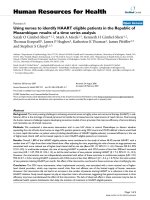
each mutation with respect to the UL20p topology [30] is
shown in Figure 1.
Complementation assay for infectious virus production
It was previously shown that deletion of the HSV-1 UL20
and the PRV UL20 genes resulted in up to two logs reduction in infectious virus production relative to their parental wild type strains. The targeted set of single or double
UL20 mutants and UL20p truncations were tested for
their ability to complement the HSV-1(KOS) UL20-null
virus. Complementation experiments involved transfection of Vero cells with plasmids encoding wild-type or
mutant UL20 genes, followed by infection with the UL20null virus as reported previously [27,30] and described in
Materials and Methods. A complementation ratio was calculated for each mutant UL20 plasmid as a percent ratio
to complementation levels provided by the wild-type
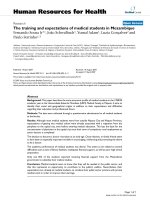
UL20 gene. The UL20 wild-type gene effectively complemented UL20-null virus infectious virus production,
while the UL20 mutants targeted in this study failed to
complement the UL20-null virus (Fig. 2). Furthermore,
Page 2 of 12
(page number not for citation purposes)
Virology Journal 2007, 4:120
/>
Table 1:
Domain
Mutation Name
WT aa Sequence
Mut. aa Sequence
I
I
I
I
I
I
I
IV
V, C-Truncation
V, C-Truncation
V, C-Truncation
CL38
CL49
Y38A
Y49A
CL38-CL49
Y38A-Y49A
CL61
CL153
204t
211t
216t
YGT
YSR
YGT
YSR
YGT-YSR
YGT-YSR
SKR
ETFSPD
SANFF
RFWTR
AILNA
AGA
AAA
AGT
ASR
AGA-AAA
AGT-ASR
SKA
AAFAPA
SANG
RFWG*
AILG*
*Indicates stop codon
the CL2 and Y117A mutations complemented the UL20null virus to wild-type levels (not shown).
the complementation for infectious virus production
results shown in figure 2.
Complementation for virus-induced cell-to-cell fusion
We previously showed that syncytial mutations in either
gB or gK failed to cause virus-induced cell fusion in the
absence of the UL20 gene [27]. Furthermore, a panel of 31
different UL20 mutants revealed that UL20 domains that
functioned in infectious virus production segregated from
those that functioned in virus-induced cell fusion [30].
The panel of UL20 mutants shown in Table 1 was tested
for the ability to complement UL20-null viruses containing syncytial mutations in either gB (syn3) or gK (syn1)
for virus-induced cell fusion as described previously [30].
Briefly, confluent Vero monolayers were transfected with
plasmids encoding either wild type or mutant UL20p, and
subsequently infected with either Δ20 gKsyn1 or Δ20
gBsyn3 viruses. Viral plaques appearing as larger plaques
in a background of uniformelly small UL20-null viral
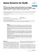
plaques were stained with anti-HSV-1 polyclonal antibody as described in Materials and Methods (Fig. 3). In
this complementation assay, 20–40% of all viral plaques
appeared considerably larger than the uniformly small
UL20-null plaques (not shown). The CL2 UL20 mutant
(Fig. 3) and Y117A (not shown) complemented effectively both gB and gK-mediated virus-induced cell fusion,
as evidenced by the appearance of viral plaques similar in
size to those produced by the wild-type UL20 gene. As previously described [30], and as shown here, the CL49 and
Y49A mutations partially complemented virus spread and
virus-induced cell fusion caused by syncytial mutations in
either gB or gK, as evidenced by the production of visibly
larger than the UL20-null viral plaques (Fig. 3). The CL38,
Y38A, and the double mutants CL38-CL39 and Y38AY39A failed to complement for either infectious virus production or virus spread, as evidenced by the appearance of
very small viral plaques (Fig. 3). These results confirmed
Intracellular transport and TGN localization of UL20p
mutants and gK
Transport and localization of UL20p and gK was further
assessed by transient coexpression of gK and UL20p and
simultaneous detection of the TGN compartment. We
showed previously that in the absence of UL20p, gK was
localized exclusively to reticular-like compartments and
was absent from the Golgi and TGN. A similar pattern was
detected for UL20p in the absence of gK [31]. In contrast,
coexpression of gK and UL20p significantly altered the
distribution pattern of both gK and UL20p with UL20p
and gK colocalized in intracellular compartments that
stained for the TGN marker TGN46. Overall, these results
showed that gK and UL20p intracellular transport and
TGN localization were functionally interdependent
strongly suggesting that gK and UL20p physically interacted [31]. Similar confocal colocalization assays were
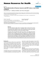
performed to test the ability of each UL20 mutant to facilitate transport and colocalization with gK. The CL38CL49, Y38-Y49, Y38A and Y49A UL20 mutants produced
similar patterns to those of the wild-type UL20 gene, since
they effectively colocalized with gK (Fig. 4: rows 1–3). In
addition, gK was colocalized with TGN46 (Fig. 4: rows 4–
6), indicating that these UL20 mutations did not affect
intracellular transport and TGN colocalization of the
mutant UL20ps with gK. Similar assays were performed
for the UL20p carboxyl terminal truncations 216t, 211t,
and 204t (Fig. 5). The UL20p mutants CL153 and CL61
that were previously shown not to complement for either
infectious virus production or virus-induced cell fusion
[30] were also tested as negative controls, while the wildtype UL20 gene served as the positive control. Both 216t
and 211t UL20 truncations enabled efficient colocalization of UL20 and gK in TGN compartments, while the
Page 3 of 12
(page number not for citation purposes)
Virology Journal 2007, 4:120
/>
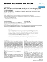
Figure 1
UL20 mutations discussed in this UL20p of UL20 mutations described
Predicted membrane topology of manuscript (larger fonts, underlined) previously [30, 31] (small fonts), and new and other
Predicted membrane topology of UL20p of UL20 mutations described previously [30, 31] (small fonts), and
new and other UL20 mutations discussed in this manuscript (larger fonts, underlined). UL20p domains where
cluster-to-alanine mutations are located are indicated by a shaded oval. Naming of cluster mutations is based on the first amino
acid mutated in each cluster. Single amino acid replacements are indicated with the amino acid position bracketed on the left by
the targeted amino acid and on the right by the changed amino acid i.e. Y38A. Carboxyl terminal truncations are indicated by
the let (t) following the terminal amino acid of the truncated UL20p. Transmembrane region (TM), Cluster mutant (CL).
204t UL20 truncation failed to transport and colocalize
with gK in TGN compartments (Fig. 5). Figure 5 represents
a three color confocal microscopy experiment, while Figure 4 was a two-color confocal microscopy experiment.
The effect of UL20 carboxyl terminal truncations on
UL20p and gK TGN localization after endocytosis from
cell surfaces
We reported previously that UL20 and gK are co-expressed
on infected cell surfaces and co-internalize to TGN cytoplasmic membranes. Similar findings were produced in
transient co-transfection experiments with both UL20 and
gK genes [31]. Similar endocytosis assays were performed
for the UL20p carboxyl terminal truncations. Briefly, in
these experiments, Vero cells that coexpressed gK and
UL20p were reacted with anti-V5 antibody under live conditions (see Materials and Methods). The fate of the internalized V5-tagged gK and FLAG-tagged UL20ps was
assessed at different times post-labeling. By 6 h post-labeling, wild-type gK and UL20p labeled under live conditions on the transfected cell-surfaces were internalized
and colocalized with TGN compartments (Fig. 6). The
216t, 211t, and CL61 mutants produced similar colocalization profiles of UL20p with gK in TGN membranes,
while 204t and CL153 failed to colocalize UL20p and gK
to TGN membranes following endocytosis (Fig. 6).
Page 4 of 12
(page number not for citation purposes)
Virology Journal 2007, 4:120
/>
Figure 2
Complementation ratios produced by mutant UL20p genes
Complementation ratios produced by mutant UL20p
genes. Vero cells were transfected with plasmids encoding
wild-type or mutant UL20 genes under the UL20 promoter
and then infected with the HSV-1(KOS) UL20-null (Δ20)
virus. Viral stocks were prepared at 24 hours post infection
and tittered on Vero cells (see Materials & Methods). The
error bars shown represent the maximum and minimum
complementation ratios obtained from three independent
experiments, and the bar height represents the average complementation ratio.
Discussion
We showed previously that UL20 and gK are functionally
interdependent for their intracellular transport, cell-surface expression and TGN localization [31] and that this
interaction plays pivotal role in cytoplasmic virion envelopment and egress from infected cells [27]. In this study,
we investigated the ability of selected UL20 mutations
reported previously, as well as a new set of UL20 mutants,
on their ability to transport and colocalize with gK on cellsurfaces and in TGN-labeled intracellular compartments:
Previously, we characterized a series of carboxyl terminal
truncations including the 204t and 211t encoding carboxyl terminal truncations of 18 and 11 aa respectively.
These two UL20p truncations failed to complement for
infectious virus production and virus-induced cell fusion,
while the 216t coding for a 6 aa truncation enabled virusinduced cell fusion, but failed to complement for infectious virus production [30]. We show here that the inability to complement for virus-induced cell fusion was not
due to defects in intracellular transport and TGN localiza-
Figure 3
viruses phenotypes of representative viral plaques obtained
after rescue of the Δ20 gK, Δ20 gK syn1, or Δ20 gKsyn3
Plaque
Plaque phenotypes of representative viral plaques
obtained after rescue of the Δ20 gK, Δ20 gK syn1, or
Δ20 gKsyn3 viruses. Vero cell monolayers were transfected with plasmids expressing the mutant UL20 genes, and
24 hours later, they were infected with the respective Δ20
gK-null viruses carrying either the syn1 (gK) or gB(syn3)
mutation. Viral plaques were visualized by immunohistochemistry at 24 hpi.
Page 5 of 12
(page number not for citation purposes)
Virology Journal 2007, 4:120
/>
The effect of UL20p amino terminal mutations on UL20p and gK colocalization in TGN cellular compartments
Figure 4
The effect of UL20p amino terminal mutations on UL20p and gK colocalization in TGN cellular compartments. Vero cells were co-transfected with gK tagged with the V5 epitope (D1V5), as well as with plasmids encoding wildtype or different mutant UL20ps tagged with the 3 × FLAG epitope (UL20D1FLAG). Thirty-six hours post-transfection, cells
were washed thoroughly, fixed, and processed for confocal microscopy. After permeabilization, rabbit anti-FLAG (α FLAG)
mAb was used to detect UL20p, mouse anti-V5 (α V5) epitope was used to detect gK, and sheep aTGN46 mAb was used to
identify the TGN. First three rows of the confocal pictures show co-localization of UL20p with gK, while rows 4–6 show colocalization of gK with TGN46.
tion, because 216t, as well as both 204t and 211t were efficiently transported to cell-surfaces and co-localized with
gK in TGN-labeled membranes. Therefore, intracellular
transport, cell-surface expression and TGN localization of
UL20p and gK are not sufficient for infectious virus production. Based on these results, we can conclude that the
Page 6 of 12
(page number not for citation purposes)
Virology Journal 2007, 4:120
/>
Figure 5
The effect of UL20p carboxyl terminal truncations on UL20p and gK colocalization in TGN cellular compartments
The effect of UL20p carboxyl terminal truncations on UL20p and gK colocalization in TGN cellular compartments. As with figure 4, Vero cells were co-transfected with gKD1V5, as well as with plasmids encoding wild-type or mutant
UL20DIFLAG proteins. Thirty-six hours post-transfection, cells were washed thoroughly, fixed, and processed for confocal
microscopy. After permeabilization, antibodies a3xFLAG, aV5 and aTGN46 were used to identify, UL20p, gK and TGN46,
respectively.
Page 7 of 12
(page number not for citation purposes)
Virology Journal 2007, 4:120
/>
Confocal microscopy of gK cell-surface expression and endocytosis to the TGN mediated by selected UL20p mutants
Figure 6
Confocal microscopy of gK cell-surface expression and endocytosis to the TGN mediated by selected UL20p
mutants. Vero cells were co-transfected with gKD1V5 as well as with plasmids encoding wild-type or mutant UL20p, as with
figures 4 and 5. Twenty-four hours post-transfection, cells were incubated under live conditions with aV5 (gK) mAb for 6
hours. Cells were washed thoroughly, fixed, and processed for confocal microscopy. After permeabilization, antibodies a3 ×
FLAG, aV5 and aTGN46 were used to identify, UL20p, gK and TGN46, respectively.
Page 8 of 12
(page number not for citation purposes)
Virology Journal 2007, 4:120
carboxyl terminal six amino acids of UL20p function
exclusively in intracellular virion envelopment and infectious virus production, while the UL20p domain spanning amino acids 204–211is important for both
intracellular transport and virus-induced cell fusion.
Domain I is the largest domain (63 aa) and it includes
stretches of acidic amino acid (D, E) clusters, which could
form electrostatic interactions with other proteins [30].
Furthermore, the amino terminus of UL20p contains
acidic clusters, as well as the amino acid motif YXXΦ
(YSRL), which have been shown to function in endocytosis of alphaherpesvirus envelope proteins from plasma
membranes to the TGN [32-36]. The acidic cluster motifs
appear to direct TGN localization by binding to a cellular
connector protein, PACS-1, which connects the glycoproteins to the AP-1 complex [37], while the YXXΦ motif
binds adaptor proteins directly [2,3,40]. The YXXΦ
(YSRL) amino acid sequence overlapping the CL49
mutated sequence, is conserved in HSV-1, HSV-2, and cercopithecine herpesvirus 1 and 2, but not in varicella zoster
(VZV) or pseurodabies virus (PRV) (not shown). Mutagenesis of the Y residue of a YXXΦ(YTKI) motif within gK
domain IV, shown to lie in the cytoplasmic side of membranes, produced a gK-null phenotype [20]. Similarly,
mutagenesis of either Y38 or Y49, or both residues
resulted in loss of infectious virus production, while the
UL20p mutants carrying either mutation or a combination of both mutations allowed efficient intracellular
transport and TGN localization. This result is similar to
the results obtained with the UL20p carboxyl terminal
domains and suggests that amino terminal domains of
UL20p that function in cytoplasmic virion envelopment
can be functionally separated from those that function in
UL20p and gK intracellular transport and TGN localization. Interestingly, the Y49A mutant allowed some virusinduced cell fusion caused by either the gBsyn3 or gKsyn1
mutation suggesting that the requirement of this residue
for infectious virus production is more stringent that the
requirement for virus-induced cell fusion.
We reported previously that the Y49A, CL49 and 216t
mutant viruses produced syncytial plaques, although their
ultrastructural phenotypes seemed to be similar to that of
the UL20-null virus [30]. We show here these phenotypes
are consistent with the findings that these UL20p mutations allowed efficient intracellular transport, cell-surface
expression and TGN localization. However, mutagenesis
of both Y38 and Y49 amino acid residues in the amino terminus of UL20p, inhibited virus-induced cell fusion,
while allowing efficient intracellular transport and TGN
localization. This result suggests that the Y38 and Y49 residues together play important roles in cytoplasmic virus
envelopment, but they are not required for proper UL20p/
gK intracellular transport. The Y38A mutation seemed to
/>
affect both virion production and virus-induced cell
fusion, although the Y49A mutation appeared to inhibit
virion production, but allowed some cell fusion to occur.
As is the case with the carboxyl terminus of UL20p discussed earlier, these results suggest that the amino terminus of UL20p contains functionally separable domains
involved in cytoplasmic virion envelopment and intracellular glycoprotein transport. Furthermore, the Y49A mutation allowed some virus-induced cell fusion, but not
infectious virus production to occur suggesting that
domains within the UL20p amino-terminus involved in
cytoplasmic virion envelopment may be functionally separated from domains functioning in UL20p/gK intracellular transport and virus-induced cell fusion.
Conclusion
These results show that UL20p domains required for
UL20p and gK intracellular transport and TGN localization can be functionally segregated from domains
involved in infectious virus production and virus-induced
cell fusion. The results suggest that virus-induced cell
fusion mechanisms are not required for cytoplasmic virion envelopment.
Materials and methods
Cells and viruses
African green monkey kidney (Vero) cells were obtained
from ATCC (Rockville, MD). The Vero-based UL20 complementing cell line, G5, was a gift of Dr. P. Desai, (John
Hopkins Medical Center) [38]. Cells were maintained as
previously described [20,29,38]. The parental wild-type
strain used in this study HSV-1 (KOS) was originally
obtained from P. A. Schaffer (Harvard Medical School).
Δ20DIV5, Δ20gBsyn3 and Δ20gKsyn1DIV5 viruses were
as described previously [27]. Virus stocks were grown on
the UL20 complementing cell line Fd20-1, the construction of which was described previously [30]. In this paper,
for simplification purposes, the Δ20DIV5 virus is referred
to as Δ20 virus and the Δ20syngK1DIV5 virus is referred to
as Δ20gKsyn1 virus [30].
Plasmids
pCR2.1-UL20, which was used as the parental vector for
UL20 mutagenesis, was generated by cloning a 773 bp
DNA fragment containing the UL20 gene, obtained by
PCR amplification of HSV-1(KOS) viral DNA, into
pCR2.1/TOPO (Invitrogen) as described in detail previously [30]. The generation of UL20 cluster to alanine
mutants CL38, CL49, CL153, and CL209, the single point
mutant Y49A, and truncation mutants, 204t, 211t, 216t
were reported previously [30]. A set of new UL20 mutants
generated for this study included a UL20 mutant containing both the CL38 and CL49 mutations (CL38 – CL49),
the alanine cluster UL20 mutant CL61, the single point
mutant Y38A, and the UL20 mutant Y-Y containing both
Page 9 of 12
(page number not for citation purposes)
Virology Journal 2007, 4:120
the Y38A and Y49A mutations. The cluster mutations, the
additional single point UL20 mutants, as well as the double mutants were generated by splice-overlap extension
(SOE) PCR [39] as described previously [30]. All mutations were verified by sequencing of the final plasmid construct.
UL20 complementation assay for infectious virion
production
Confluent Vero monolayers in six well plates were transfected with 2 μg of wild-type or mutant UL20 plasmid
with Lipofectamine 2000 as described by the manufacturer (Invitrogen). Six hours post-transfection, the monolayers were infected with a UL20-null virus at an MOI of 1.
Infections were placed on a rocker for 1 hour at 4°C, and
then transferred to 37°C for 2 hours. Residual virus was
inactivated using an acid wash (PBS containing .5 M glycine, pH3) for 2 min, and monolayers were subsequently
washed 3 times with DMEM to restore the pH to a normal
level. Infections were incubated at 37°C for 24 hours.
After repeated freeze/thaw cycles, virus stocks were titered
in triplicate on Fd20-1 cells, which effectively complement the UL20-null defect [30]. The complementation
ratio for each mutant was calculated with the formula
(virus titer of mutant/virus titer of negative control).
UL20 complementation assay for virus-induced cell-to-cell
fusion and virus spread
The complementation assay was performed essentially as
we described previously for addressing the role of the
HSV-1 UL11 protein in virion morphogenesis [40].
Briefly, confluent Vero monolayers in six-well plates were
transfected with 2 μg of wild-type or mutant UL20 plasmid with Lipofectamine 2000 as described by the manufacturer (Invitrogen). 18 hours post transfection, the
monolayers were infected at an MOI of 0.1 with either
Δ20gKsyn1 or Δ20gBsyn3 viruses. Infections were placed
on a rocker at room temperature for 1 hour, then transferred to 37°C for 30 minutes. Cells were overlaid with
DMEM containing 1% methylcellulose. 24 hours postinfection, cell fusion was determined by visualization of
syncytia formation by light microscopy. Cells were stained
with a polyclonal HRP conjugated HSV-1 antibody as
directed by the manufacturer (DakoCytomation). Briefly,
cells were washed with PBS to remove methylcellulose
media, and fixed with 4°C methanol for 15 minutes. TBS
containing a 1:750 dilution of the polyclonal HSV-1 antibody was added to the cells and placed on a rocker at 4°C
for 1 h. Cells were washed with TBS and developed using
the Vector NovaRED peroxidase substrate kit as directed
by the manufacturer (Vector, Inc). In this assay,
Complementation of the UL20-null virus by transient
expression of the wild-type UL20 gene caused the production of up to 40% of total viral plaques appearing to have
/>
similar morphology and size to the HSV-1(F) parental
virus.
Confocal microscopy
Cell monolayers were grown on coverslips in six-well
plates. Cell monolayers were transfected with the indicated UL20 and/or gK plasmid combinations by using
Lipofectamine 2000 (Invitrogen) according to the manufacturer's instructions and prepared for confocal microscopy approximately 30 h posttransfection. Cells were
washed with TBS and fixed with electron microscopygrade 3% paraformaldehyde (Electron Microscopy Sciences, Fort Washington, Pa.) for 15 min, washed twice
with phosphate-buffered saline-50 mMglycine, and permeabilized with 1.0% Triton X-100. Monolayers were
subsequently blocked for 1 h with 7% normal goat serum
and 7% bovine serum albumin in TBS (TBS blocking
buffer) before incubation for 2 h with either anti-V5 (Invitrogen, Inc.), for recognition of gK, or anti-FLAG (Sigma
Chemical, Inc.), for recognition of UL20p, diluted 1:500
in TBS blocking buffer. Alternatively, simultaneous detection of gK and UL20p in cotransfected cells was accomplished by concurrent incubation with murine anti-V5
and rabbit anti-FLAG (Sigma Chemical, Inc.) diluted
1:500 in TBS blocking buffer. Cells were then washed
extensively and incubated for 30 min with Alexa Fluor
594 and/or Alexa Fluor 647-conjugated anti-immunoglobulin G diluted 1:500 in TBS blocking buffer. After
incubation, excess antibody was removed by washing five
times with TBS. TGN were identified with a donkey antiTGN46 primary antibody and an Alexa Fluor 488-conjugated sheep anti-donkey secondary antibody [41]. Specific immunofluorescence was examined using a Leica
TCS SP2 laser scanning confocal microscope (Leica Microsystems, Exton, Pa.) fitted with a CS APO 63× Leica objective (1.4 numerical aperture). Individual optical sections
in the z axis, averaged six times, were collected at the indicated zoom in series in the different channels at 1,024- by
1,024-pixel resolution as described previously [27,29,42].
Images were compiled and rendered with Adobe Photoshop. Image analyses were generated and analyzed
using the Leica confocal microscopy software package and
were modified from protocols described previously [43].
UL20p/gK cell surface internalization assay
Internalization assays were modified from similar assays
performed previously [35,44,45]. Briefly, Vero cells were
transfected with pgKDIV5 and either pUL20-3 × FLAG or
a variant containing the indicated UL20 mutation [29].
Twenty hours posttransfection, cells were incubated under
live conditions for 6 h at 37°C with mouse anti-V5. Cells
were extensively washed, fixed with paraformaldehyde,
and processed for confocal microscopy as described
above, with the exception that the internalized antibodies
served as the primary antibody for gK (mouse anti-V5).
Page 10 of 12
(page number not for citation purposes)
Virology Journal 2007, 4:120
Competing interests
/>
19.
The author(s) declare that they have no competing interests.
20.
Authors' contributions
J. Melancon performed most of the experiments. K. G.
Kousoulas wrote the manuscript.
21.
22.
Acknowledgements
This work was supported by a grant from the National Institute of Allergy
and Infectious Diseases (AI43000) to K.G.K. J. M. M. and P. A. F. were supported by Louisiana Economic Development Graduate Assistant Fellowships. We acknowledge financial support by the LSU School of Veterinary
Medicine to BIOMMED.
References
1.
2.
3.
4.
5.
6.
7.
8.
9.
10.
11.
12.
13.
14.
15.
16.
17.
18.
Roizman B, Knipe DM: Herpes Simplex Viruses and Their Replication. In Fields Virology Volume 2. Third edition edition. Edited by:
Knipe DM, Howley PM. Philadelphia, PA , Lippincott-Williams &
Wilkins; 2001:2399-2459.
Spear PG: Membrane fusion induced by herpes simplex virus.
In Viral fusion mechanisms Edited by: Bentz J. Boca Raton, Fla , CRC
Press.; 1993:201-232.
Spear PG: Entry of alphaherpesviruses into cells. Seminars in
Virology 1993, 4:167-180.
Spear PG: Herpes simplex virus: receptors and ligands for cell
entry. Cell Microbiol 2004, 6(5):401-410.
Spear PG, Eisenberg RJ, Cohen GH: Three classes of cell surface
receptors for alphaherpesvirus entry. Virology 2000, 275(1):1-8.
Spear PG, Longnecker R: Herpesvirus entry: an update. J Virol
2003, 77(19):10179-10185.
Mettenleiter TC: Herpesvirus assembly and egress. J Virol 2002,
76(4):1537-1547.
Mettenleiter TC, Klupp BG, Granzow H: Herpesvirus assembly: a
tale of two membranes. Current opinion in microbiology 2006,
9(4):423-429.
Baines JD, Ward PL, Campadelli-Fiume G, Roizman B: The UL20
gene of herpes simplex virus 1 encodes a function necessary
for viral egress. J Virol 1991, 65(12):6414-6424.
MacLean CA, Efstathiou S, Elliott ML, Jamieson FE, McGeoch DJ:
Investigation of herpes simplex virus type 1 genes encoding
multiply inserted membrane proteins. J Gen Virol 1991, 72(Pt
4):897-906.
Jacobson JG, Chen SH, Cook WJ, Kramer MF, Coen DM: Importance of the herpes simplex virus UL24 gene for productive
ganglionic infection in mice. Virology 1998, 242(1):161-169.
Sanders PG, Wilkie NM, Davison AJ: Thymidine kinase deletion
mutants of herpes simplex virus type 1. J Gen Virol 1982,
63(2):277-295.
Bzik DJ, Fox BA, DeLuca NA, Person S: Nucleotide sequence of a
region of the herpes simplex virus type 1 gB glycoprotein
gene: mutations affecting rate of virus entry and cell fusion.
Virology 1984, 137(1):185-190.
Pellett PE, Kousoulas KG, Pereira L, Roizman B: Anatomy of the
herpes simplex virus 1 strain F glycoprotein B gene: primary
sequence and predicted protein structure of the wild type
and of monoclonal antibody-resistant mutants. J Virol 1985,
53(1):243-253.
Bond VC, Person S: Fine structure physical map locations of
alterations that affect cell fusion in herpes simplex virus type
1. Virology 1984, 132(2):368-376.
Debroy C, Pederson N, Person S: Nucleotide sequence of a herpes simplex virus type 1 gene that causes cell fusion. Virology
1985, 145(1):36-48.
Hutchinson L, Goldsmith K, Snoddy D, Ghosh H, Graham FL, Johnson
DC: Identification and characterization of a novel herpes
simplex virus glycoprotein, gK, involved in cell fusion. J Virol
1992, 66(9):5603-5609.
Pogue-Geile KL, Lee GT, Shapira SK, Spear PG: Fine mapping of
mutations in the fusion-inducing MP strain of herpes simplex
virus type 1. Virology 1984, 136(1):100-109.
23.
24.
25.
26.
27.
28.
29.
30.
31.
32.
33.
34.
35.
36.
37.
38.
Ryechan WT, Morse LS, Knipe DM, Roizman B: Molecular genetics
of herpes simplex virus. II. Mapping of the major viral glycoproteins and of the genetic loci specifying the social behavior of infected cells. J Virol 1979, 29:677-697.
Foster TP, Kousoulas KG: Genetic analysis of the role of herpes
simplex virus type 1 glycoprotein K in infectious virus production and egress. J Virol 1999, 73(10):8457-8468.
Fuchs W, Klupp BG, Granzow H, Mettenleiter TC: The UL20 gene
product of pseudorabies virus functions in virus egress. J Virol
1997, 71(7):5639-5646.
Hutchinson L, Johnson DC: Herpes simplex virus glycoprotein K
promotes egress of virus particles.
J Virol 1995,
69(9):5401-5413.
Jayachandra S, Baghian A, Kousoulas KG: Herpes simplex virus
type 1 glycoprotein K is not essential for infectious virus production in actively replicating cells but is required for efficient envelopment and translocation of infectious virions
from the cytoplasm to the extracellular space. Journal of Virology 1997, 71(7):5012-5024.
Tomishima MJ, Smith GA, Enquist LW: Sorting and transport of
alpha herpesviruses in axons. Traffic 2001, 2(7):429-436.
Johnson DC, Huber MT: Directed egress of animal viruses promotes cell-to-cell spread. J Virol 2002, 76(1):1-8.
Ward PL, Campadelli-Fiume G, Avitabile E, Roizman B: Localization
and putative function of the UL20 membrane protein in cells
infected with herpes simplex virus 1.
J Virol 1994,
68(11):7406-7417.
Foster TP, Melancon JM, Baines JD, Kousoulas KG: The Herpes
Simplex Virus Type 1 UL20 Protein Modulates Membrane
Fusion Events during Cytoplasmic Virion Morphogenesis and
Virus-Induced Cell Fusion. J Virol 2004, 78(10):5347-5357.
Dietz P, Klupp BG, Fuchs W, Kollner B, Weiland E, Mettenleiter TC:
Pseudorabies virus glycoprotein K requires the UL20 gene
product for processing. J Virol 2000, 74(11):5083-5090.
Foster TP, Alvarez X, Kousoulas KG: Plasma membrane topology of syncytial domains of herpes simplex virus type 1 glycoprotein K (gK): the UL20 protein enables cell surface
localization of gK but not gK-mediated cell-to-cell fusion. J
Virol 2003, 77(1):499-510.
Melancon JM, Foster TP, Kousoulas KG: Genetic analysis of the
herpes simplex virus type 1 (HSV-1) UL20 protein domains
involved in cytoplasmic virion envelopment and virusinduced cell fusion. J Virol 2004, 78(14):7329-7343.
Foster TP, Melancon JM, Olivier TL, Kousoulas KG: Herpes simplex
virus type-1 (HSV-1) glycoprotein K (gK) and the UL20 protein are interdependent for intracellular trafficking and
trans-Golgi
network
localization.
J
Virol
2004,
78(23):13262-13277.
Alconada A, Bauer U, Hoflack B: A tyrosine-based motif and a
casein kinase II phosphorylation site regulate the intracellular trafficking of the varicella-zoster virus glycoprotein I, a
protein localized in the trans-Golgi network. Embo Journal
1996, 15(22):6096-6110.
Alconada A, Bauer U, Sodeik B, Hoflack B: Intracellular traffic of
herpes simplex virus glycoprotein gE: characterization of the
sorting signals required for its trans-Golgi network localization. J Virol 1999, 73(1):377-387.
Brideau AD, del Rio T, Wolffe EJ, Enquist LW: Intracellular trafficking and localization of the pseudorabies virus Us9 type II
envelope protein to host and viral membranes. J Virol 1999,
73(5):4372-4384.
Tirabassi RS, Enquist LW: Mutation of the YXXL endocytosis
motif in the cytoplasmic tail of pseudorabies virus gE. J Virol
1999, 73(4):2717-2728.
Zhu Z, Hao Y, Gershon MD, Ambron RT, Gershon AA: Targeting
of glycoprotein I (gE) of varicella-zoster virus to the transGolgi network by an AYRV sequence and an acidic amino
acid-rich patch in the cytosolic domain of the molecule. J Virol
1996, 70(10):6563-6575.
Wan L, Molloy SS, Thomas L, Liu G, Xiang Y, Rybak SL, Thomas G:
PACS-1 defines a novel gene family of cytosolic sorting proteins required for trans-Golgi network localization. Cell 1998,
94(2):205-216.
Desai P, DeLuca NA, Glorioso JC, Person S: Mutations in herpes
simplex virus type 1 genes encoding VP5 and VP23 abrogate
Page 11 of 12
(page number not for citation purposes)
Virology Journal 2007, 4:120
39.
40.
41.
42.
43.
44.
45.
/>
capsid formation and cleavage of replicated DNA. J Virol 1993,
67(3):1357-1364.
Aiyar A, Xiang Y, Leis J: Site-directed mutagenesis using overlap
extension PCR. Methods Mol Biol 1996, 57:177-191.
Fulmer PA, Melancon JM, Baines JD, Kousoulas KG: UL20 protein
functions precede and are required for the UL11 functions of
herpes simplex virus type 1 cytoplasmic virion envelopment.
Journal of virology 2007, 81(7):3097-3108.
McMillan TN, Johnson DC: Cytoplasmic domain of herpes simplex virus gE causes accumulation in the trans-Golgi network, a site of virus envelopment and sorting of virions to
cell junctions. J Virol 2001, 75(4):1928-1940.
Foster TP, Rybachuk GV, Alvarez X, Borkhsenious O, Kousoulas KG:
Overexpression of gK in gK-transformed cells collapses the
Golgi apparatus into the endoplasmic reticulum inhibiting
virion egress, glycoprotein transport, and virus-induced cell
fusion. Virology 2003, 317(2):237-252.
Demandlox D, Davoust J: Multicolour analysis and local image
correlation in confocal microscopy.
J Microscopy 1997,
185:21-36.
Brideau AD, Eldridge MG, Enquist LW: Directional transneuronal
infection by pseudorabies virus is dependent on an acidic
internalization motif in the Us9 cytoplasmic tail. J Virol 2000,
74(10):4549-4561.
Tirabassi RS, Enquist LW: Role of envelope protein gE endocytosis in the pseudorabies virus life cycle. J Virol 1998,
72(6):4571-4579.
Publish with Bio Med Central and every
scientist can read your work free of charge
"BioMed Central will be the most significant development for
disseminating the results of biomedical researc h in our lifetime."
Sir Paul Nurse, Cancer Research UK
Your research papers will be:
available free of charge to the entire biomedical community
peer reviewed and published immediately upon acceptance
cited in PubMed and archived on PubMed Central
yours — you keep the copyright
BioMedcentral
Submit your manuscript here:
/>
Page 12 of 12
(page number not for citation purposes)