Báo cáo sinh học: " Oral administration of IFN-a2b-transformed B. longum protects the Balb/c mice against coxsackievirus B3-induced myocarditis" pptx
Bạn đang xem bản rút gọn của tài liệu. Xem và tải ngay bản đầy đủ của tài liệu tại đây (362.13 KB, 15 trang )
This Provisional PDF corresponds to the article as it appeared upon acceptance. Fully formatted
PDF and full text (HTML) versions will be made available soon.
Oral administration of IFN-a2b-transformed B. longum protects the Balb/c mice
against coxsackievirus B3-induced myocarditis
Virology Journal 2011, 8:525 doi:10.1186/1743-422X-8-525
Yu Zhijian ZY ()
Huang Zhen ZH ()
Shao Chongwen CS ()
Huang Yuanjian YH ()
Zhang Fan FZ ()
Yang Jin JY ()
Deng Lili LD ()
Zeng Zhongming ZZ ()
Deng Qiwen QD ()
Zeng Weiseng WZ ()
ISSN 1743-422X
Article type Research
Submission date 22 July 2011
Acceptance date 8 December 2011
Publication date 8 December 2011
Article URL />This peer-reviewed article was published immediately upon acceptance. It can be downloaded,
printed and distributed freely for any purposes (see copyright notice below).
Articles in Virology Journal are listed in PubMed and archived at PubMed Central.
For information about publishing your research in Virology Journal or any BioMed Central journal, go
to
/>For information about other BioMed Central publications go to
Virology Journal
© 2011 Zhijian et al. ; licensee BioMed Central Ltd.
This is an open access article distributed under the terms of the Creative Commons Attribution License ( />which permits unrestricted use, distribution, and reproduction in any medium, provided the original work is properly cited.
/>Virology Journal
© 2011 Zhijian et al. ; licensee BioMed Central Ltd.
This is an open access article distributed under the terms of the Creative Commons Attribution License ( />which permits unrestricted use, distribution, and reproduction in any medium, provided the original work is properly cited.
Oral administration of interferon-α2b-
transformed Bifidobacterium longum protects
BALB/c mice against coxsackievirus B3-
induced myocarditis
ArticleCategory :
Research
ArticleHistory :
Received: 22-Jul-2011; Accepted: 22-Nov-2011
ArticleCopyright
:
© 2011 Yu et al.; licensee BioMed Central Ltd. This is an Open Access
article distributed under the terms of the Creative Commons Attribution
License ( which permits
unrestricted use, distribution, and reproduction in any medium, provided
the original work is properly cited.
Zhijian Yu,
Aff1
Email:
Zhen Huang,
Aff1
Email:
Chongwen Shao,
Aff2
Email:
Yuanjian Huang,
Aff2
Email:
Fan Zhang,
Aff1
Email:
Jin Yang,
Aff1
Email:
Lili Deng,
Aff1
Email:
Zhongming Zeng,
Aff1
Email:
Qiwen Deng,
Aff1
Corresponding Affiliation: Aff1
Phone: +86-775-2655311126402
Fax: +86-755-26565025
Email:
Weiseng Zeng,
Aff2
Corresponding Affiliation:
Aff2
Phone: +86-20-61647073
Email:
Aff1
Department of Infectious Diseases, the Affiliated Shenzhen Nanshan
Hospital of Guangdong Medical College, No 89, Taoyuan Road,
Nanshan district, 518052 Shenzhen, China
Aff2
Department of Cell Biology, Southern Medical University, No 1023,
Satai Raod, Baiyun district, 510515 Guangzhou, China
Abstract
Multiple reports have claimed that low-dose orally administered interferon (IFN)-α is beneficial
in the treatment of many infectious diseases and provides a viable alternative to high-dose
intramuscular treatment. However, research is needed on how to express IFN stably in the gut.
Bifidobacterium may be a suitable carrier for human gene expression and secretion in the
intestinal tract for the treatment of gastrointestinal diseases. We reported previously that
Bifidobacterium longum can be used as a novel oral delivery of IFN-α. IFN-transformed B.
longum can exert an immunostimulatory role in mice; however the answer to whether this
recombinant B. longum can be used to treat virus infection still remains elusive. Here, we
investigated the efficacy of IFN-transformed B. longum administered orally on coxsackie virus
B3 (CVB3)-induced myocarditis in BALB/c mice. Our data indicated that oral administration of
IFN-transformed B. longum for 2 weeks after virus infection reduced significantly the severity of
virus-induced myocarditis, markedly down regulated virus titers in the heart, and induced a T
helper 1 cell pattern in the spleen and heart compared with controls. Oral administration of the
IFN-transformed B. longum, therefore, may play a potential role in the treatment of CVB3-
induced myocarditis.
Keywords
Bifidobacterium, Coxsackievirus B, Enterovirus, Interferon, Myocarditis, Oral administration
Introduction
The oral use of low doses of interferon (IFN)-α has been shown to exhibit beneficial effects in
mice or human with acquired immunodeficiency syndrome (AIDS) [1], hepatitis B [2], aphthous
stomatitis [3], and measles [4]. These studies have indicated that IFN-α/-β given orally provides
a viable alternative to the current high-dose treatment intramuscularly [5,6]. The number of dairy
and probiotic products that contain bifidobacteria has developed rapidly in recent years, and its
probiotic properties have been extended further by the production of the recombinant
Bifidobacterium-containing products [7-11]. Genetically engineered Bifidobacterium has been
reported as a successful exogenous gene delivery carrier for the treatment of many diseases. We
previously constructed a recombinant B. longum (IFN-transformed B. longum) that was inducible
by arabinose to express efficiently secreted IFN-α2b in vitro [12]. Moreover, oral administration
of IFN-transformed B. longum to mice increased intestinal sIgA and serum IFN-α2b levels,
which suggested the potential clinical value of this bacterium as a kind of oral interferon in the
treatment of virus infection [13].
Human IFN can inhibit coxsackievirus B3 (CVB3) replication in vitro and protects murine
models from CVB3-induced myocarditis [14-19]. However, it is not clear whether oral
administration of IFN can treat CVB3-induced myocarditis in vivo. In this study, the effect of
IFN-transformed B. longum by oral administration on the development of CVB3-induced
myocarditis in mice was evaluated.
Material and methods
Cells and viruses
CVB3 (Nancy strain) was obtained from Prof. Wang at the Department of Biotechnology of
Ginan University, China [20]. African green monkey kidney (Vero) cells were cultured in
Dulbecco’s modified Eagle’s medium (DMEM) that contained 8% fetal calf serum (Gibco,
Rockville, USA). Confluent cultures of Vero cells were infected with CVB3 and incubated at
37°C until an extensive cytopathic effect was observed (generally at 3–5 days post-infection).
Subsequently, the culture media were collected, the cell debris was pelleted by centrifugation
and removed and the supernatant was aliquoted and stored at−80°C.
Bacteria culture
IFN-transformed B. longum were constructed by transforming B. longum with pBAD-SPIFN
(BSPIFN) as reported [12]. Briefly, BSPIFN plasmids consisted of a fusion gene of the
arabinosidase signal peptide and human IFN-α2b (hIFNα2b). Recombinant IFN-transformed B.
longum contained an L-arabinose promoter and displayed highly efficient IFN-α2b expression
[12]. The control plasmid-transformed B. longum bacteria were transformed with the control
plasmid (pBAD-gIIIA) without the insertion of hIFN-α2b gene. Recombinant BSPIFN- and
control plasmid-transformed B. longum were cultured anaerobically and prepared as described in
our previous report [12]. A 10-µl suspension of bacteria was seeded onto BL agar plates (Nissui)
that contained 100 g L
−1
ampicillin to determine the actual number of viable bacilli in the
inocula. Colonies were counted after 24 h of anaerobic culture.
Interventions and groups
This study was approved by the Ethics Committee of Southern Medical University (Guangzhou,
China). Four-week-old male BALB/c mice (weight, 15 ± 0.5 g; Southern Medical University,
USA) were inoculated i.p. with a 50% cell-culture infectious dose of CVB3 at 5 × 10
6
(as
determined by plaque assay on Vero cells). Infected animals given this virus dosage survived for
at least 6 months post-infection. We studied the efficacy of IFN-transformed B. longum on
coxsackievirus B3-induced myocarditis. Forty BALB/c mice were inoculated with the virus and
were divided into four groups. ‘BIFN’ group and ‘Control’ group animals were administered
orally with IFN- and control plasmid- transformed B. longum for 2 weeks respectively after the
inoculation of the virus. The ‘IFN’ group was injected i.m. with a therapeutic dose (1.5 µg kg
−
1
week
−1
) of pegylated IFN α2b (PegIntron). The ‘saline’ group was administered i.p. once daily
with sterile saline after infection. Three mice were kept under the same conditions to act as the
normal control. Recombinant bacteria were given to the mice orally once every 2 days using a
tuberculin syringe attached to a 20-gauge olive-tip steel feeding tube, passed through the oral
cavity and esophagus. All animals were killed at day 14 post-infection (following ether
anesthesia). Up to day 14 post-infection, half of the murine hearts were dissected aseptically for
virus titration and RNA extraction for cytokine quantity. The other half of the heart was used for
hematoxylin–eosin (H&E) staining. The spleen was removed surgically to isolate mononuclear
cells (MNCs).
Morphometry
Hematoxylin–eosin staining was performed according to the standard techniques. The selected
surfaces of myocarditis lesions studied were considered to be representative of the relative
inflammatory area in the entire heart volume, because of the relatively homogeneous distribution
of the myocarditis lesions in the affected hearts. The number of myocarditis lesions and their
surface proportion were determined with H&E-stained sections of the hearts of the untreated and
other groups. The proportion of the surface occupied by myocarditis lesions was determined by
means of a conventional point-counting method, as reported earlier [21], by using an ocular grid
that contained 121 equally spaced points. The surface proportion was taken to be an estimate of
the percentage of heart tissue that was affected by focal myocarditis. Counting was performed on
three sections per heart and the sections were evaluated at a magnification of less than ×200.
Virus titration from heart homogenate
The aseptic hearts of the animals were weighed and homogenized in the minimal essential
medium of 2 ml phosphate-buffered saline (PBS). The supernatant was subjected to three freeze–
thaw cycles and centrifugation at 5000 rpm for 8 min, then was absorbed and diluted sequentially
10-fold in RPMI 1640 medium. Vero cells were grown to confluence in microtiter trays, infected
with serial dilutions of the homogenates, and incubated for 3 h at 37°C. The cells were cultured
for 72 h of cultivate, then the monolayers were fixed in 10% phosphate-buffered formalin and
stained with crystal violet (Invitrogen); the numbers of plaques were counted. Virus titers were
determined by standard plaque formation assay and expressed as per organ weight (in grams).
Quantitation of transcript level of cytokines and Mx1
The total RNA of the heart tissues was extracted with Trizol reagent (Invitrogen, Carlsbad, CA,
USA) and reverse transcribed into cDNA according to the manufacturer’s protocol (Invitrogen,
Carlsbad, CA, USA). Transcription of IFN-γ, TNF-α, Mx1 or the housekeeping gene β-actin was
detected by real-time polymerase chain reaction (PCR) using a SYBR Green Master Mix
(Applied Biosystems). Thermocycler conditions included an initial denaturation step at 94°C for
2 min; a three-step cycle procedure was carried out (denaturation, 94°C, 20 s; annealing, 58°C,
20 s; and extension, 72°C, 30 s) for 35 cycles. All reactions were performed in at least duplicate
for each sample. Data were collected and analyzed quantitatively on an ABI Prism 7900
sequence detection system (Applied Biosystems). The β-actin gene was used as an endogenous
control to normalize for differences in the amount of total RNA in each sample and the relative
mRNA expression was calculate by normalization to the value of the β-actin transcripts. Primers
for IFN-γ, TNF-α, Mx1 and the housekeeping gene β-actin have been reported previously
[13,21].
Preparation of mononuclear cells (MNCs) and cytokine detection
Spleens were removed surgically, and the MNCs were isolated as described previously [22].
Suspensions of MNCs from the spleen were prepared with RPMI-1640 culture medium that
contained heat-inactivated fetal bovine serum (50 mL L
−1
), L-glutamine (2 mM), penicillin
(1 × 10
5
U L
−1
), streptomycin (100 mg L
−1
), and HEPES (25 mM) (all from Life Sciences). The
MNCs were seeded into 24-well plates (each well had 2 × 10
8
cells L
−1
) and stimulated
subsequently with 100 µL of Con A (5 mg L
−1
) for 72 h at 37°C, 5% CO
2
in air, and 95%
humidity. The levels of IFN-γ, TNF-α and IL-10 in the supernatants were measured by OptEIA
commercial enzyme-linked immunosorbent assay (ELISA) kits (BD Pharmingen), following the
manufacturers’ instructions. The detection limits of the ELISA assays were as follows: 2500 pg
ml
−1
for IFN-γ, 825 pg ml
−1
for TNF-α and 650 pg ml
−1
for IL-10.
Statistical analysis
Data were shown as the mean ± standard error of the mean (SEM). Statistical analyses of the
data were performed by one-way analysis of variance (ANOVA), and the correlation between
two variables was tested by bivariate correlation analysis using SPSS11.0; a p-value < 0.01 was
considered to be statistically significant.
Results
Evaluations for the severity of myocarditis and virus replication
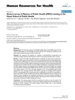
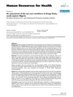
The prominent cardiac inflammation area is observed in Figure 1. The percentage of the
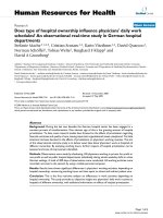
pathological area of the heart sections in the BIFN, B, IFN-α and saline groups was elevated and
is compared in Figure 2a. The pathological area of heart sections in the BIFN group was
significantly lower compared with the B and saline groups (p < 0.01) respectively, but markedly
high compared with the IFN group. The levels of cardiac CVB3 titers and CVB3 RNA in the
cardiac tissues of the BIFN group were significantly lower compared with the B and saline
groups respectively and markedly higher compared with IFN group (p < 0.01) (Figure 2b).
Figure 1 Evaluation of the severity of myocarditis. (a–e) are representative of histopathologic
images in heart tissue from saline, B, BIFN and interferon (IFN) groups respectively
(hemotoxylin and eosin (H&E) staining, original magnification × 200). Ten mice per group were
analyzed in this study
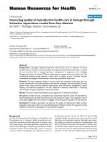
Figure 2 Pathological areas of the heart. The percentage of pathological areas in different
groups was shown in (a). The coxsackievirus B3 (CVB3) titration detected by standard plaque
formation assay was indicated in (b). The levels of interferon (IFN)-γ and tumor necrosis factor
(TNF)-α mRNA were determined in (c) and (d) respectively. The concentration of, IFN-γ and
TNF-α and were shown in (e, f and g) respectively. The detection of IFN-induced Mx1 mRNA
was determined in (h). **versus B group, p
> 0.05; *versus BIFN group, p < 0.01
Enhanced levels of IFN-γ and TNF-α
We evaluated the T helper (Th) cell patterns induced by oral-administered IFN-transformed B.
longum by measurement of the levels of two typical Th1 cytokines (IFN-γ and TNF-α). Our data
showed that the cardiac IFN-γ and TNF-α mRNA levels in the BIFN group were enhanced
significantly compared with that in the saline and B groups but were markedly reduced compared
with that in the IFN-α group (p < 0.01; Figure 2c, d). Furthermore, we detected the levels of IFN-
γ, TNF-α and IL-10 in the supernatant from the cultured MNCs from murine spleen. The levels
of supernatant IFN-γ and TNF-α in the BIFN group were markedly raised compared with that in
the saline and B groups (p < 0.01). Moreover, the levels of serum IL-10 in BIFN group were also
markedly decreased compared with that in the saline and B groups (p < 0.01; Figure 2g).
Increased expression of Mx1 mRNA in cardiac tissues
The Mx1 gene is induced typically by IFN. Its intracellular gene transcription level in a tissue
samples can represent the relative amount of local type I IFN that stimulates the cells or tissues
[23,24]. We measured the Mx1 gene transcription levels in cardiac tissues using real-time PCR to
evaluate the local type I IFN concentration and activity. High Mx1 mRNA transcript levels were
detected in the BIFN group compared with the saline and B groups respectively (p < 0.01; Figure
2h), a finding that was suggestive of a possibly high type I IFN concentration in this organ.
Discussion
In this experiment, we proved the efficacy of the BIFN-transformed B. longum cells on the
CVB3-induced myocarditis. Oral administration of IFN-transformed B. longum cells can reduce
significantly the cardiac inflammatory area of CVB3-infected mice by day 14 compared with the
B and saline groups respectively, which suggested that BIFN-transformed B. longum cells can
improve the severity of disease. It has been demonstrated that the dominant pathogenic process
in the early stages of CVB3 infection is the direct attack on myocardial cells by the virus,
therefore antivirus treatment at this phase is very important to improve the development of virus
infection [25]. Our data indicated that the cardiac virus titers in the murine heart of BIFN group
were decreased significantly compared with B group, which indicated that this recombinant B.
longum may improve cardiac inflammation partly by inhibition of virus replication at the early
stage of CVB3 infection. Classical theories suggest that CD4
+
Th1 cells play a vital role against
virus infection in adaptive immune responses by production of IFN-γ for effective clearance of
virus invasion. In this study, IFN-transformed B. longum increased the expression of Th1
cytokines (IFN-γ and TNF-α) mRNA in cardiac tissue and enhanced the secretion of Th1
cytokines (IFN-γ and TNF-α) from splenocytes, which suggested that this recombinant B.
longum is able to induce expression of CD4
+
Th1 cells against virus infection.
Our former studies have shown that hIFN-α2b from IFN-transformed B. longum is expressed
mainly as a mature secretory cytokine and that serum hIFN-α2b level can be enhanced in the
mice that have been administrated orally with B. longum [12,13]. As we know, the expression of
hIFN-α2b in IFN-transformed B. longum is mainly induced by L-arabinose, which is a
component of biopolymers such as hemicellulose and pectin [26]. The administration of IFN-
transformed B. longum has been demonstrated to increase the serum and intestinal IFN-α2b level
and we hypothesize that IFN expression by this bifidobacteria in mice might be induced
persistently by L-arabinose in MRS or by the administered food and then enter the blood
circulation by gastrointestinal absorption [13]. In this study, we compared mice either treated
with saline and control B. longum mice respectively. The Mx1 mRNA levels, which represent
the local tissue IFN concentration, were increased significantly in cardiac tissues in the BIFN
group, which suggested that IFN-transformed B. longum can increase the level of active type 1
IFN locally. Further study is needed to ascertain how to control the expression of IFN stably in
gut and whether these bacteria affect the microbial flora.
Bifidobacterium has many beneficial effects on human health that include prevention of
infection, immunomodulation, promotion of lactose digestion and protection against colon
cancer [9-11]. Recently, genetically engineered Bifidobacterium has been used successfully as an
exogenous gene delivery carrier for bowel disease and cancer gene therapy [9-11,27]. This
finding suggests that Bifidobacterium may be a suitable carrier for human gene expression and
secretion in the intestinal tract for the treatment of gastrointestinal diseases. Here, we
demonstrated the efficacy of BIFN-transformed B. longum to CVB3-induced myocarditis in the
mice. Our data showed, compared with IFN-transformed B. longum, that IFN-α2b administered
intramuscularly could reduce significantly virus infection, decrease the severity of virus-induced
myocarditis, and induce a robust Th1 pattern in the spleen and heart. Nevertheless, IFN-
transformed B. longum has its own advantages that include localization in the gastrointestinal
cavity and spread of the physiological role locally [13]. Further experimentation is needed to
evaluate whether IFN-transformed B. longum can be added to probiotic yogurt or diet and
whether it can protect high-risk people who eat these products from the virus myocarditis. The
model in this study was to inoculate with CVB3 intraperitoneally and this route may affect the
therapeutic efficacy of IFN-transformed B. longum compared with IFN-α2b given
intramuscularly. The preventive or therapeutic roles of IFN-transformed B. longum in virus
diseases need to be studied further.
In conclusion, oral administration of IFN-transformed B. longum can decrease the severity of
virus-induced myocarditis, reduce the virus titers in the heart and induce a Th1 pattern in the
spleen and heart in vivo. IFN-transformed B. longum may play a potential role in the treatment of
coxsackie virus B3-induced myocarditis. However, the advantages of the IFN-transformed B.
longum in the treatment and prevention of enterovirus infection need to be studied further.
Competing interests
The authors declare that they have no competing interests.
Authors’ contributions
QD and WZ conceived the study and QD wrote the paper. ZY, ZH, CS, YH, FZ, JY, LD and WZ
participated in the laboratory studies. All authors read and approved the final manuscript.
Acknowledgments
This work was supported by two grants from Shenzhen scientific Research Program of the
People’s Republic of China (NO. 200801020 and NO. 201001023) and Science and Technology
Planning Project of Guangdong Province, China (No. 2010B011000005).
References
1. Katabira ET, Sewankambo NK, Mugerwa RD, Belsey EM, Mubiru FX, Othieno C, Kataaha
P, Karam M, Youle M, Perriens JH, Lange JM: Lack of efficacy of low dose oral interferon
alfa in symptomatic HIV-1 infection: a randomised, double blind, placebo controlled trial.
Sex Transm Infect 1998, 74(4):265–270.
2. Cummins J, Beilharz M, Krakowka S: Oral use of interferon. J Interferon Cytokine Res
1999, 19:853–857.
3. Hutchinson VA, Mok WL, Angenend JL, Cummins JM, Richards AB: Chronic major
aphthous stomatitis: oral treatment with low-dose alpha-interferon. Mol Biother 1990,
2(4):217–220.
4. Lecciones JA, Abejar NJ, Dimaano EE, Bartolome R, Cinco S, Mariano N, Yerro ME, Cobar
S, Fuggan B: A pilot double-blind, randomized, and placebo-controlled study of orally
administered IFN-α -n1 (Ins) in pediatric patients with measles. J Interferon Cytokine Res
1998, 18:647–652.
5. Kim Y, Thapa M, Hua DH, Chang KO: Biodegradable nanogels for oral delivery of
interferon for norovirus infection. Antiviral Res 2011, 89(2):165–173.
6. Dec M, Puchalski A: Use of oromucosally administered interferon-alpha in the
prevention and treatment of animal diseases. Pol J Vet Sci 2008, 11:175–186.
7. Hu B, Kou L, Li C, Zhu LP, Fan YR, Wu ZW, Wang JJ, Xu GX: Bifidobacterium longum as
a delivery system of TRAIL and endostatin cooperates with chemotherapeutic drugs to
inhibit hypoxic tumor growth. Cancer Gene Ther 2009, 16:655–663.
8. Reyes Escogido ML, De León Rodríguez A, Barba de la Rosa AP: A novel binary
expression vector for production of human IL-10 in Escherichia coli and Bifidobacterium
longum. Biotechnol Lett 2007, 29:1249–1253.
9. Shkoporov AN, Efimov BA, Khokhlova EV, Kafarskaia LI, Smeianov VV: Production of
human basic fibroblast growth factor (FGF-2) in Bifidobacterium breve using a series of
novel expression/secretion vectors. Biotechnol Lett 2008, 30:1983–1988.
10. Tang W, He Y, Zhou S, Ma Y, Liu G: A novel Bifidobacterium infantis-mediated
TK/GCV suicide gene therapy system exhibits antitumor activity in a rat model of bladder
cancer. J Exp Clin Cancer Res 2009, 28:155.
11. Yazawa K, Fujimori M, Nakamura T, Sasaki T, Amano J, Kano Y, Taniguchi S:
Bifidobacterium longum as a delivery system for gene therapy of chemically induced rat
mammary tumors. Breast Cancer Res Treat 2001, 66:165–170.
12. Deng Q, Zeng W, Yu Z: Signal peptide of Arabinosidase enhances secretion of
interferon-alpha2b protein by Bifidobacterium longum. Arch Microbiol 2009, 191:681–686.
13. Yu Z, Zeng Z, Huang Z, Lian J, Yang J, Deng Q, Zeng W: Increased mRNA expression of
interferon-induced Mx1 and immunomodulation following oral administration of IFN-α2b-
transformed B. longum to mice. Arch Microbiol 2010, 192:633–638.
14. Heim A, Grumbach I, Pring-Akerblom P, Stille-Siegener M, Muller G, Kandolf R, Figulla
HR: Inhibition of coxsackievirus B3 carrier state infection of cultured human myocardial
fibroblasts by ribavirin and human natural interferon-alpha. Antivir Res 1997, 34:101–111.
15. Kandolf, R., A. Canu, and P. H. Hofschneider: Coxsackie B3 virus can replicate in
cultured human fetal heart cells and is inhibited by interferon. J Mol Cell Cardiol 1985,
17:167–181.
16. Okada, I., A. Matsumori, Y. Matoba, M. Tominaga, T. Yamada, and C. Kawai:
Combination treatment with ribavirin and interferon for coxsackievirus B3 replication. J
Lab Clin Med 1992, 120:569–573.
17. Padalko E, Nuyens D, De Palma A, Verbeken E, Aerts JL, De Clercq E, Carmeliet P, Neyts
J: The interferon inducer ampligen [poly(I)-poly(C12U)] markedly protects mice against
coxsackie B3 virus-induced myocarditis. Antimicrob Agents Chemother 2004, 48(1):267–274.
18. Deonarain R, Cerullo D, Fuse K, Liu PP, Fish EN: Protective role for interferon-beta in
coxsackievirus B3 infection. Circulation 2004, 110:3540–3543.
19. Wang YX, da Cunha V, Vincelette J, White K, Velichko S, Xu YF, Gross C, Fitch RM,
Halks-Miller M, Larsen BR: Antiviral and myocyte protective effects of murine interferon-
beta and -alpha2 in coxsackievirus B3-induced myocarditis and epicarditis in BALB/c
mice. Am J Physiol Heart Circ Physiol 2007, 293:H69–H76.
20. Wang YF, Wang XY, Ren Z, Qian CW, Li YC, Kaio K, Wang QD, Zhang Y, Zheng LY,
Jiang JH, Yang CR, Liu Q, Zhang YJ: Phyllaemblicin B inhibits coxsackie virus B3 induced
apoptosis and myocarditis. Antiviral Res 2009, 84(2):150–158.
21. Yuan J, Yu M, Lin QW, Cao AL, Yu X, Dong JH, Wang JP, Zhang JH, Wang M, Guo HP,
Cheng X, Liao YH: Th17 cells contribute to viral replication in coxsackievirus B3-induced
acute viral myocarditis. J Immunol 2010, 185(7):4004–4010.
22. Alignani D, Maletto B, Liscovsky M, Rópolo A, Morón G, Pistoresi-Palencia MC. Orally
administered OVA/CpG-ODN induces specific mucosal and systemic immune response in
young and aged mice. J Leukocyte Biol 2005, 77:898–905.
23. Bollati-Fogolín M, Müller W: Virus free, cell-based assay for the quantification of
murine type I interferons. J Immunol Methods 2005, 306:169–175.
24. Petry H, Cashion L, Szymanski P, Ast O, Orme A, Gross C, Bauzon M, Brooks A, Schaefer
C, Gibson H, Qian H, Rubanyi GM, Harkins RN: Mx1 and IP-10: biomarkers to measure
IFNbeta activity in mice following gene-based delivery. J Interferon Cytokine Res 2006,
26:699–705.
25. Dennert R, Crijns HJ, Heymans S: Acute viral myocarditis. Eur Heart J 2008, 29:2073–
2082.
26. David RL: CRC Handbook of Chemistry and Physics. 88th edition. Boca Raton: CRC Press;
2007–2008:110 p.
27. Yao J, Wang JY, Lai MG, Li YX, Zhu HM, Shi RY, Mo J, Xun AY, Jia CH, Feng JL, Wang
LS, Zeng WS, Liu L: Treatment of mice with dextran sulfate sodium-induced colitis with
human interleukin 10 secreted by transformed Bifidobacterium longum. Mol Pharm 2011,
8(2):488–497.
A
B
D E
C
Figure 1
Saline B BIFN IFN
0.0
0.1
0.2
0.3
0.4
0.5
**
*
*
*
A
Relative inflammatory area
Saline
B
BIFN
IFN
0
1
2
3
4
5
6
**
*
*
*
B
CVB3 titers
Saline
B
BIFN
IFN
0.0
0.4
0.8
1.2
1.6
2.0
**
*
*
*
C
relative IFN-¦ÃmRNA result
Saline
B
BIFN
IFN
0.0
0.5
1.0
1.5
2.0
2.5
3.0
3.5
4.0
4.5
**
*
*
*
D
Relative TNF-¦ÁmRNA result
Saline
B
BIFN
IFN
0
250
500
750
1000
1250
1500
1750
*
*
*
**
E
IFN-¦Ã(pg/ml)
Saline
B
BIFN
IFN
0
100
200
300
400
**
*
*
*
F
TNF-¦Á(pg/ml)
Saline
B
BIFN
IFN
0
40
80
120
160
200
240
**
*
*
*
G
IL-10(pg/ml)
Saline
B
BIFN
IFN
0.0
0.5
1.0
1.5
2.0
2.5
**
*
*
*
H
Relative Mx1 mRNA result
Figure 2