Báo cáo sinh học: " Downregulation of APOBEC3G by Xenotropic Murine Leukemia-Virus Related Virus (XMRV) in Prostate Cancer Cells docx
Bạn đang xem bản rút gọn của tài liệu. Xem và tải ngay bản đầy đủ của tài liệu tại đây (440 KB, 12 trang )
Virology Journal
This Provisional PDF corresponds to the article as it appeared upon acceptance. Fully formatted
PDF and full text (HTML) versions will be made available soon.
Downregulation of APOBEC3G by Xenotropic Murine Leukemia-Virus Related
Virus (XMRV) in Prostate Cancer Cells
Virology Journal 2011, 8:531
doi:10.1186/1743-422X-8-531
Abhinav Dey ()
Chinmay K Mantri ()
Jui Pandhare-Dash ()
Bindong Liu ()
Siddharth Pratap ()
Chandravanu Dash ()
ISSN
Article type
1743-422X
Short report
Submission date
26 September 2011
Acceptance date
12 December 2011
Publication date
12 December 2011
Article URL
/>
This peer-reviewed article was published immediately upon acceptance. It can be downloaded,
printed and distributed freely for any purposes (see copyright notice below).
Articles in Virology Journal are listed in PubMed and archived at PubMed Central.
For information about publishing your research in Virology Journal or any BioMed Central journal, go
to
/>For information about other BioMed Central publications go to
/>
© 2011 Dey et al. ; licensee BioMed Central Ltd.
This is an open access article distributed under the terms of the Creative Commons Attribution License ( />which permits unrestricted use, distribution, and reproduction in any medium, provided the original work is properly cited.
Downregulation of APOBEC3G by Xenotropic
Murine Leukemia-Virus Related Virus (XMRV)
in Prostate Cancer Cells
ArticleCategory
: Short Report
ArticleHistory
: Received: 26-Sep-2011; Accepted: 22-Nov-2011
© 2011 Dey et al; licensee BioMed Central Ltd. This is an Open Access
article distributed under the terms of the Creative Commons Attribution
ArticleCopyright : License ( which permits
unrestricted use, distribution, and reproduction in any medium, provided
the original work is properly cited.
Abhinav Dey,Aff1†
Chinmay Kumar Mantri,Aff1†
Jui Pandhare-Dash,Aff1†
Bindong Liu,Aff1
Siddharth Pratap,Aff2
Chandravanu Dash,Aff1
Corresponding Affiliation: Aff1
Phone: +1-615-3276996
Fax: +1-615-3276929
Email:
Aff1
From The Laboratory of Retrovirology and Epigenetics, Center For
AIDS Health Disparities Research, Vanderbilt-Meharry Center For
AIDS Research (CFAR), Department of Biochemistry and Cancer
Biology, 1050 Dr. DB Todd Jr. Blvd, Old Hospital Building, Room
5027, Nashville TN 37208, TN, USA
Aff2
Mass Spectrometry Core, Meharry Medical College School of
Medicine, Nashville, TN, USA
†
These authors have contributed equally
Emails:
Abhinav Dey:
Chinmay Kumar Mantri:
Jui Pandhare-Dash:
Bindong Liu:
Siddharth Pratap:
Abstract
Background
Xenotropic murine leukemia virus (MLV)-related virus (XMRV) is a gammaretrovirus that was
discovered in prostate cancer tissues. Recently, it has been proposed that XMRV is a laboratory
contaminant and may have originated via a rare recombination event. Host restriction factor
APOBEC3G (A3G) has been reported to severely restrict XMRV replication in human
peripheral blood mononuclear cells. Interestingly, XMRV infects and replicates efficiently in
prostate cancer cells of epithelial origin. It has been proposed that due to lack off or very low
levels of A3G protein XMRV is able to productively replicate in these cells.
Findings
This report builds on and challenges the published data on the absence of A3G protein in prostate
epithelial cells lines. We demonstrate the presence of A3G in prostate epithelial cell lines
(LNCaP and DU145) by western blot and mass spectrometry. We believe the discrepancy in
A3G detection is may be due to selection and sensitivity of A3G antibodies employed in the
prior studies. Our results also indicate that XMRV produced from A3G expressing LNCaP cells
can infect and replicate in target cells. Most importantly our data reveal downregulation of A3G
in XMRV infected LNCaP and DU145 cells.
Conclusions
We propose that XMRV replicates efficiently in prostate epithelial cells by downregulating A3G
expression. Given that XMRV lacks accessory proteins such as HIV-1 Vif that are known to
counteract A3G function in human cells, our data suggest a novel mechanism by which
retroviruses can counteract the antiviral effects of A3G proteins.
Keywords
XMRV, APOBEC3G, Retrovirus, Prostate
Findings
Xenotropic murine leukemia-virus related virus (XMRV) is a member of the gammaretrovirus
family that was first detected in human prostate tumors [1]. Although initial studies supported
the presence of XMRV in prostate cancer tissues [2-4], since then several laboratories have
failed to detect the virus in cohorts of prostate cancer patients [5-9]. Very recently, Paprotka et
al. (2011) have challenged an association of XMRV with human diseases [10]. These authors
have reported that XMRV may have originated by a rare recombination event during tumor
passaging in mice. Therefore, it has been proposed that XMRV is a laboratory contaminant and
not a human pathogen. Nevertheless, being a newly discovered gammaretrovirus and having the
ability to infect human cells, XMRV may serve as a model gammaretrovirus to further our
understanding of retroviral biology.
Apolipoprotein B mRNA-editing enzyme catalytic polypeptide-like 3 (APOBEC3) proteins,
APOBEC3A to APOBEC3G are a class of cytidine deaminases that has been reported to restrict
retroviral replication in humans [11]. In case of HIV-1, the restriction by APOBEC3G (A3G)
and APOBEC3F (A3F) are counteracted by Vif that degrades A3 proteins via proteosomal
degradation [12]. It has been reported that XMRV replication can be inhibited by A3 proteins
such as A3G, A3B, A3F, and murine APOBEC3 (mA3) [13-17]. Although hypermutation of
XMRV genome in A3G/A3F-expressing peripheral blood mononuclear cells (PBMCs) severely
restrict XMRV replication in human blood, infected PBMCs have been suggested to serve as
source of infectious XMRV [17]. Given that XMRV is a simple retrovirus and does not encode
accessory proteins that are known to counteract A3G/A3F, these observations suggest XMRV
cannot survive the restriction of innate immunity for productive infection in humans.
XMRV replicates efficiently in prostate epithelial cell lines specifically in LNCaP cells [3,18]. In
addition, the prostate cancer cell line 22Rv1 has been shown to be chronically infected with
XMRV and produces highly infectious virus [19]. Since host restriction factor A3G is able to
restrict XMRV, the question is how XMRV replicates efficiently in these human prostate cell
lines. There are at least three studies that have suggested that XMRV efficiently replicates in
prostate epithelial cancer cell lines since these cells lack or express undetectable levels of A3G
[13-16]. In this report, we demonstrate that prostate epithelial cell lines LNCaP and DU-145
express detectable levels of A3G by western blot analysis. We confirm the presence of A3G in
LNCaP cells by mass spectrometry. We believe the results described in earlier reports on the
absence of A3G in these cells may be due to the sensitivity of antibody used in their western blot
analysis.
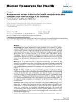
We detected A3G in LNCaP and DU145 cells (Figure 1A) using a polyclonal antibody (antiApoC29) raised against the 29 amino acid (aa) of the C-terminal end of A3G protein (NIH AIDS
Reagent Program Catalog # 10201). We used lysates of CD4+ T cells and CEM cells as positive
controls and CEM-SS cells as negative control for A3G expression by western analysis (Figure
1A). These results contradict the previous reports that were unable to detect A3G in these cells
[13-16]. We realized that the antibody used in the previous reports was a polyclonal antibody
(anti-ApoC17) raised against the 17aa of the C-terminal of A3G protein (NIH AIDS Reagent
Program Catalog # 9968). Therefore, we repeated our western analysis using anti-ApoC17 and
similar to the earlier reports we could not detect A3G in LNCaP and DU-145 cells (Figure 1B).
Therefore, we examined whether the band detected in Figure 1A using anti-ApoC29 contains
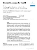
A3G protein. To confirm this, we carried out Electrospray Mass Spectrometry (ESI) analysis by
cutting out A3G band from the coommassie stained gels. After a comparative analysis of the
spectra with purified A3G, we could map more than 85% of A3G peptides in the A3G band
detected in the lysates of the LNCaP cells (Figure 2). Since the anti-ApoC29 used in our study
has been used by several laboratories to detect A3G, the reason previous reports were unable to
detect A3G in LNCaP cells may be due to the sensitivity of the antibody used in their western
analysis. In addition, it is possible that the epitopes of endogenous A3G in prostate epithelial
cells are not recognized by the anti-ApoC17. It is important to point out that scanning for m/z
peptides from our mass spectrometry data revealed presence of peptides mapped to A3B, A3D
and A3F. This is not surprising given that A3B, A3D and A3F have molecular weights similar to
A3G. However, the molecular weights of A3A, A3C and A3H (~23 kDa) are substantially lower
than A3G and detection of A3A/A3C/A3H in the A3G band are highly unlikely.
Figure 1 Detection of A3G in prostate cancer cells of epithelial origin A3G was detected using
two different polyclonal rabbit sera obtained from the NIH AIDS Reagent Program. AntiApoC17 was raised against a synthetic peptide comprising of the 17 C-terminal residues of A3G
(Cat. No. 10082), while the other was (Anti-ApoC29) Anti-A3G C-terminal antisera raised
against a C-terminal peptide representing the last 29 amino acids of human A3G coupled to a
hapten (Cat. No 10201). As a loading control β-actin (Sigma Co., USA) was used. For
immunoblot analysis, cell lysates (5 µg) were subjected to SDS-polyacrylamide gel
electrophoresis and western blot analysis with the appropriate antibodies. Western blot analysis
using, (A) Anti-ApoC29 and (B) Anti-ApoC17.
Figure 2 Proteomic Analysis for detection of A3G in LNCaP cells The cell lysate from LNCaP
cells was loaded onto SDS-polyacrylamide gel (4–12% gradient gel) and resolved through
electrophoresis as described in Figure 1. Pure recombinant A3G was also run on a separate gel.
Both the gels were stained with Coomassie brilliant blue. One section of the gel was subjected to
immunoblotting using anti-ApoC29 to assist in A3G identification in the gel for mass
spectrometry. The gel bands corresponding to A3G protein were excised and were subjected to
in-gel trypsin digestion based on the manufacturer’s protocol (Thermo Scientific). The resulting
peptides were analyzed using a Thermo Finnigan LTQ ion trap instrument ESI. Peptides were
separated on a packed capillary tip (Polymicro Technologies, 100 àm ì 11 cm) with Jupiter C18
resin (5 µm, 300 Å, Phenomenex) using an in-line solid-phase extraction column (100 àm ì 6
cm) packed with the same C18 resin. The total ion chromatogram for the digested pure A3G and
A3G from LNCaP cells were compared. The common regions of similar retention time (≈2
minutes) were analyzed to search for m/z peaks corresponding to A3G in the LNCaP sample.
The representative peaks were matched with the m/z peaks corresponding to pure A3G. This
search revealed that more than 85% of the sequence could be identified in the spectra in the A3G
band detected in LNCaP cells. (A) Spectra of purified A3G (m/z 800 to 1100); (B) Mass spectra
of A3G from LNCaP cell lysate (m/z 800 to 1100); (C) Spectra of purified A3G (m/z 1100 to
1500); and (D) Spectra of A3G from LNCaP cell lysate (m/z 1100 to 1500). The detected
peptides from A3G sequence have been shown in red. The X-axis represents m/z and the Y-axis
represents intensity.
Given that XMRV replicates efficiently in LNCaP and DU-145 cells [3], we examined the effect
of XMRV infection on the A3G levels in these cells. We used culture supernatants from
chronically infected LNCaP cells with XMRV as the source of virus. We infected LNCaP and
DU-145 cells and confirmed infection by detecting XMRV p30 in these cells. We used goat
polyclonal anti-Rauscher MLV p30 Gag (a gift from Dr. Sandra Ruscetti, NCI-Frederick) that
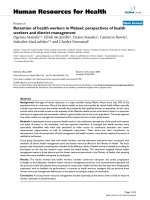
reacts with XMRV p30 and its precursor Gag in our experiment (Figure 3A). Subsequently, we
analyzed A3G expression in these cells by western blot analysis. Intriguingly, XMRV infection
resulted in a substantial downregulation of A3G expression in both of these cell types (Figure
3B). Our densitometry analysis revealed that A3G was downregulated almost 60% in infected
LNCaP cells whereas the downregulation was ~ 40% in infected DU145 cells (Figure 3C). It is
important to point out that the virus used to infect these cells were produced from the LNCaP
cells that express A3G. It has been shown that A3G can be efficiently packed into XMRV
virions [25]. Paportka et al. have reported that XMRV produced from PBMCs with high levels of
A3G proteins are infection competent and can infect new cells [17]. Since we detected robust
XMRV p30 bands in our infection experiments (Figure 3B), our data suggest XMRV produced
from A3G containing cells have the ability to productively infect and replicate in target cells.
Therefore our findings challenge the notion that the prostate epithelial cell lines were able to
produce infectious XMRV because these cells do not express A3G. Given that our results
demonstrate downregulation of A3G in XMRV infected prostate cancer cells, we believe XMRV
has the ability to counteract A3G antiviral function in prostate cancer cells. These results are
highly significant given that XMRV is a simple retrovirus and does not encode accessory
proteins that are known to degrade A3G.
Figure 3 XMRV induced downregulation of A3G in prostate cancer cells We used culture
supernatant from chronically infected LNCaP cells with XMRV as the source of infectious
XMRV. Virus infections were performed using cells plated 1 day before infection. Cells were at
50% confluency at the time of infection. On the day of infection, fresh media containing 5 µg/ml
polybrene was added to the cells and virus was layered on the cells and incubated for 6 h to
allow virus adsorption. Cells were then washed once with PBS, and fresh media containing FBS
were added. After 1–2 weeks, XMRV infection was confirmed by detecting XMRV p30 protein
by using goat polyclonal anti-Rauscher MLV p30 Gag in uninfected (−) and XMRV infected (+)
LNCaP cells (A) and DU-145 cells (B). (C) A3G expression was determined by the protocol
described in Figure 1 using anti-ApoC29. (D) Densitometry of A3G expression as determined by
three independent experiments.
In the absence of Vif-like accessory proteins, retroviruses such as Human T cell lymphoma virus
(HTLV) and Murine leukemia virus (MLV) have developed alternative mechanisms to evade
host restriction by A3 proteins. A motif of HTLV nucleocapsid (NC) prevents packaging of A3G
into the virion [20]. Therefore exclusion of A3G has been proposed to be a common mechanism
for Vif-deficient retroviruses to counteract A3G restriction. MLV virions have also been reported
to exclude mA3 [21]. XMRV has been demonstrated to package A3G [16], therefore a role for
exclusion mechanism is unlikely. XMRV produced from LNCaP cells show signatures of
hypermutation that are characteristics of A3F [15,17]. Therefore, it is plausible XMRV is
somehow resistant to A3G restriction in these cell types. XMRV may achieve this either by
downregulating A3G levels or by evading A3G restriction. Given that MLV has been reported to
inactivate mA3 by viral protease [22], a similar mechanism for XMRV cannot be ruled out.
Furthermore, certain polymorphic alleles of A3G have been reported to increase the
susceptibility to HIV infection [23]. Therefore, we are investigating whether A3G produced from
prostate epithelial cells have mutations that can be assigned for cell specific susceptibility to
XMRV infection. The other mechanism that could possibly explain our results is that A3G
remains as high-molecular-mass (HMM) ribonucleoprotein complex in prostate epithelial cells.
It has been reported that A3G in resting CD4+ cells and monocytes are predominantly in its lowmolecular-mass (LMM) active form making these cells refractory to HIV-1 infection [24,25].
Conversion of LMM to the inactive HMM complex, when the CD4+ cells are activated or
monocytes are differentiated into macrophages, makes these cells prone to HIV infection. Since
HMM forms of A3G are reported to be enzymatically inactive, if A3G remains in HMM
complex in prostate epithelial cells, it may not be able to restrict XMRV replication.
In summary, this report demonstrates the presence of A3G in prostate epithelial cell lines
(LNCaP and DU145) that support efficient XMRV replication. Since XMRV packages A3G in
its virions and lacks Vif-like accessory proteins, our findings on XMRV-induced downregulation
of A3G may represent a new pathway by which retroviruses counteract antiviral effects of A3
proteins in human cells. Our data warrants further studies to decipher the mechanism by which
XMRV may counteract restriction by A3 proteins.
Competing interests
The authors declare that they have no competing interests.
Authors’ contributions
JP, CM, AD, BL, and CD conceived the ideas, and designed the experiments. AD, CM and JP
carried out all the experiments and participated in the discussion of the data. AD and SP carried
our Mass spec analysis. CD coordinated the study, and wrote the manuscript. All authors read
and approved the final manuscript.
Acknowledgements
We thank Dr. Sandra Ruscetti (NCI-Frederick) for the antibodies. We thank Dr. James Hildreth
(UC-Davis) for technical guidance. This work is supported in part by grants to CD
(R00DA024558, R03DA30896) from NIH, (UL1 RR024975) from the Vanderbilt CTSA Grant
and (U54RR026140) Meharry Translational Research Center (MeTRC).
References
1. Urisman A, Molinaro RJ, Fischer N, Plummer SJ, Casey G, Klein EA, Malathi K, MagiGalluzziC,Tubbs RR, Ganem D, Silverman RH, DeRisi JL: Identification of a novel
gammaretrovirus in prostate tumors of patients homozygous for R462Q RNASEL variant.
PLoSPathog 2006, 2:e25.
2. Arnold RS, Makarova NV, Osunkoya AO, Suppiah S, Scott TA, Johnson NA, Bhosle SM,
Liotta D, Hunter E, Marshall FF et al.: XMRV infection in patients with prostatecancer:
novel serologic assay and correlation with PCR andFISH. Urology 2010, 75:755–761.
3. Dong B, Kim S, Hong S, Das Gupta J, Malathi K, Klein EA, Ganem D, DeRisi JL, Chow SA,
Silverman RH: An infectious retrovirus susceptible to an IFN antiviral pathway from
human prostate tumors. ProcNatlAcadSci USA 2007, 104:1655–60.
4. Schlaberg R, Choe DJ, Brown KR, Thaker HM, Singh IR: XMRV ispresent in malignant
prostatic epithelium and is associated with prostate cancer,especially high-grade tumors.
ProcNatlAcadSci U S A 106:16351–16356.
5. Sabunciyan S, Mandelberg N, Rabkin CS, Yolken R,Viscidi R: No difference in antibody
titers against xenotropicMLV related virus in prostate cancer cases and cancerfreecontrols. Mol. Cell. Probes 2010,25:134–136.
6. Fischer N, Hellwinkel O, Schulz C, Chun FK, Huland H, Aepfelbacher M, Schlomm T:
Prevalence of human gammaretrovirus XMRV in sporadic prostate cancer. J ClinVirol
2008, 43:277–83.
7. Hohn O, Krause H, Barbarotto P, Niederstadt L, Beimforde N, DennerJ, Miller K, Kurth R,
Bannert N: Lack of evidence for xenotropic murine leukemia virus-related virus (XMRV) in
German prostate cancer patients. Retrovirology 2009,6:92.
8. Verhaegh GW, de Jong AS, Smit FP et al.: Prevalence of human xenotropic murine
leukemia virus-related gammaretrovirus (XMRV) in Dutch prostate cancer patients.
Prostate 2010, 71:415–20.
9. Aloia AL, Sfanos KS, Isaacs WB et al.: XMRV: a new virus in prostate cancer? Cancer
Res 2010, 70:10028–33.
10. Paprotka T, Delviks-Frankenberry KA, Cingöz O, Martinez A, Kung H-J, Tepper CG, Hu
W-S, Fivash Jr. MJ, Coffin MJ, Pathak VK: Recombinant Origin of the Retrovirus XMRV
Science 2011, Paper in press. 31 May 2011/Page 1/10.1126/science.1205292
11. Sheehy, A. M., N. C. Gaddis, J. D. Choi, and M. H. Malim.: Isolation of a human gene
that inhibits HIV-1 infection and is suppressed by the viral Vif protein. Nature
2002,418:646–650.
12. Gabuzda, D. H., et al.: Role of vif in replication of human immunodeficiencyvirus type 1
in CD4 T lymphocytes. J Virol 1992, 66:6489–6495.
13. Bogerd, H. P., F. D. B. P. Zhang, and B. R. Cullen.: Human APOBEC3proteins can
inhibit xenotropic murine leukemia virus-related virus infectivity. Virology 2011, 410:234–
239.
14. Groom, H. C., M. W. Yap, R. P. Galao, S. J. Neil, and K. N. Bishop.: Susceptibility of
xenotropic murine leukemia virus-related virus (XMRV) to retroviral restriction factors.
Proc. Natl. Acad. Sci. U. S. A. 2010, 107:5166–5171.
15. Paprotka, T., et al.: Inhibition of xenotropic murine leukemia virusrelated virus by
APOBEC3 proteins and antiviral drugs. J Virol 2010, 84:5719–5729.
16. Stieler, K., and N. Fischer.: Apobec 3 G efficiently reduces infectivity of the human
exogenousgammaretrovirus XMRV. PLoS One 2010, 5:e11738.
17. Paprotka, T., et al.: Inhibition of xenotropic murine leukemia virusrelatedvirus by
APOBEC3 proteins in PBMCS. J Virol 2011, 85:4888–4897.
18. Rodriguez JJ, Goff SP: Xenotropic murine leukemia virus-related virus establishes an
efficient spreading infection and exhibits enhanced transcriptional activity in prostate
carcinoma cells. J Virol 2010 Mar, 84(5):2556–62.
19. Knouf EC et al.: Multiple integrated copies and high-level production of the human
retrovirus XMRV from 22Rv1 prostate carcinoma cells. J Virol 2009, 83:7353–7356.
20. Derse D, Hill SA, Princler G, Lloyd P, Heidecker G.: Resistance of human T cell leukemia
virus type 1 to APOBEC3G restriction is mediated by elements in nucleocapsid. Proc Natl
Acad Sci U S A. 2007, 104:2915–2920.
21. Doehle BP, Schafer A, Wiegand HL, Bogerd HP, Cullen BR.: Differential sensitivity of
murine leukemia virus to APOBEC3-mediated inhibition is governed by virion exclusion. J
Virol 2005, 79:8201–8207.
22. Abudu A, Takaori-Kondo A, Izumi T, Shirakawa K, Kobayashi M, et al.: Murine
retrovirus escapes from murine APOBEC3 via two distinct novel mechanisms. Curr Biol
2006, 16:1565–1570.
23. An P, Bleiber G, Duggal P, Nelson G, May M, Mangeat B, Alobwede I, Trono D, Vlahov D
Donfield S, Goedert JJ, Phair J, Buchbinder S,O’Brien SJ, Telenti A and Winkler CA:
APOBEC3G Genetic Variants and Their Influence on the Progression to AIDS.J Virol
2004, 78:11070–11076.
24. Chiu Y, Soros VB, Kreisberg J, Stopak K, Yonemoto Wes, Greene WC.: Cellular
APOBEC3G restricts HIV-1 infection in resting CD4+ T cells. 2005. Nature, 435:108–114.
25. Triques K and Stevenson M.: Characterization of restrictions to human
immunodeficiency virus type 1 infection of monocytes. J Virol 2004, 78:5523–5527.
Figure 1
Figure 2
"
Figure 3