Báo cáo sinh học: "Time-course of sFlt-1 and VEGF-A release in neutropenic patients with sepsis and septic shock: a prospective study" pptx
Bạn đang xem bản rút gọn của tài liệu. Xem và tải ngay bản đầy đủ của tài liệu tại đây (641.75 KB, 8 trang )
RESEARCH Open Access
Time-course of sFlt-1 and VEGF-A release in
neutropenic patients with sepsis and septic
shock: a prospective study
Brunna E Alves
1
, Silmara AL Montalvao
1
, Francisco JP Aranha
1
, Irene Lorand-Metze
2
, Carmino A De Souza
2
,
Joyce M Annichino-Bizzacchi
2
, Erich V De Paula
1*
Abstract
Background: Septic shock is the most feared complication of chemotherapy-induced febrile neutropenia. So far,
there are no robust biomarkers that can stratify patients to the risk of sepsis complications. The VEGF-A axis is
involved in the control of microvascular permeability and has been involved in the pathogenesis of conditions
associated with endothelial barrier disruption such as sepsis. sFlt-1 is a soluble variant of the VEGF-A receptor
VEGFR-1 that acts as a decoy receptor down-regulating the effects of VEGF-A. In animal models of sepsis, sFlt-1
was capable to block the barrier-breaking negative effects of VEGF-A and to significantly decrease mortality. In
non-neutropenic patients, sFlt-1 has been shown to be a promising biomarker for sepsis severity.
Methods: We prospectively evaluated conce ntrations of sFlt-1 and VEGF-A at different time-points during febrile
neutropenia, and evaluated the association of these levels with sepsis severity and septic shock development.
Results: Neutropenic patients that evolved with septic shock (n = 10) presented higher levels of sFlt-1 and VEGF-A
measured 48 hours after fever onset than patients with non-complicated sepsis (n = 31) and levels of these
biomarkers correlated with sepsis severity scores. Estimation of the diagnostic accuracy of sFlt-1 levels for the
discrimination of patients that evolved to septic shock yielded promising results in our study population.
Discussion: Our data suggest that sFlt-1 and VEGF-A could be useful biomarkers for sepsis severity in patients with
febrile neutropenia. In addition, the kinetics of sFlt-1 release in patients that evolve to septic shock suggest that
the sFlt-1 could be a salvage compensatory mechanism in patients with septic shock, but that the magnitude of
the sFlt-1 release observed in human sepsis is not sufficient to reproduce the beneficial anti-VEGF-A effects
observed in animal models of sepsis.
Background
Patients with hematological malignancies submitted to
intensive chemotherapy present a higher risk of sepsis and
sepsis complications. Febrile neutropenia (FN) in these
patients is considered a medical emergency, and a standar-
dized management approach including wide-spectrum
antibiotics and admission is usually implemented for all
patients. So far, there are no reliable laboratory markers to
indicate whether FN patients will recover uneventfully or
rapidly deteriorate to sepsis, septic shock and death [1,2].
Vascular endothelial growth factor (VEGF-A) is an
endothelial growth factor that is widely known for its
key role in the regulation of embryonic and post-natal
angiogenesis. However, VEGF-A was first characterized
by its endothelial barrier-breaking properties, as a potent
stimulator of endothelial permeability [3]. This capability
to disrupt the integrity of an endothelial cell tube is in
fact very important during angiogenesis, as new cells
have to be incorporated in a growing vessel. Recently,
this property has been explored as a putative common
downstream mechanism in pathological conditions asso-
ciated with loss of endothelial barrier function. In line
with this hypot hesi s, several authors have demonstrated
elevated VEGF-A levels in intensive care units (ICU)
* Correspondence:
1
Hematology and Hemotherapy Center, University of Campinas, Campinas,
SP, Brazil
Full list of author information is available at the end of the article
Alves et al. Journal of Translational Medicine 2011, 9:23
/>© 2011 Alves et al; licensee BioMed Central Ltd. This is an Open Access article distributed under the terms of the Creative Commons
Attribution License (http://creative commons.org/licenses/by/2.0), which permits unrestricted use, distribution, and reproduction in
any medium, provided the original work is properly cited.
patients with sepsis, as well as associations betwe en
VEGF-A levels and sepsis severity [4-6].
sFlt-1 is a natural splice variant of the tyrosine-kinase
receptor Flt-1, which is an endothelial cell receptor for
VEGF-A. sFlt-1 binds free VEGF-A and acts as its
antagonist [7]. In animal models of sepsis, sFlt-1 has
been shown to attenuate the severity of the inflamma-
tory response and to antagonize the barrier-breaking
properties of VEGF-A, thus suggesting a therapeutic
role for this protein [8]. The potential value of sFlt-1 as
a biomarker for sepsis severity has been demonstrated
in studies with non-neutropenic patients [6,9].
Here we prospectively evaluated the serial expression
of sFlt-1 and VEGF-A in patients with hematological
malignancies and chemotherapy-related FN, to gain
insights about both potential rolesofsFlt-1inpatients
with febrile neutropenia and sepsis, as a biomarker or as
a therapeutic tool.
Methods
Patient’s eligibility criteria
Recruitment took place at the Bone Marrow Transpl an-
tation Unit of our University hospital between March
2008 and March 2009. Inclusion criteria were: (1) diag-
nosis o f hematological malignancies, and (2) admission
as inpatients for intensive chemothera py (induction for
acut e leukem ia or high-dose sequential therapy for lym-
phomas) or hematopoietic stem-cell transplantation
(HSCT). Patients were invited to participate before the
initiation of chemotherapy. The study was performed in
accordance with the Declaration of Helsinki and
approved by the local ethics committee and informed
written consent was obtained from all patients. Fever
(T≥38.0°C) at admission for chemotherapy was the only
exclusio n criteria, but only patients that presented fever
during neutropenia (defined as a neutrophil count
<500⁄μl) were included in the second phase (see labora-
tory measurements). Clinical data were obtained from
the medical records.
Sepsis definitions and risk stratification scores
Sepsis, in this population, was defined by the presence of
two or more of the following: (1) temperature > 38.0°C, (2)
heart rate > 90 beats/min, (3) respiratory rate > 20 breaths/
min o r PaCO
2
< 3 2 mmHg; and a microbi o logically proven
or clinically evident source of infection [10]. In accordance
with current management protocols, an infectious etiology
was assumed for all FN patients, and broad-spectrum anti-
biotics were initiated immediately after cultures were
obtained [11]. Septic shock was present in patients in
which sepsis was complicated with hypoperfusion or
hypotension (systolic arterial pressure <90 mmHg or a
reduction in systolic blood pressure of >40 mmHg from
baseline), despite adequate volume resuscitation. Severity
of illness was assessed by calculating the Sequential Organ
Failure Assessment (SOFA) score [12] daily after the devel-
opment of fever. Patients were also stratified by the Multi-
national Association for Supportive Care In Cancer
(MASCC) score at the time o f fever [13,14]
Laboratory measurements
Venous blood was drawn within 12 hours after first epi-
sode of neutropenic fever, and 48 hours thereafter.
Serum levels of VEGF- A and sFl t-1 were measured in
duplicate using a commercial enzyme-linked immuno-
sorbent assay (ELISA) kit (Quantikine, R&D Systems,
Minneapolis, MN, USA) according to the manufacturer’s
instructions.
Statistical Analysis
Patients were divided in two outcome subgroups
according to t he presence of absence of septic shock at
any time point before the resolution of neutropenia and
before 3 0 days. Differences in continuous and categori-
cal variables were analyzed using the Mann-Whitney or
Fisher’s exact test respectively. Data are expressed as
median and range unless otherwise stated. Correlation
analysis (Spearman’s rank correlation) was performed
between sepsis severity scores and VEGF-A e sFlt-1 con-
centrations. Receiver operator characteristics (ROC) pro-
cedures were used to estimate diagnostic accuracy. A P
value less than or equal to 0.05 was considered statisti-
cally significant. All statistical analyses were performed
with the GraphPad Prism Software (GraphPad Prism
Software Inc. San Diego, California, USA).
Results
Patients Characteristics
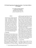
Of 60 patients that were included in the study, only 41
experienced neutropenic fever and completed the study
(Figure 1). Patient characteristics are shown in Table 1.
Septic shock during the period of neutropenia requiring
mechanical ventilation was present in 10 patients, but was
not present at study entry in any of the patients. Median
time to septic shock development was 4.1 days (range 1 -
7 days) after the first episode of neutropenic fever, and in
only one patient, septic shock onset occurred in the first
48 hours after study entry. The median time between the
onset of septic shock and the need for mechanical ventila-
tion was 1 day (range 0-2 days). Eight patients died from
complications of sepsis within the first 30 days after the
onset of fever, yielding an overall 30-day mortality of
13.3%. Clinical significant differences between patients
with non-complicated sepsis and septic shock included
age, presence of bloodstream infection, SOFA score
48 hours after fever onset and MASCC score at FN onset.
Gram-negative and Gram-positive organisms were isolated
in 7 and 6 patients respectively, whereas fungi were
Alves et al. Journal of Translational Medicine 2011, 9:23
/>Page 2 of 8
isolated in 3 patients. Isolated microorganisms included:
A. baumannii, E. coli, K. pneumoniae, P. aeruginosa,
E. cloacae, S. aureus, S. epidermidis, S. viridans, Fusarium
and Aspergillus. Four patients had blood cultures positives
for two pathogens.
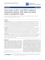
Time-course of sFlt-1 and VEGF-A expression in FN
At the time of fever onset no statistical signific ant differ-
ence could be detected between VEGF-A levels in patients
with non-complicated sepsis (20.7 pg/ml, range 7.9-129.3
pg/ ml) or with septic shock (20.0 pg/ml, range 9.3-158.9
pg/ml; P = 0.9). However, after 48 hours, VEGF-A levels
were higher in patients with septic shock (33.0 pg/ml,
range 13.0-241.9 pg/ml) compared to patients with non-
complicated sepsis (20.9 pg/ml, range 5.6-124.4 pg/ml; P =
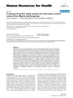
0.03) (Figure 1). Similar to VEGF-A, no difference could
be observed between sFlt-1 levels at the time of neutrope-
nic fever between patients with non-complicated sepsis
(47.3 pg/ml, range 20.8-117.6 pg/ml) and septic shock
(49.2 pg/ml, range 29.6-91.1 pg/ml; P = 0.3). However, 48
hours after neutropenic fe ver a marked difference could
be observed between patients with and without septic
shock, with increased sFlt-1 concentrations in patients
with septic shock (116.0 pg/ml, range 42.7-208.4 pg/ml)
compared to patients with non-complicated sepsis (42.9
pg/ml, range 25.9-472.9 pg/ml; P = 0.002) (Figure 2).
Association of serum sFlt-1 and VEGF-A levels with sepsis
prognosis
To explore a potential association of sFlt-1 and VEGF-A
levels with sepsis outcome in patients with FN, we first
evaluated whethe r serum VEGF-A and sFlt-1 levels cor-
related with sepsis severity scores. As shown in Tab le 2,
sFlt-1 measured at fever onset was significantly
Figure 1 Study flowchart.
Table 1 Patient characteristics
Sepsis ¥
(n = 31)
Septic shock
(n = 10)
P
Gender 0.16 **
Male 13 (42%) 7 (70%)
Female 18 (58%) 3 (30%)
Age (median, range) 37 (16-55) 55 (24-62) P < 0.01 *
Disease status 0.12 **
Complete remission 13 (42%) 1 (10%)
Active disease 18 (58%) 9 (90%)
Treatment 0.48 **
Intensive CTx (includes autologous HSCT) 17 (55%) 6 (60%)
Allogeneic HSCT 14 (45%) 4 (40%)
Neutrophils/μl - Fever (median, range) 60 (0 - 290) 50 (20 - 470) 0.40 *
Days of neutropenia (median, range) 12 (4 - 22) 14 (7 - 30) 0.36 *
Platelets ×10
3
/μl - fever (median, range) 25 (6 - 169) 38 (12 - 90) 0.16 *
Days with fever (median, range) 4 (1 - 12) 5 (1 - 12) 0.65 *
SOFA score - fever onset (median, range) 3 (0 - 7) 4 (2 - 8) 0.31 *
SOFA score - 48 hours (median, range) 4 (2 - 7) 7 (4 - 16) P = 0.01 *
MASCC score (median, range) 21 (16 - 23) 18 (11 - 24) P = 0.03 *
Agent isolation in bloodstream P < 0.001 **
Yes 4 (13%) 8 (80%)
No 27 (87%) 2 (20%)
¥ Non-complicated sepsis; * Mann-Whitney test; ** Fisher’s exact test. HSCT: Hematopoietic stem cell transplantation; CTx: chemotherapy.
Alves et al. Journal of Translational Medicine 2011, 9:23
/>Page 3 of 8
correlated with both MASCC and SOFA scores. VEGF-
A level (measured at fever onset) correlated with SOFA
score calculated 48 hours after fever onset (Table 2).
Next, we explored whether the individual or combined
analysis of sFlt-1 and VEGF-A levels could help in risk
stratification of patients with FN. In order to do so, we
plotted simultaneously the values of sFlt-1 and VEGF-A
in patients with non-complicated sepsis and septic
shock, dichotomizing marker levels by their median
values. The graphic representation of this analysis seems
to indicate that 48 ho urs after fever onset, patients that
evolvetosepticshockaremorelikelytopresentabove
median levels of both sFlt-1 and VEGF-A, than patients
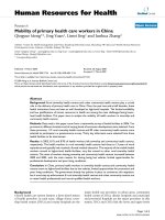
with non-complicated sepsis (Figur e 3). Furthermore,
the relative risk for septic shock development in patients
with both levels above the median compared to all other
patients was 6.9 (1.67-28.5; P = 0.004 ; Fisher’sexact
test).
Fina lly, we estim ated the diagnostic accuracy of sFlt-1
and VEGF-A levels using ROC procedures in our study
popu lation. When measured at fever onset, neither sFlt-
1 nor VEGF-A levels yielded area under the ROC curve
values that indicated any diagnostic capacity to discrimi-
nate patients that would evolve to non-complicated sep-
sis or to septic shock patients. However, when measured
48 hours after fever onset, when clinical signs of se ptic
shock were still not present in any but one patient, both
markers yielded area under the ROC curve values that
suggest a diagnostic capacity for the discrimination of
FN that evolve to septic shock (Table 3).
Figure 2 Serum sFlt-1 and VEGF-A levels in FN. Serum sFlt- 1
and VEGF-A levels in patients with FN. Box plots representing
serial concentrations of sFlt-1 and VEGF-A in patients with FN with
non-complicated sepsis (n = 31) or septic shock (n = 10) at fever
onset and 48 hours thereafter. Mann-Whitney test.
Table 2 Correlation of sFlt-1 and VEGF-A with severity of
illness
MASCC SOFA (Fever
onset)
SOFA (48
hours)
VEGF-A (fever
onset)
Rs = - 0.18
P = 0.31
Rs = - 0.21
P = 0.23
Rs = - 0.17
P = 0.33
VEGF-A (48 hours) Rs = -
0.43
P = 0.03
- Rs = 0.09
P = 0.66
sFlt-1 (Fever onset) Rs = -
0.42
P < 0.01
Rs = 0.33
P = 0.04
Rs = 0.32
P = 0.04
sFlt-1 (48 hours) Rs = - 0.11
P = 0.52
- Rs = 0.25
P = 0.16
The Spearman’s correlation coefficients (Rs) for sFlt-1 and VEGF-A mea sured at
fever onset and after 48 hours with the severity of illness scores are shown.
The correlation of the markers after 48 hours of fever onset with scores at the
time of fever onset was not assessed.
Figure 3 sFlt-1 and VEGF-A levels in patients with FN .
Combined analysis sFlt-1 and VEGF-A levels in patients with
FN. Actual sFlt-1 and VEGF-A serum levels obtained at fever onset
and after 48 hours are plotted simultaneously as well as median
values for each marker at each time point (dotted lines). At fever
onset, cases with non-complicated sepsis (empty circles) and septic
shock (full circles) are spread evenly across the median values for
both parameters (3a-b). After 48 hours, cases that evolved to septic
shock seem to localize more frequently in the right upper quadrant
(high VEGF-A and high sFlt-1) than cases with non-complicated
sepsis, in which levels of both biomarkers change very little.
Alves et al. Journal of Translational Medicine 2011, 9:23
/>Page 4 of 8
Discussion
Despite improvements in sup portive care, complications
of sepsis are still one of the main challenges in the man-
agement of patients submitted to intensive chemother-
apy. Patients with FN are particularly prone to sepsis
complications, and because no clinical or laboratory
marker can reliably identify patients at lower risk of sep-
tic shock, immediate admission and broad-spectrum
antibiotics is still the most widely strategy used in the
management of patients with FN. Although the use of
oral antibiotics in low risk patients has been shown to
be safe [15], a robust definition of a low risk patient is
still not available [16]. The MASCC score, which is the
most studied model, seems to yield, in limited studies, a
71% sensitivity and a 91% positive predictive value to
identify low-risk patients [17]. However, subjectivity in
clinical assessment of the “disease burden” parameter,
the rarity of chronic obstructive lung disease in children
and young adul ts, and its limited validation in the out-
patient setting and in p atients with acute leukemia still
preclude its widespread adoption for the management of
FN patients. This opens room for the search of biomar-
kers that could reliably stratify patients w ith higher risk
of sepsis complications. In addition, preliminary data
from animal studies suggest that sFlt-1 could play an
important role in the treatment of sepsis, as an endothe-
lial barrier stabilizing agent, provi ded that the VEGF-A
and sFlt-1 axis indeed play clinical relevant roles in the
pathogenesis of sepsis comp lications in humans. There-
fore we explored the time-course and the significance of
serum levels of sFlt-1 and VEGF-A in patients with FN
and hematological malignancies.
The endothelial barrier-breaking properties of VEGF-
A are less widely characterized than its mitogenic effects
on endothelial cells. Rather than an independent func-
tion, this barrier-breaking property is i ndeed an impor-
tant part of VEGF-A’s role in the regulation of
angiogenesis, as the disassemble of an intact endothelial
line is necessary for the incorporation of new endothe-
lial cells during vessel sprout. The clinical relevance of
this effect in humans was observed more than a decade
ago, when patients treated with low dose VEGF-A to
boost revascularization in critical limb ischemia pre-
sented peripheral edema as a consistent adverse event
[18]. Following this observation , elevated VEGF-A levels
have been associated with a variety of con ditions that
share the disruption of the endothelial barrier as a com-
mon pathogenic mechanism, including sepsis [19,20]. In
intensive car e unit patients w ith sepsis, levels of VEGF-
A have also been associated with disease severity and
mortality [4-6,21]. More recently, VEGF-A levels were
evaluated in a smaller s tudy with patients with FN,
which observed higher VEGF-A levels in patients that
evolved to severe sepsis compared to patients with non-
complicated sepsis [22]. VEGF-A acts by binding to two
tyrosine-kinase transmembrane receptors: VEGFR-1
(Flt-1) and VEGFR-2, mainly expressed in endothelial
cells. VEGF-A and its receptors act in conjunction with
other regulators of angiogenesis such as the angio-
poiein/Tie-2 axis, which has also been associated with
sepsis diagnosis and o utcome by we and others [23-26].
sFlt-1 is a splic e variant of the receptor VEGFR-1. sFl t-1
is secreted in soluble form, binds VEGF-A and acts as a
decoy receptor, down-regulating its cellular effects. sFlt-
1 has been shown to protect mice from VEGF-A
induced sepsis [27]. Antagonism of VEGF-A by sFlt-1
has a lso been explored therapeutically in the treatment
of pathogenic vessel growth in cancer and other diseases
[28,29]. In our study we demonstrated that patients with
FN that evolve to septic shock present higher serum
levels o f VEGF-A compared to patients with non-com-
plicated sepsis, when measured 48 hours after fever
onset. Our observation confirms, in a larger population
of patients with septic shock (10 patients), a recent
study in patients with FN in which only 1 patient
evolved to septic shock [2 2]. We also describe for the
first time that sFlt-1 levels are higher in severely neutro-
penic patients that evolve to septic shock compared to
patients with non-complicated sepsis, and that this
increase is only present 48 hours after fever onset. This
observation is consistent with recent studies from one
group that evaluated sFlt-1 levels in non-neutropenic
patients with sepsis and septic sho ck [6,9] that also
observed higher sFlt-1 levels in patients with septic
shock, and that sFlt-1 could be a useful biomarker for
sepsis severity. Furthermore, we demonstrated that both
VEGF-A and sFlt-1 levels correlated with sepsis severity
scores.
An interesting finding of our study is the divergent
trend of sFlt-1 level s observed in patients that evolve to
septic shock (towards higher levels) compared to
patients that recover uneventfully (unchanged levels)
(Figure 2). This trend seems to indicate that higher sFlt-
1 serum levels in the former group of patients could be
Table 3 Diagnostic accuracy of sFlt-1 and VEGF-A levels
for septic shock development
Biomarker
Time-point
sFlt-1 VEGF-A
Fever onset
AUC * 0.61 (0.41-0.81);
P = 0.81
0.58 (0.38-0.77);
P = 0.47
48 hours after fever onset
AUC * 0.87 (0.73-1.00);
P < 0.01
0.76 (0.55-0.97);
P = 0.02
*AUC: area under ROC curve; sFlt-1 and VEGF-A thresholds correspond to the
median value for each time point. AUC expressed with 95% confidence
interval (CI95%).
Alves et al. Journal of Translational Medicine 2011, 9:23
/>Page 5 of 8
the expression of an additional compensatory mechan-
ism, triggered by the failure of sFl t-1-independent
mechanisms that maintain endothelial barrier in patients
of the latter group. In animal models of sepsis, over-
expression of sFlt-1 was capable to completely block the
barrier-breaking effects of VEGF-A and to reduce mor-
tality, suggesting that sFlt-1 could be used as a regulator
of vascular per meability in pathological conditions.
However,thiswaspossiblebyusingagenetransfer
strategy that resulted in a more than 100-fold increase
in sFlt-1 levels [8]. Our data demonstrate that the up to
10-fold elevation of sFlt-1 serum concentration observed
in humans was not sufficient to block the development
of septic shock in patients with FN. Whether several-
fold higher e levations of sFlt-1 could effectively block
the endothelial barrier disruption present in patients
with septic shock is an exciting scientific question that
remains to be answered.
In our study, i nitial samples were collected very early
after fever onset, when no signs of sepsis complications
were present. Median time to septi c shock development
in our study was 4 days, and even samples collected 48
hours after fever were stil l obtained before the develop-
ment of overt septic shock, in all but one patient. This
was only possible because of the in-hospital design of
our study in which patients were under strict monitor-
ing for fever signs, and contrasts with studies of sFlt-1
and VEGF-A levels in non-neutropenic patients, which
were mostly performed in intensive care units, after the
development of sepsis complications. Even though this
specific characteristic of our study does not reproduce
real-lifepracticewhereabiomarkerwouldbeused,it
probably allows a more comprehensive evaluation of the
kinetics of sFlt-1 an d VEGF-A release in human sepsis.
In our study, differences in sFlt-1 and VEGF-A levels
could not be demonstrated at fever onset, and were only
present 48 hours thereafter. This is also in co ntrast with
the observation of higher sFlt-1 and VEGF-A levels in
non-neutropenic patients with sepsis at “early” time
points. Again, we believe that rather than a difference in
the kinetics of sFlt-1 and VEGF-A release in patients
with neutro penia, this difference reflects the earlier eva-
luation of these biomarker levels in our patients com-
pared t o previous studies. Indeed, the fact that none of
the observed di fferences in biomarker l evels were pre-
sent at fever onset h as important implications. First, it
suggests that VEGF-A and sFlt-1 increases are a rela-
tively later consequence of the cascade of events that
leadstosepticshock.Thishypothesisissupportedby
the intuitive assumption that VEGF-A acts as one of the
final downs tream elements in the pathogenesis of septic
shock, and is consistent with our previous hypothesis
that sFlt-1 is released as a salvage compensatory
mechanism to restore barrier function. A second clinical
impl ication of our re sults refers to the use of sFlt-1 and
VEGF-A for risk stratification of patients with FN. In
our study, the estimation of diagnostic accuracy of
VEGF-A and sFlt-1 yielded promising results only when
levels were measured 48 hours after fever onset. An
ideal biomarker for risk stratification in FN should not
require serial sampling, as this would not allow early
discharge of low risk patients. Future studies with higher
number of patients and under a less controlled environ-
ment (including outpatients) are warranted to check
whether levels of these biomarkers obtained in a “real
world” setting will be able to capture this increase in
sFlt-1and/or VEGF-A levels of patients with a worse
prognosis and accurately discriminate FN patients with
different outcomes.
The major sources of VEGF-A in sepsis are still a
matter of debate, and no information on the source of
sFlt-1 in sepsis had b een published so far. A study with
healthy volunteers suggested that platele ts, and mainly
granulocytes are the sources of more than 90% of circu-
lating VEGF-A [30]. In contrast, in an animal model of
sepsis VEGF-A levels increased in liver, kidney and
heart, and no difference could be detected between
VEGF-A levels in serum and plasma, arguing against a
majorroleofplateletsasasourceofVEGF-A[8].In
our study, VEGF-A and sFlt-1 serum levels were
approximately 5 and 10-fold lower respectively than
plasma levels in non-neutropenic septic patients, thus
suggesting that platelets and granulocyt es do represent
an important source of these citokynes in sepsis [ 6].
However, no difference in neutrophil and platelet counts
could be demonstrated between patients with non-com-
plicat ed sepsis and septic shock at fever onset (Table 1),
and no statistical significant correlation could be
demonstrated between VEGF-A and sFlt-1 levels with
platelet and neutrophil counts at any time-point (data
not shown).
Our study has several limitations including a relatively
low number of patients, which precludes subgroup ana-
lysis, a single-center design and the fact that the in-hos-
pital setting does not reproduce the real-life conditions
whereabiomarkerwouldbeuseful.However,our
exploratory studied wa s not aimed to definitively prove
or rule out the usefulness of sFlt-1 and VEGF-A deter-
minations as biomarkers of sepsis severity in FN, but
rather to test whether these biomarkers showed diagnos-
tic promised under controlled and ideal c onditions. In
other words, the limitations of our study could also be
regarded as its strengths, if it is acknowledged that it
was designed to answer a “phase 2 question” in the hier-
archy of diagnostic research, setting the stage for a
future and planned validating study [31], as well as to
gain insights about the time-course of sFlt-1 and VEGF-
A release during the very initial phase of sepsis.
Alves et al. Journal of Translational Medicine 2011, 9:23
/>Page 6 of 8
Conclusions
In conclusion, our study demonstrates that patients with
hematological malignancies and post-chemotherapy FN
tha t evolve to septic shock pres ent higher levels of sFlt-
1 and VEGF-A than patients with non-complicated sep-
sis, and that levels of these biomarkers correlate with
sepsis severity scores. In addition, the time-course of
sFlt-1 release suggests that is could represent a salvage
compensatory mec hanism in patients that evolve to sep-
tic shock. Additional studies are warranted to explore
the validity of these observat ions, as well as the feasibil-
ity of t heir incorporation into risk stratification models
for neutropenic patients or, in the future, as therapeutic
tools in sepsis.
Acknowledgements
This study was financially supported by Fapesp and CNPq, Brazil. The
Hematology and Hemotherapy Center - Hemocentro UNICAMP, forms part
of the National Institute of Science and Technology of Blood, Brazil (INCT do
Sangue CNPq/MCT/FAPESP).
Author details
1
Hematology and Hemotherapy Center, University of Campinas, Campinas,
SP, Brazil.
2
Faculty of Medical Sciences, University of Campinas, Campinas, SP,
Brazil.
Authors’ contributions
BEA enrolled patients, recorded clinical data, performed laboratory analysis
and contributed to manuscript production; SALM Performed laboratory
analysis; FJPA performed statistical analysis and reviewed the manuscript; IL,
CADS and JMA contributed to the study design and reviewed the
manuscript; EVDP designed the study, analyzed data and contributed to
manuscript production. All authors read and approved the final manuscript.
Competing interests
The authors declare that they have no competing interests.
Received: 31 October 2010 Accepted: 3 March 2011
Published: 3 March 2011
References
1. Ellis M: Febrile Neutropenia. Ann N Y Acad Sci 2008, 1138:329-350.
2. Pierrakos C, Vincent JL: Sepsis biomarkers: a review. Crit Care 2010, 14:R15.
3. Senger DR, Galli SJ, Dvorak AM, Perruzzi CA, Harvey VS, Dvorak HF: Tumor
cells secrete a vascular permeability factor that promotes accumulation
of ascites fluid. Science 1983, 219:983-985.
4. Pickkers P, Sprong T, Eijk L, Hoeven H, Smits P, Deuren M: Vascular
endothelial growth factor is increased during the first 48 hours of
human septic shock and correlates with vascular permeability. Shock
2005, 24:508-512.
5. van der Flier M, van Leeuwen HJ, van Kessel KP, Kimpen JL, Hoepelman AI,
Geelen SP: Plasma vascular endothelial growth factor in severe sepsis.
Shock 2005, 23:35-38.
6. Shapiro NI, Yano K, Okada H, Fischer C, Howell M, Spokes KC, Ngo L,
Angus DC, Aird WC: A prospective, observational study of soluble FLT-1
and vascular endothelial growth factor in sepsis. Shock 2008,
29:452-457.
7. Kendall RL, Wang G, Thomas KA: Identification of a natural soluble form
of the vascular endothelial growth factor receptor, FLT-1, and its
heterodimerization with KDR. Biochem Biophys Res Commun 1996,
226:324-328.
8. Yano K, Liaw PC, Mullington JM, Shih SC, Okada H, Bodyak N, Kang PM,
Toltl L, Belikoff B, Buras J, et al: Vascular endothelial growth factor is an
important determinant of sepsis morbidity and mortality. J Exp Med
2006, 203:1447-1458.
9. Shapiro N, Schuetz P, Yano K, Sorasaki M, Parikh SM, Jones AE, Trzeciak S,
Ngo L, Aird WC: The association of endothelial cell signaling, severity of
illness, and organ dysfunction in sepsis. Crit Care 2010, 14:R182.
10. Bone RC, Balk RA, Cerra FB, Dellinger RP, Fein AM, Knaus WA, Schein RM,
Sibbald WJ: ACCP/SCCM Consensus Conference Committee. Definitions
for sepsis and organ failure and guidelines for the use of innovative
therapies in sepsis. The ACCP/SCCM Consensus Conference Committee.
American College of Chest Physicians/Society of Critical Care Medicine.
1992. Chest 2009, 136:e28.
11. Hughes WT, Armstrong D, Bodey GP, Bow EJ, Brown AE, Calandra T, Feld R,
Pizzo PA, Rolston KV, Shenep JL, Young LS: 2002 guidelines for the use of
antimicrobial agents in neutropenic patients with cancer. Clin Infect Dis
2002, 34:730-751.
12. Vincent JL, Moreno R, Takala J, Willatts S, De Mendonca A, Bruining H,
Reinhart CK, Suter PM, Thijs LG: The SOFA (Sepsis-related Organ Failure
Assessment) score to describe organ dysfunction/failure. On behalf of
the Working Group on Sepsis-Related Problems of the European Society
of Intensive Care Medicine. Intensive Care Med 1996, 22:707-710.
13. Klastersky J, Paesmans M, Rubenstein EB, Boyer M, Elting L, Feld R,
Gallagher J, Herrstedt J, Rapoport B, Rolston K, Talcott J: The Multinational
Association for Supportive Care in Cancer risk index: A multinational
scoring system for identifying low-risk febrile neutropenic cancer
patients. J Clin Oncol 2000, 18:3038-3051.
14. Uys A, Rapoport BL, Anderson R: Febrile
neutropenia: a prospective study
to validate the Multinational Association of Supportive Care of Cancer
(MASCC) risk-index score. Support Care Cancer 2004, 12:555-560.
15. Vidal L, Paul M, Ben-Dor I, Pokroy E, Soares-Weiser K, Leibovici L: Oral
versus intravenous antibiotic treatment for febrile neutropenia in cancer
patients. Cochrane Database Syst Rev 2004, CD003992.
16. Phillips B, Wade R, Stewart LA, Sutton AJ: Systematic review and meta-
analysis of the discriminatory performance of risk prediction rules in
febrile neutropaenic episodes in children and young people. Eur J Cancer
46:2950-2964.
17. Kern WV: Risk assessment and treatment of low-risk patients with febrile
neutropenia. Clin Infect Dis 2006, 42:533-540.
18. Baumgartner I, Pieczek A, Manor O, Blair R, Kearney M, Walsh K, Isner JM:
Constitutive expression of phVEGF165 after intramuscular gene transfer
promotes collateral vessel development in patients with critical limb
ischemia. Circulation 1998, 97:1114-1123.
19. Harada M, Mitsuyama K, Yoshida H, Sakisaka S, Taniguchi E, Kawaguchi T,
Ariyoshi M, Saiki T, Sakamoto M, Nagata K, et al: Vascular endothelial
growth factor in patients with rheumatoid arthritis. Scand J Rheumatol
1998, 27:377-380.
20. Taha Y, Raab Y, Larsson A, Carlson M, Loof L, Gerdin B, Thorn M: Vascular
endothelial growth factor (VEGF)–a possible mediator of inflammation
and mucosal permeability in patients with collagenous colitis. Dig Dis Sci
2004, 49:109-115.
21. Karlsson S, Pettila V, Tenhunen J, Lund V, Hovilehto S, Ruokonen E: Vascular
endothelial growth factor in severe sepsis and septic shock. Anesth Analg
2008, 106:1820-1826.
22. Hamalainen S, Juutilainen A, Matinlauri I, Kuittinen T, Ruokonen E, Koivula I,
Jantunen E: Serum vascular endothelial growth factor in adult
haematological patients with neutropenic fever: a comparison with C-
reactive protein. Eur J Haematol 2009, 83:251-257.
23. Giuliano JS Jr, Lahni PM, Harmon K, Wong HR, Doughty LA, Carcillo JA,
Zingarelli B, Sukhatme VP, Parikh SM, Wheeler DS: Admission angiopoietin
levels in children with septic shock. Shock 2007, 28:650-654.
24. Kumpers P, Lukasz A, David S, Horn R, Hafer C, Faulhaber-Walter R, Fliser D,
Haller H, Kielstein JT: Excess circulating angiopoietin-2 is a strong predictor
of mortality in critically ill medical patients. Crit Care 2008, 12:R147.
25. Siner JM, Bhandari V, Engle KM, Elias JA, Siegel MD: Elevated serum
angiopoietin 2 levels are associated with increased mortality in sepsis.
Shock 2009, 31:348-353.
26. Alves BE, Montalvao SA, Aranha FJ, Siegl TF, Souza CA, Lorand-Metze I,
Annichino-Bizzacchi JM, De Paula EV: Imbalances in serum angiopoietin
concentrations are early predictors of septic shock development in
patients with post chemotherapy febrile neutropenia. BMC Infect Dis
2010, 10:143.
27. Tsao PN, Chan FT, Wei SC, Hsieh WS, Chou HC, Su YN, Chen CY, Hsu WM,
Hsieh FJ, Hsu SM: Soluble
vascular endothelial growth factor receptor-1
protects mice in sepsis. Crit Care Med 2007, 35:1955-1960.
Alves et al. Journal of Translational Medicine 2011, 9:23
/>Page 7 of 8
28. Bagri A, Kouros-Mehr H, Leong KG, Plowman GD: Use of anti-VEGF
adjuvant therapy in cancer: challenges and rationale. Trends Mol Med
16:122-132.
29. Schmucker C, Ehlken C, Hansen LL, Antes G, Agostini HT, Lelgemann M:
Intravitreal bevacizumab (Avastin) vs. ranibizumab (Lucentis) for the
treatment of age-related macular degeneration: a systematic review.
Curr Opin Ophthalmol 2010, 21:218-226.
30. Kusumanto YH, Dam WA, Hospers GA, Meijer C, Mulder NH: Platelets and
granulocytes, in particular the neutrophils, form important
compartments for circulating vascular endothelial growth factor.
Angiogenesis 2003, 6:283-287.
31. Sackett DL, Haynes RB: The architecture of diagnostic research. Bmj 2002,
324:539-541.
doi:10.1186/1479-5876-9-23
Cite this article as: Alves et al.: Time-course of sFlt-1 and VEGF-A release
in neutropenic patients with sepsis and septic shock: a prospective
study. Journal of Translational Medicine 2011 9:23.
Submit your next manuscript to BioMed Central
and take full advantage of:
• Convenient online submission
• Thorough peer review
• No space constraints or color figure charges
• Immediate publication on acceptance
• Inclusion in PubMed, CAS, Scopus and Google Scholar
• Research which is freely available for redistribution
Submit your manuscript at
www.biomedcentral.com/submit
Alves et al. Journal of Translational Medicine 2011, 9:23
/>Page 8 of 8