Báo cáo sinh học: "Shipping blood to a central laboratory in multicenter clinical trials: effect of ambient temperature on specimen temperature, and effects of temperature on mononuclear cell yield, viability and immunologic function" potx
Bạn đang xem bản rút gọn của tài liệu. Xem và tải ngay bản đầy đủ của tài liệu tại đây (452.41 KB, 13 trang )
RESEARCH Open Access
Shipping blood to a central laboratory in
multicenter clinical trials: effect of ambient
temperature on specimen temperature, and
effects of temperature on mononuclear cell yield,
viability and immunologic function
Walter C Olson
1*
, Mark E Smolkin
2
, Erin M Farris
3
, Robyn J Fink
4
, Andrea R Czarkowski
5
, Jonathan H Fink
6
,
Kimberly A Chianese-Bullock
1,7
, Craig L Slingluff Jr
1,7
Abstract
Background: Clinical trials of immunologic therapies provide opportunities to study the cellular and molecular
effects of those therapies and may permit identification of biomarkers of response. When the trials are performed
at multiple centers, transport and storage of clinical specimens become important variables that may affect
lymphocyte viability and function in blood and tissue specimens. The effect of temperature during storage and
shipment of peripheral blood on subsequent processing, recovery, and function of lymphocytes is understudied
and represents the focus of this study.
Methods: Peripheral blood samples (n = 285) from patients enrolled in 2 clinical trials of a melanoma vaccine
were shipped from clinical centers 250 or 1100 miles to a central laboratory at the sponsoring institution. The yield
of peripheral blood mononuclear cells (PBMC) collected before and after cryostorage was correlated with
temperatures encountered during shipment. Also, to simulate shipping of whole blood, heparinized blood from
healthy donors was collected and stored at 15°C, 22°C, 30°C, or 40°C, for varied intervals before isolation of PBMC.
Specimen integrity was assessed by measures of yield, recovery, viability, and function of isolated lymphocytes.
Several packaging systems were also evaluated during simulated shipping for the ability to maintain the intern al
temperature in adverse temperatures over time.
Results: Blood specimen containers experienced temperatures during shipment ranging from -1 to 35°C. Exposure
to temperatures above room temperature (22°C) resulted in greater yields of PBMC. Reduced cell recovery
following cryo-preservation as well as decreased viability and immune function were observed in specimens
exposed to 15°C or 40°C for greater than 8 hours when compared to storage at 22°C. There was a trend toward
improved preservation of blood specimen integrity stored at 30°C prior to processing for all time points tested.
Internal temperatures of blood shipping containers were maintained longer in an acceptable range when warm
packs were included.
Conclusions: Blood packages shipped overnight by commercial carrier may encounter extreme seasonal temperatures.
Therefore, considerations in the design of shipping containers should include protecting against extreme ambient
temperature deviations and maintaining specimen temperature above 22°C or preferably near 30°C.
* Correspondence:
1
Human Immune Therapy Center, University of Virginia, Charlottesville, VA,
USA
Full list of author information is available at the end of the article
Olson et al. Journal of Translational Medicine 2011, 9:26
/>© 2011 Olson et al; licensee BioMed Central Ltd. This is an Open Access article distribute d under the terms of the Creative Commons
Attribution Licens e ( censes/by/2.0), which permits unrestricted use, distribution, and reproduction in
any medium, provided the original work is properly ci ted.
Background
Cell-based immunological assays are integral to moni-
toring the effects of immunotherapy clinical trials. The
main clinical specimen obtained for these assays is
whole blood collected in heparinized vacuta iner tubes
from which peripheral blood mononuclear cells (PBMC)
are isolated. Assays of cellular immune responses to
immune therapy depend on functional and viable
PBMC. It is critical that outside factors, other than
study parameters, do not introduce significant variability
in the immune assays due to compromised PBMC integ-
rity. Therefore, trials utilizing multiple cl inical centers
present challenges in how to best process and transport
whole blood and tissue samples.
The need for specific guidelines for the shipment of
biological specimens is of great concern for the conduct
of multi-center clinical trails at the national and interna-
tional level [1-3]. Both complex processing and delay
before processing by individu al laboratories increase the
variability in specimen performance [4]. In contrast,
central laboratory processing lessens the variability
introduced by mu ltiple processing protocols but is more
costly and may not be available f or all inv estigators. It
therefore becomes a critical issue in the design of multi-
center clinical trials to determine whether biological
specimens should be processed immediately, the same
day, or after shipment to a central laboratory.
Early studies have demonstrated how time and tem-
perature of storage affect lymphocyte viability and phe-
notype when whole blood is stored overnight at 4°C
[5-7]. Storage at room tempera ture prior to processing
also affects viability and blastogenic responses [8] as
well as lymphocyte separation by Ficoll density centrifu-
gation [9,10]. T he importance of establishing standard
shipping parameters has been stressed in the infectious
disease setting, in which a profound impact of shipping
was noted on the lymphoproliferative responses to
microbial antigens in both HIV-infected and healthy
donors [11,12]. Single cell-based techniques such as ELI-
spot assays [13-15], intracellular cytokine staining
[16-19], and HLA-specific multimeric assays [20-22] are
widely used and depend on optimal conditions for speci-
men handling in order to detect rare populations of
peptide specific lymphocytes in response to immu-
notherapy. Several studies have confirmed that cryopre-
served PBMC can be used reliably in these assays
[23-26]. Use of cryopreserved samples, however,
depends on optimal s ample handling before and after
cryopreservation. Some studies have defined optimal
time intervals between venipuncture and cryopreserva-
tion [26-29] and o ptimal conditions for freezing [30].
Also, handling and storage of cryopreserved PBMC have
been evaluated, showing that fluctuations in sub-zero
freezing temperatures can alter the viability and function
of recovered lymphocytes; shipping conditions for frozen
samples have also been addressed [31,32]. However, the
effect of ambient temperature changes during shipping
or storage prior to cryopreservation has not been
addressed.
It has been suggested that a n interval of whole blood
storage exceeding 8 hours (h) causes a significant
decrease in cellular immune function [27]. This finding
provides rationale for immediate isolation and cryopre-
servation of PBMC at each participating clinical center
and indeed, optimization of cryopreserv ation media and
of thawing practices has improved recovery of immuno-
logical responses at the single cell level [25,3 0]. How-
ever, processing of blood and cryopreservation of PBMC
at off-site locations is expensive and requires oversight
and quali ty control of the processing lab at each center.
Thus, for many multicenter clinical trials of cancer vac-
cines and other therapies, all off-site whole blood speci-
mens are shipped to a central laboratory according to a
standard operating protocol, and monitored strictly for
quality control and quality assurance. Our concern that
shipping whole blood in different seasons, in various cli-
mates, may impact PBMC viability and functio n
prompted this study. Specifically, we have addressed the
effect of shipping temperatures on cell viability, recovery
and function, and have modeled these in vitro when
controlling for temperature.
Methods
Blood collection, processing and storage
Patients’ blood specimens were derived from p artici-
pants enrolled in one of three studies. Participan ts were
enrolled in the clinical studies following informed con-
sent, and with Institutional Review Board for Health
Sciences Research approval (IRB-HSR# 10598, 10524,
and11491) and review by the FDA (BB-IND# 9847 and
12191). Patients’ blood specimens from 2 clinical trials
(HSR# 1524(HSR# 10524 and 11491) were monitored
during a 9 month period from late summer , through
fall, winter and early spring. Two hundred and eighty-
five blood specimens collected at participating clinical
trial centers in Houston, TX and Philadelphia PA, were
shipped to Charlottesville VA. Clinical laboratory ana-
lyses, including complete blood counts (CBC) and differ-
ential hematological counts, were performed at the
individual centers and the results incorporated into a
trial database. An additional 60 ml of blood were col-
lected in 10cc heparinized vacutainer tubes (BDBios-
ciences, Franklin Lakes, NJ) and were shipped, in
insulated packaging, by overnight courier at ambient
temperature to the Biorepository and Tissue Research
Facility (BTRF) at the University of Vi rginia (UVa) for
processing and cryo-preservation, on the day they
arrived, for fut ure immunological testing in cell-based
Olson et al. Journal of Translational Medicine 2011, 9:26
/>Page 2 of 13
assays. Shipments of patients’ blood specimens were
continuously monitored using the TempCheck Sensor
(Marathon Products, Inc., San Leandro, CA) to deter-
mine the temperature range to which blood samples
were exposed when shipped overnight by commercial
carrier, and to evaluate the effects of those temperatures
on cell yield. Temperature gauges recorded the maxi-
mum and minimum temperatures attained inside the
packages during shipment. Blood drawn at UVa was
processed either the same day or the following d ay,
depending on when in the day it was drawn. The
volume of blood collected, and the number of viable
PBMC isolated were recorded by the BTRF. These
values were used to determine the cell yield before cryo-
preservation. In all cases, the PBMC fraction of whole
blood was collected from Leucosep™ (Greiner Bio-One,
Monroe, NC) tubes following centrifugation for 10 min-
utes at 1000 × g.
The exp ected cell yield for each sample was calculated
from the CBC and differential tests performed on whole
blood at the originating clinical laboratory. The absolute
lymphocyte and absolute monocyte counts calculated
from the CBC and differentia l were combined and mul-
tiplied by the volume of blood collected to represent the
expected total PBMC in the blood ( expected cell yield).
Additional File 1 provides a table of cell count data
from each center. The table shows the calculated per-
centage (mean, m edian, and quartiles) of lymphocytes
and monocytes derived from differential and complete
cell counts. The number of PBMC isolat ed by Ficoll
separation, divided by the expected cell yield provides
the ratio cell yield. Ratio cell yields of less than 1 are
expected due to losses in Ficoll separation. However,
becausetheFicollseparationsweredonebythesame
central laboratory and according to a consistent proto-
col, differences in ratio cell yields in different subgroups
of specimens are primarily attributed to effects of ship-
ping conditions.
Incubation conditions for whole blood
In one set of experiments, approximately 7-8 ml whole
blood were collected into each of eleven heparinized
vacutainer tubes from six healthy donors according to
IRB protocol 10598 and were labelled to define the tem-
perature conditions to which they would be exposed.
Each tube was incubated at various temperatures over a
24hperiodatconditionsintendedtomodelwhatmay
happen in overnight ship ping conditions (Additional File
2). A fter a 1-2 h equilibratio n period at room tempera-
ture (RT, 22°C), tubes from each sample were placed in
each of the 4 conditions: (a) temperature-controlled
refrigerated centrifuge set at 15°C, (b) 22°C as a control
condition, (c) water bath set at 30°C, or (d) water bath
set at 40°C. In addition, one tube was placed in a 50°C
water bath for 2 h, but this condition invariably led to
hemolysis and the samples were not evaluabl e. For each
temperature condition (other than RT), one tube
was exposed to t hat low or high temperature for 2, 8 or
12 h, and then each was returned to RT for the remain-
ing 24 h study period. Thus, one tube served as an
untreated control and was at kept at RT for the whole
24 h. After these incubations, PBMC we re isolated from
each blood sample by Ficoll density gradient as
described above. Viable cell numbers were determined
by trypan dye exclusion. PBMC were cryopreseved in
freezing medium (90% FCS, 10% DMSO) overnight at
-80°C, then transferred to vapor phase liquid nitrogen
for 1-4 weeks before thawing for analysis.
ELIspot Assay
Cells producing IFNg after antigen specific and non-
specific stimulation were enumerated by ELIspot assay
as described previously [33,34]. In brief, PBMC were
thawed in pre-warmed RPMI1640 (Invitrogen, Carlsbad
CA) containing 10% human AB serum (HuAB; Gemini)
and 100 Units/mL of DNase I (Worthington Biochem-
ical Corp., Lakewood, NJ). Cells were centrifuged at
350 × g and adjusted to the desired cell density in
RPMI 1640 supple mented with 10% HuAB serum and
plated into PVDF-membrane plates coated with anti-
interferon gamma antibody (Pierce-Endogen, Thermo
Scientific, Rockford IL). Phytohemagglutinin (PHA),
phorbol myristate acetate (PMA and ionomycin were
obtained from Sigma-Aldrich (St. Louis, MO). A pool of
35 MHC Class I restricted peptides consisting of pep-
tides from cytomegalovirus, E pstein-Barr and influenza
virus proteins (CEF peptide pool; [35]; Anaspec, Fre-
mont CA) or media alone were added in quadruplicate
andculturesincubatedovernightat37°Cina5%CO
2
atmosphere. Spots were developed according to standard
protocol and enumerated on a BioReader 4000 (Bio-Sys,
Karben, Germany) plate reader.
Flow cytometry
CD3, CD4, CD8 and CD56 positive lymphocyte popula-
tions were enumerated by flow cytometry using fluores-
cent-labelled antibodies (BDBiosci ences, San Diego, CA).
Cells were washed, suspended in PBS (Invitrogen) con-
taining 0.1% BSA (Sigma) and 0.1% sodium azide
(Sigma). Titrated amounts of each reagent were added to
cells, incubated, washed free of excess stain , and fixed in
paraformaldehyde. To determine whether there was an
increase in apoptosis due to different storage conditions,
thawed PBMC were incubated overnight at 37°C in 5%
CO
2
in RPMI 1640 + 10% Hu man AB serum
.
The next
day, PBMC were surface stained with fluorescently
labeled antibodies to CD3, CD4, and CD8, then stained
with Annexin V accordi ng to manufacturer’s instructions
Olson et al. Journal of Translational Medicine 2011, 9:26
/>Page 3 of 13
(BDBioscience, San Diego, CA) and 7-AAD (EMD Che-
micals, Inc., Gibbstown, NJ) to determine the level of
apoptosis [36-38]. Cells were acquired on a FACSCalibur
flow cytometer maintained by the Flow Cytometry core
facility of the University of Virginia. Data were analyzed
with FlowJo software (Treestar, Ashland OR).
Testing of Blood Shipping Packages
The standard shipping container used in our clinical
trials was obtained from Safeguard Technologies Corp.
(Conshohocken, PA). It consist ed of a white corrugated
box fitted with a hydrophilic foam-lined clear plastic
snap-lock case inserted into a plastic zip lock bag. Th is
was placed inside a cardboard shipping container lined
with 1” thick Styrofoam. An alternate packaging design
was provided by JVI (Charlottesville VA) and consisted
of a 14” ×11” ×5” box of 200# corrugated cardboard
insulated with Control Temp Packaging foam of 1”
thickness. Inside was placed a 12” ×9” ×3” clamshell
type clear plastic bo x containing a n 11” ×8.5” ×5/8”
foam vial holder.
Each type of shipping container was tested for its abil-
ity t o maintain temperature in cold ambient conditions
(e.g.: during winter months). Forty heparinized vacutai-
ners were filled with water and equilibrated to 37°C.
Ten vacutainers were placed inside each of 4 packages
(2 of each type). Each package type received a gel pack
conditionedateither37°Cor22°Cwhichwasthen
placed alongside the v acutainer holder. One probe of an
indoor/outdoor thermometer (Taylor Precision Pro-
ducts, Oak Brook IL) was placed inside the package
while another remained outside to monitor external
ambient temperature. Packages were place d either in a
cold room at 4°C for a minimum of 12 h or were
handled in a manner to model the experience of a pack-
age being shipped via motor vehicle overnight in a non-
heated compartment. Temperatures were recorded every
15 minutes during the first hour, and 30-60 minutes
thereafter.
Additional testing of the JVI packaging material was
performed by R.N.C. Industries Inc. (Norcross GA
30071) at high external package temperature. The clam-
shell foam holder containing vials of liquid was placed
inside the package. Tw o 12 oz Control Temp gel packs
conditioned at 20°C were placed in the clamshell onto
which the foam vial holder (including the 1/4” foam
above and below) containing five 5/8” vials f illed with
water conditioned at 20°C was placed inside. The pack-
age was closed, put at 45°C and the internal package
temperature was monitored for 48 hours using an
Omega OMB-DAQ-55 USB data acquisition system,
serial number #156772. T thermocouples were cali-
brated 2 months earlier using a stirred water bath
calibration.
Statistical analysis
The MIXED procedure in SAS 9.1.3 (SAS Institute,
Cary, NC) was used to analyze the effects of tempera-
ture (3 levels) and duration (3 levels) on outcomes
including ELIspot, phenotype, and viability. These effects
were modeled jointly (main effects plus interactions) for
each outcome me asure and outcome measurements
were first normalized by division of the raw data by the
donor value at RT for 24 h. Since donors served as
blocks and contributed an observation from each condi-
tion (i.e. each combination of temperature and duration
level), intra-donor correlation was modeled assuming a
compound symmetry structure in the residual covar-
iance matrix. Degrees of freedom were calculated using
the Kenward-Roger method. To assess the effects of sto-
rage under different temperature conditions on apopto-
sis among CD4 a nd CD8 populations, a modeling
scheme similar to the one above was performed using
calculated logits as the outcome measure. This is
defined as the log
e
([p
i
/1-p
i
]/[p
c
/1-p
c
]) where p = the
proportion of cells that are apoptotic or necrotic (as
defined by Annexin V and 7AAD staining); i = the sto-
rage conditions of the whole blood specimen; and c =
the storage condition of the control specimen at R T for
24 hours. All tests were assessed at a = 0.05.
Results
Effect of shipping temperatures and extreme changes in
temperature on the cell yield for clinical trial specimens
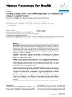
Package temperatures were lowest in winter months and
highest in summer months, suggesting that the tempera-
tures experienced during shipping varied by ambient
seasonal temperatures (Figure 1A). The extreme tem-
peratures ranged from about -1°C to 35°C with 91% fall-
ing completely within the range of 4°C and 32°C.
There was a trend to lower PBMC yields in colder
months from November throug h February (Figure 1B),
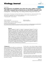
although outliers were noted. Lower minimum temperature
was associated with lower cell yield (p = 0.001, Figure 2A),
whereas higher maximum temperature correlated with
higher ce ll yield (p = 0.04, Figure 2B). The range in shipping
temperatures during the winter was typically bounded by a
high temperature of 22°C, and during the warmer months
by 22°C as a low temperature. The ma ximum change
(deviation) in temperature from 22°C observed during ship-
ment was determine d us ing the high or low temperature
furthest from 22°C. This represents an estimate of the
degree of temperature fluctuation encountered during ship-
ment and is plotted against the yield in Figure 2C, where
there was a correlation with warmer temperatures (p <
0.001). Overall, warmer temperatures favored greater cell
yiel ds. These observations led us to ini tiate control led in
vitro studies on the impact of storage temperature on cell
recovery, viability, and immunological function.
Olson et al. Journal of Translational Medicine 2011, 9:26
/>Page 4 of 13
Effect of temperature on cell yield before
cryopreservation
To determine whether exposure to extreme tempera-
tures impacts the overall integrity of PBMC, blood spe-
cimens from 6 normal volunteers were stored a t
temperatures in a range encountered during blood ship-
ment or varying lengths of time and were assessed for
cell yield, cell recovery and cell function (Additional File
2). Blood was exposed to temperatures of 15°, 30°, 40°,
or 50°C for 2 h, 8 h, and 12 h and left at room tempera-
ture after that exposure for a total of 24 h after collec-
tion. Significant and unacceptable lysis and cell loss
were associated with incubation 2 h at 50°C ; thus, these
were not analyzed further (unpublished observat ion).
Adequate data already exist for the negative effects of
refrigeration at 2-8°C [6,7,9]; s o this temperature was
not a ssessed here. Blood stored 24 h at room tempera-
ture (22°C) was used as a reference for comparison.
A significant decrease in the PBMC cell yield was
observed for samples stored at 15°C for 12 h (p < 0.003;
Table 1). Blood stored at 30°C had PBMC yields almost
identical to the RT standard. Exposure to high or low
temperature for 8 h, followed by RT incubation was
associated with no significant decrement in cell yields at
any of the temperatures. There was a trend to lower cell
yields with 12 h at 40°C, but it was not significant.
Effect of Temperature on Cell Recovery after
cryopreservation
We hypothesized that shipping temperatures may
impact ce ll recovery and viability after storage in liquid
nitrogen. The total number of viable cells (trypan blue
dye exclusion) was recorded for each of the PBMC
M
o
n
t
h
40
30
20
10
0
Cell Yield
Temperature (°C)
A
B
1.6
1.2
0.8
0.4
0
SAu ApONDJ F M
Figure 1 Recorded internal package temperatures during
shipment and cell yields of blood from off-site cancer centers.
(A) High (+) and low (●) package temperatures recorded between
August, 2005 through April, 2006. (B) Yield of PBMC (cell yield)
obtained from specimens shipped during this time after Ficoll
separation. The ratio cell yield is expressed as a ratio of total
number of PBMC collected after Ficoll divided by the number of
PBMC (lymphocytes and monocytes) estimated from the differential
WBC recorded on the same specimens before shipment. The
dashed line represents 100% recovery of PBMC after Ficoll as a ratio
cell yield of one.
1.6
1.2
0.8
0.4
-5 0 10 20 30 15 20 25 30 35 40
-
30
-
20
-
10 0 10 20 30
Max Deviation from RT
(
°C
)
Cell Yield
Minimum Temperature
(
°C
)
Maximum Temperature
(
°C
)
ABC
Figure 2 The recovery of cells after Ficoll separation increased as shipping temperature increased. (A) Correlation of the ratio cell yield
with minimum temperature during transport; p = 0.001. (B) Correlation of the ratio cell yield with maximum temperature during transport; p =
0.04. (C) Correlation of the ratio cell yield as a function of maximum temperature deviation from room temperature (22°C) during shipment;
p < 0.001
Olson et al. Journal of Translational Medicine 2011, 9:26
/>Page 5 of 13
samples exposed to varied temperatures as reported
above. Percent recovery was calculated as the ratio of
recovered viable cells to the number of viable cells initi-
ally frozen. E ach condition was compared to storage at
RT for 24 h. Significant reduction of PBMC recovery
was associated with storage of blood 12 h at 15°C or 40°
C but not with e ither 2 h or 8 h (Table 1). However, at
30°C, the trend favored higher recoveries of PBMC, at
all time points, than that seen at RT.
Effect of temperature on viability and phenotype after
cryo-storage
These samples were also assessed by flow cytometry
for evaluable PBMC populations and the selective loss
of T lymphocyte sub-populat ions after cryo-preserva-
tion. Changes in the PBMC population were not
reflected in the proportion of CD 4
+
and CD8
+
lympho-
cyte sub-populations (Additional file 3) or in the pro-
portion of CD56
+
lymphocytes (data not shown)
compared to that seen when whole blood is stored
overnight at RT.
However, damage to cells as a result of extreme ship-
ping temperatures may not be evident at the time of
collection or immediately after cryo-storage, but rather
during subsequent incubation [39]. Therefore, PBMC
were assessed for viability using Annexin and 7AAD to
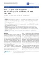
measure apoptosis [36-38] after an overnight rest. Sig-
nificant decreases in viable PBMC (Figure 3A) were
observed i n blood specimens incubated at 40°C for 8 h
(p = 0.002) and 12 h (p < 0.001). This was not seen at
the other temperature condit ions tested, even at 12 h of
incubation. CD8 populations (Figure 3B) showed signifi-
cant decreases in viability at 40°C for 8 h (p = 0.013)
and after 12 h (p = 0.03). CD4 viability (Figure 3C) was
significantly reduced after 12 h at 40°C (p = 0.03). A
greater proportion of CD4 T cells (Figure 4A and 4B)
were in early stages of apoptosis (Annexin V+, 7AAD-)
whereas a greater proportion of CD8 T cells (Figure 4C
and 4D) were in t he later stages of apoptosis (Annexin
V+, 7AAD+ ) under these same conditions. Estimates of
the odds ratio for CD4 and CD8 populations to undergo
apoptotic or necrotic cell death after exposure to 40°C
Table 1 Effect of exposure to different incubation conditions on PBMC isolation from whole blood and recovery after
cryo-preservation
Cell Yield before Cryopreservation Cell Recovery after Thawing
Exptl
RT
2h
22 h
8h
16 h
12 h
12 h
2h
22 h
8h
16 h
12 h
12 h
15°C 0.85 (0.60, 1.10)
p = 0.22
0.88 (0.63, 1.13)
p = 0.32
0.59 (0.34, 0.84)
p = 0.003
1.00 (0.70, 1.30)
p = 0.99
1.02 (0.72, 1.32) p = 0.88 0.66 (0.36, 0.97) p = 0.031
30°C 0.90 (0.65, 1.15)
p = 0.40
1.02 (0.76, 1.27)
p = 0.90
1.00 (0.75, 1.25)
p=1
1.12 (0.82, 1.42)
p = 0.41
1.20 (0.90, 1.50) p = 0.19 1.19 (0.88, 1.49) p = 0.21
40°C 0.87 (0.62, 1.12)
p = 0.30
0.83 (0.58, 1.08)
p = 0.16
0.79 (0.53, 1.06)
p = 0.12
0.91 (0.61, 1.22)
p = 0.56
0.78 (0.48, 1.08) p = 0.14 0.63 (0.32, 0.95) p = 0.026
Blood from 6 normal donors was incubated 24 h at RT (22°C, control), and for 2-12 h at 15, 22, 30 or 40°C (Exptl = Experimental), then at RT for the remainder of
24 h. PBMC were harvested and, cryopreserved, and thawed at least 1 week later. The estimated means and 95% confidence intervals of ratios of cell yield to the
control sample (RT × 24 h) are shown, both before cryopreservation and upon recovery of cells after cryopreservation. P-values are in boldface when statistically
significant.
Incubation Time
(
hours
)
and Temperature
(
C
)
of Whole Blood
A
B
C
Percent Viable
0
25
50
75
100
** ***
15° 22° 30° 40°
24 2 8 1228122812
*
15° 22° 30° 40°
24 2 8 1228122812
*
15° 22° 30° 40°
24 2 8 1228122812
Figure 3 Viability of PBMC 24 hours after thawing from liquid nitrogen. After whole blood was incubated at different temperatures f or
varying lengths of time, PBMC were isolated and cryopreserved. Samples were thawed and rested overnight at 37°C before staining with CD4,
CD8, Annexin V and 7-AAD. The viable populations were defined as Annexin V negative and 7AAD negative and are expressed as a percentage
of the respective populations of (A) PBMC, (B) CD8 and (C) CD4 lymphocytes. Shaded area on graph represents the control condition of
incubating whole blood at 22°C for 24 hours to which all other conditions were compared. (*) p = 0.003; (**) p = 0.03.
Olson et al. Journal of Translational Medicine 2011, 9:26
/>Page 6 of 13
at 8 and 12 hours, had significance levels of p = 0.0335
and p = 0.0035 for CD4 populations, and p < 0.006 for
CD8 when compared to the control storage condition
(24 h @ RT).
Effect of temperature on cell function after cryostorage
The principal cell based assay for monitoring our clini-
cal trials of immunotherapy is the ELIspot assay which
measures specific T cell r esponses by enumerating T
cells secreting cytokine (IFN-gamma) after peptide sti-
mulation. We determined whether there was an adverse
effect of temperature on t he function of l ymphocytes in
our standard ELIspot procedure. Thawed PBMC from
each temperature condition were stimulated overnight
with PMA, PHA, or CEF or were left un-stimulated.
The follo wing day, plates were developed and the num-
ber of spots recorded for each condition. Relative to
blood incubated overnight at RT, whole blood initially
incubated at 40°C for 8 h and 12 h resulted in signifi-
cant decreases in the number of IFN-gamma producing
T c ells in response to PMA (Figure 5A; p≤004). Lower
spot counts to PHA (Figure 5B) and to CEF (Figure 5C)
were observed with whole blood exposed to either 40°C
or 15°C, respectively, for 12 h, but were not statistically
significant. Incubation at 30°C for up to 12 h was
equivalent to 22°C for measures of function by ELIspot.
Package testing in high and low ambient temperatures
Packagi ng was designed by JVI ( Charlottesville, VA) for
shipping blood specimens in vacutainer tubes where
high or low ambient temperatures may be encountered
during shipping. Testing in our laboratory compared
the internal temperatures in shipping co ntainers
designed by JVI w ith that of our prior shipping con-
tainer (SafeGuard) under winter temperature conditions.
Three of the four tests are presented in Figure 6. Pre-
warmed gel packs (RT or 37C) were included to delay a
rapid decrease in the internal temperature. Each s hip-
ping container was fitted with internal and external
temperature probes and placed at 4°C or outside. I n
each condition, the internal temperatures in both types
of containers fell at approximately the same rate (Figure
6A-C, representing 3 of 4 experiments that were per-
formed). The JVI shipping container, compared to the
SafeGuard container, maintained internal temperatures
above 15°C more consistently. Gel packs conditioned at
Incubation Time
(
h
)
and Temperature
(
C
)
of Whole Blood
2 8 12 24 2 8 12 2 8 12
15° 22° 30° 40°
0
25
50
75
0
25
50
75
2 8 12 24 2 8 12 2 8 12
15° 22° 30° 40°
AB
C D
%
Annexin V+ 7AAD
% Annexin V 7AAD
Figure 4 CD8 T cells show greater susceptibility to apoptosis than CD4 T cells. The percentage of cells in different stages of apoptosis was
evaluated for CD4 and CD8 T cell populations. (A) Percentage of CD4 lymphocytes in early stages of apoptosis (Annexin V+, 7AAD-) and (B) late
stages of apoptosis (Annexin V+, 7AAD+); (C) CD8 lymphocytes in early stages of apoptosis (Annexin V+, 7AAD-) and (D) late stages of apoptosis
(Annexin V+, 7AAD+). Shaded region indicates control condition as described in Figure 3.
Olson et al. Journal of Translational Medicine 2011, 9:26
/>Page 7 of 13
37°C maintained an internal temperature above 15°C for
approximately 2 hours longer than RT-conditioned gel
packs when packages were placed at a constant external
temperature of 4°C (Table 2). When exposed to outside
temperatures as would occur during shipment in winter,
gel packs pre-warmed at 37°C helped maintain an inter-
nal temperature above 15°C for 1.8 hours longer than
gel packs conditioned at room temperature. Thereafter,
the decline of the internal temperature was similar in
all packaging conditions tested. After moving the
packages to RT, the rates at which the internal tempera-
tures increased were similar for each condition (Figure
6A, C).
The ef fects of extreme high ambient temperatures on
maintaining internal temperatures within th e range of
15-35°C was tested on the newly de signed JVI shipping
container. The shipping container was placed at 45°C
(Figure 7) and for 45 hours, the temperature remained
under 35°C. For at least 21 hours, the internal tempera-
ture stayed between 20° and 30°C.
2 8 12 24 2 8 12 2 8 12
15° 22° 30° 40°
2 8 12 24 2 8 12 2 8 12
15° 22° 30° 40°
1000
10
100
10000
2 8 12 24 2 8 12 2 8 12
15° 22° 30° 40°
SFC / 200,000 PBMC
Incubation Time
(
hours
)
and Temperature
(
C
)
of Whole Blood
A
B
C
*
*
Figure 5 Mitogen and antigen-activated PBMC responses as detected by IFNgamma secretion in an ELIspot assay. After thawing from
liquid nitrogen, PBMC were incubated 18 hours at 37°C with (A) PMA/ionomycin, (B) PHA or (C) CEF peptide pool and then tested for IFNg
secretion by ELIspot assay. Results are presented as SFC per 200,000 PBMC for PMA and PHA. CEF SFC are adjusted for the percentage of CD8+
T cells and presented as SFC per 200,000 CD8 T cells. Each condition is compared to the control condition (arrows) as described in Figure 3. (*)
p < 0.004.
A
-10
Temperature (°C)
B
C
0
10
20
30
0 5 10 15 20 25 30
Time
(
hours
)
2 4 6 8 10 12 14 16 18 5 1015202530354
0
Figure 6 Internal temperature change over time in containers designed for shipping blood specimens. Ten water-filled vacutainer vials
were pre-warmed to 37°C placed inside the JVI Control Temp shipping container or in the Safeguard (SG) shipping container, surrounded with
pre-warmed gel packs, placed inside an insulated corrugated cardboard container, and sealed with tape for testing at low external temperatures.
Internal package temperatures were continuously monitored inside JVI and SG shipping containers while placed (A) at a constant low
temperature of 4°C for 22 hours followed by 22°C for 8 hours; (B) outdoors in ambient winter temperatures for 16 hours; and (C) outdoors in
ambient winter temperatures for 18 hours followed by placement of package at 22°C for 20 hours. (green diamond) External package (ambient)
temperature; internal package temperatures: (red triangle), JVI with 37°C thermal pack; (purple square), SG with 37°C thermal pack; (blue triangle),
JVI with 22°C thermal pack; (blue square), SG with 22°C thermal pack. Solid black line indicates 15°C; dashed line denotes 0°C.
Olson et al. Journal of Translational Medicine 2011, 9:26
/>Page 8 of 13
Discussion
Recently much-needed attention has be en given to the
conditions under which blood specimen s, collected for
correlativ e studies of immune therapy, are handled prior
to PBMC isolation. How samples are processed and
shipped from trial sites as whole blood or separated
PBMC can affect the outcome of immunological moni-
toring of vaccine-based immunotherapeutic clinical
trials. Arguably, it is optimal to assay a blood sample
immediately and at the site where it is collected, as is
done for most routine clinical laborat ory tests. However,
for novel or experimental correlative studies, this is not
usually feasible, since expertise for those tests requires
specialized laboratories. Also, an argument can be made
for evaluating pre- and post-treatment blood samples in
the same assay to provide internal controls. Thus, blood
samples often are shipped to centralized laboratories for
correlative studies where they are often cryopreserved
for later batch analysis. Another question is whether
cryopreservation should be done at each site, or whether
whole blood should be shipped to t he central lab for
processing there. Several details of cryopreserva tion
methods can impact PBMC function and viability [30];
so if cell isolation and cryopreservation is done at each
site, there needs to be intensive training and quality
assurance to confirm comparable methods and results.
Though it is an o ption, this approach often is infeasible
for financial and organizational reasons. Thus, it is com-
mon for whole blood to be shipped from multiple sites
to a central laboratory for PBMC isolation and cryopre-
servation, for later analysis. However, the possible
impact of temperature during shipping, and prior to
processing, has not been systematically ad dressed. In
this study, we have focused on the effect of temperature
during shipping to assess its variation based on season
of the year, and to assess the impact of temperature on
PBMC viability and function.
In multiple studies in the HIV literature, delayed pro-
cessing of whole blood has been identified as a major
factor affecting PBMC performance in cell-based immu-
nological assays [26-29]. Delay in processing during
overnight shipping (at least 24 h) decreased responses to
microbial antigens in lymphoproliferative assays [12]
indicating the need for defined transportation conditions
for speci fic antigens. However, that study did not assess
the impact of temperature during shipping. The same
investigators also demonstrated that the way in which
frozen PBMC are thawed, and how long PBMC are
cryopreserved, will impact lymphoproliferative responses
to specific antigens [26]. Bull et al. found that the time
from phlebotomy to crypreservation should be less than
8 hours for optimal performance in cell based assays
such as ELIspot and intracellular cytokine staining
assays [27]. Delaying processing of w hole blood by 6
hours a lso impaired the response of antigen-presenting
cells to Toll-like receptor ligands [40]. On the other
hand, Whiteside et al. [41] showed the phenotype and
function of dendritic cell populations derived from
apheresis products shipped overnight were not markedly
different from DC generated from cells immediately fro-
zen after elutriation. Smith et al. showed that delayed
processing of blood resulted in a decrease in cell viabi-
lity as well as a marked reduction in IFNg SFC in
response to varicella zoster antigen [29]; the presence of
DNase partially restored the response [42]. K ierstead et
al. [28] demonstrated that cryopreservation of PBMC
should be done within 12 hours of phlebotomy. How-
ever , in these two prior studies , the whole blood [29] or
Table 2 Pre-warmed gel packs extend the time above
15°C when shipping at cold temperatures
Outside Temperature Gel Pack Temperature Hours
Safe-Guard JVI
4°C 37°C 3.5 4.5
RT 1.8 2.0
Ambient 37°C 3.4 5.9
RT 2.5 3.2
Gel packs pre-warmed at RT or 37°C were packed inside blood specimen
shipping containers along with probes to measure the internal and
temperature after sitting overnight in a constant 4°C cold room or outdoors
where temperatures fell below freezing. The number of hours the internal
temperature remained above 15°C is show.
0
10
20
30
40
50
0 5 10 15 20 25 30 35 40 45
H
ou
r
s
Degrees
C
entigrade
Figure 7 Temperature performance test of the JVI Control
Temp shipping container. Five vials, filled with water conditioned
at 20°C, were suspended inside the foam vial holder and placed
inside the plastic clamshell plastic box fitted with small foam pads.
Two of the vials each had a T thermocouple taped to it. The
clamshell package was put inside the insulated corrugated cardboard
box in which two 12 oz. Control Temp gel packs conditioned at 20°C
were also placed inside and taped shut. The shipping container was
set inside a 45°C chamber for forty-five hours and the internal
package temperature recorded as described in Methods. The red line
indicates the external temperature of the chamber. The blue line
represents the average internal temperature of the shipping
container obtained from duplicate temperature probes.
Olson et al. Journal of Translational Medicine 2011, 9:26
/>Page 9 of 13
PBMC[28]wasstoredorshippedat4°Covernight
before cryopreservation. It is not known whether there
were negative effects from storing or shipping at 4°C.
Our data show that there is better viability, cell yield,
and function when cells are shipped at room tempera-
ture (22°C) or 30°C than at 15°C, and it is generally
accepted that storage of whole blood at 4°C negatively
impacts cell viability [5], function [29], and population
recovery [6,7,43,44]. Acknowledging the range of data in
the literature, in a separate study, we are also evaluating
the function of PBMC processed the same day (< 8 h)
or after overnight shipping or storage (manuscript in
preparati on). However the current manuscript focuses
on the impact of temperature during shipping in those
cases when overnight shipping is necessary.
In some prior studies, statistical differences between
immediate and delayed processing of specimens were
influencednotonlybythedelayinprocessingbutalso
by the method o f processing and by the type of antic-
oagulant used [27]. Thus, although there was a statis-
tical decrease in viability and recovery when whole
blood was collected in heparin and isolated by Accus-
pin technology (centrifuge tube divided into two
chambers by means of a porous high-density polyethy-
lene barrier, known as a frit), no significant d ecrease
was evident when PBMC were collected at the inter-
face of plasma and Ficoll. Similarly, significant differ-
ences in viability (but not recovery) between fresh and
delayed samples were evident when collected in ACD
or EDTA anticoagulants but not in heparin when
PBMC were isolated directly onto a Ficoll cushion.
Furthermore, the functionality of PBMC was not sig-
nificantly impaired by either method when measured
in an IFNg-ELIspot assay in response to the CEF pool
of peptides.
The observations leading to the present study come
from the multi-center clinical trials we have conducted
at the U niversity of Virginia in collaboration with Can-
cer c enters in Houston TX and Philadelphia PA. Blood
specimens shipped from these locations encounter
extreme seasonal climate conditions. On the other hand,
blood specimens at the on-site location are, for t he
most part, collected, stored and processed with no expo-
sure to extreme temperatures and pr ocessed either on
the same d ay or after storage overnight at room tem-
perature. This study has addressed 1) the seasonal
changes in temperature inside packages of blood speci-
mens during shipping in the U.S., 2) changes in tem-
perature i nside packages simulat ing hot or cold ambient
temperatures during shipping, and 3) the effects of tem-
peratures above and below room temperature on PBMC
numbers, viability, and function. These studi es ar e rele-
vant to shipping blood specimens for correlative studies
in many settings.
We are not aware o f prior work tracking temperature
ranges encountered within blood shipping containe rs or
their variation by season of the year. We found that
shipping of blood in insulated containers by contracted
overnight carriers is associated with large seasonal varia-
tions in temperature inside the packaging, ranging from
-1°C in winter to 35°C in summer, with most in the
range of 4°-32°C. Thus, blood samples in transit are fre-
quently e xposed to high temperatures at or above 30°C
and low temperatures that approach or go below freez-
ing temperatures at least transiently. The monitoring
devices used in these shipments recorded the minimum
and maximum temperatures but not the duration of
each temperature. Thus, we also studied the changes
over time in a dynamic manner in hot or cold condi-
tions designed to mimic changes that may occur during
shipping, and found that insulation maintains internal
temperature below 30°C for up to 21 hours in ambient
temperatures that likely exceed those experienced dur-
ing shipping (45°C). We found in very cold ambient
conditions, that the insulated containers maintained the
internal temperatures above 15°C for almost 6 hours
and above 20°C for over 3 hours, with the aid of thermal
packs pre-warmed at 37°C.
We have found that incubation of whole blood at 50°C
caused unacceptably high loss of PBMC (data not
shown). Storage at 15°C or 40°C for 12 h causes signifi-
cant decreases in cell yields, viability and/or function
but exposure to those temperatures for 2 hours, or in
some cases even 8 hours is associated with PBMC
yields, viabili ty and function comparable to those found
from blood stored at RT. The apoptosis rates in this
study of about 30-35% in thawed cells incubated over-
night are higher than observed in prior work where
apoptosis was measur ed directly after thawing [32]. It is
not u ncommon, however, that cells undergo a delayed-
onset cell death (reviewed by Baust [39]) which may
account for the increase in apoptosis measured here.
Other studies also confirm that the total viability
decreases after overnight incubation [28]. Regardless, we
find that there is function in the PBMC that are viable
after overnight incubation .Incubationat15or30°Cis
associated with comparable T cell f unction assessed by
ELIspot assay to that seen with PBMC stored at RT.
Interestingly, we found that i ncubation at 30°C for peri-
ods up to 12 h was even associated with equivalent or
better yields, viability and function compared to samples
left at RT. However, incubation at 15°C or 40°C for 8-12
h was associated with decreased viability and function.
Colder temperature (15°C) primarily affected cell yield
after Ficoll separation and reduced recovery following
cryopreservation. Recovery may be due to a perturbation
in cell density [7] or formation of cell aggregates [5,45].
No increase in apoptosis relative to that seen when
Olson et al. Journal of Translational Medicine 2011, 9:26
/>Page 10 of 13
blood was stored at room temperature was noted at
lower temperatures which is consistent with other data
showing reduced apoptotic rate of PBMC held at 4°C
for 24 hours [46].
We currently endea vor to keep samples in the range
of 20-30°C during shipping. In winter months, it helps
to start with samples warm (e.g. 37°C) and we recom-
mend shipping blood at 30-37°C, and recommend
including a “warm pack” at 37°C in the container, which
we found provides an extra 1-2 h ours protection from
extreme cold during winter ambient conditions
(Table 3). On the other hand, during summer months,
we recommend shipping at RT, to allow for some
increase before exceeding 30°C. Inclusion of a fluid pack
at RT may a lso help to buffer the temperature changes
during shipping in the summer.
We propose that controlled and monit ored shipping
temperatures may mitigate negative effects of shipping
blood in multicenter trials. Careful attention to the ship-
ping containers and testing in ambient temperature is
recommended. Certainly o ne way to prevent negative
effects of cold or hot te mperatures during shipping of
blood specimens is to isolate PBMC or other cellular
elements prior to shipping, and either to cry opreserve
them or to assay them on site. This introduces other
sources of error and substantial costs, by the need to
maintain quality control assay validation across multiple
laboratori es, which is problematic. We believ e there is a
role for shipping blood specimens for centralized assays
where those assays can be performed in batches with
appropriate controls, but attention to details of shipping
conditions are warranted in such circumstances, to max-
imize the reliability of the results. It also is appropriate,
in multicenter trials, to stratify patients by institution to
control for systematic variations in temperature during
shipping that may be encountered depending on the
latitude of the institution and the shipping distance.
Conclusions
Blood packages shipped overnight by commercial carrier
may encounter extreme seasonal temperatur es. Warmer
temperatures favor greater cell yields of shipped blood
specimens whereas colder temperatures for long periods
of time lower cell recovery and viability. Temperatures
≥40C for ≥8 hours reduces cell viability and f unctional-
ity after cryo-preservation. In the design of containers
for blood shipment, maintaining an ambient tempera-
ture between 22°C and 30°C should be considered.
Additional material
Additional file 1: Comparable cell numbers were derived from
complete and differential blood counts at each of the 3 hospital
trial centers participating in this study. The mean, median, 25
th
and
75
th
quartiles for lymphocyte and monocyte populations in the
peripheral blood are presented. Values are expressed as million of cells
per mL of blood. 1-Virginia; 7-Texas; 9-Pennsylvania.
Additional file 2: Flow diagram depicting the sequence of events in
the in Vitro study on time and temperature of whole blood storage
prior to cryopreservation and functional analysis. Approximately 60
mL of whole blood from six healthy donors were collected into
heparinized vacutainers. Aliquots were divided equally among ten
conditions: nine experimental conditions in which blood was exposed to
various temperatures for a defined length of time, then placed at RT (22°
C) for the remainder of the 24 h storage period and one reference
condition in which whole blood was stored overnight at RT. After
storage, PBMC were collected after Ficoll separation, counted and
cryopreserved. After 1-4 weeks, PBMC were removed for liquid nitrogen
and cell recovery, viability, phenotype, and function were determined.
Additional file 3: Proportions of CD4 and CD8 populations in PBMC
did not change under various temperature conditions, compared to
RT control conditions. Blood from 6 normal donors was incubated 24 h
at RT (22°C, control), and for 2-12 h at 15, 22, 30 or 40°C (Exptl), then RT
for the remainder of 24 h. PBMC were harvested, cryo-preserved, and
then thawed at least 1 week later. Samples were stained with
fluorescently labeled anti-CD3, anti-CD4 and anti-CD8 antibodies before
flow cytometric analysis. The proportion of CD4
+
cells among PBMC was
measured as the number of CD3
+
CD4
+
cells divided by the total PBMC.
Similarly, the proportion of CD8+ cells among PBMC was measured as
the number of CD3
+
CD8
+
cells divided by the total PBMC. The ratios of
these CD4 and CD8 proportions are reported in this table, for each
temperature condition, compared to control samples left at RT for 24 h.
The estimated means, 95% confidence intervals and p-value of these
ratios are shown for CD4 and CD8 populations.
Acknowledgements
The authors express their gratitude for R.N.C. Industries Inc. Lab, 640
Langford Drive, Norcross GA 30071 in conducting the temperature
performance test for the JVI Control Temp shipping container. This study
was funded by NIH/NCI grants R01 CA118386 and R21 CA103528 (to C.L.S).
Support was also provided by the University of Virginia Cancer Center
Support Grant (NIH/NCI P30 CA44579: Clinical Trials Office, Biorepository and
Tissue Research Facility, Flow Cytometry Core, and Biomolecular Core
Facility); the UVA General Clinical Research Center (NIH M01 RR00847). Also,
philanthropic support was provided from the Commonwealth Foundation
for Cancer Research and Alice and Bill Goodwin. Additional philanthropic
support was provided by Frank and Jane Batten, the James and Rebecca
Craig Foundation, George S. Suddock, Richard and Sherry Sharp, and the
Patients and Friends Research Fund of the University of Virginia Cancer
Center. No corporate funding support was provided for this study.
Author details
1
Human Immune Therapy Center, University of Virginia, Charlottesville, VA,
USA.
2
Dept. of Public Health Sciences, University of Virginia, Charlottesville,
VA, USA.
3
Atlantic Research Group, 125 S. Augusta Street, Suite 3000,
Staunton, VA, USA.
4
1901 E. Market Street, Charlottesville, VA, USA.
5
9652 S.
Michigan, Chicago, IL, USA.
6
JVI, LLC, 615 Cami Lane, Charlottesville, VA, USA.
7
Dept. of Surgery, University of Virginia, Charlottesville, VA, USA.
Authors’ contributions
WCO performed the in vitro studies, data analyses and writing the
manuscript. MES performed all the statistical analysis, writing relevant
Table 3 Recommendations for shipping whole blood
specimens
Time of Year Ambient Temperature Packing
Winter 30-37°C 37°C warm packs
Summer RT RT packs
Olson et al. Journal of Translational Medicine 2011, 9:26
/>Page 11 of 13
sections of the manuscript and editing. EF assisted in the gathering and
organization of shipping data. RJF was instrumental in concept of study.
ARC assisted in the in vitro stud ies and data analysis. JHF designed and
tested the shipping container from JVI. KAC-B developed the plan for the in
vitro studies. CLS conceived study, participated in its design and
coordination, and helped draft the manuscript. All authors read and
approved the final manuscript.
Competing interests
JVI is a corporate entity based in Charlottesville, VA, that was contracted to
make packaging for blood shipment. The CEO of JVI is Jon Fink, who is
included as a co-author for his scientific contributions to package design. He
is married to Robyn Fink who was a UVA employee with this research team
and who managed the multicenter trials including the tracking and
monitoring of blood samples shipped from outside sites. The packaging
prepared by JVI to meet specifications of the research team was purchased
by the University of Virginia Human Immune Therapy Center and used
(when, relative to these data) for shipping blood specimens.
Received: 26 October 2010 Accepted: 8 March 2011
Published: 8 March 2011
References
1. Vaught JB: Blood collection, shipment, processing, and storage. Cancer
Epidemiol Biomarkers Prev 2006, 15:1582-1584.
2. Vaught JB, Caboux E, Hainaut P: International efforts to develop
biospecimen best practices. Cancer Epidemiology Biomarkers & Prevention
2010, 19:912-915.
3. Hallmans G, Vaught JB: Best Practices for Establishing a Biobank. In
Methods in Biobanking. Edited by: Dillner J. Springer Science+Business
Media, LLC; 2011:241-260.
4. Leyland-Jones BR, Ambrosone CB, Bartlett J, Ellis MJC, Enos RA, Raji A,
Pins MR, Zujewski JA, Hewitt SM, Forbes JF, Abramovitz M, Braga S,
Cardoso F, Harbeck N, Denkert C, Jewell SD: Recommendations for
collection and handling of specimens from group breast cancer clinical
trials. J Clin Oncol 2008, 26:5638-5644.
5. Ashmore LM, Shopp GM, Edwards BS: Lymphocyte subset analysis by flow
cytometry. Comparison of three different staining techniques and effects
of blood storage. Journal of Immunological Methods 1989, 118:209-215.
6. Garraud O, Moreau T: Effect of blood storage on lymphocyte
subpopulations. Journal of Immunological Methods 1984, 75:95-98.
7. Weiblen BJ, Debell K, Giorgio A, Valeri CR: Monoclonal antibody testing of
lymphocytes after overnight storage. Journal of Immunological Methods
1984, 70:179-183.
8. Kaplan J, Nolan D, Reed A: Altered lymphocyte markers and blastogenic
responses associated with 24 hour delay in processing of blood
samples. Journal of Immunological Methods 1982, 50:187-191.
9. Bongers V, Bertrams J: The influence of common variables on T cell
subset analysis by monoclonal antibodies. Journal of Immunological
Methods 1984, 67:243-253.
10. Nicholson JKA, Jones BM, Cross GD, McDougal JS: Comparison of T and B
cell analyses on fresh and aged blood. Journal of Immunological Methods
1984, 73:29-40.
11. Betensky RA, Connick E, Devers J, Landay AL, Nokta M, Plaeger S,
Rosenblatt H, Schmitz JL, Valentine F, Wara D, Weinberg A, Lederman M:
Shipment impairs lymphocyte proliferative responses to microbial
antigens. Clin Vaccine Immunol 2000, 7:759-763.
12. Weinberg A, Betensky RA, Zhang L, Ray G: Effect of shipment, storage,
anticoagulant, and cell separation on lymphocyte proliferation assays for
human immunodeficiency virus-infected patients. Clin Vaccine Immunol
1998, 5:804-807.
13. Lewis JJ, Janetzki S, Schaed S, Panageas KS, Wang S, Williams L, Meyers M,
Butterworth L, Livingston PO, Chapman PB, Houghton AN: Evaluation of
CD8+ T-cell frequencies by the Elispot assay in healthy individuals and
in patients with metastatic melanoma immunized with tyrosinase
peptide. Int J Cancer 2000, 87:391-398.
14. Scheibenbogen C, Lee K-H, Stevanovic S, Witzens M, Waldmann V,
Naeher H, Rammensee H-G, Keilholz U: Analysis of the T cell response to
tumor and viral peptide antigens by an IFN[gamma]-elispot assay. Int J
Cancer 1997,
71:932-936.
15.
Schmittel A, Keilholz U, Scheibenbogen C: Evaluation of the interferon-
[gamma] ELISPOT-assay for quantification of peptide specific T
lymphocytes from peripheral blood. Journal of Immunological Methods
1997, 210:167-174.
16. Jung T, Schauer U, Heusser C, Neumann C, Rieger C: Detection of
intracellular cytokines by flow cytometry. Journal of Immunological
Methods 1993, 159:197-207.
17. Letsch A, Scheibenbogen C: Quantification and characterization of
specific T-cells by antigen-specific cytokine production using ELISPOT
assay or intracellular cytokine staining. Methods 2003, 31:143-149.
18. Maecker HT, Dunn HS, Suni MA, Khatamzas E, Pitcher CJ, Bunde T,
Persaud N, Trigona W, Fu TM, Sinclair E, Bredt BM, McCune JM, Maino VC,
Kern F, Picker LJ: Use of overlapping peptide mixtures as antigens for
cytokine flow cytometry. Journal of Immunological Methods 2001,
255:27-40.
19. Maino VC, Maecker HT: Cytokine flow cytometry: a multiparametric
approach for assessing cellular immune responses to viral antigens.
Clinical Immunology 2004, 110:222-231.
20. Britten C, Janetzki S, Ben-Porat L, Clay T, Kalos M, Maecker H, Odunsi K,
Pride M, Old L, Hoos A, Romero P, for the HLA-peptide Multimer Proficiency
Panel of the CVC-CRI Immune Assay Working Group: Harmonization
guidelines for HLA-peptide multimer assays derived from results of a
large scale international proficiency panel of the Cancer Vaccine
Consortium. Cancer Immunology, Immunotherapy 2009, 58:1701-1713.
21. Frelinger J, Ottinger J, Gouttefangeas C, Chan C: Modeling flow cytometry
data for cancer vaccine immune monitoring. Cancer Immunology,
Immunotherapy 2010, 59:1435-1441.
22. Altman JD, Moss PAH, Goulder PJR, Barouch DH, Heyzer-Williams MG, Bell JI,
McMichael AJ, Davis MM: Phenotypic analysis of antigen-specific T
lymphocytes. Science 1996, 274:94-96.
23. Axelsson S, Faresj÷ M, Hedman M, Ludvigsson J, Casas R: Cryopreserved
peripheral blood mononuclear cells are suitable for the assessment of
immunological markers in type 1 diabetic children. Cryobiology 2008,
57:201-208.
24. Kreher CR, Dittrich MT, Guerkov R, Boehm BO, Tary-Lehmann M: CD4+ and
CD8+ cells in cryopreserved human PBMC maintain full functionality in
cytokine ELISPOT assays. Journal of Immunological Methods 2003,
278:79-93.
25. Maecker H, Moon J, Bhatia S, Ghanekar S, Maino V, Payne J, Kuus-Reichel K,
Chang J, Summers A, Clay T, Morse M, Lyerly HK, DeLaRosa C, Ankerst D,
Disis M: Impact of cryopreservation on tetramer, cytokine flow
cytometry, and ELISPOT. BMC Immunology 2005, 6:1-17.
26. Weinberg A, Song LY, Wilkening C, Sevin A, Blais B, Louzao R, Stein D,
Defechereux P, Durand D, Riedel E, Raftery N, Jesser R, Brown B, Keller MF,
Dickover R, McFarland E, Fenton T, for the Pediatric ACTG Cryopreservation
Working Group: Optimization and limitations of use of cryopreserved
peripheral blood mononuclear cells for functional and phenotypic T-cell
characterization. Clin Vaccine Immunol 2009, 16:1176-1186.
27. Bull M, Lee D, Stucky J, Chiu YL, Rubin A, Horton H, McElrath MJ: Defining
blood processing parameters for optimal detection of cryopreserved
antigen-specific responses for HIV vaccine trials. Journal of Immunological
Methods
2007, 322:57-69.
28.
Kierstead LS, Dubey S, Meyer B, Tobery TW, Mogg R, Fernandez VR, Long R,
Guan L, Gaunt C, Collins K, Sykes KJ, Mehrotra DV, Chirmule N, Shiver JW,
Casimiro DR: Enhanced rates and magnitude of immune responses
detected against an HIV vaccine: Effect of using an optimized process
for isolating PBMC. AIDS Research and Human Retroviruses 2007, 23:86-92.
29. Smith JG, Levin M, Vessey R, Chan ISF, Hayward AR, Liu X, Kaufhold RM,
Clair J, Chalikonda I, Chan C, Bernard M, Wang WW, Keller P, Caulfield MJ:
Measurement of cell-mediated immunity with a varicella-zoster virus-
specific interferon-+¦ ELISPOT assay: Responses in an elderly population
receiving a booster immunization. J Med Virol 2003, 70:S38-S41.
30. Disis ML, dela Rosa C, Goodell V, Kuan LY, Chang JCC, Kuus-Reichel K,
Clay TM, Kim Lyerly H, Bhatia S, Ghanekar SA, Maino VC, Maecker HT:
Maximizing the retention of antigen specific lymphocyte function after
cryopreservation. Journal of Immunological Methods 2006, 308:13-18.
31. Weinberg A, Song LY, Wilkening CL, Fenton T, Hural J, Louzao R, Ferrari G,
Etter PE, Berrong M, Canniff JD, Carter D, Defawe OD, Garcia A, Garrelts TL,
Gelman R, Lambrecht LK, Pahwa S, Pilakka-Kanthikeel S, Shugarts DL,
Tustin NB: Optimization of storage and shipment of cryopreserved
peripheral blood mononuclear cells from HIV-infected and uninfected
Olson et al. Journal of Translational Medicine 2011, 9:26
/>Page 12 of 13
individuals for ELISPOT assays. Journal of Immunological Methods 2010,
363:42-50.
32. Smith JG, Joseph HR, Green T, Field JA, Wooters M, Kaufhold RM,
Antonello J, Caulfield MJ: Establishing acceptance criteria for cell-
mediated-immunity assays using frozen peripheral blood mononuclear
cells stored under optimal and suboptimal conditions. Clin Vaccine
Immunol 2007, 14:527-537.
33. Slingluff CL, Petroni GR, Chianese-Bullock KA, Smolkin ME, Hibbitts S,
Murphy C, Johansen N, Grosh WW, Yamshchikov GV, Neese PY,
Patterson JW, Fink R, Rehm PK: Immunologic and clinical outcomes of a
randomized phase II trial of two multipeptide vaccines for melanoma in
the adjuvant setting. Clin Cancer Res 2007, 13:6386-6395.
34. Slingluff CL, Petroni GR, Olson WC, Smolkin ME, Ross MI, Haas NB,
Grosh WW, Boisvert ME, Kirkwood JM, Chianese-Bullock KA: Effect of
granulocyte/macrophage colony-stimulating factor on circulating CD8+
and CD4+ T-cell responses to a multipeptide melanoma vaccine:
outcome of a multicenter randomized trial. Clin Cancer Res 2009,
15:7036-7044.
35. Currier JR, Kuta EG, Turk E, Earhart LB, Loomis-Price L, Janetzki S, Ferrari G,
Birx DL, Cox JH: A panel of MHC class I restricted viral peptides for use
as a quality control for vaccine trial ELISPOT assays. Journal of
Immunological Methods 2002, 260:157-172.
36. Martin SJ, Reutelingsperger CPM, McGahon AM, Rader JA, van Schie RC,
LaFace DM, Green DR: Early redistribution of plasma membrane
phosphatidylserine is a general feature of apoptosis regardless of the
initiating stimulus: inhibition by overexpression of Bcl-2 and Abl. Journal
of Experimental Medicine 1995, 182:1545-1556.
37. Schmid I, Uittenbogaart CH, Keld B, Giorgi JV: A rapid method for
measuring apoptosis and dual-color immunofluorescence by single laser
flow cytometry. Journal of Immunological Methods 1994, 170:145-157.
38. Vermes I, Hannen C, Steffens-Nakken H: A novel assay for apoptosis. Flow
cytometric detection of phosphatigylserine expression on early
apoptotic cells using fluorescein labelled Annexin V. Journal of
Immunological Methods 1995, 184:39-51.
39. Baust JM: Molecular mechanisms of cellular demise associated with
cryopreservation failure. Cell Preservation Technology 2002, 1:17-31.
40. Meier A, Fisher A, Sidhu HK, Chang JJ, Wen TF, Streeck H, Alter G,
Silvestri G, Altfeld M: Rapid loss of dendritic cell and monocyte responses
to TLR ligands following venipuncture. J Immunol Methods 2008,
339:132-140.
41. Whiteside TL, Griffin DL, Stanson J, Gooding W, McKenna D, Sumstad D,
Kadidlo D, Gee A, Durett A, Lindblad R, Wood D, Styers D: Shipping of
therapeutic somatic cell products. Cytotherapy 2010, 1-13, Early Online.
42. Smith JG, Liu X, Kaufhold RM, Clair J, Caulfield MJ: Development and
validation of a gamma interferon ELISPOT assay for quantitation of
cellular immune responses to varicella-zoster virus. Clin Vaccine Immunol
2001, 8:871-879.
43. McKenna KC, Beatty KM, Vicetti Miguel R, Bilonick RA: Delayed processing
of blood increases the frequency of activated CD11b+ CD15+
granulocytes which inhibit T cell function. Journal of Immunological
Methods 2009, 341
:68-75.
44. Van Lambalgen R, Van Meurs GJE: Lymphocyte subpopulations do not
alter during blood storage at 4C. Journal of Immunological Methods 1985,
80:39-43.
45. De Paoli P, Villalta D, Battistin S, Gasparollo A, Santini G: Letter to the
editor. Journal of Immunological Methods 1983, 61:259-260.
46. Bergman M, Bessler H, Salman H, Djaldetti M: Relationship between
temperature and apoptosis of human peripheral blood mononuclear
cells. International Journal of Hematology 2003, 77:351-353.
doi:10.1186/1479-5876-9-26
Cite this article as: Olson et al.: Shipping blood to a central laboratory
in multicenter clinical trials: effect of ambient temperature on specimen
temperature, and effects of temperature on mononuclear cell yield,
viability and immunologic function. Journal of Translational Medicine 2011
9:26.
Submit your next manuscript to BioMed Central
and take full advantage of:
• Convenient online submission
• Thorough peer review
• No space constraints or color figure charges
• Immediate publication on acceptance
• Inclusion in PubMed, CAS, Scopus and Google Scholar
• Research which is freely available for redistribution
Submit your manuscript at
www.biomedcentral.com/submit
Olson et al. Journal of Translational Medicine 2011, 9:26
/>Page 13 of 13