Báo cáo sinh học: "Enhanced presentation of MHC class Ia, Ib and class II-restricted peptides encapsulated in biodegradable nanoparticles: a promising strategy for tumor immunotherapy" docx
Bạn đang xem bản rút gọn của tài liệu. Xem và tải ngay bản đầy đủ của tài liệu tại đây (1.4 MB, 10 trang )
RESEARC H Open Access
Enhanced presentation of MHC class Ia, Ib and
class II-restricted peptides encapsulated in
biodegradable nanoparticles: a promising
strategy for tumor immunotherapy
Wenxue Ma
1*
, Trevor Smith
2
, Vladimir Bogin
3
, Yu Zhang
4
, Cengiz Ozkan
4
, Mihri Ozkan
4
, Melanie Hayden
1
,
Stephanie Schroter
1
, Ewa Carrier
1
, Davorka Messmer
1
, Vipin Kumar
2
and Boris Minev
1,5,6*
Abstract
Background: Many peptide-based cancer vaccines have been tested in clinical trials with a limited success, mostly
due to difficulties associated with peptide stability and delivery, resulting in inefficient antigen presentation.
Therefore, the development of suitable and efficient vaccine carrier systems remains a major challenge.
Methods: To address this issue, we have engineered polylactic-co-glycolic acid (PLGA) nanoparticles incorporating:
(i) two MHC class I-restricted clinically-relevant peptides, (ii) a MHC class II-binding peptide, and (iii) a non-classical
MHC class I-binding peptide. We formulated the nanoparticles utilizing a double emulsion-solvent evaporation
technique and characterized their surface morphology, size, zeta potential and peptide content. We also loaded
human and murine dendritic cells (DC) with the peptide-containing nanoparticles and determined their ability to
present the encapsulated peptide antigens and to induce tumo r-specific cytotoxic T lymphocytes (CTL) in vitro.
Results: We confirmed that the nanoparticles are not toxic to either mouse or human dendritic cells, and do not
have any effect on the DC maturation. We also demonstrated a significantly enhanced presentation of the
encapsulated peptides upon internalization of the nanoparticles by DC, and confirmed that the improved peptide
presentation is actually associated with more efficient generation of peptide-specific CTL and T helper cell
responses.
Conclusion: Encapsulating antigens in PLGA nanoparticles offers unique advantages such as higher efficiency of
antigen loading, prolonged prese ntation of the antigens, prevention of peptide degradation, specific targeting of
antigens to antigen presenting cells, improved shelf life of the antigens, and easy scale up for pharmaceutical
production. Therefore, these findings are highly significant to the development of synthetic vaccines, and the
induction of CTL for adoptive immunotherapy.
Background
In recent years, peptides derived from tumor-associated
antigens (TAA) have been identified for a variety of
human cancers [1]. Thus far, however, effective peptide
vaccination of patients with cancer has been limit ed to
very few trials [2]. The relative paucity of responsiveness
after conventional peptide vaccination is mostly due to
the high levels of protein degradation, limiting antigen
delivery. Polymeric nanoparticles (NP) may allow encap-
sulation of peptides inside a polymeric matrix, protect-
ing them against enzymatic and hydrolytic degradation.
In addition, the nanoparticle vaccine approach offers the
possibility of developing tailor-made vaccines containing
specific targets or molecules that may improve their
function [3].
Dendritic c ells (DC) are the most potent professional
antigen-presenting cells (APC), having the ability to
initiate primary immune responses [4]. Therefore,
immunotherapy utilizing DC has become a promising
* Correspondence: ;
1
Moores UCSD Cancer Center, University of California San Diego
Full list of author information is available at the end of the article
Ma et al. Journal of Translational Medicine 2011, 9:34
/>© 2011 Ma et al; licensee BioMed Central Ltd. This is an Open Access article distributed under the terms of the Creative Commons
Attribution License ( whi ch permi ts unrestricted use, distribution, and reproduction in
any medium, provided the original work is properly cited.
therapeutic modal ity in recent years [5-7]. However, the
lack of efficient and long-lasting antigen presentation by
DC in vivo has been a major difficulty in the develop-
ment of effective vaccines. These obstacles could be cir-
cumvented through the development of nanoparticles,
which can efficiently d eliver the antigenic peptides into
the APC.
Our recent studies have characterized distinct subsets
of regulatory CD4+FOXP3- and CD8aa+TCRab+T
cells that target autoaggressive Vb8.2+ T cell responses
for down-regulation and protection against autoimmune
disease [8-13]. Several non-classical MHC class I, Qa-
1a-restricted CD8aa+TCRab+ T cell and MHC class
II-restricted CD4+ T cell clones and lines have been
characterized [8,9,11]. The CD4+ T cells recognize a
TCR-derived peptide in the context of a class II MHC
molecule, I-Au [11]. However, CD8aa+TCRab+ T cells
are cytotoxic and recognize another TCR-derived peptide
bound to a class Ib molecule, Qa-1 [8,9]. These cells are
physiologically primed and operate in unison to assist in
recovery from T cell-mediated experimental autoimmune
encephalomyelitis in H-2
u
mice [10,13,14]. Interestingly,
recent data suggest that class Ib MHC-restricted cyto-
toxic CD8+ T cells also play an important role in anti-
tumor immunity [15,16]. Therefore, it is important to
examine whether CTL can be effectively primed using
nanoparticle containing class Ib-binding peptides.
To engineer our nanoparticle-based vaccines, we uti-
lized the biodegradable and biocompatible polymer
PLGA, which is already approved by the US FDA
[17,18]. PLGA is easy to formulate into different devices
and has generated immense interest because of its favor-
able properties, which include good biocompatibility,
biodegradability, and mechanical strength [17,18].
Here we demonstrate for t he first time efficient nano-
particle-facilitated loading of class I-restricted, clinically
relevant TAA-derived peptides to human DC, and the
development of nanopartic le s incorporating MHC class
II and non-classical MHC class I, Qa-1-binding peptides
that are able to stimulate helper CD4+ and cytotoxic
CD8aa+TCRab+ T cells. Importantly, we confirmed
that the enhanced peptide presentation by the NP-
loaded DC is associated with more efficient generation
of antitumor CTL.
Methods
Antibodies and reagents
Antibodies: Anti-human IFN-g (mAb 1-D1K) - Mabtech
Inc. (Mariemont, OH), anti-HLA-A2-FITC, anti-HLA-DR-
PE, anti-CD83-PE, anti-CD80-FITC, anti-CD86-FITC,
mouse-IgG1-FITC, mouse-IgG1-PE, and mouse-IgG2a-PE
all from BD (San Diego, CA). Cytokines: GM-CSF - BER-
LEX (Richmond, CA), Interleukin 4 (IL-4) - PeproTech
(Rocky Hill, NJ), IL-2 - Chiron (Emeryville, CA). Peptides:
MART-1
27-35
,gp100
209-217
and mSTEAP
326-335
-Gen-
Script Corp. (Piscataway, NJ), murine TCR Vb8.2 chain
peptides: B5 (76-101) - Caltech (Pasadena, CA), and p42
(42-50) - Synthetic Biomolecules (San Diego, CA). PLGA -
Birmingham Polymers (Birmingham, AL), bovine serum
albumin (BSA) and Poly (vinyl alcohol) (PVA) - Sigma-
Aldrich (St. Louis, MO), coumarin 6 - Polyscience
(Warrington, PA).
Cell lines
T2 cell line [19] was purchased from ATCC (Manassas,
VA). Human melanoma cell lines 624 and 1351, as well
as human tumor-infiltrating lymphocytes (TIL) cell lines
TIL1235 an d TIL1520 were kindly provided by Dr. John
R Wunderlich (NIH/NCI, Bethesda, MD). CD4+TCRab
+(B5.1)andCD8aa+TCRab+ (XT14), [8] T cell lines
were generated in H-2u background and were specific
for the Vb8.2TCR-derived peptides, B5 and p42-50,
respectively. The CD4+TCRab+ (B5.1) cell line was gen-
erated from the draining lymph nodes of a H-2u mouse
immunized i.p. with 20 μg of TCR peptide B5.
Nanoparticle formulation
Peptide-containing NP were formulated using a double
emulsion-solvent evaporati on techni que as we described
previously [20]. For optimizing the peptide dose
entrapped in the NP, 300 μg, 600 μg and 1 mg of each
peptide was formulated into the PLGA polymer in each
NP batch. Some NP were formulated with a f luorescent
dye (coumarin 6) by adding 100 μg of coumarin 6 to
the polymer solution prior to emulsification.
Nanoparticle characterization
To characterize the surface morphology of the NP we
utilized a scanning electron microscope (SEM). Particle
size analysis and zeta potential determination was carried
out using a Zetasizer (Malvern, Worcestershire, UK). The
peptide content of the peptide-loaded NP was deter-
mined by HPLC using a C18 column (Waters, Milford,
MA). Specifically, the peptides and nanoparticles were
separated and identified using ultraviolet (UV) detection
and known standards, at a wavelength of 280nm
(attenuation 0.002 AU). An aliquot (50 μl) was injected
onto the column and eluted with a mobile phase contain-
ing a gradient mixture of reagent A, 0 .1% trifluoroacetic
acid (TFA) in water (Sigma Aldrich St Louis, MO, USA),
and reagent B, 0.1% TFA in Acetonitrile. The gradient
times were as follows: 0-23 minutes, 75% A and 25% B;
23 -25 minutes, 0% A and 100% B. Total run tim e was 25
minutes at a flow rate of 0.8 ml per minute.
Generation of Human DC
Peripheral blood mononuclear cells (PBMC) isolated
from buffy coats of healthy donors were allowed to
Ma et al. Journal of Translational Medicine 2011, 9:34
/>Page 2 of 10
adhere in 6-well plates for 1 hour. The adherent cells
were cultured with 1000 U/ml GM-CSF and 300 U/ml
IL-4, with cytokines added o n days 2, 4, and 6. Lipopo-
lysaccharide (LPS) was added to the culture medium on
day 7, and two days later, the mature DC (mDC) were
harvested and characterized by FACS using antibodies
against HLA-DR, CD80, CD83, and CD86.
Generation of murine bone marrow-derived dendritic
cells (BMDC)
Murine DC were derived from tibias and femurs by flush-
ing out the bone marrow with RPMI medium as described
[21] and cultured with 10 ng/ml IL-4 and 25 ng/ml GM-
CSF for 5 days, with cytokines added on day 3.
Nanoparticle uptake imaging studies
Human immature DC (imDC) were seeded overnight on
sterile cover slips and incubated with NP containing
coumarin-6 for 1-hr at 37°C. Then, the cover slips were
washed and observed with a fluorescent microscope. For
confocal microscopy, imDC were incubated in 4-well
chamber slides for 1 hour at 37°C with 100 μg/ml Cou-
marin 6-containing NP, washed and fixed with parafor-
maldehyde. After washing and staining with DAPI,
imDC were mounted on glass slides. Confocal images
were obtained with a Leica TCS SP2 UV confocal
microscope.
FACS analysis of NP-loaded DC
NP-loaded and non-loaded DC were stained with th e
following antibodies for 30 min at 4°C: PE-anti-human
HLA-DR, PE-anti-human CD83, FITC-anti-human
CD80, and FITC-anti-human CD86. All data was ana-
lyzed using the Cell Quest software.
Antigen presentation by the NP-loaded DC
Human imDC were collect ed and pul sed with the pep-
tides MART-1
27-35
,orgp100
209-217
,orincubatedfor1
hour with 100 μ g/ml nanoparticles formulated with the
same peptides at 300 μg, 600 μg or 1 mg peptide per
batch. LPS (100 ng/ml) was subsequently added. Two
days later, the mDC were harvested, tested by FACS,
and co-cultured for 20 hours with TIL1235 (recognizing
MART-1
27-35
) or TIL1520 (recognizing gp100
209-217
),
and the efficiency of the antigen presentation was evalu-
ated in an IFN-g ELISPOT assay.
Induction of CTL with the NP-loaded mDC
The mDC wer e mixed with HLA-A2
+
/CD8
+
T cells at a
ratio of 1:10 in complete medium, and incubated at 37°
C. Four days later, 20 U/ml of IL-2 and 30 U/ml of IL-7
were added. On days 7 and 14, the cultures were re-
stimulated with peptide-pulsed adherent autologous
CD8
−
cells in complete medium. Specifically, irradiated
CD8
−
cells were incubated for 2 hours with b-2 micro-
globulin (at 5 μg/ml) and peptide (at 5 μg/ml), washed
once and used as stimulators of the CTL. Seven days
later, the CTL were tested by IFN-g ELISPOT assays
and CytoTox96 cytotoxicity assays.
Elispot
The frequency of cytokine-secreting cells was measured
in a human IFN-g ELISPOT assay as we previously
described [ 22]. The responder cells (TIL1235, TIL1520
or CTL) were incu bated with the DC c ultu res at a ratio
of 1:1 for 20 hours, and the spots were counted using
ImmunoSpot
®
(CTL, Cleveland OH).
Cytotoxicity Assays
The CTL cytotoxic activity was determined by Cyto-
Tox96 cytotoxicity assays (Promega) according to manu-
facturer’s protocol.
Nanoparticle stimulation assays using murine BMDC
BMDC were harvested after 5 days culture in IL-4 and
GM-CSF, and incubated with 100 μg/ml nanoparticles
(B5 or control) or 10 μg/ml B5 peptide for one hour.
DC were then removed from nanoparticle/peptide
supernatant by positive selection using anti-CD11c
beads (Miltenyi), and split into aliquots. Day 0 DC ali-
quots were used straight away i n a cell proliferation
assay and 2x10
4
B5-reactive CD4+ T cells (B5.1) were
co-cultured with 3.3 to 100x10
3
DC. A 72-hour assay,
with
3
H-thymidine added for the last 8 hours was per-
formed. Day 2 DC aliquots were cultured for 2 days in
GM-CSF and IL-4, and a proliferation assay was per-
formed. For the p42-50 nanoparticle assay imDC were
incubated with 100 and 200 μg/ml of p42-50 NP or con-
trol NP, or with 20 μg/ml p42-50 peptide. DC were then
removed from nanoparticle/peptide supernatant by posi-
tive selection using anti-CD11c beads, treated with LPS
for 12 hours, washed, a nd co-cultured in proliferation
assays with 2 × 10
4
CD8+ (XT14) T cells [8]. At 48 hrs,
supernatants from the assay wells were removed and
IFN-g measured by ELISA.
Statistical analysis
Data were analyzed by descriptive statistics, calculating
the mean and standard deviation for continuous vari-
ables. The paired Student’s t test was used to evaluate
differences between NP-loaded versus non-loaded pairs
of cell cultures. The P values of <0.05 were considered
significant.
ResultS
Nanoparticle characterization
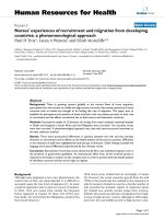
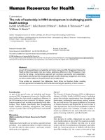
SEM images revealed that the nanoparticles were spheri-
cal in shape, with a smooth surface (Figure 1A). The
Ma et al. Journal of Translational Medicine 2011, 9:34
/>Page 3 of 10
size distribution of the nanoparticles was in the range of
181-282 nm, with a mean diameter of 215.46 ± 48.6
nm, and an average zeta potential of -20.2 mV.
The peptide content of three different nanoparticle
preparations, as determined by HPLC, is (i) 1.588 micro-
grams of peptide per milligram of the nanoparticle pre-
paration formulated with 300 micrograms of peptide, (ii)
3.176 microgra ms of peptide per milligram of the nano-
particle preparation formulated with 600 micrograms of
peptide, and (iii) 5.293 micrograms of peptide per milli-
gram of the nanoparticle preparation formulated with
1000 micrograms of peptide. In preliminary experiments
we selected NP formulation prepared with 600 μgof
peptide f or our in vitro immunization experiments . As
we routinely use 100 μg/ml peptide-loaded NP for DC-
loading, this amount corresponds to 0.3176 μgofpep-
tide per ml of DC-loading medium. In comparison, the
amount of peptide u sed for pulsing of the control DC
group is 1 μg/ml or abou t three times as much as those
in the NP formulation.
Human dendritic cells can efficiently internalize
nanoparticles
Coumarin-6-containing NP were visible inside DC after
just 1-hour of co-incubation (Figure 1C). However, no
fluorescence was observed when the same DC were
incubated with free coumarin-6 (Figure 1B). These stu-
dies showed that 100% of the observed human imDC
internalized nanoparticles. Nuclear staining revealed that
the NP were most likely localized in the cytoplasm or
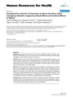
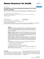
endoplasmic reticulum of t he DC (Figure 1D). Confocal
microscopy using the coumarin-6-containing NP
revealed an intense cytoplasmic fluorescence (6-
coumarin) in the DC (Figure 2), confirming that our pep-
tide-containing NP are avidly internalized by the imDC.
Effect of PLGA nanoparticle uptake on the maturation
status of the human DC
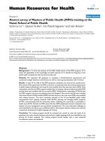
The tested DC surface markers CD80, CD83, CD86 and
HLA-DR were not upregulated after incubation with our
PLGA nanoparticles in several repeated experiments
(Figure 3). In contrast, incubation with LPS induced sig-
nificant upregulation of these markers. These results
indicate that the NP uptake did not influence DC
phenotype and their ability to mature.
A
A
B
B
C
C
D
D
Figure 1 Nanoparticle internalization by immature DC.
(A) Nanoparticles observed with a SEM. Magnification 60,000×. NP-
loaded imDC examined under a fluorescence microscope after a 1-h
incubation with free coumarin-6 (B), or with NP containing
coumarin-6 (C). NP-loaded imDC incubated with Hoechst nuclear
stain (D). Magnification 400×.
A
B
Figure 2 Confocal microscope analysis. A single immature DC
observed after a 1-hour incubation with NP containing peptide
Mart-1
27-35
and coumarin-6. Overlaid confocal images using DAPI,
FITC (A), and reflection (B) channels are shown. The bar represents
10 μm.
Ma et al. Journal of Translational Medicine 2011, 9:34
/>Page 4 of 10
Enhanced antigen presentation by human DC loaded with
NP containing class I-restricted peptides
We prepared NP containing the peptides MART-1
27-35
,
and gp100
209-217
. Peptide-pulsed or NP-loaded DC were
compared for their ability to present these peptides to
TIL lines recognizing MART-1
27-35
, (TIL1235) and
gp100
209-217
(TIL1520) four days after the NP-loading.
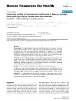
NP-loaded DC were recognized by TILs much better
than the DC pulsed with the same peptides or DC
loaded with empty nanoparticles (Figure 4A, B). These
findings suggest that the significantly enhanced presen-
tation of the peptides loaded with NP resulted from
improved loading and sustained release of these peptides
after the internalization of the NP.
Improved generation of peptide-specific CTL
with NP-loaded DC
To address the question whether enhanced presentation
of the peptides actually improves the generation of CTL,
we initiated in vitro cultures of NP-loaded DC with
responder CD8+ T lymphocytes. Spe cifi cally, we det er-
mined whether our NP vaccin es formulated with the
clinically-relevant melanoma peptide MART-1
27-35
could
induce potent CTL capable of recognizing and killing
peptide-pulsed target cells and melanoma tumor cells
in vitro. CTL induced with peptide-pulsed DC
were compared to CTL induced with NP-loaded DC
(Figure 4C). We found that the CTL induced with the
melanoma peptide M ART-1
27-35
encapsulated into our
nanoparticles were able to recognize and kill specifical ly
not only the peptide-pulsed T2 cells, but also the HLA-
A2-positive melanoma c ells 624. In contrast, the CTL
induced with the peptide-pulsed DC were less efficient
in killing these target cells, and the HLA-A2-negative
melanoma cells 1351 were not recognized (Figure 4C).
These experiments confirmed that our nanoparticle-
based vaccine could expand precursor CTL i n PBMC of
HLA-A2+ donors and induce MHC class I-restricted,
specific CTL responses against the melanoma cells.
Enhanced presentation of murine MHC class II-restricted
peptide encapsulated into nanoparticles
Murine imDC were incubated for 1hr with B5-loaded
nanoparti cles (B5-NP), contro l nanoparticles or B5 pep-
tide. Equivalent levels of proliferation in B5-reactive
CD4+ T cell line, B5.1 on co-c ulture with B5-NP or
peptide pulsed DC was observed ( Figure 5A.) We had
previously predicted that B5 peptide encapsulated into
our NP would be pr otected from degradation and
released slowly into the dendritic cell’s antigen proces-
sing pathways. This would allow for an increased dura-
tion of antigen presentation compared to naked peptide
that would be quickly degraded. To investigate this
imDC were incubated with NP and peptide for 1 hr,
before separation from the non-captured NP or peptide.
DC were then incubated alone for 48 hrs before being
co-cultured with the CD4+ T cell line, B5.1. Indeed, we
observed an enhanced proliferation of the CD4+ T cells
co-cultured with B5 nanoparticle-treated DC in compar-
ison to the B5 peptide-treated DC (Figure 5B). This
finding suggests that the nanoparticles increase the
duration for which antigenic peptides can be presented
by the DC.
Presentation of murine nonclassical MHC class I, Qa-1-
restricted peptide encapsulated into nanoparticles
imDC loaded with peptide p42 encapsulated into NP
were co-cultured with Qa-1-resticted CD8aa+TCRab
+T cell line (XT-14). We found that DC loaded with NP
containing the peptide p42 could stimulate XT-14 T cell
line to produce IFN-g (Figure 5C). No significant stimu-
lation was observed with the empty control NP. These
experiments clearly show that imDC loaded with NP
vehicles carrying non-classi cal MHC class I peptides can
present efficiently these Qa-1-restricted peptides.
Discussion
Epitope-based peptide vaccines can be designed to
include multiple epitopes from one or several antigens,
i
mD
C
i
ncubated w
i
th: mD
C
medium NP
Figure 3 Phenotype of NP-loaded DC.ImmatureDC(imDC)
analyzed 30 hours after a 1-hour incubation with NP containing
Mart-1
27-35
and coumarin-6, and compared to mature LPS-
stimulated DC (mDC). Open area plots - DC stained with isotype
controls; solid area plots - DC stained with antibodies for HLA-DR,
CD80, CD83, and CD86.
Ma et al. Journal of Translational Medicine 2011, 9:34
/>Page 5 of 10
and can be easily analyzed for purity and produced eco-
nomically on a large scale. Currently however, there are
no human peptide-based cancer vaccines on the market,
mostly resulting from difficulties associated with their
poor immunogenicity, stability and delivery. We [22,23]
and others [24,25] have described strategies to enhance
the peptides’ immunogenicity and stability. Direct pep-
tide delivery to dendritic cells using particulate delivery
systems is a promising new approach. In addition to
having a depot effect on the peptide antigens, inherent
properties of the particles themselves engender immu-
nogenicity of the peptides, and allow uptake of an
immunogenic package of peptides and other molecules
[26]. This approach is exemplified with the use of lipo-
somes and immunost imulatory complexes [27], as well
as virosomes [28] and exosomes [29].
Cultured DC are very suitable for the delivery of pep-
tide vaccines, as their direct loading bypasses the pro-
cessing requirements and allows for precise delivery of
peptide antigens to the immune system [30]. However,
the potency of DC-based vaccines is significantly
reduced by the short persistence of the MHC/peptide/
b2 -microglobulin complexes on the D C surface, espe-
cially when the antigen-derived peptides are bound from
the outside and not processed [31].
It is therefore essential that the vaccine-carrier sys-
tems are capable of delivering the vaccines inside the
APCs, in order to facilitate a potent and prolonged anti-
gen presentation. Liposome-based systems are com-
monly used, but their delivery efficiency is sub-optimal,
and the duration of their effects is relatively short [32].
In addition, the liposome is not a thermodynamically
stable system and therefore has multiple physical sta bi-
lity issues such as rapid drug leakage, merging of vesi-
cles and low loading efficiency. The viral vector delivery
is limited by a difficult large- scale production and
potential for toxicity [33], immune and inflammatory
responses [34], as well as insertional mutagenesis and
oncogenic effects [35].
Nanoparticle-based vaccin e deliver y system s offer sig-
nificant advantages due to their safety profile, ease of
manufacture and storage, and most importantly, their
versatility in designing customized products for specific
targeting applications [36]. We devel oped previously
A
B
C
Figure 4 Enhanced antigen presentation and CTL induction by NP-loaded DC. DC loaded with nanoparticles containing the peptides: (A)
MART-1
27-35
,or(B) gp100
209-217
. DC were incubated with: soluble peptide (DC+peptide); empty nanoparticles (CNP); or with nanoparticles
formulated with the same peptides using 300 μg (DC+NP300) or 600 μg (DC+NP600) peptide per batch. Four days later, DC were co-cultured
for 20 hours with TIL1235 (recognizing MART-1
27-35
) or TIL1520 (recognizing gp100
209-217
) cells, and the antigen presentation was evaluated in an
IFN-g ELISPOT assay. (C) Cytotoxic activity of CTL induced in vitro with peptide-pulsed or NP-loaded dendritic cells: Dendritic cells were pulsed
with the peptide MART-1
27-35
or with MART-1
27-35
-containing NP and used as APC to induce MART-1
27-35
-specific CTL. The experimental groups
include: T2 target cells incubated with peptide-DC induced CTL (□), or with NP-DC induced CTL (■); peptide-pulsed T2 cells incubated with
peptide-DC induced CTL (∓), or with NP-DC induced CTL (ℓ); HLA-A2
+
melanoma cells 624 incubated with peptide-DC induced CTL (△), or with
NP-DC induced CTL (▲); and HLA-A2
-
melanoma cells 1351 incubated with peptide-DC induced CTL (▽), or with NP-DC induced CTL (▼). The CTL
lines were incubated with the target cells for 4 hours and the cytotoxicity was determined with a standard LDH-release assay (Promega). Data is
representative of 3 independent experiments; bars, SD. *, significant differences (P < 0.05) between experimental and control cultures (non-
pulsed DC or CNP-loaded DC).
Ma et al. Journal of Translational Medicine 2011, 9:34
/>Page 6 of 10
PLGA nanoparticles designed for sustained release of
drugs or DNA to human umb ilical vein endothelial cells
[37], and prostate cancer cells [20]. In the current study,
we used PLGA-based nanoparticles as a delivery system
to load clinically-relevant, tumor antigen-derived pep-
tides into human DC, and found that 100% of imDC
internalized NP after just 1-hour co-incubation with the
nanoparticles (Figure 2). Using our new PLGA nanopar-
ticles containing MHC class I peptides as a vaccine
delivery system offers distinct advantages over the
administration of the corresponding soluble peptides.
These NP are composed of solid PLGA polymers, there-
fore avoiding the drug-leakage problems involved in
liposome formulations, thus preventing the proteolytic
degradation of the antigen. These polymeric NP can
also be easily lyophilized for long-term storage with
better stability than liposome and other liquid carrier
systems.
We also considered the influence of PLGA uptake on
the properties of the DC, and its potential negative
effect on some important parameters for the DC func-
tion. The hydrolysis of PLGA leads to the liberation of
lactic and glycolic acids, and therefore we expected that
the resulting acidification could negatively affect the cel-
lular functions of the PLGA-loaded DC. It this study we
did not observe any negative effects and/or reduced via-
bility of the PLGA-loaded DC. We also found that our
NP formulations did not have an effect on the matura-
tion of human or murine DC. Our findings are in agree-
ment with a similar study, which reported that
immature DC loaded with PLGA particles exhibited a
similar DC phenotype to those without any loading [38].
A
C
B
Figure 5 Enhanced stimulation of MHC class II-restricted and non-classical MHC class I-restricted T cell lines by NP-loaded DC. (A-B)
MHC class II-restricted CD4+ T cell lines: imDC incubated with: 100 μg/ml NP containing B5 peptide (▲); empty nanoparticles (▼); or 10 μg/ml
B5 peptide (■) for one hour. (A) B5-reactive CD4+ T cells co-cultured with imDC. A 72-hour assay was performed, with
3
H-thymidine added for
the last 8 hours. (B) DC cultured for another 48 hours before co-culture with the responder T cells and a
3
H-thymidine assay. (C) MHC class I-
restricted CD8+ T cell lines: imDC incubated with 100/200 μg/ml empty nanoparticles (CNP), or with nanoparticles containing the peptide p42
(NP42) for one hour. Subsequently, p42-reactive CD8+ T cells were co-cultured with DC, and 48-hour later, IFN-gamma was measured by ELISA.
Data is representative of 3 independent experiments; bars, SD. *, Significant differences (P < 0.05) between experimental and control cultures.
Ma et al. Journal of Translational Medicine 2011, 9:34
/>Page 7 of 10
In contrast, another study showed that a maturation
process has been induced by polystyrene nanospheres,
as the maturation markers HLA-DR and CD86 were
upregulated [39]. A similar result was observed using
PLGA nanoparticle formulations in cord blood derived
DC[40],aswellasmurinebonemarrowderivedDC
[41]. These discrepancies are most likely due to the dif-
ferent culture conditions and differences in the nanopar-
ticle preparations in these studies. We conclude that the
intake of our PLGA NP does not adversely affect impor-
tant DC functions required for their use as vaccines in
the clinic.
In the present study, the efficiency of the antigen pre-
sentation by human DC was significantly enhanced after
just 1-hour incubation with our NP containing class I-
restricted peptides (Figure 4A and 4b). The level of anti-
gen presentation was related to the amount of peptide
incorporated inside the NP. Importantly, in these studies
we used patient-derived TIL lines and peptides used in
many clinical trials, w hich suggests a speedy utilization
of these data in clinical trial designs.
For the presentation of MHC class I restricted T cell
epitopes from PLGA-encapsulated peptides, the involved
antigen presenting cells must be able to “cross present”
the exogenous peptides onto MHC class I molecules by
either the classical proteasome and TAP-dependent
pathway [42], or by an alternative TAP-independent
pathway [22,23] of antigen presentation. DC, macro-
phages and some endothelial cells have been shown to
be able to cross present [43]. Cross presentation of solu-
ble proteins by DC can occur, but it is extr emely ineffi-
cient, as it usually requires the incubation with high
concentrations of protein antigens. Remarkably, in this
study the amount of peptides encapsulated inside our
NP was significantly lower than the amount of peptides
used to pulse the DC externally. Still, we observed a
greatly enhanced antigen presentation by the NP-loaded
DC. We suggest that this enhancement of antigen pre-
sentati on is due to the slow hydrolysis of the PLGA NP
in the endosomes the DC, which pro vides a continuous
supply of peptide ligands for newly synthesized MHC
class I and II molecules. We also demonstrated that the
NP-loadedDCwereabletoinducemorepotentCTL
than the DC pulsed with the same peptides externally
(Figure 4C). The resulting CTL were able to recognize
and kill efficiently not only peptide-pulsed target cells,
but also HLA-matched melanoma cells expressing the
corresponding antigens. Similarly, murine DC could pre-
sent antigens more efficiently as a result of the antigen
encapsulation inside our nanoparticles (Figure 5). These
results provide further confirmation of the usefulness of
this approach for i nduction of potent and specific anti-
tumor responses, and its potential for clinical application.
Our nanoparticles were also effective in stimulating
class II-restricted CD4 + T cells as well as no n-classical
class I-restricted CD8aa+TCRab+ cytotoxic T cells.
Notably, these CD4+ and CD8aa+TCRab+ T cells are
involved in the negative feed back regulation of autoim-
munity [8,9,11-13]. Our earlier studies have shown that
priming of these regulatory T cells following peptide or
DNA immunization results in immune regulatio n
[8,11,12,44]. Data presented in this paper further indi-
cate that nanoparticles containing appropriate peptides
can be used to generate effective vaccines not only
against tumors but also in the intervention of autoim-
mune diseases. Class Ib MHC-restricted cytotoxic
T cells also play an important part in the anti-tumor
responses [15,16]. It is clear from our data that nanopar-
ticles containing class Ib MHC (Qa-1 or HLA-E in
humans) binding tumor-associated antigens can also be
designed.
Conclusions
The development of nanoparticle-based vaccines derived
from clinically relevant tumor antigens holds great pro-
mise. Encapsulating antigens in PLGA nanoparticles
offers unique advantages such as higher efficiency of
ant igen loading, prolonged presentation of the antigens,
prevention of peptide degradation, specific targeting o f
antigens to APC, improved shelf life of the antigens, and
easy scale up for pharmaceutical production. In addition,
a variety of targeting strategies may be readily utilized,
including ligand-receptor mediated targeting, antibody-
antigen interaction, lectin-carbohydrate interaction, etc.
Recent advances in polymer c hemistry also allow for
many variations of the nanoparticle design, including
simultaneous delivery of a combination of vaccines,
immunomodulators, drugs or other compounds, creating
a potent multivalent therapeutic strategy. This paper is
therefore highly s ignificant to the development of opti-
mized clinical grade v accines, and the induction of CTL
for adoptive immunotherapy of cancer.
Abbreviations
APC: antigen-presenting cells; CTL: cytotoxic T lymphocytes; DC: dendritic
cells; GM-CSF: granulocyte-macrophage colony-stimulating factor; HLA:
human leukocyte antigen; MART-1: melanoma antigen recognized by T cell
1; NP: polymeric nanoparticles; PLGA: polylactic-co-glycolic acid; TAA: tumor-
associated antigens; TAP: transporter associated with antigen processing;
TCR: T cell receptor
Acknowledgements and funding
We thank Neal Sekiya and Judy Nordberg for their assistance on the FACS
data. Funding: Department of Defense grant PC041024 (B. Minev), NCI grants
U54CA132384 and U54CA132379 (B. Minev), and NIH R01 AI052227 (V.
Kumar)
Author details
1
Moores UCSD Cancer Center, University of California San Diego.
2
Laboratory
of Autoimmunity, Torrey Pines Institute for Molecular Studies.
3
MediStem,
Ma et al. Journal of Translational Medicine 2011, 9:34
/>Page 8 of 10
Inc., San Diego, CA.
4
Laboratory of Biomaterials and Nanotechnology,
University of California Riverside.
5
Division of Neurosurgery, University of
California San Diego.
6
Genelux Corporation, San Diego, CA.
Authors’ contributions
WM carried out and participated in all of the studies, including nanoparticle
preparation and characterization, DC isolation and loading, CTL induction in
vitro, data analysis and manuscript preparation. TS carried out murine DC
isolation and loading, analysis of presentation of murine MHC class II-
restricted peptides and murine non-classical MHC class I, Qa-1-restricted
peptides encapsulated into nanoparticles, and data analysis. VB participated
in the design of the study, helped with the statistical analysis and
manuscript preparation. YZ participated in nanoparticle characterization, DC
loading and imaging and data analysis. CO YZ participated in nanoparticle
characterization, DC imaging and data analysis. MO participated in
nanoparticle characterization, DC loading and imaging and data analysis. MH
participated in DC loading and CTL induction. SS participated in DC loading
and CTL induction. EC participated in the design of the study, helped with
the statistical analysis and manuscript preparation. DM participated in
nanoparticle characterization, DC loading and data analysis. VK participated
in data analysis and supervised studies related to murine class II and Qa-1-
restricted T cell presentation. BM designed, supervised and coordinated the
study, performed the statistical analysis and drafted the manuscript. All
authors read and approved the final manuscript.
Competing interests
The authors declare that they have no competing interests.
Received: 23 November 2010 Accepted: 31 March 2011
Published: 31 March 2011
References
1. Wang RF: Human tumor antigens: implications for cancer vaccine
development. Journal of Molecular Medicine 1999, 77:640-655.
2. Marincola FM: A balanced review of the status T cell-based therapy
against cancer. J Transl Med 2005, 3:16.
3. Heurtault B, Saulnier P, Pech B, Proust JE, Benoit JP: Physico-chemical
stability of colloidal lipid particles. Biomaterials 2003, 24:4283-4300.
4. Banchereau J, Pascual V, Palucka AK: Autoimmunity through cytokine-
induced dendritic cell activation. Immunity 2004, 20:539-550.
5. Rosenblatt J, Kufe D, Avigan D: Dendritic cell fusion vaccines for cancer
immunotherapy. Expert Opin Biol Ther 2005, 5:703-715.
6. Steinman RM, Banchereau J: Taking dendritic cells into medicine. Nature
2007, 449:419-426.
7. Drake CG, Antonarakis ES: Update: immunological strategies for prostate
cancer. Curr Urol Rep 11:202-207.
8. Tang X, Maricic I, Purohit N, Bakamjian B, Reed-Loisel LM, Beeston T,
Jensen P, Kumar V: Regulation of immunity by a novel population of Qa-
1-restricted CD8alphaalpha+TCRalphabeta+ T cells. J Immunol 2006,
177:7645-7655.
9. Tang X, Maricic I, Kumar V: Anti-TCR antibody treatment activates a novel
population of nonintestinal CD8 alpha alpha+ TCR alpha beta+
regulatory T cells and prevents experimental autoimmune
encephalomyelitis. J Immunol 2007, 178:6043-6050.
10. Smith TR, Kumar V: Revival of CD8+ Treg-mediated suppression. Trends
Immunol 2008, 29:337-342.
11. Kumar V, Sercarz EE: The involvement of T cell receptor peptide-specific
regulatory CD4+ T cells in recovery from antigen-induced autoimmune
disease. J Exp Med 1993, 178:909-916.
12. Kumar V, Aziz F, Sercarz E, Miller A: Regulatory T cells specific for the
same framework 3 region of the Vbeta8.2 chain are involved in the
control of collagen II-induced arthritis and experimental autoimmune
encephalomyelitis. J Exp Med 1997, 185:1725-1733.
13. Kumar V: Homeostatic control of immunity by TCR peptide-specific
Tregs. J Clin Invest 2004, 114:1222-1226.
14. Madakamutil LT, Maricic I, Sercarz E, Kumar V: Regulatory T cells control
autoimmunity in vivo by inducing apoptotic depletion of activated
pathogenic lymphocytes. J Immunol 2003, 170:2985-2992.
15. Seliger B, Abken H, Ferrone S: HLA-G and MIC expression in tumors and
their role in anti-tumor immunity. Trends
Immunol 2003, 24:82-87.
16. van Hall T, Laban S, Koppers-Lalic D, Koch J, Precup C, Asmawidjaja P,
Offringa R, Wiertz EJ: The varicellovirus-encoded TAP inhibitor UL49.5
regulates the presentation of CTL epitopes by Qa-1b1. J Immunol 2007,
178:657-662.
17. Rokkanen P, Bostman O, Vainionpaa S, Vihtonen K, Tormala P, Laiho J,
Kilpikari J, Tamminmaki M: Biodegradable implants in fracture fixation:
early results of treatment of fractures of the ankle. Lancet 1985,
1:1422-1424.
18. Bercovy M, Goutallier D, Voisin MC, Geiger D, Blanquaert D, Gaudichet A,
Patte D: Carbon-PGLA prostheses for ligament reconstruction.
Experimental basis and short-term results in man. Clin Orthop Relat Res
1985, 159-168.
19. Salter RD, Cresswell P: Impaired assembly and transport of HLA-A and -B
antigens in a mutant TxB cell hybrid. EMBO J 1986, 5:943-949.
20. Sahoo SK, Ma W, Labhasetwar V: Efficacy of transferrin-conjugated
paclitaxel-loaded nanoparticles in a murine model of prostate cancer. Int
J Cancer 2004, 112:335-340.
21. Inaba K, Inaba M, Romani N, Aya H, Deguchi M, Ikehara S, Muramatsu S,
Steinman RM: Generation of large numbers of dendritic cells from
mouse bone marrow cultures supplemented with granulocyte/
macrophage colony-stimulating factor. J Exp Med 1992, 176:1693-1702.
22. Minev BR, Chavez FL, Dudouet BM, Mitchell MS: Synthetic insertion signal
sequences enhance MHC class I presentation of a peptide from the
melanoma antigen MART-1. Eur J Immunol 2000, 30:2115-2124.
23. Minev BR, McFarland BJ, Spiess PJ, Rosenberg SA, Restifo NP: Insertion
signal sequence fused to minimal peptides elicits specific CD8+ T-cell
responses and prolongs survival of thymoma- bearing mice. Cancer Res
1994, 54:4155-4161.
24. Sotiriadou NN, Kallinteris NL, Gritzapis AD, Voutsas IF, Papamichail M, von
Hofe E, Humphreys RE, Pavlis T, Perez SA, Baxevanis CN: Ii-Key/HER-2/neu
(776-790) hybrid peptides induce more effective immunological
responses over the native peptide in lymphocyte cultures from patients
with HER-2/neu+ tumors. Cancer Immunol Immunother 2007, 56:601-613.
25. Zirlik KM, Zahrieh D, Neuberg D, Gribben JG: Cytotoxic T cells generated
against heteroclitic peptides kill primary tumor cells independent of the
binding affinity of the native tumor antigen peptide. Blood 2006,
108:3865-3870.
26. Liang MT, Davies NM, Blanchfield JT, Toth I: Particulate systems as
adjuvants and carriers for peptide and protein antigens. Curr Drug Deliv
2006, 3:379-388.
27. Kersten GF, Crommelin DJ: Liposomes and ISCOMs. Vaccine 2003,
21:915-920.
28. Westerfeld N, Zurbriggen R: Peptides delivered by immunostimulating
reconstituted influenza virosomes. J Pept Sci 2005, 11:707-712.
29. Taieb J, Chaput N, Zitvogel L:
Dendritic cell-derived exosomes as cell-free
peptide-based
vaccines. Crit Rev Immunol 2005, 25:215-223.
30. Ridolfi R, Petrini M, Fiammenghi L, Stefanelli M, Ridolfi L, Ballardini M,
Migliori G, Riccobon A: Improved overall survival in dendritic cell
vaccination-induced immunoreactive subgroup of advanced melanoma
patients. J Transl Med 2006, 4:36.
31. Ludewig B, McCoy K, Pericin M, Ochsenbein AF, Dumrese T, Odermatt B,
Toes RE, Melief CJ, Hengartner H, Zinkernagel RM: Rapid peptide turnover
and inefficient presentation of exogenous antigen critically limit the
activation of self-reactive CTL by dendritic cells. J Immunol 2001,
166:3678-3687.
32. Liang MT, Davies NM, Toth I: Encapsulation of lipopeptides within
liposomes: effect of number of lipid chains, chain length and method of
liposome preparation. Int J Pharm 2005, 301:247-254.
33. Ferber D: Gene therapy. Safer and virus-free? Science 2001, 294:1638-1642.
34. Lamfers M, Idema S, van Milligen F, Schouten T, van der Valk P,
Vandertop P, Dirven C, Noske D: Homing properties of adipose-derived
stem cells to intracerebral glioma and the effects of adenovirus
infection. Cancer Lett 2009, 274:78-87.
35. Lee KY, Kwon IC, Kim YH, Jo WH, Jeong SY: Preparation of chitosan self-
aggregates as a gene delivery system. J Control Release 1998, 51:213-220.
36. Vinogradov S: The second annual symposium on nanomedicine and
drug delivery: exploring recent developments and assessing major
advances. 19-20 August 2004, Polytechnic University, Brooklyn, NY, USA.
Expert Opin Drug Deliv 2004, 1:181-184.
37. Davda J, Labhasetwar V: Characterization of nanoparticle uptake by
endothelial cells. Int J Pharm 2002, 233:51-59.
Ma et al. Journal of Translational Medicine 2011, 9:34
/>Page 9 of 10
38. Moffatt S, Cristiano RJ: Uptake characteristics of NGR-coupled stealth PEI/
pDNA nanoparticles loaded with PLGA-PEG-PLGA tri-block copolymer
for targeted delivery to human monocyte-derived dendritic cells. Int J
Pharm 2006, 321:143-154.
39. Matsusaki M, Larsson K, Akagi T, Lindstedt M, Akashi M, Borrebaeck CA:
Nanosphere induced gene expression in human dendritic cells. Nano
Lett 2005, 5:2168-2173.
40. Diwan M, Elamanchili P, Lane H, Gainer A, Samuel J: Biodegradable
nanoparticle mediated antigen delivery to human cord blood derived
dendritic cells for induction of primary T cell responses. J Drug Target
2003, 11:495-507.
41. Elamanchili P, Diwan M, Cao M, Samuel J: Characterization of poly(D, L-
lactic-co-glycolic acid) based nanoparticulate system for enhanced
delivery of antigens to dendritic cells. Vaccine 2004, 22:2406-2412.
42. Kovacsovics-Bankowski M, Rock KL: A phagosome-to-cytosol pathway for
exogenous antigens presented on MHC class I molecules. Science 1995,
267:243-246.
43. von Euw EM, Barrio MM, Furman D, Bianchini M, Levy EM, Yee C, Li Y,
Wainstok R, Mordoh J: Monocyte-derived dendritic cells loaded with a
mixture of apoptotic/necrotic melanoma cells efficiently cross-present
gp100 and MART-1 antigens to specific CD8+ T lymphocytes. J Transl
Med 2007, 5:19.
44. Kumar V, Maglione J, Thatte J, Pederson B, Sercarz E, Ward ES: Induction of
a type 1 regulatory CD4 T cell response following V beta 8.2 DNA
vaccination results in immune deviation and protection from
experimental autoimmune encephalomyelitis. Int Immunol 2001,
13:835-841.
doi:10.1186/1479-5876-9-34
Cite this article as: Ma et al.: Enhanced presentation of MHC class Ia, Ib
and class II-restricted peptides encapsulated in biodegradable
nanoparticles: a promising strategy for tumor immunotherapy. Journal
of Translational Medicine 2011 9:34.
Submit your next manuscript to BioMed Central
and take full advantage of:
• Convenient online submission
• Thorough peer review
• No space constraints or color figure charges
• Immediate publication on acceptance
• Inclusion in PubMed, CAS, Scopus and Google Scholar
• Research which is freely available for redistribution
Submit your manuscript at
www.biomedcentral.com/submit
Ma et al. Journal of Translational Medicine 2011, 9:34
/>Page 10 of 10