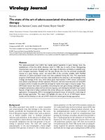
Báo cáo sinh học: "Gene therapy with tumor-specific promoter mediated suicide gene plus IL-12 gene enhanced tumor inhibition and prolonged host survival in a murine model of Lewis lung carcinoma" pptx
Bạn đang xem bản rút gọn của tài liệu. Xem và tải ngay bản đầy đủ của tài liệu tại đây (7.76 MB, 10 trang )
RESEARCH Open Access
Gene therapy with tumor-specific promoter
mediated suicide gene plus IL-12 gene enhanced
tumor inhibition and prolonged host survival in a
murine model of Lewis lung carcinoma
Yu Xu
1,2
, Jinxuan Hou
2,3
, Zhengchun Liu
1,2
, Haijun Yu
1,2
, Wenjie Sun
1,2
, Jie Xiong
1,2
, Zhengkai Liao
1,2
,
Fuxiang Zhou
1,2
, Conghua Xie
1,2
and Yunfeng Zhou
1,2*
Abstract
Background: Gene therapy is a promising therapeutic approach for cancer. Targeted expression of desired
therapeutic proteins within the tumor is the best approach to reduce toxicity and improve survival. This study is to
establish a more effective and less toxic gene therapy of cancer.
Methods: Combined gene therapy strategy with recombinant adenovirus expressing horseradish peroxidase (HRP)
mediated by human telomerase reverse transcr iptase (hTERT) promoter (AdhTERTHRP) and murine interleukin-12
(mIL-12) under the control of Cytomegalovirus (CMV) promoter (AdCMVmIL-12) was developed and evaluated
against Lewis lung carcinoma (LLC) both in vivo and in vitro. The mechanism of action and systemic toxicities were
also investigated.
Results: The combination of AdhTERTHRP/indole-3-acetic acid (IAA) treatment and AdCMVmIL-12 resulted in
significant tumor growth inhibition and survival improvement compared with AdhTERTHRP/IAA alone (tumor
volume, 427.4 ± 48.7 mm
3
vs 581.9 ± 46.9 mm
3
, p = 0.005 on day 15; me dian overall survival (OS), 51 d vs 33 d) or
AdCMVmIL-12 alone (tumor volume, 362.2 ± 33.8 mm
3
vs 494.4 ± 70.2 mm
3
, p = 0.046 on day 12; median OS, 51 d
vs 36 d). The combination treatment stimulated more CD4
+
and CD8
+
T lymphocyte infiltration in tumors,
compared with either AdCMVmIL-12 alone (1.3-fold increase for CD4
+
T cells and 1.2-fold increase for CD8
+
T cells,
P < 0.01) or AdhTERTHRP alone (2.1-fold increase for CD4
+
T cells and 2.2-fold increase for CD8
+
T cells, P < 0.01).
The ap optotic cells in combination group were significantly increased in comparison with AdCMVmIL-12 alone
group (2.8-fold increase, P < 0.01) or AdhTERTHRP alone group (1.6-fold increase, P < 0.01). No significant
systematic toxicities were observed.
Conclusions: Combination gene therapy with AdhTERTHRP/IAA and AdCMVmIL-12 could significantly inhibit tumor
growth and improve host survival in LLC model, without significant systemic adverse effects.
Background
Over the last years, gene therapy has emerged as a pro-
mising strategy for cancer treatment [1]. However, some
limitations are associated with its clinical application, the
reduced specificity to deliver functional therapeutic genes
into tumor cells being the ma jor one [2]. Th erefore,
research in gene therapy has been focused on t he devel-
opment of targeting strategies.
Tissue- or cell-specific promoters represent one of the
main methods of gene targeting. T he human telomerase
reverse transcriptase (hTERT) promoter ha s been widely
used in gene therapy for targeting cancer cells, which is
highly active in human cancer cells but not in normal dif-
ferentiated human cells [3-6].Therefore, it fulfilled the
characteristic of tumor origins with marked heterogeneity.
Meanwhile, it was demonstrated that hTERT promoter
* Correspondence:
1
Department of Radiation and Medical Oncology, Zhongnan Hospital of
Wuhan University, Wuhan 430071, PR China
Full list of author information is available at the end of the article
Xu et al. Journal of Translational Medicine 2011, 9:39
/>© 2011 Xu et al; licensee BioMed Central Ltd. This is an Open Access article distributed under the terms of the Creat ive Commons
Attribution License (http://creativec ommons.org/licenses/by/2.0), which permits unrestricted use, distribution, and reproduction in
any medium, provided the original work is properly cited.
had high transcriptional activity in a variety of human
cancer cell lines, but not in normal human cells in adeno-
virus mediated transgene experiments [7,8]. Furthermore,
Gu et al[9] showed that hTERT promoter could efficiently
use mouse transcription machiner y despite the apparent
distinct regulatory mechanisms, and that hTERT promoter
was highly active in murine tumor cells, but quiescent in
normal murine cells and tissues. These findings indicated
that hTERT promo ter should be usef ul for targeting the
pharmaceutical effects of a therapeutic gene to cancer
cells.
Gene directed enzyme/prodrug therapy (GDEPT) or
suicide gene therapy using viral vectors is an attracti ve
alternative approach to cancer therapy, with the potential
to give therapeutic ratios superior to standard chemo-
and radiotherapy [10]. The horseradish peroxidase
(HRP)/indole-3-acetic acid (IAA) system is a novel
GDEPT system , which has shown great efficacy in killing
tumor cells. In this setting, a viral vector expressing a
therapeutic enzyme (HRP) is delivered to the tumor cells.
The nontoxic prodrug (IAA) is administered systemically
by intravenous injection or loc ally by intraperitoneal or
intratumoral injection to maximize its concentration
within the tumor, and converted into cytotoxic metabo-
lites by HRP. It was demonstrated that HRP/IAA system
was more cytotoxic to tumors than the well-known HSV-
tk/GCV system [11,12]. Furthermore, HRP is normally
absent in mammalian cells and IAA is a poor substrate
for mammalian peroxidases, thus systemic toxicity is
avoided [13].
To optimize the therapeutic efficacy of suicide gene
therapy, it is important to explore n ew strategies of
combined therapy, which employ targeted suicide gene
in combina tion with immunotherapy that cooperatively
enhance the antitumor effects while mitigating side
effects. Immunotherapy uses the transfer of genes of
various cytokines and co-stimulatory molecule into
tumor cells to stimulate an antitumoral immune
response in experimental animals [14]. Interleukin-12
(IL-12) is a heterodimetic cytokine, composed of
35 KDa (p35) and 40 KDa (p40) subunits, whi ch bind to
receptors present on NK and T cells [15]. IL-12 plays
multiple roles in the immune system, such a s au gment-
ing the proliferation and cytotoxic activity of T cells and
NK cells and initiating Th1-type immune responses by
activation of CD4
+
and CD8
+
cells [16].
In the present study, we investigated a combined tar-
get suicide gene therapy and immunomodulating gene
therapy approach for Lewis lung carcinoma (LLC),
based on the delivery of HRP/IAA and murine IL-12 by
the same adenovirus vector. These studies were per-
formed both in vitro, by measuring cell viability, and
in vivo, by determining the tumor size and animal survi-
val, assessing both tumoral histology and infiltration of
T-lymphocytes, and evaluating toxic studies. The data
showed that combination gene therapy increased the
therapeutic efficiency in the murine LLC model used in
this study.
Materials and methods
Cell culture and animals
LLC and A549 cell lines were obtained from the Cell
Bank of the Chinese Academy of Science (Shanghai,
China) and maintained in 5% CO
2
at 37°C in Dulbecco’s
minimum essential medium (DMEM) containing 10%
fetal bovine serum (FBS), 100 U/m l penicillin and
100 mg/ml strepto mycin. All culture reagent s were pur-
chased from Hyclone (Logan, UT, USA) or Invitrogen
(Gaithersburg, MD, USA). Being syngenic with LLC,
male C57BL/6 mice (6-week old) obtained from Shang-
hai SLAC Laboratory Animal Co. Ltd (Shanghai, China)
were housed in specific pathogen-free condition at the
Animal Experimental Center of Wuhan University. The
facilities and the protocol were consistent with the regu-
latio ns on animal use for biomedical experiments issued
by the Ministry of Science and Technology of China,
and approved by the Animal Care Committee of Wuhan
University.
Recombinant adenoviruses
The plasmid phTERTHRP constructed in our lab as
described [17-19] was digested with MluI and BamHI,
and subcloned into the same site of pAdTrack-C (which
was modified by inserting MluI and BamHI clone sites
based on pAdTrack) to generate pAdhTERTHRP. The
murine IL-12 obtained from pUMVC3-mIL12 (Aldevron
Inc., Fargo, USA) was subcloned into pAdTrackCMV to
generate pAdCMVmIL-12 by digesting with SalI and
NotI. The shuttle vector pAdTrack and Escherichia coli
AdEasy-1 were kindly provided by Dr. JG Wu (State
Key Laboratory of Virology, College of Life Sciences at
Wuhan University, China). For recombinant prAdh-
TERTHRP and prAdCMVIL-12, homologous recombi-
nation was performed as described previously [20].
Recombinant adenoviruses were packaged by GeneChem
Co., Ltd (Shanghai, Chin a). Briefly, recombinant plas-
mids were transfected into 293 cells to obtain adeno-
virus prestocks. Virus was purified by double cesium
chloride gradient ultracentrifugation. Viral titer was
determined by plaque assay and expressed as plaque-
forming units (pfu). Purified virus aliquots were stored
at -80°C.
In vitro studies
For adenoviral gene transduct ion efficiency in vitro,
A549 and LLC cells were infected with AdCMV(-) at
multiplicity of infection (MOI) of 1, 10, 100 and 1,000.
After incubation for 48 hours, the cells were analyzed
Xu et al. Journal of Translational Medicine 2011, 9:39
/>Page 2 of 10
using flow cytometry (FC500, Beckman coulter, CA,
USA) for green fluorescent protein (GFP) expression.
Subsequently, LLC cells were transduced with AdCMV
(-), AdCMVmIL-12 and AdhTERTHRP alone or in
combination at appropriate MOI. Cell proteins w ere
harvested and the expression of HRP was detected by
western blot. Culture supernatants were collected for
determination of IL-12 concentration by a sandwich
enzyme-linked immunosorbant assay (ELISA).
For cytotoxicity of HRP/IAA system, LLC cells (2 × 10
3
/
well) were plated in 96 -well plates and allowed to adhere
overnight. The cells were transduced as described above
and incubated for 16 hours. Then fresh media containing
IAA (Sigma, MO, USA) at a concentration of 0-5 mM were
exchanged every 48 hours. Cell viability was determined by
MTT assay (Invitrogen, CA, USA) 120 hours later and the
optical density value was measured by a microplate reader
(Turner BioSystems, CA, USA).
In vivo studies
Atotalof5×10
6
LLC cells were inoculated subcuta-
neously in the right flank of C57BL/6 mice. After
14 days, the tumor was isolated, prepared to cell suspen-
sion and inoculated into new mice. When the tumors
reached 5-6 mm in diameter (Day 10), the mice were
randomized to 4 groups (n = 13 each): group I, AdCMV
(-) (1 × 10
9
pfu); group II, single-agent AdhTERTHRP
(5 × 10
8
pfu); group III, single-agent AdCMVmIL-12
(5 × 10
8
pfu); group IV, combination AdCMVmIL-12
and AdhTERTHRP (5 × 10
8
pfu + 5 × 10
8
pfu). The
adenoviruses were diluted in 30 μl phosphate buffered
saline (PBS). 48 hours after virus injection (Day 12),
3 mice of each group were sacrificed for examining
HRP and IL-12 expression in tumor tissues. Meanwhile,
IAA (50 mg/kg daily) was administered to the rest mice
by intraperitoneal injection for 7 days from Day 12 to
18. Five mice from each group were sacrificed for evalu-
ating the effects of various treatments on Da y 19. In
addition, survival studies were s et up with different
treatment groups of an imals (n = 5) in an identical
manner. Tumor size was measured using caliper every
3 days and the volume was calculated using the follow-
ing f ormula: (L × W
2
)/2, where L equals length and W
equals width. Animals with very high tumor volume
(exceeded 3500 mm
3
) were sacrificed for ethical reasons
and this was recorded as the date of death for survival
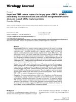
studies. The general scheme of in vivo experiment was
outlined in Figure 1.
Western blot analysis
HRPexpressioninLLCcellsandsubcutaneoustumors
infected with AdCMV(-), AdhTERTHRP and AdCMVmIL-
12 alone or in combination was determined by Western
blot. Transduced cells and tumor tissues were lysed in
2× sample buffer (100 mM Tris-HCl pH6.8, 200 mM DTT,
4% SDS, 20% glycerol and 0.2% bromoplenol blue) and
separated by 10% SDS-PAGE. Proteins were transferred to
PVDF membranes (Millipore, MA, USA) and then
immersed in a blocking solution co ntaining 5% non-fat
milk and 0.1% tween-20 for 1 hour. Afterwards, the mem-
branes were incubated with mouse anti -HRP (dilution,
1:500) or mouse anti-b-actin (dilution, 1:1000) for 2 hours
and with goat anti-mouse secondary ant ibody (dilution,
1:10000) for 1 hour at room temper ature. All the an tibodies
were purchased from San ta Cruz Biotechnology (Santa
Cruz, USA). Enhanced chemiluminescence (Beyotime,
Shanghai, China) was used to visualize the immunoreactive
bands.
ELISA
Culture supernatants of transduced LLC cells and tumor
tissue lysates of treate d mice were collected. The IL-1 2
concentration was determined using a sandwich ELISA
(R&D systems, CA, USA) according to the manufacturer’s
instructions.
Immunohistochemical analysis and apoptosis assay
Tumor tissues were formalin fixed and 4 μm s ections were
stained with hematoxylin and eosin for routine histological
analysis. For immunohistochemical analysis, acetone fixed
fresh-frozen sections were stained for infiltration T lym-
phocytes (CD4
+
and CD8
+
) with specific antibodies (BD
PharMingen, CA, USA) following standard method as
described [21]. For apoptosis assay, formalin fixed sections
were analyzed for DNA fragmentation by terminal deoxy-
necleo tidyl transferase-mediated dUTP nick-end labeling
(TUNEL) assay (Roch, NJ, USA) according to the manufac-
turer’s instructions. Cell proliferation was also determined
using anti-Ki-67 antibody (Santa Cruz Biotechnology,
Santa Cruz, USA). Positive staining was scored by light
microscopy. After initial scanning under × 100 magnifica-
tion, positively stained cells in ten fields under × 400
(0.15 mm
2
) magnification were counted and the mean
number/high power field (HPF ± SEM) was determined.
In vivo toxicity studies
Sera were collec ted from the treated animals to measure
the biochemistry markers including alanine transami-
nase (ALT), aspartate aminotransferase (AST), b lood
urea nitrogen (BUN) and creatine (Cr) using commercial
kits (Sigma, MO, USA). For histological examination,
some tissues were harvested, fixed with formalin and
stai ned with hemato xylin and eosin. 3 mice without any
treatment were used as normal control.
Statistical analysis
The significance of differences between experimental
groups was calculated using Student’s t-test or one-way
Xu et al. Journal of Translational Medicine 2011, 9:39
/>Page 3 of 10
ANOVA analysis a s appropriate. Kaplan-Meier curves
were compared using the log-rank test. In all cases,
P values less than 0.05 were considered statistically sig-
nificant. Analysis was performed with the GraphPad
Prism 5 (version 5.01, GraphPad software, Inc.).
Results
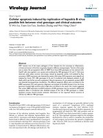
Gene transfer efficiency in LLC cells with Adenoviruses
LLC cells were relatively resistant to infecti on b y
AdCMV(-) compared with A549 cells. Only a few LLC
cells were infected at MOI of 10. When the MOI
increased to 100 and 1000, the percentages of trans-
duc ed cells were up to 10-15% and 40-45% (Figure 2A).
Thus the MOI of 1000 was chosen for the subsequent
studies.
HRP and IL-12 expression in vitro
LLC cells transduced with AdCMV(-), AdhTERTHRP
and AdCMVmIL-12 alone or in combination were har-
vested and determined by western blot for the expression
of HRP. Results showed that the HRP expression was
observed in AdhTERTHRP alone group and combination
groupwhereasnotinAdCMV(-)orAdCMVmIL-12
alone groups (Figure 2B). The expression levels of IL-12
in culture supernatant were measured by ELISA assay.
LLC cells in AdCMVmIL-12 alone and in combination
groups resulted in secretionofupto5.8ng/ml,butno
IL-12 expression was detected in supernatants from
AdCMV(-) or AdhTERTHRP alone groups (Figure 2C).
Cytotoxicity of the HRP/IAA system in vitro
To validate the biological activity of exogenous HRP,
transduced LLC cells we re treated with IAA at indicated
concentrations in Figure 2D. The results were s hown as
the percentage of cell viability with respect to control cells
without IAA treatment. LLC cells in AdhTERTHRP alone
group and combination group exhibited a dose-dependent
manner of cytotoxicity, and both the IC
50
of IAA was
about 3.0 mM. The results indicated that HRP/IAA system
had efficient cytotoxic effects on LLC cells. However, high
concentration of IAA (more than 5 mM) showed mild
toxicity on HRP-negative cells.
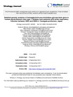
In vivo antitumor effect of gene therapy
The LLC mouse model was used to assess the in vivo anti-
tumor activity of AdhTERTHRP and AdCMVmIL-12 as
single agent or in combination. First of all, the expression
of HRP and IL-12 in tumors was deter mined and similar
results were found as in vitro (Figure 3A and 3B). For
tumor growth, the data clearly showed remarkable inhibi-
tion of combining AdCMVmIL-12 and AdhTERTHRP
treatment in comparison with AdCMV(-), AdCMVmIL-12
and AdhTERTHRP alone (Figure 3C). AdhTERTHRP
treatment ( group II) significantly suppre ssed tumor growth
through day 6 to 24 compared with AdCMV(-) treatment
(group I) (groups II vs I, p = 0.024). AdCMVmIL-12 treat-
ment (group III) was associated with more potent antitu-
mor effects (groups III vs II, p > 0.05; groups III vs I, p =
0.047 on day 9 to 24). Combination treatment with
AdhTERTHRP and AdCMVmIL-12 (group IV) was asso-
ciated with the most marked suppression of tumor growth
(groups IV vs III, p = 0.046 on day 12 to 24; groups IV vs
II, p = 0.005 on day 15 to 24; groups IV vs I, p = 0.029
on day 6 to 24). These results indicate d that both
AdhTERTHRP alone and AdCMVmIL-12 alone sup-
pressed tumor growth, and the combination showed syner-
gistic antitumor effects. As a consequence, five animals
from each group were monitored and survival curves were
established (Figure 3D). Mice treated with the combination
regimen had a significant survival advantage w ith the med-
ian survival increase to 51 versus 36 days for AdCMVmIL-
12 alone-treated mice, 33 days for AdhTER THRP alone-
treated mice and 24 days for AdCMV(-) control treated
mice (Figure 3B). Statistical comparison (log-rank test)
showed a significant difference (P = 0.0001) and a signifi-
cant trend between treatment g roups (P = 0.0003).
T lymphocyte infiltration in tumors
The antitumoral activity of immuno-gene therapy strate-
gies involved the activation of the immune system
Figure 1 General outline of the in vivo experimentation with mouse model of LLC.
Xu et al. Journal of Translational Medicine 2011, 9:39
/>Page 4 of 10
against the neoplastic tissue. To evaluate the effects of
different treatments on immune cell infiltration in local
tumors, immunohistochemistry was performed against
CD4
+
and CD8
+
T cells (Figure 4; Table 1). The results
rev ealed that the comb ination therapy (AdhTERTHR P +
AdCMVmIL-12) led to extensive tumor infiltration by
CD4
+
T cells (P < 0.0001, 9.4-fold) and CD8
+
T cells (P <
0.0001, 8.6-fold) compared with AdCMV(-) group. The
infiltration of CD4
+
and CD8
+
T cells in combination
group was also substantially greater than that observed in
tumors given either AdCMVmIL-12 (P = 0.002, 1.3-fold
for CD4
+
T cells; P = 0.001, 1.2-fold for CD8
+
T cells) or
AdhTERTHRP (P < 0.0001, 2.1-fold for CD4
+
T cells; P <
0.0001, 2.2-fold for CD8
+
T cells) alone.
Apoptosis and proliferation in tumors
To examine potential mechanism of treatment-related
antitumor effects, apoptosis and proliferation were assessed
in tumors from different treatment groups. Apoptotic cells
with brown nuclei were counted under a light microscope
in randomly chosen f ields. The results showed that a signif-
icant increase in the apoptotic cells in combination group
Figure 2 Transduction of adenoviruses in LLC cells in vitro. A, adenoviral gene transduction efficiencies in murine LLC and human A549 cell
lines at indicated MOIs. B, HRP expression of LLC cells in various treatment groups was analyzed by western blot. C, IL-12 expression in the
supernatant of LLC cells infected with AdCMV(-), AdhTERTHRP and/or AdCMVmIL-12 was quantified using mIL-12p70 ELISA kit following the
manufacturer’s instructions. N.D., no detection. D, effects of transduction with different adenoviruses followed by IAA treatment in LLC cells in
vitro. Transduced cells were incubated with various concentrations of IAA for 5 days, and cell viability was determined by MTT assay. IC
50
was
calculated as the concentration of drug which inhibited cell growth by 50%. Data were representative of three independent experiments. Each
point represented the means ± SEM and was expressed as percentage relative to drug-free cells.
Xu et al. Journal of Translational Medicine 2011, 9:39
/>Page 5 of 10
(P < 0.0001, Figure 4; Table 1) compared with all other
groups. The trend, combination > AdhTERTHRP >
AdCMVmIL-12 > AdCMV( -), implied that the apoptosis
inducing effects of combination strategy were more potent
than those induced by either AdCMVmIL-12 (P <0.001,
2.8-fold) or AdhTERTHRP (P < 0.001, 1.6-fold) alone in
LLC tumors. However, analysis of cell proliferation using
anti-Ki-67 antibody staining in tumor s did not show any
significant differences between groups.
In vivo toxicity studies
To evaluate in vivo toxicity of various strategies with ade-
noviruses, biochemistry markers of liver and kidney in
sera and histological changes of key tissues were examined
from treate d mice. The results showe d that the liver and
kidney function was not impaired in each treatment group
(Figure 5A). Meanwhile, there were no obvious pathologi-
cal changes in heart, liver, spleen, lung and kidney of trea-
ted mice in comparison with animals without treatment
(Figure 5B-F). Neither serum markers nor histology dif-
fered between virus treatment groups versus normal con-
trol mice, suggesting that intratumoral administrat ion
with AdhTERTHRP and/or AdCMVmIL-12 did not cause
detectable system toxicity.
Discussion
Gene therapy has been used extensively to cure a variety
of tumors in different experimental models [22]. How-
ever, the specificity of therapeutic gene expression was
unsatisfied [4]. To prevent the toxicity of suicide genes
in normal cells, tumor specific promoters including
hTERT promoter have been utilized to drive the specific
expression of ‘toxic’ genes in tumors of certain origins
[23-25]. hTERT is transcriptionally repressed in normal
human adult tissues but up-regulated in the majority of
human tumors from all tissues, which prompted the
investigations on the use of hTERT promoter to re strict
the expression of delivered genes to cancer cells and the
results were encouraging [4,26-28].
Suicide gene delivered by viral vectors was demonstrated
to be an effecti ve approach for cancer treatment. Besides
direct killing effect on transduced cells, the bystander
effect of suicide gene therapy plays a crucial role in cancer
treatment due to it is impossible to transfer the s uicide
Figure 3 In vivo evaluation of antitumor effect in a murine model of LLC. A, HRP express ion in tumor tissues of various treatment groups
was analyzed by western blot. B, IL-12 expression in tumor tissue lysates of different treatment groups was quantified by ELISA. N.D., no
detection. C, tumor volumes were measured at the indicated time points after intratumoral injection with AdCMV(-), AdhTERTHRP, AdCMVmIL-12
alone or in combination. Each data point represented the mean tumor volume in that group. Error bars represented means ± SEM. B, long-term
survival of animals after treatment with different strategies. (n = 5 per group).
Xu et al. Journal of Translational Medicine 2011, 9:39
/>Page 6 of 10
gene into all tumor cells. Meanwhile, it was reported that
the the host’s immune system plays an impor tant role in
the bystander effect in vivo [29,30]. Thus, the improve-
ment of host’ s immunit y could enhance the b ystander
effect of the sui cide gene therapy. IL-12 is an important
macrophage-derived cytokine that can drive IFN-g pro-
duction, which exerted direct effects on the tumor or
recruited endogenous APCs (antigen present cells) and
effector T cells to the tumor site [31,32]. Local expression
of IL-12 (to maintain low serum concentrations to reduce
Figure 4 Effects of different treatments on tumor infiltration by immune cells, apoptosis and proliferation in LLC tumors. Treated mice
were sacrificed 24 hours after last IAA administration. Tumor sections were determined by immunohistochemical staining using specific antibodies
against CD4, CD8, Ki-67 and TUNEL assay. The positive cells were scored through light microscopy. After initial scanning under × 100 magnification,
positive stained cells in 10 filed under × 400 magnification were counted and the mean number of stained cells was averaged over 10 fields.
Table 1 Analyses of LLC tumor sections showing effects of different treatments on tumor infiltration by immune cells
and apoptosis
Treatment group Mean number/HPF ± SEM╪
Th cells (CD4) Cytolytic T cells (CD8) Apoptotic cells (TUNEL) Proliferating cells (Ki-67)
AdCMV(-) 14.8 ± 2.8 20.6 ± 4.9 2.1 ± 1.2 437.4 ± 39.7
AdhTERTHRP 67.2 ± 7.7 79.1 ± 9.4 25.8 ± 3.3 471.5 ± 51.7
AdCMVmIL-12 109.6 ± 11.0 143.1 ± 38.7 14.4 ± 1.7 409.6 ± 36.1
Combination 139.1 ± 17.6 177.2 ± 12.6 40.2 ± 4.4 463.3 ± 55.4
P* < 0.0001 < 0.0001 < 0.0001 > 0.05
╪After initial scanning under × 100 magnification, positive stained cells in 10 fields under × 400 (0.15 mm
2
) magnification were counted and the mean number/
high-power field (HPF) was determined.
*Statistical analyses were conducted with one-way ANOVA for all the parameters. P values less than 0.05 were considered statistically significant.
Xu et al. Journal of Translational Medicine 2011, 9:39
/>Page 7 of 10
systemic toxicity) could be readily achieved by gene ther-
apy vectors [33]. Local concentration of IL-12 could not
only reduce toxicity but might be crucial for the establish-
ment of antitumoral immunity [34].
Therefore, strategies combined suicide gene with
immune-gene could enhance the antitumor effect than
either alone, which was demonstrated in several experi-
mental models [15,16,35,36]. In the present study, we
evaluated the therapeutic efficiency of target suicide
gene therapy mediated by hTE RT promoter in combina-
tion with immuno-gene therapy in a murine m odel of
LLC both in vitro and in vivo.
Figure 5 In vivo toxicity studies. A, biochemistry markers in serum. The data were presented as means ± SEM of fiver animals per group. B-F,
representative images for histology of treated heart (B), liver (C), spleen (D), lung (E) and kidney (F) from combination group. (B-F), Magnification,
× 200; Scale bar, 50 μm. (B-F inserts), Magnification, × 400; Scale bar, 20 μm.
Xu et al. Journal of Translational Medicine 2011, 9:39
/>Page 8 of 10
The suicide gene (AdhTERTHRP) and immune-gene
(AdCMVmIL-12) were constructed based on adenovirus
due to its high transgene efficiency. However, the results
showed that murine LLC cells were relatively resistant to
adenovirus infection compared with human A549 cells
(Figure 2A), w hich was consistent with previous reports
[37]. The present study employed LLC cell line since it is
syngenic with the immunocompetent mouse model. The
expression of HRP protein was detected in AdhTERTHRP
alone and combination groups but not in AdCMV(-) or
AdCMVmIL-12 alone groups in vitro (Figure 2B) and
in vivo (Figure 3A). In the contrast, IL-12 was detected in
culture supernatant (Figure 2C) and tumor tissues (Figure
3B) in AdCMVmIL-12 and combination groups while not
in AdCMV(-) and AdhTERTHRP groups. The results
indicated that therapeutic genes were successfully deliv-
ered by adenovirus vectors and the tumor specific promo-
ter (hTERT) was efficient to drive target gene expression.
In addition, the HRP/IAA system was reported to be more
cytotoxic to tumor cells than the well-known HSV-tk/
GCV system, especially in the anoxia condition [11,12].
Strong cytotoxicity of HRP/IAA system in vitro was also
observed in the study with a dose dependent manner,
which further suggested the biological activity of HRP
coded by exogenous genes (Figure 2D).
For in vivo study, formation of tumor nodule and trans-
gene expression before IAA administration in the study
indicated that the present experimental system might
mimic some clinical situations and might be very suitable
for the purpose of assessing therapeutic effects. The com-
bination of AdhTERTHRP with AdCMVmIL-12 not only
showed significantly stronger tumor suppression effects
(Figure 3C), but also remarkab ly prolonged the survival
of animals (Figure 3D) compared with AdCMVmIL-12
alone, AdhTERTHRP alone and AdCMV(-).These find-
ings taken together with the fact that the tumor specific
hTERT promoter was sufficient to drive suicide gene
expression indicated that adenovirus-mediated HRP gene
therapy combined with cytokine IL-12 gene therapy
might be clinically therapeutic and useful for lung cancer.
We further investigated the possible mechanisms of
the e nhanced antitumor effects by HRP/IAA and IL-12
combination gene therapy (Figure 4, Table 1). The
results indicated that the activities of HRP/IAA could
not only generate significantly cytotoxic activities locally,
but also potentially maximize tumor antigen presenta-
tion through its necrotic and apoptotic effects. The
addition of adenoviral vector-del iver ed locally active IL-
12 could p otentially maximize the infiltr ation of specific
immune cells and cause them to be activated and
mature into active effectors, and thus effectively co-
operate with HRP/IAA gene therapy. Another major
focus of the present study was to evaluate whether sui-
cide gene therapy plus immuno-gene therapy had any
toxicity on the treated animals. The results showed that
there was no obviously systemic toxicity, as shown by
serum analysis for biochemical markers of liver and
kidney and the histological examination of hematoxylin
and eosin stained major organs (Figure 5). Taking all
these data into consideration, it appears t hat combina-
tion therapy of AdhTERTHRP/IAA and AdCMVmIL-12
had the best therapeutic effect in terms of tumor
growth, survival in the LLC tumor model and without
detectable system toxicity.
Conclusions
In summary, the concept of using targeted suicide gene
therapy in combination with immuno-gene therapy is
attractive for many malignancies. The present study
could be concluded that the combination therapy of
HRP/IAAandIL-12wereabletoreducethetumor
growth and enhanced animal survival in the LLC
model. This combined system could provide a more
effective and less toxic therapy for cancer, although
further studies and clinical trials will be necessary in the
future.
List of abbreviations
hTERT: human telomerase reverse transcriptase; GDEPT: Gene directed
enzyme/prodrug therapy; HRP: horseradish peroxidase; IAA: indole-3-acetic
acid; IL-12: interleukin-12; CMV: Cytomegalovirus; LLC: Lewis lung carcinoma;
DMEM: Dulbecco’s minimum essential medium; FBS: fetal bovine serum; pfu:
plaque-forming units; MOI: multiplicity of infection; ELISA: enzyme-linked
immunosorbant assay; TUNEL: terminal deoxynecleotidyl transferase-
mediated dUTP nick-end labeling; ALT: alanine transaminase; AST: aspartate
aminotransferase; BUN: blood urea nitrogen; Cr: Creatinine.
Acknowledgements
This work was supported by grants from the National Natural Science
Foundation of China (No. 30672438) and the Hubei Provincial Natural
Science Foundation of China (Nos. 2006ABC009 and JX4A06).
Author details
1
Department of Radiation and Medical Oncology, Zhongnan Hospital of
Wuhan University, Wuhan 430071, PR China.
2
Hubei Key Laboratory of
Tumor Biological Behaviors, Wuhan 430071, PR China.
3
Department of
Oncology, Zhongnan Hospital of Wuhan University, Wuhan 430071, PR
China.
Authors’ contributions
YX selects the research topic, conducts most experiments, statistical analysis
and writes manuscript. JH and ZL conduct the pathological examination. HY,
WS, JX, ZL, FZ and CX supply technique assistance and review the
manuscript. YZ conceives the study project, organizes the whole study
process, provides financial support, and finalizes the manuscript. All authors
have read and approved the final manuscript.
Conflicts of interests
The authors declare that they have no competing interests.
Received: 12 January 2011 Accepted: 11 April 2011
Published: 11 April 2011
References
1. Alexandrova R: Experimental strategies in gene therapy of cancer. J Buon
2009, 14(Suppl 1):S23-32.
Xu et al. Journal of Translational Medicine 2011, 9:39
/>Page 9 of 10
2. Fukazawa T, Matsuoka J, Yamatsuji T, Maeda Y, Durbin ML, Naomoto Y:
Adenovirus-mediated cancer gene therapy and virotherapy (Review).
Int J Mol Med 2010, 25:3-10.
3. Wirth T, Kuhnel F, Kubicka S: Telomerase-dependent gene therapy. Curr
Mol Med 2005, 5:243-251.
4. Fakhoury J, Nimmo GA, Autexier C: Harnessing telomerase in cancer
therapeutics. Anticancer Agents Med Chem 2007, 7:475-483.
5. Kim NW, Piatyszek MA, Prowse KR, Harley CB, West MD, Ho PL, Coviello GM,
Wright WE, Weinrich SL, Shay JW: Specific association of human
telomerase activity with immortal cells and cancer. Science 1994,
266:2011-2015.
6. Shay JW, Bacchetti S: A survey of telomerase activity in human cancer.
Eur J Cancer 1997, 33:787-791.
7. Gu J, Kagawa S, Takakura M, Kyo S, Inoue M, Roth JA, Fang B: Tumor-
specific transgene expression from the human telomerase reverse
transcriptase promoter enables targeting of the therapeutic effects of
the Bax gene to cancers. Cancer Res 2000, 60:5359-5364.
8. Koga S, Hirohata S, Kondo Y, Komata T, Takakura M, Inoue M, Kyo S,
Kondo S: A novel telomerase-specific gene therapy: gene transfer of
caspase-8 utilizing the human telomerase catalytic subunit gene
promoter. Hum Gene Ther 2000, 11:1397-1406.
9. Gu J, Andreeff M, Roth JA, Fang B: hTERT promoter induces tumor-
specific Bax gene expression and cell killing in syngenic mouse tumor
model and prevents systemic toxicity. Gene Ther 2002, 9:30-37.
10. Springer CJ, Niculescu-Duvaz I: Approaches to gene-directed enzyme
prodrug therapy (GDEPT). Adv Exp Med Biol 2000, 465:403-409.
11. Greco O, Dachs GU, Tozer GM, Kanthou C: Mechanisms of cytotoxicity
induced by horseradish peroxidase/indole-3-acetic acid gene therapy.
J Cell Biochem 2002, 87:221-232.
12. Greco O, Folkes LK, Wardman P, Tozer GM, Dachs GU: Development of a
novel enzyme/prodrug combination for gene therapy of cancer:
horseradish peroxidase/indole-3-acetic acid. Cancer Gene Ther 2000,
7:1414-1420.
13. Folkes LK, Candeias LP, Wardman P: Toward targeted “oxidation therapy”
of cancer: peroxidase-catalysed cytotoxicity of indole-3-acetic acids. Int J
Radiat Oncol Biol Phys 1998, 42:917-920.
14. VandenDriessche T, Bakkus M, Toussaint-Demylle D, Thielemans K,
Verschueren H, De Baetselier P: Tumorigenicity of mouse T lymphoma
cells is controlled by the level of major histocompatibility complex class
I H-2Kk antigens. Clin Exp Metastasis 1994, 12:73-83.
15. Toda M, Martuza RL, Rabkin SD: Combination suicide/cytokine gene
therapy as adjuvants to a defective herpes simplex virus-based cancer
vaccine. Gene Ther 2001, 8
:332-339.
16. Yamazaki M, Straus FH, Messina M, Robinson BG, Takeda T, Hashizume K,
DeGroot LJ: Adenovirus-mediated tumor-specific combined gene therapy
using Herpes simplex virus thymidine/ganciclovir system and murine
interleukin-12 induces effective antitumor activity against medullary
thyroid carcinoma. Cancer Gene Ther 2004, 11:8-15.
17. Liao ZK, Zhou FX, Luo ZG, Zhang WJ, Xiong J, Bao J, Han G, Zhang MS,
Xie CH, Zhou YF: Radio-activation of hTERT promoter in larynx squamous
carcinoma cells: an ‘indirected-activator’ strategy in radio-gene-therapy.
Oncol Rep 2008, 19:281-286.
18. Liao Z, Huang C, Zhou F, Xiong J, Bao J, Zhang H, Sun W, Xie C, Zhou Y:
Radiation enhances suicide gene therapy in radioresistant laryngeal
squamous cell carcinoma via activation of a tumor-specific promoter.
Cancer Lett 2009, 283:20-28.
19. Xiong J, Zhou YF, Wang WF, Sun WJ, Liao ZK, Zhou FX, CH X: Radiobiology
effects of radiation-induced horseradish peroxidase/indole-3-acetic
suicide gene expression in lung cancer cells. Chin J Radiat Oncol 2010,
19:364-368.
20. Luo J, Deng ZL, Luo X, Tang N, Song WX, Chen J, Sharff KA, Luu HH,
Haydon RC, Kinzler KW, et al: A protocol for rapid generation of
recombinant adenoviruses using the AdEasy system. Nat Protoc 2007,
2:1236-1247.
21. Khatri A, Zhang B, Doherty E, Chapman J, Ow K, Pwint H, Martiniello-
Wilks R, Russell PJ: Combination of cytosine deaminase with uracil
phosphoribosyl transferase leads to local and distant bystander effects
against RM1 prostate cancer in mice. J Gene Med 2006, 8:1086-1096.
22. Cao S, Cripps A, Wei MQ: New strategies for cancer gene therapy:
progress and opportunities. Clin Exp Pharmacol Physiol 2010, 37:108-114.
23. Latham JP, Searle PF, Mautner V, James ND: Prostate-specific antigen
promoter/enhancer driven gene therapy for prostate cancer:
construction and testing of a tissue-specific adenovirus vector. Cancer
Res 2000, 60:334-341.
24. Cai X, Zhou J, Chang Y, Sun X, Li P, Lin J: Targeting gene therapy for
hepatocarcinoma cells with the E. coli purine nucleoside phosphorylase
suicide gene system directed by a chimeric alpha-fetoprotein promoter.
Cancer Lett 2008, 264:71-82.
25. Luo XR, Li JS, Niu Y, Miao L: Targeted killing effects of double CD and TK
suicide genes controlled by survivin promoter on gastric cancer cell. Mol
Biol Rep 2011, 38(2):1201-7.
26. Cong YS, Wen J, Bacchetti S: The human telomerase catalytic subunit
hTERT: organization of the gene and characterization of the promoter.
Hum Mol Genet 1999, 8:137-142.
27. Horikawa I, Cable PL, Afshari C, Barrett JC: Cloning and characterization of
the promoter region of human telomerase reverse transcriptase gene.
Cancer Res 1999, 59:826-830.
28. Takakura M, Kyo S, Kanaya T, Hirano H, Takeda J, Yutsudo M, Inoue M:
Cloning of human telomerase catalytic subunit (hTERT) gene promoter
and identification of proximal core promoter sequences essential for
transcriptional activation in immortalized and cancer cells. Cancer Res
1999, 59:551-557.
29. Holder JW, Elmore E, Barrett JC: Gap junction function and cancer.
Cancer
Res 1993, 53:3475-3485.
30. Freeman SM, Ramesh R, Marrogi AJ: Immune system in suicide-gene
therapy. Lancet 1997, 349:2-3.
31. Rakhmilevich AL, Janssen K, Turner J, Culp J, Yang NS: Cytokine gene
therapy of cancer using gene gun technology: superior antitumor
activity of interleukin-12. Hum Gene Ther 1997, 8:1303-1311.
32. Chang CJ, Chen YH, Huang KW, Cheng HW, Chan SF, Tai KF, Hwang LH:
Combined GM-CSF and IL-12 gene therapy synergistically suppresses
the growth of orthotopic liver tumors. Hepatology 2007, 45:746-754.
33. Berraondo P, Prieto J, Gonzalez-Aseguinolaza G: Advances in interleukin-12
gene therapy for acquired liver diseases. Curr Gene Ther 2009, 9:62-71.
34. Colombo MP, Vagliani M, Spreafico F, Parenza M, Chiodoni C, Melani C,
Stoppacciaro A: Amount of interleukin 12 available at the tumor site is
critical for tumor regression. Cancer Res 1996, 56:2531-2534.
35. Faneca H, Cabrita AS, Simoes S, Pedroso de Lima MC: Evaluation of the
antitumoral effect mediated by IL-12 and HSV-tk genes when delivered
by a novel lipid-based system. Biochim Biophys Acta 2007, 1768:1093-1102.
36. Nasu Y, Bangma CH, Hull GW, Yang G, Wang J, Shimura S, McCurdy MA,
Ebara S, Lee HM, Timme TL, Thompson TC: Combination gene therapy
with adenoviral vector-mediated HSV-tk+GCV and IL-12 in an orthotopic
mouse model for prostate cancer. Prostate Cancer Prostatic Dis 2001,
4:44-55.
37. Fukunaga M, Takamori S, Hayashi A, Shirouzu K, Kosai K: Adenoviral herpes
simplex virus thymidine kinase gene therapy in an orthotopic lung
cancer model. Ann Thorac Surg 2002, 73:1740-1746.
doi:10.1186/1479-5876-9-39
Cite this article as: Xu et al.: Gene therapy with tumor-specific promoter
mediated suicide gene plus IL-12 gene enhanced tumor inhibition and
prolonged host survival in a murine model of Lewis lung carcinoma.
Journal of Translational Medicine 2011 9:39.
Submit your next manuscript to BioMed Central
and take full advantage of:
• Convenient online submission
• Thorough peer review
• No space constraints or color figure charges
• Immediate publication on acceptance
• Inclusion in PubMed, CAS, Scopus and Google Scholar
• Research which is freely available for redistribution
Submit your manuscript at
www.biomedcentral.com/submit
Xu et al. Journal of Translational Medicine 2011, 9:39
/>Page 10 of 10