Báo cáo sinh học: "15 kDa Granulysin versus GM-CSF for monocytes differentiation: analogies and differences at the transcriptome level" docx
Bạn đang xem bản rút gọn của tài liệu. Xem và tải ngay bản đầy đủ của tài liệu tại đây (4.64 MB, 12 trang )
RESEARC H Open Access
15 kDa Granulysin versus GM-CSF for monocytes
differentiation: analogies and differences at the
transcriptome level
Luciano Castiello
1
, David F Stroncek
1*
, Michael W Finn
2
, Ena Wang
3
, Francesco M Marincola
3
, Carol Clayberger
2
,
Alan M Krensky
2
and Marianna Sabatino
1
Abstract
Background: Granulysin is an antimicrobial and proinflammatory protein with several isoforms. While the 9 kDa
isoform is a well described cytolytic molecule with pro-inflammatory activity, the functions of the 15 kDa isoform is
less well understood. Recently it was shown that 15 kDa Granulysin can act as an alarmin that is able to activate
monocytes and immature dendritic cells. Granulocyte Macropha ge Colony Stimulating Factor (GM-CSF) is a growth
factor widely used in immunotherapy both for in vivo and ex vivo applications, especially for its proliferative effe cts.
Methods: We analyzed gene expression profiles of monocytes cultured with 15 kDa Granulysin or GM-CSF for 4,
12, 24 and 48 hours to unravel both similarities and differences between the effects of these stimulators.
Results: The analysis revealed a common signature induced by both factors at each time point, but over time, a
more specific signature for each factor became evident. At all time points, 15 kDa Granulysin induced immune
response, chemotaxis and cell adhesion genes. In addition, only 15 kDa Granulsyin induced the activation of
pathways related to fundamental dendritic cell functions, such as co-stimulation of T-cell activation and Th1
development. GM-CSF specifically down-regulated genes related to cell cycle arrest and the immune response.
More specifically, cytokine production, lymphocyte mediated immunity and humoral immune response were
down-regulated at late time points.
Conclusion: This study provides important insights on the effects of a novel agent, 15 kDa granulysin, that holds
promise for therapeutic applications aimed at the activation of the immune response.
Background
Many immunotherapies are based o n the use of immu-
nomodulators for the activation or suppression of the
immune response. These immunomodulators include
cytokines, chemokines and growth factors that act on
specific subsets of immune cells in vivo or ex vivo, alone
or in combination, to modulate an immune response.
GM-CSF is a growth factor encoded by the CSF2 gene
[1]. It is a glycoprotein naturally produced by lympho-
cytes and monocytes that induces the ex vivo prolifera-
tion of hematopoietic progenit or cells to form colonies of
mature blood cells[2]. In addition, GM-CSF induces the
proliferation of monocytes-macrophages and se cretion of
inflammatory cytokines such as tumor necrosis factor
(TNF) and interleukin 1 (IL-1) [3]. It plays an impo rtant
role in the activation of dendritic cells (DCs), T cells and
natural killer (NK) cells[2]. Because of its rol e in modu-
lating b oth the innate and adaptive immune responses,
GM-CSF has been used for immunotherapies both
in vivo and ex vivo. In vivo alone and in combination
with other cytokines, it e nhances antigen presenta tion of
cancer cells [4,5] and stimulates autologous immune
responses [1,2]. It has also been used as a tumor vaccine
adjuvant [1]. Ex vivo applications of GM-CSF are mainly
related to the differentiation of monocytes into immature
DCs in combination with IL-4 [6], IL-15 [7], interferon a
(IFN- a) [8], or as a single agent [9]. At a molecu lar level,
GM-CSF induces monocyte expression of IL-10 [10],
* Correspondence:
1
Cell Processing Section, Department of Transfusion Medicine, Clinical
Center, National Institutes of Health, Bethesda, MD 20892, USA
Full list of author information is available at the end of the article
Castiello et al. Journal of Translational Medicine 2011, 9:41
/>© 2011 Castiello et al; licensee BioMed Central Ltd. This is an Open Access article distributed under the terms of the Cr eative Commons
Attribution License (http://creativecomm ons.org/licenses/by/2.0), which permits unrestricted use, distribution, an d reproduction in
any medium, provided the original work is pro perl y cited.
IL-3R [11], CD23 (FCER2) [12], C D1 [13] and r egulates
the expression of MHC class II antigens [14]. However,
the molecular effects of GM-CSF on monocytes in vitro
have not yet been completely characterized.
Granulysin is a member of the saposin-like protein
(SAPLIP) family [15] and colocalizes in the granular
compartments of human cytotoxic T lymphocytes (CTL)
and NK cells along with granzymes and perforin [16]. It
is encoded by GNLY and is a glycoprotein with at least
4 different isoforms [15]. The “mature” granulysin pro-
tein (9 kDa) results from the proteolytic maturation of a
“secretory” 15 kDa precursor. The 9 kDa isoform is a
well characterized proinflammatory cyt okine with cytoli-
tic act ivity [17]. It is able to induce cytolysis of various
types o f tumors and microbes and induces the expres-
sion of several cytokines, such as CCL5 (RANTES),
CCL2 (MCP1), CCL4 (MIP-1b), IFNa,andIL-1[17].
The 15 kDa protein is constitutiv ely secreted but its
physiological roles have only recently b een elucidated
[18]. Several diseases, including infections, cancer, auto-
immune and skin ailments, are characterized b y an
abnormal level of expression of Granulysin, suggesting a
possible role in regulating immune response and the
normal physiology [17]. Recently it has been shown t hat
both 9 and 15 kDa recombinant Granulysin are able to
activate antigen presenting cells and act as immune alar-
mins [18]. In fact, they induced in vitro chemotaxis and
activation of both human and mice DCs and inflamma-
tory leukocytes [18]. Of note, 15 kDa Granulysin is
much more potent in chemotaxis and proinflammatory
activities than the 9 kDa isoform [18] and while the
9 kDa isoform is a potent antimicrobial and tumoricidal
agent, the 15 kDa form has no cytolytic activity in vitro
(Clayberger et al., submitted for publication).
In the present study, we performed gene expression
analysis of monocytes cultured fo r 4, 12, 24 and
48 hours in presence of either GM-CSF or 15 kDa
Granulysin. This analysis showed that a common signa-
ture could be identified at each time point, but over
time, different specific effects could be assigned to each
of the cytokines relevant to monocyte d ifferentiation
and potential therapeutic use. In particular, GM-CSF
specifically modulated the expression of several genes
involved in the cell differentiation, whereas Granulysin
specifically induced the expression of proinflammatory
cytokines.
Methods
15 kDa Granulysin expression and purification
A detailed description of the procedure has been pre-
viously described by Finn et al, 2011 [19]. Briefly, a cDNA
clone of the 15 kDa Granulysin gene was generated from
human peripheral blood cells and cloned into a pet28A
E. coli expression vector. After being engineered for insect
expression and secretion, the vector was transfected in
Hi5insectcellsandafter2daysofcultureat21Cthe
supernatant was filtered using a 0.45 μM filter and applied
to a 5 ml HiTra p Heparin HP (GE Health Care, Uppsala,
Sweden). Fractions containing the 15 kDa Granulysin
were pooled, purified on 1 ml Resource S column
(GE Health Care), concentrated and stored at -80°C.
Cell Culture
Human peripheral blood from three healthy donors was
collected by apheresis in the Department of Transfusion
Medicine of the Clinical Center (NIH) using Amicus
Separator (Baxter Healthcare Corp., Fenwal Division,
Deerfield, IL). The monocyte fraction was immediately
separated by elutriation (Elutra
®
, Gambro BCT, Lake-
wood, CO, USA) according to the manufacturer’ s
instructions and the purity achieved was greater than
80%. Fresh monocytes were cultured in 6-well plates
(Corning Costar, Corning Incorporated, Corning, NY,
USA) at a concentration of 2 ×10
6
cell/ml in 90%
RPMI-1640 media, 10% AB heat inactivated plasma,
10 mcg/ml gentamicin in the presence of 15 kDa Gran-
ulysin (10 nM) or GM-CSF (Leukine Sagramostin,
10 ng/ml, 56 IU/ml, Genzyme, Cambridge, MA, USA)
and harvested at 4, 12, 24 and 48 hours.
RNA extraction
At times 0, 4 h, 12 h, 24 h and 48 h 20 ×10
6
cells from
each culture condition were used for total RNA extraction
using miRNA Easy Kits (Qiagen, Valencia, CA, USA). RNA
quantity and quality were assessed by ND-1000 Spectro-
photometer (NanoDrop Technologies, Wilmington, DE,
USA) and Agilent 2100 Bioanalyser (Agilent Technologies,
Waldbronn, Germany), respectively.
Microarray Analysis
Samples and universal Human Reference RNA (Strata-
gene, Santa Clara, CA, USA) were amplified and labeled
using Agilent kit according to the manufacturer’ s
instructions and hybridized on Agilent Chip (Whole
Human genome, 4 × 44 k, Agilent Technologies, Santa
Clara, CA, USA). The arrays were scanned with Agilent
Microarray Scanner and the images were analyzed using
Agilent Feature Extraction Software 9.5.1.1. Resulting
data were uploaded onto mAdb Gateway http://madb.
nci.nih.gov, retrieved and analyzed with BRB Array
Tools ArrayTools.html. The
raw data set was filtered according to a standard proce-
dure to exclude spots below a minimum intensity of 20
in b oth fluorescence channels. If the fluorescence inten-
sity of one channel was higher t han 20, but the other
was below 20, the fluorescence of the low intensity
Castiello et al. Journal of Translational Medicine 2011, 9:41
/>Page 2 of 12
channel was arbitrarily set to 20. Flagged spots were also
excluded from the analysis. A total of 33757 genes
passed the filter and were used for the analysis.
Real Time PCR Analysis
A total of 0.5 μg of purified RNA was used to synthesize
cDNA using Random Hexamers (Qiagen, Valencia, CA,
USA) and Superscript II RT (Invitrogen, Carlsbad, CA,
USA) according to the manufacturer’s instruction. The
expression of CCL2, CCR7, CD209 and PIM1 were
tested using specific TaqMan Gene Expression Assays
(Applied B iosystems, Carlsbad, CA, USA). HPRT1 was
selected as the housekeeping gene, due to the fact that
it has been described as a housekeeping gene in mono-
cytes [20] and it showed low variability in our microar-
ray dataset. RT-PCR reactions were setup with TaqMan
Universal PCR Master Mix (Applied Biosystems) in
384-well plates in a final reaction volume of 10 μl. PCR
was conducted using a 7900 HT Sequence Detection
System (Applied Biosystems) and data were analyzed
using SDS 2.3 software package (Applied Biosystems).
Statistical Analysis
Class comparison was conducted with BRB Array Tools
using a random variance model. Significant genes were
defined as p-value < 0.001 and FDR < 0.1. Hierarchical
cluster analysis and TreeView software were used for
data visualization (Eisen Lab, ) [21].
Partek Genomic Suite 6.4 (Partek Inc., St. Louis, MO,
USA) was used for the Principal Component Analysis.
Database for A nnotation, Visualization and Integr ated
Discovery (DAVID) 2008 software [22,23] was used for
Gene Ontology (GO) enrichment analysis. For the analy-
sis of specific pathways related to DC functions a ll the
genes that, according to Biocarta (carta.
com), are part of a specific pathway were selected. For
each pathway, similarly to Chaussabeletal2008[24]a
less stringent p-value (0.05) and FDR (0.15) filter was
applied and the remaining number of genes was ar ith-
metically computed according to their up/down-
regulation.
Results
GM-CSF and 15 kDa Granulysin induce partially
overlapping monocyte signatures
Elutriate d monocytes were cultured in presence o f GM-
CSF (10 ng/ml, 56 IU/ml) or 15 kDa Granulysin
(10 nM). At 4, 12, 24 and 48 hours RNA was isolated
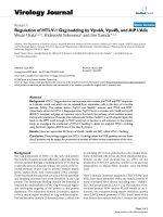
and used for global gene expression analysis. Principal
component analysis of the entire dataset (Figure 1a)
revealed that GM-CSF and 15 kDa Granulysin induced
a r esponse in monocytes that was similar at early time
points (4 hours) but strongly differed at later time
points (12, 24 and 48 hours). In particular, principal
component (PC) #1 which accounted for 31.5% of the
variability of the dataset did not separate the samples
cultured with Granulysin from those cultured with GM-
CSF, but clearly placed the 4 and 48 hour samples at
the extremes with the other samples in between and
closer to the 48 hour samples. This indicated that the
two agents induced one group of genes at 4 hours and a
second set at lat er times. P C #2, w hich accounted for
14.8% of the variability, split the G M-CSF and Granuly-
sin samples into two distinct groups at later time points,
indicating that the differences between t he GM-CSF-
and Granulysin-cultured monocytes became more evi-
dent at later time points. The third PC (14.1% of the
variability) segregated time 0 samples, the untreated
monocytes, from the other samples indicating that both
agents induced major changes at the transcriptome level
when compared to time 0 samples.
In order to stratify changed transcripts associated with
treatment and time in an unbiased fashion, the complete
gene set was further filtered to i nclude genes with
expression levels ≥ 1.75-fold from the median in at least
20% of the samples [25]. 9951 out of 337 57 genes were
obtained and used for an unsupervised hierarchical clus-
ter analysis which clearly separate d early time point
samples (T0 and T4) from the late time point samples
(Figure 1b). Moreover, within the cluster of the late
time point samples, three subclust ers emerged: all
12-hour samples, the late 15 KDa Granulysin and late
GM-CSF samples. This analysis revealed that GM-CSF
and Granulysin induce in monocytes similar changes at
the transcriptome level at early time points, but differ-
ences become more evident at later time points.
GM-CSF and 15 kDa Granulysin induce the expression of
several genes related to apoptosis and cell differentiation
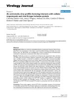
To analyze genes significantly induced by both GM-CSF
and Granulysin compared to time 0 monocytes, we
selected only the genes that at each time point were com-
monly induced following treatment by both agents com-
pared to time 0 monocytes (t-test with p-value < 0.001
and FDR < 0.1). A total of 3191, 2416, 1534 and 1738
genes were induced by both GM-CSF and Granulysin at 4,
12, 24 and 48 hours respectively. We then evaluated gene
ontology (GO) families that were statistically overrepre-
sented among up- and down-regulated genes at each time
point (Figure 2a, b, c, d). Genes related to apoptosis and
cell differentiation were significantly enriched at almost all
time points. In particular, genes that negatively regulate
apoptosis were up-regulated at 4 hours, whereas at later
time points those involved with positive induction of
apoptosi s were mainly down-regulated, suggesting a gen-
eral down-regulation of apoptosis at each time point. The
opposite was observed regarding proliferation related
genes, with proliferation related genes mainly up-regulated
Castiello et al. Journal of Translational Medicine 2011, 9:41
/>Page 3 of 12
at 4 hours and the negative regulation of proliferation
related genes down-regulated at later time points, pointing
to a ge neral induction of cell proliferation. Moreover, at
later time points, genes encoding zinc finger proteins were
up-regulated and those encoding ribosomal proteins were
down-regulated. Interestingly, at 12 hours both GM-CSF
and Granulysin induced genes related to the regulation of
the adaptive immune response, including CD40, CD80,
PVR, PVRL2 and IDO1. This initial activation of the
immune system was followed at 24 and 48 hours by the
down-regulation of genes involved in leukocyte activation
and proliferation, such as IL-8, IL-15, RAB27A, BCL11,
FYN and CLCF1.
The GM-CSF-specific gene expression signature
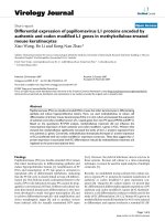
To identify genes specifically induced by GM-CSF at each
time point we sel ected only the genes that were differen-
tially expressed (p-value<0.001andFDR<0.1)byGM-
CSF-treated monocytes versus both time 0 monocytes
and cells treated with Granulysin at the same time points.
A total of 98, 768, 756 and 467 genes were specifically
induced in GM-CSF-treated monocytes at 4, 12, 24 and
48 hours, respectively (Figure 3). Gene functional cate-
gories defined by Gene Ontology (GO) families at each
time point were analyzed and only those overrepresented
in both up- and down-regulated genes were illustrated
(Figure 3). Interestingly, GM-CSF-treated monocytes spe-
cifically down-regulated immune related genes at each
time point, among which were IL-10, CXCL1, CXCL2,
CXCR4, CXCR5, and the co-stimulatory molecules
CD27, CD28, FYB (ADAP) and TNFSF4 (OX40L). In
particular, cytokine production, lymphocyte mediated
immunity and humoral immune response GO families
were overrepresente d among the down-regulated genes
at late t ime points. In contrast, at 48 hours, antigen pro-
cessing and presentation were specifically up-regulated,
including the overexpression of the genes CD1A, CD1B,
CD1E, and HLA-DQA1. Moreover, at 12 hours, GM-CSF
specifically up-regulated genes involved in myeloid cell
differentiation, including IRF4, CSF1 (GCSF), RUNX1,
CBFB and PPARG. In addition, at 12 hours, GM-CSF
specifically induced the down-regulation of genes related
to cell cycle arrest (among which were the cyclin-depen-
dent kinase inhibitors CDKN1B, CDKN2B, CDNK1C),
and thus favored cell proliferation . However, at 48 hours,
anti-apoptotic genes, such as PIM3, THBS1, HGF and
SERPINB2, were mainly down-regulated. Additionally,
among the up-regulated genes specifically induced by
GM-CSF at early time points were angiogenesis genes,
while at late time points lipid biosynthetic process genes
were up-regulated and several histone genes were down-
regulated.
The 15 kDa Granulysin-specific gene expression signature
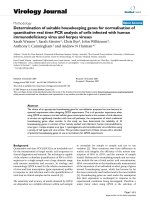
A t otal of 152, 498, 429 and 598 genes were specifically
induced in Granulysin treated monocytes at 4, 12, 24 and
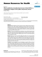
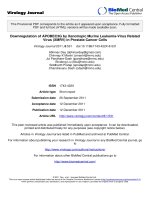
Figure 1 Gene expression analysis of monocytes cultured with GM-CSF or 15 kDa Granulysin. a) Principal component analysis of all
samples based on the entire dataset (33757 genes); b) Dendrogram of the unsupervised cluster of 9951 genes that were present in at least
22 samples and whose expression differed in at least 5 samples by more than 1.75-fold from the median.
Castiello et al. Journal of Translational Medicine 2011, 9:41
/>Page 4 of 12
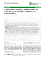
Figure 2 Monocyte genes induced by both GM-CSF and 15 kDa granulysin. Hierarchical clustering of the 3191, 2416, 1534 and 1738 genes
induced by both factors at 4 (a), 12 (b), 24 (c) and 48 (d) hours respectively (p-value < 0.001, FDR < 0.1) and the related GO analysis. The
hierarchical clustering was T0 corrected; the black bar indicates T0 monocytes, the fuchsia bar GM-CSF-treated monocytes, and the light blue bar
Granulysin-treated monocytes. GO analyses were made with DAVID. The bars indicate -Log10 of the p-value of the overrepresentation of genes
induced in each GO family. Green bars indicate down-regulated genes, while red bars indicate up-regulated genes. The orange line indicates the
threshold of statistical significance (p-value = 0.05).
Castiello et al. Journal of Translational Medicine 2011, 9:41
/>Page 5 of 12
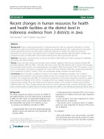
Figure 3 Monocyte genes specifically induced by GM-CSF. Hierarchical clustering of the 98, 768, 756 and 467 genes induced by GM-CSF at 4
(a), 12 (b), 24 (c) and 48 (d) hours respectively (p-value < 0.001, FDR < 0.1) and the related GO analysis. The expression of each of these genes
differed in GM-CSF treated monocytes compared to both time 0 monocytes and cells treated with Granulysin at the same time points. The
hierarchical clustering was T0 corrected; the black bar indicates T0 monocytes, the fuchsia bar GM-CSF-treated monocytes, and the light blue bar
Granulysin-treated monocytes. GO analyses were made with DAVID. The bars indicate -Log10 of the p-value of the overrepresentation of
induced genes in each GO family. The green bars indicate down-regulated genes, while the red bars indicate up-regulated genes. The orange
line indicates the threshold of statistical significance (p-value = 0.05).
Castiello et al. Journal of Translational Medicine 2011, 9:41
/>Page 6 of 12
48 hours, respectively, versus both time 0 monocytes and
cells treated with GM-CSF at the same time points (p-
value < 0.001 and FDR < 0.1, Figure 4). GO analysis
showed immune response genes were up-regulated by
Granulysin at ea ch time point. In particular, a coordi-
nated and time-dependent induction of immune related
genes could be detected. At 12 hours, innate immunity
related genes were up-regulated, but were later down-
regulated. At 48 hours humoral and lymphocyte prolif-
era tive genes were mostly up-regulated, including CCL2,
TNFRSF4,CD38,EBI3,C2,andC3.Inaddition,cell
adhesion genes, including 4 integrins (ITGB8, ITGA9,
ITGAV, ITGB5 ) and the chemokine C-C motif receptor
7 (CCR7), were specifically up-regulated especially at
late time points. In addition, chemotaxis related genes
were up-regulated at almost all time points, although
the involved genes ch anged markedly between 4 and
48 hours. In fact, CXCL1, CXCL11, CCL20 and IL-6
were up-regulated at 4 hours, whereas CXCL3, CXCL12,
CCL2, CCRL2, NRP2 and SEMA3A were induced at 48
hours. Of special note was the induction of cell prolifera-
tion genes: after the negative regulation of cell prolifera-
tion genes at 4 hours, a positive regulation of cell
proliferation genes was most prominent at 48 hours.
Granulysin, but not GM-CSF, activated pathways are
related to DC function and common-host-response
Since one of the main in vitro therapeutic uses of GM-
CSF is the differentiation, in combination with IL-4, of
monocytes into DCs a nd considering that our results
suggest a partially similar response of monocytes when
cultured with GM-CSF or 15 kDa Granulysin, we focused
on 6 specific Biocarta pathways primarily involved in DC
function (Figure 5). To evaluate the level of activation of
each pathway we used gene lists with a less stringent cut
off (p-value < 0.05 and FDR < 0.15) [24] and calculated
the percentage of genes in each pathway induced by each
treatment versus T0 monocytes. GM-CSF- and Granuly-
sin-treated monocytes showed a similar number of genes
in the Antigen Processing and Presentat ion, and Mono-
cyte a nd Surface Molecules Pathways, although the for-
mer pathway revealed a constant up-regulation of genes,
whereas for the latter pathway a down-regulation at late
time points. In contrast, differences were observed
regarding the other four pathways, reinforcing the obser-
vations described above. GM-CSF-treated monocytes
clearly showed a unique down-regulation of the IL-10
Anti-Inflammatory Signaling and the Co-stimulatory
Signal during T-cel l Activ ation Pathways, whereas Gran-
ulysin-treated monocytes showed an up-regulation of
genes in the latter pathway as well as those in the IL-12
and Stat4 Dependent Signal ing in Th1 Development and
Dendritic Cells in Regulating Th1 and Th2 Development
Pathways. Almost the same conclusions could be
outlined by focusing on the fold c hange of the genes in
each pathway instead of the percentage of genes (dat a
not shown).
To v alidate the microarray data, we p erformed real-
time PCR on CCL2, CCR7, PIM1 and CD209 genes.
The selection of CCL2 and CCR7 was based on their
up-regulation in Granulysin-treated, but not GM-CSF-
treated monocytes in array data. PIM1 was selected
because it has been descr ibed as being induced by GM-
CSF[26], and CD209 was selected since it is a marker of
DC differentiation. The analysis was perf ormed only on
untreated T0 monoc ytes and hour 4 and 48 GM-CSF-
and Granulysin-treated monocytes. Both CCL2 and
CCR7 were statistically up-regulated by both agents at 4
hours, however, at 48 hours they were only up-regulated
by Granulysin (p-value < 0.01) with a fold change
greater than 70 for CCR7 and greater than 800 for
CCL2 compared to time 0 monocytes, confirming the
finding b y microarray analysis (Additional file 1). At 4
hours the expression of both CCL2 and CCR7 was
greater in Granulysin-treated monocytes than in mono-
cytes treated with GM-CSF, with a fold change in Gran-
ulysin samples more than doubled for CCR7 and more
than quadrupled for CCL2 compared to GM-CSF.
Although PIM1 was filtered out in our analysis, RT-PCR
showed a statistically significant induction of PIM1 at
48 hours (p-value < 0.05) by both GM-CSF and Granu-
lysin, but its expression was greater in G M-CSF treated
cells. This difference can be easily ascribed to the high
stringency we used for statistical analysis of the microar-
ray data (p-value < 0.001) where we preferred to select
and analyze only those genes showing strong induction
compared time 0 monocytes. In addition, we observed
that CD209 was up-regulated by both agents at 48
hours (p-value < 0.01, with fold changes between 5 and
20 versus time 0 monocytes), which is similar to what
we observed in the microarray dataset (both agents
increased the expression CD209 genes with p-values <
0.0001).
Discussion
GM-CSF has been used for immunotherapy both in vivo
and ex vivo because of its s timulatory effect on immune
system cells. Its main application for ex vivo immu-
notherapy is the differentiation of monocytes into DCs
[9]. The broad utilization of GM-CSF in experimental
conditions as well as in clinical use is partially due t o
the lack of alternative agents with similar activity. In
this study, we performed a functional characterization of
15kDaGranulysinsidebysidewithGM-CSFand
reported their impact on gene expression changes and
kinetics in monocytes. Consideri ng the stronger reliabil-
ity of analyses of functional modules of gene s compared
to the analysis of single genes [24,27,28], we focused our
Castiello et al. Journal of Translational Medicine 2011, 9:41
/>Page 7 of 12
Figure 4 Monocyte genes specifically induced by 15 kDa Granulysin. Hierarchical clustering of the 152, 498, 429 and 598 genes induced by
15 kDa Granulysin at 4 (a), 12 (b), 24 (c) and 48 (d) hours respectively (p-value < 0.001, FDR < 0.1) and the relative GO analysis. The expression
of each of these genes differed in Granulysin treated monocytes compared to both time 0 monocytes and cells treated with GM-CSF at the
same time points. The hierarchical cluster analysis were T0 corrected, the black bar indicates T0 monocytes, the fuchsia bar GM-CSF-treated
monocytes, and the light blue bar Granulysin-treated monocytes. GO analyses were made with DAVID. The bars in the GO analysis indicate
-Log10 of the p-value of the overrepresentation of the induced genes for each GO family. The green bars indicate down-regulated genes, while
red ones indicate up-regulated genes. The orange line indicates the threshold of statistical significance (p-value = 0.05).
Castiello et al. Journal of Translational Medicine 2011, 9:41
/>Page 8 of 12
analysis only on the pathways overrepresente d among
genes differently expressed with highly stringent
p-values. Although it could be argued that several genes
were not included in the analysis due to the high strin-
gency,theuseofthesecriteriaensuredhighsensitivity
and specificity[29].
Our analysis showed that GM-CSF and 15 kDa Granuly-
sin share similar functional p roperty illustrated by their
induction of large number of gene expression changes at
different time points. The genes common to bo th agents
were mainly related to cell differentiation and a poptosis;
these genes enhanced the differentiation of monocytes and
negatively impacted apoptosis. In addition, the common
signature included immune response genes that were initi-
ally up-regulated in a similar fashion by both cytokines
and were then down-regulated. However, beyond these
overlapping functional characteristics, two different signa-
tures specific to the agent were de tected. The GM-CSF-
specific signature revealed a down-regul ation of immune
response genes, among which were several co-stimulatory
molecules. In contrast, Granulysin specifically and strongly
induced genes relat ed to the immune response with an
initial activation on innate immune related genes followed
by lymphocyte proliferative genes at later time points. In
addition, cell adhesion genes were also specifically induced
by Granulysin.
GM-CSF is a growth factor whose cellular effects had
been studied for more than twenty years [30]. At low con-
centrations (< 1 pM) it induces only cell survival, but at
higher concentrat ions it leads to monocyte proliferation,
differentiation and functional activation [31]. We found
that, although both GM-CSF and Granulysin induced
genes related to cell differentiation and silenced genes
related to cell death, only GM-CSF treated monocytes
showed the down-regulation of cell cycle arrest genes, as
previously described [31,32] and the up-regulation of genes
involved in the myeloid cell differentiation. Moreover, our
gene expression analysis not only confirmed the induction
by GM-CSF of previously described genes, such as the
anti-apoptotic gene IRF4 [33], the proliferative gene PIM1
[26], CSF1 [34] and the macrophage inducer PPARG
[35,36]; but also showed the up-regulation of the prolifera-
tion/differentiation regulator dimer RUNX1 -CBFB.
RUNX1 -CBFB has not been previously reported to be up-
regulated by GM-CSF and this observation merits further
investigation.
Monocytes cultured in presence of GM-CSF alone are
able to differentiate into iDCs, although these iDCs show
a reduced ability to induce an effective activation of lym-
phocytes after maturation [36-38]. Our gene expr ession
analysis clearly showed that GM-CSF leads to a specific
down-regulation of several immune-related genes.
Although we observed that GM-CSF induced a specific
up-regulation of the well-known CD1 family genes [13],
which play an important role in lipid antigen presentation;
gene profiling also revealed a specific down-regulation of
the co-stimulator y genes CD27, CD28, FYB (ADAP) and
TNFSF4 (OX40L). Recent studies have shown how the
proteins encoded by these genes are fundamental for the
interaction of monocyte-derived dendritic cells and T and
B cells [39-44]. In particular, GM-CSF derived DCs show
a reduced ability to secret IL-12 after maturation [9,37].
Consistent with this, we observed a general specific down-
regulation of the IL-12 and STAT 4 Dependent Signaling
Pathway in Th1 Development and the Co-stimulatory Sig-
nal during T-cell Activation Pathway. While these data
suggest that GM-CSF treated monocytes might have a
diminished ability to positively stimulate lymphocytes fol-
lowing antigen presentation, further focu sed functional
studies are needed to test this hypothesis. Of particular
interest is the observation that in the setting tested, GM-
CSF specifically down-regulated IL-10, both the gene and
the pathway, whereas previous results suggest that mono-
cytes cultured in presence of GM-CSF produce high
amounts of IL-10 once stimulated with LPS, IFN-g, TNFa
or anti-CD40 Ab [9,37]. This dis crepancy could be the
result of the differences in the concentration of GM-CSF
used in the monocyte culture conditions or it may be that
the higher expression of IL-10 by GM-CSF cultured
monocytes is only subsequent to the stimulation with
maturating agents.
15 kDa Granulysin is constitutively secreted in vivo by
CTL and NK cells, but its function is still i ncompletely
Figure 5 DC related Biocarta pathway level analysis.The
percentages of genes statistically induced by each treatment and at
each time point compared to time 0 monocytes are displayed in a
grid (p-value < 0.05, FDR < 0.15). The position of each time-
treatment in the grid is described in the bottom right corner,
whereas the bottom left indicates the scale of intensity of the colors
in the grid.
Castiello et al. Journal of Translational Medicine 2011, 9:41
/>Page 9 of 12
defined [15,17]. The ability of Granulysin to replicate
some GM-CSF-induced monocyte responses is shown
by the observation that between 4 and 48 hours thou-
sands of genes were induced by both GM-CSF- and
Granulysin. On the other hand, the gene expression
analysis revealed that Granulysin, but not GM-CSF,
treated monoc ytes showed an overexpression of several
immune-related genes at e ach time point. Moreover,
our data showed that Granulysin induced a specific
time-coordinated activation of the immune system. At
early time points, several genes involved in the activa-
tion of the innate immune system were induced
whereas, at later time points, lymphocyte proli feration
genes and humoral immune response were up-regulated.
In addition, the pathway analysis clearly demonstrated
that Granulysin-treated monocytes specifically induced
the IL-12 and Stat4 Dependent Signaling Pathway in
Th1 Development, suggesting that Granulysin might
induce a shift towards Th1 T cell differentiation.
Recently, co-stimulatory molecules have been shown
toplayaroleinchemotaxis[45].Wefoundthat,in
contrast to GM-CSF-treatment, Granulysin treatment
did not lead to the down-regulation of co-stimulatory
molecules; rather Granulysin specifically showed an up-
regulation of the co-stimulatory pathways and overex-
pressed chemotactic genes at each time point. In parti-
cular, Granulysin induced the expression of a wide
group of chemokines that are able to attract ne utrophils
(CXCL1, CXCL3) [46], memory and activated T cells
(CXCL11, CCL20, CCR7) [47,48], monocytes (CCL2,
CCL20) [49], macrophages and dendritic cells (NRP2)
[50]. Several studies have shown that chemokines act
synergistically [51,52], strengthening their signals and
overcoming eventual antagonists secreted by pathogens
[53,54]. Interestingly a partially overlapping time-
fashioned chemokine induction has been described by
myeloid and plasmacytoid D Cs exposed to influenza
virus [55]. This observation might indicate that 15 kDa
Granulysin plays an important role in activating the
immune system in response to pathogens by inducing
monocytes to recruit other immune cells. Moreover, the
observation that Granulysin acts as an alarmin strengthen
this hypothesis [16,18].
Conclusions
In conclusion, the analysis of gene expression profiles of
monocytes cultured in presence of GM-CSF and 15 kDa
Granulysin revealed that although both induce many of
the same genes, these two cytokines induce two differ-
ent monocyt e responses. Considering the greater induc-
tion of several immune related functions by 15 kDa
Granulysin, this study suggests that 15 kDa Granulysin
may prove a useful therapeutic immunomodulator for
in vitro production of Th-1 polarized monocyte-derived
DCs for adoptive immunotherapy.
Additional material
Additional file 1: Quantitative real time PCR analysis of selected
genes. Relative quantification of CCR7, CCL2, PIM1 and CD209 genes are
represented. HPRT1 was used as a housekeeping gene. One sample of
time 0 monocytes was set to the unitary value (1) and used as calibrator.
Values from the 3 different donors were averaged and the standard
deviation is represented for each bar. The light blue columns represent
GM-CSF-treated monocytes and the purple bar Granulysin-treated
monocytes.
Acknowledgements and funding
This work is supported by the Intramural Programs of the National Institutes
of Health Clinical Center and National Cancer Institute.
Author details
1
Cell Processing Section, Department of Transfusion Medicine, Clinical
Center, National Institutes of Health, Bethesda, MD 20892, USA.
2
Laboratory
of Cellular and Molecular Biology, National Cancer Institute, National
Institutes of Health, Bethesda, MD 20892, USA.
3
Infectious Disease and
Immunogenetics Section, Department of Transfusion Medicine, Clinical
Center, and Center for Human Immunology (CHI), National Institutes of
Health, Bethesda, MD 20892, USA.
Authors’ contributions
LC performed experiments and data analysis; MWF expressed and purified
the 15 kDa Granulsyin; DFS, MS, FMM, EW, CC, AMK contributed to
experimental design and data analysis; LC, DFS compiled the manuscript;
MS, FMM, EW, CC, AMK revised the manuscript. All of the authors have read
and approved the final manuscript.
Competing interests
AMK and CC hold patents on granulysin. The remaining authors declare no
competing interests.
Received: 10 March 2011 Accepted: 18 April 2011
Published: 18 April 2011
References
1. Waller EK: The role of sargramostim (rhGM-CSF) as immunotherapy.
Oncologist 2007, 12(Suppl 2):22-26.
2. Everly JJ, Lonial S: Immunomodulatory effects of human recombinant
granulocyte-macrophage colony-stimulating factor (rhuGM-CSF):
evidence of antitumour activity. Expert Opin Biol Ther 2005, 5:293-311.
3. Mitsuyasu RT, Golde DW: Clinical role of granulocyte-macrophage colony-
stimulating factor. Hematol Oncol Clin North Am 1989, 3:411-425.
4. Boyer MW, Waller EK, Bray RA, Unangst T, Johnson TS, Phillips C, Jurickova I,
Winton EF, Yeager AM: Cytokine upregulation of the antigen presenting
function of acute myeloid leukemia cells. Leukemia 2000, 14:412-418.
5. Arellano ML, Langston A, Winton E, Flowers CR, Waller EK: Treatment of
relapsed acute leukemia after allogeneic transplantation: a single center
experience. Biol Blood Marrow Transplant 2007, 13:116-123.
6. Sallusto F, Lanzavecchia A: Efficient presentation of soluble antigen by
cultured human dendritic cells is maintained by granulocyte/
macrophage colony-stimulating factor plus interleukin 4 and
downregulated by tumor necrosis factor alpha. J Exp Med 1994,
179:1109-1118.
7. Mohamadzadeh M, Berard F, Essert G, Chalouni C, Pulendran B, Davoust J,
Bridges G, Palucka AK, Banchereau J: Interleukin 15 skews monocyte
differentiation into dendritic cells with features of Langerhans cells.
J Exp Med 2001, 194:1013-1020.
8. Santini SM, Lapenta C, Logozzi M, Parlato S, Spada M, Di PT, Belardelli F:
Type I interferon as a powerful adjuvant for monocyte-derived dendritic
Castiello et al. Journal of Translational Medicine 2011, 9:41
/>Page 10 of 12
cell development and activity in vitro and in Hu-PBL-SCID mice. J Exp
Med 2000, 191:1777-1788.
9. Conti L, Gessani S: GM-CSF in the generation of dendritic cells from
human blood monocyte precursors: recent advances. Immunobiology
2008, 213:859-870.
10. Lehmann MH: Recombinant human granulocyte-macrophage colony-
stimulating factor triggers interleukin-10 expression in the monocytic
cell line U937. Mol Immunol 1998, 35:479-485.
11. Smith WB, Guida L, Sun Q, Korpelain en EI, van den HC, Gillis D,
Hawrylowicz CM , Vadas MA, Lopez AF: Neutrophils activated by
granulocyte-macrophage colony-stimulating factor express receptors
for interleukin-3 which mediate cla ss II expression. Blood 1995,
86:3938-3944.
12. Alderson MR, Tough TW, Ziegler SF, Armitage RJ: Regulation of human
monocyte cell-surface and soluble CD23 (Fc epsilon RII) by granulocyte-
macrophage colony-stimulating factor and IL-3. J Immunol 1992,
149:1252-1257.
13. Kasinrerk W, Baumruker T, Majdic O, Knapp W, Stockinger H: CD1 molecule
expression on human monocytes induced by granulocyte-macrophage
colony-stimulating factor. J Immunol 1993, 150:579-584.
14. Hornell TM, Beresford GW, Bushey A, Boss JM, Mellins ED: Regulation of the
class II MHC pathway in primary human monocytes by granulocyte-
macrophage colony-stimulating factor. J Immunol 2003, 171:2374-2383.
15. Hanson DA, Kaspar AA, Poulain FR, Krensky AM: Biosynthesis of granulysin,
a novel cytolytic molecule. Mol Immunol 1999, 36:413-422.
16. Zitvogel L, Kroemer G: The multifaceted granulysin. Blood 2010,
116:3379-3380.
17. Krensky AM, Clayberger C: Biology and clinical relevance of granulysin.
Tissue Antigens 2009, 73:193-198.
18. Tewary P, Yang D, de la RG, Li Y, Finn MW, Krensky AM, Clayberger C,
Oppenheim JJ: Granulysin activates antigen-presenting cells through
TLR4 and acts as an immune alarmin. Blood 2010, 116:3465-3474.
19. Finn MW, Clayberger C, Krensky AM: Expression and purification of 15 kDa
granulysin utilizing an insect cell secretion system. Protein Expr Purif 2011,
75:70-74.
20. Mane VP, Heuer MA, Hillyer P, Navarro MB, Rabin RL: Systematic method
for determining an ideal housekeeping gene for real-time PCR analysis.
J Biomol Tech 2008, 19:342-347.
21. Eisen MB, Spellman PT, Brown PO, Botstein D: Cluster analysis and display
of genome-wide expression patterns. Proc Natl Acad Sci USA 1998,
95:14863-14868.
22. Huang dW, Sherman BT, Zheng X, Yang J, Imamichi T, Stephens R,
Lempicki RA: Extracting biological meaning from large gene lists with
DAVID. Curr
Protoc Bioinformatics 2009, 13, Unit.
23. Dennis G Jr, Sherman BT, Hosack DA, Yang J, Gao W, Lane HC, Lempicki RA:
DAVID: Database for Annotation, Visualization, and Integrated Discovery.
Genome Biol 2003, 4:3.
24. Chaussabel D, Quinn C, Shen J, Patel P, Glaser C, Baldwin N, Stichweh D,
Blankenship D, Li L, Munagala I, et al: A modular analysis framework for
blood genomics studies: application to systemic lupus erythematosus.
Immunity 2008, 29:150-164.
25. Simon R: Using DNA microarrays for diagnostic and prognostic
prediction. Expert Rev Mol Diagn 2003, 3:587-595.
26. Guthridge MA, Barry EF, Felquer FA, McClure BJ, Stomski FC, Ramshaw H,
Lopez AF: The phosphoserine-585-dependent pathway of the GM-CSF/IL-
3/IL-5 receptors mediates hematopoietic cell survival through activation
of NF-kappaB and induction of bcl-2. Blood 2004, 103:820-827.
27. Mootha VK, Lindgren CM, Eriksson KF, Subramanian A, Sihag S, Lehar J,
Puigserver P, Carlsson E, Ridderstrale M, Laurila E, et al: PGC-1alpha-
responsive genes involved in oxidative phosphorylation are coordinately
downregulated in human diabetes. Nat Genet 2003, 34:267-273.
28. Segal MR, Dahlquist KD, Conklin BR: Regression approaches for microarray
data analysis. J Comput Biol 2003, 10:961-980.
29. Shi L, Jones WD, Jensen RV, Harris SC, Perkins RG, Goodsaid FM, Guo L,
Croner LJ, Boysen C, Fang H, et al: The balance of reproducibility,
sensitivity, and specificity of lists of differentially expressed genes in
microarray studies. BMC Bioinformatics 2008, 9(Suppl 9):S10.
30. Hamilton JA: Colony-stimulating factors in inflammation and
autoimmunity. Nat Rev Immunol 2008, 8:533-544.
31. Guthridge MA, Powell JA, Barry EF, Stomski FC, McClure BJ, Ramshaw H,
Felquer FA, Dottore M, Thomas DT, To B, et al: Growth factor pleiotropy is
controlled by a receptor Tyr/Ser motif that acts as a binary switch. EMBO
J 2006, 25:479-489.
32. Williams GT, Smith CA, Spooncer E, Dexter TM, Taylor DR: Haemopoietic
colony stimulating factors promote cell survival by suppressing
apoptosis. Nature 1990, 343:76-79.
33. Lehtonen A, Matikainen S, Miettinen M, Julkunen I: Granulocyte-
macrophage colony-stimulating factor (GM-CSF)-induced STAT5
activation and target-gene expression during human monocyte/
macrophage differentiation. J Leukoc Biol 2002, 71:511-519.
34. Gruber MF, Gerrard TL: Production of macrophage colony-stimulating
factor (M-CSF) by human monocytes is differentially regulated by GM-
CSF, TNF alpha, and IFN-gamma. Cell Immunol 1992, 142
:361-369.
35.
Ricote M, Huang J, Fajas L, Li A, Welch J, Najib J, Witztum JL, Auwerx J,
Palinski W, Glass CK: Expression of the peroxisome proliferator-activated
receptor gamma (PPARgamma) in human atherosclerosis and regulation
in macrophages by colony stimulating factors and oxidized low density
lipoprotein. Proc Natl Acad Sci USA 1998, 95:7614-7619.
36. Skorokhod OA, Alessio M, Mordmuller B, Arese P, Schwarzer E: Hemozoin
(malarial pigment) inhibits differentiation and maturation of human
monocyte-derived dendritic cells: a peroxisome proliferator-activated
receptor-gamma-mediated effect. J Immunol 2004, 173:4066-4074.
37. Ganguly D, Paul K, Bagchi J, Rakshit S, Mandal L, Bandyopadhyay G,
Bandyopadhyay S: Granulocyte-macrophage colony-stimulating factor
drives monocytes to CD14low CD83+ DC. Immunology 2007, 121:499-507.
38. Chitta S, Santambrogio L, Stern LJ: GMCSF in the absence of other
cytokines sustains human dendritic cell precursors with T cell regulatory
activity and capacity to differentiate into functional dendritic cells.
Immunol Lett 2008, 116:41-54.
39. Lippert U, Zachmann K, Ferrari DM, Schwarz H, Brunner E, Mahbub-Ul Latif
AH, Neumann C, Soruri A: CD137 ligand reverse signaling has multiple
functions in human dendritic cells during an adaptive immune
response. Eur J Immunol 2008, 38:1024-1032.
40. Hashimoto-Okada M, Kitawaki T, Kadowaki N, Iwata S, Morimoto C, Hori T,
Uchiyama T: The CD70-CD27 interaction during the stimulation with
dendritic cells promotes naive CD4(+) T cells to develop into T cells
producing a broad array of immunostimulatory cytokines in humans. Int
Immunol 2009, 21:891-904.
41. Hendriks J, Xiao Y, Rossen JW, van der Sluijs KF, Sugamura K, Ishii N, Borst J:
During viral infection of the respiratory tract, CD27, 4-1BB, and OX40
collectively determine formation of CD8+ memory T cells and their
capacity for secondary expansion. J Immunol 2005, 175:1665-1676.
42. Wang J, Guo Z, Dong Y, Kim O, Hart J, Adams A, Larsen CP, Mittler RS,
Newell KA: Role of 4-1BB in allograft rejection mediated by CD8+ T cells.
Am J Transplant 2003, 3:543-551.
43. Croft M, So T, Duan W, Soroosh P: The significance of OX40 and OX40L to
T-cell biology and immune disease. Immunol Rev 2009, 229:173-191.
44. Zhang X, Voskens CJ, Sallin M, Maniar A, Montes CL, Zhang Y, Lin W, Li G,
Burch E, Tan M, et al: CD137 promotes proliferation and survival of
human B cells. J Immunol 2010, 184:787-795.
45. Zhong W, Zhang Z, Hinrichs D, Wu X, Hall M, Xia Z, Rosenbaum JT: OX40
induces CCL20 expression in the context of antigen stimulation: an
expanding role of co-stimulatory molecules in chemotaxis. Cytokine 2010,
50:253-259.
46. Scimone ML, Lutzky VP, Zittermann SI, Maffia P, Jancic C, Buzzola F,
Issekutz AC, Chuluyan HE: Migration of polymorphonuclear leucocytes is
influenced by dendritic cells. Immunology 2005, 114:375-385.
47. Sallusto F, Schaerli P, Loetscher P, Schaniel C, Lenig D, Mackay CR, Qin S,
Lanzavecchia A: Rapid and coordinated switch in chemokine receptor
expression during dendritic cell maturation. Eur J Immunol 1998,
28
:2760-2769.
48.
Schutyser E, Struyf S, Van DJ: The CC chemokine CCL20 and its receptor
CCR6. Cytokine Growth Factor Rev 2003, 14:409-426.
49. Deshmane SL, Kremlev S, Amini S, Sawaya BE: Monocyte chemoattractant
protein-1 (MCP-1): an overview. J Interferon Cytokine Res 2009, 29:313-326.
50. Rey-Gallardo A, Escribano C, Delgado-Martin C, Rodriguez-Fernandez JL,
Gerardy-Schahn R, Rutishauser U, Corbi AL, Vega MA: Polysialylated
neuropilin-2 enhances human dendritic cell migration through the basic
C-terminal region of CCL21. Glycobiology 2010, 20:1139-1146.
51. Vanbervliet B, Bendriss-Vermare N, Massacrier C, Homey B, de Bouteiller O,
Briere F, Trinchieri G, Caux C: The inducible CXCR3 ligands control
plasmacytoid dendritic cell responsiveness to the constitutive
Castiello et al. Journal of Translational Medicine 2011, 9:41
/>Page 11 of 12
chemokine stromal cell-derived factor 1 (SDF-1)/CXCL12. J Exp Med 2003,
198:823-830.
52. Gouwy M, Struyf S, Proost P, Van Damme J: Synergy in cytokine and
chemokine networks amplifies the inflammatory response. Cytokine
Growth Factor Rev 2005, 16:561-580.
53. Rosenkilde MM: Virus-encoded chemokine receptors–putative novel
antiviral drug targets. Neuropharmacology 2005, 48:1-13.
54. Alcami A, Saraiva M: Chemokine binding proteins encoded by pathogens.
Adv Exp Med Biol 2009, 666:167-179.
55. Piqueras B, Connolly J, Freitas H, Palucka AK, Banchereau J: Upon viral
exposure, myeloid and plasmacytoid dendritic cells produce 3 waves of
distinct chemokines to recruit immune effectors. Blood 2006,
107:2613-2618.
doi:10.1186/1479-5876-9-41
Cite this article as: Castiello et al.: 15 kDa Granulysin versus GM-CSF for
monocytes differentiation: analogies and differences at the
transcriptome level. Journal of Translational Medicine 2011 9 :41.
Submit your next manuscript to BioMed Central
and take full advantage of:
• Convenient online submission
• Thorough peer review
• No space constraints or color figure charges
• Immediate publication on acceptance
• Inclusion in PubMed, CAS, Scopus and Google Scholar
• Research which is freely available for redistribution
Submit your manuscript at
www.biomedcentral.com/submit
Castiello et al. Journal of Translational Medicine 2011, 9:41
/>Page 12 of 12