Báo cáo sinh học: "A novel multiplex assay combining autoantibodies plus PSA has potential implications for classification of prostate cancer from non-malignant cases" docx
Bạn đang xem bản rút gọn của tài liệu. Xem và tải ngay bản đầy đủ của tài liệu tại đây (750.94 KB, 11 trang )
RESEARCH Open Access
A novel multiplex assay combining
autoantibodies plus PSA has potential
implications for classification of prostate cancer
from non-malignant cases
Chong Xie
1
, Hyun J Kim
2
, Jonathan G Haw
3
, Anusha Kalbasi
3
, Brian K Gardner
4
, Gang Li
5
, Jianyu Rao
6
, David Chia
6
,
Monty Liong
7
, Rubio R Punzalan
8
, Leonard S Marks
3
, Allan J Pantuck
3
, Alexandre de la Taille
9
, Guomin Wang
1
,
Hideki Mukouyama
10
and Gang Zeng
3*
Abstract
Background: The lack of sufficient specificity and sensitivity among conventional cancer biomarkers, such as
prostate specific antigen (PSA) for prostate cancer has been widely recognized after several decades of clinical
implications. Autoantibodies (autoAb) among others are being extensively investigated as potential substitute
markers, but remain elusive. One major obstacle is the lack of a sensitive and multiplex approach for quantifying
autoAb against a large panel of clinically relevant tumor-associated antigens (TAA).
Methods: To circumvent preparation of phage lysates and purification of recombinant proteins, we identified B
cell epitopes from a number of previously defined prostate cancer-associated antigens (PCAA). Peptide epitopes
from cancer/testis antigen NY-ESO-1, XAGE-1b, SSX-2,4, as well as prostate cancer overexpressed antigen AMACR,
p90 autoantigen, and LEDGF were then conjugated with seroMAP microspheres to allow multiplex measurement
of autoAb present in serum samples. Moreover, simultaneous quantification of autoAb plus total PSA was achieved
in one reaction, and termed the “A+PSA” assay.
Results: Peptide epitopes from the above 6 PCAA were identified and confirmed that autoAb against these
peptide epitopes reacted specifically with the full-length protein. A pilot study was conducted with the A+PSA
assay using pre-surgery sera from 131 biopsy-confirmed prostate cancer patients and 121 benign prostatic
hyperplasia and/or prostatitis patients. A logistic regression-based A+PSA index was found to enhance sensitivities
and specificities over PSA alone in distinguishing prostate cancer from nonmalignant cases. The A+PSA index also
reduced false positive rate and improved the area under a receiver operating characteristic curve.
Conclusions: The A+PSA assay represents a novel platform that integrates autoAb signatures with a conventional
cancer biomarker, which may aid in the diagnosis and prognosis of prostate cancer and others.
Background
Both the cellular and humoral arms of the human
immune system recognize tumor-associated antigens
(TAA) derived from endogenously arising cancer cells.
Of particular interest to the serological analysis of
human cancers is a panel of clin ically relevant TAA
recognized by autoAb present in the serum of cancer
patients including those with prostate cancers [1,2]. In
prostate cancer, autoAb-recognized prostate cancer-
associated antigens (PCAA) may be divided into two
categories: 1) autoAb recognize a-methylacyl-CoA
(AMACR) [3,4], p90 autoantige n [5], and lens epithe-
lium-derived growth factor p75 (LEDGF) [6], which
have low levels of expression in normal tissues, but are
overexpressed in prostate cancer; 2) autoAb react
against cancer/testis antigens such as NY-ESO-1 [7],
* Correspondence:
3
Department of Urology, David Geffen School of Medicine at UCLA, 10833
Le Conte Ave, Los Angeles, CA 90095-1738, USA
Full list of author information is available at the end of the article
Xie et al. Journal of Translational Medicine 2011, 9:43
/>© 2011 Xie et al; licensee BioMed Central L td. This is an Open Access article di stributed under th e terms of the C reative Commons
Attribution License ( s/by/2.0 ), which permits unrestricted use, distribution, and reproduction in
any medium, provided the original work is properly ci ted.
SSX-2,4 [8], and XAGE-1b [9], which are observed only
in cancer patients but not healthy donors (HD) or
patients with benign conditions. Cancer/testis antigens
are by f ar the most cancer-speci fic TAA, which are
shared by a number of solid tumors including prostate
cancer, lung cancer, and so on. In normal tissues, they
are only expressed in immune-privileged germline cells.
In this study, we focused on a panel of clinically relevant
PCAA, whose expression in prostate cancer tissues and
autoAb presence in serum samples have been verified
by multiple groups. AutoAb against these targets are
also observed prominently in prostate cancer patients
than healthy donors.
In contrast to conventional biomarkers produced by
tumor cells such as PSA, autoAb against clinically rele-
vant TAA are produced by the body in response to neo-
plastic transformation. Spontaneous autoAb present in
patients’ serum samples may reflect cancer-related
inflammation, immunocompetence of the host, and
immunogenicity of the endogenously arising cancer
[10,11]. Even though more and more studies have
shown the signif icance of circulating autoAb in serving
cancer detection, diagnosis, prognosis, and other areas
[12-14], sensitive a nd cost-effective detection of autoAb
against multiple TAA still lacks that may se rve for clini-
cal laboratories.
Currently, two main strategies are used for broad-
based profiling of circ ulating autoAb: serological surveys
using phage lysates encoding specific TAA [4], protein
array and ELISA-based approaches using purified
recombinant proteins [15-17]. The former approach
requires large amounts of sera individually pre-adsorbed
with E. coli phage lysates for reduction of background;
the latter require the purification of proteins encoding
individual TAA. To circumvent the requirement of puri-
fying phage lysates or individual TAA protein, we have
focused on targeted identification of B cell epitopes
from TAA [18], and developed a novel multiplex assay
platform that quantifies autoAb plus total PSA in a sin-
gle reaction for prostate cancer.
Methods
Prediction, screening and validation of B-cell epitopes
from PCAA
The study focused on 6 PCAA, namely NY-ESO-1,
SSX-2,4, XAGE-1b, AMACR, p90, and LEDGF. All had
been reported by multiple groups with data on gene
expression and autoAb presence in prostate cancer
patients. As previously described [18], pr ediction and
screening of peptide epitopes was conducted using clas-
sic ELISA. Peptides were considered positive based on
recognition by serum samples from prostate cancer
patients (n > 50) but not healthy donors (n > 20). Then,
peptide-reacting serum samples were verified for
recognition of the full-length or a truncated recombi-
nant protein using Western blot. Only after such a pro-
cedure, a validated peptide was conjugated onto
seroMAP microbeads for mu ltiplex measurement. All
peptides involved in this study were sy nthesized at
Genscript Inc. (Piscataway, NJ) and GeneMedicine, Inc.
(San Antonio, TX). Histori cal serum samples from can-
cer and HD as described previously [18] were used for
identification of peptide epitopes, which were indepen-
dent of those used in the subsequ ent study com paring
A+PSA index with PSA. In the case of identifying pep-
tide epitopes from shared cancer/testis antigen XAGE-
1b and SSX2,4, serum samples from NSCLC were used.
This choice was made based on higher frequency of ser-
opositive subjects in NSCLC and the fact that peptides
from shared antigens identified using one type of cancer
patients can be equally well recognized by prostate can-
cer patients [18].
Clinical and demographic characteristics of serum donors
involved in the study
All serum samples were collected under institutional
review board-approved protocols from UCLA (IRB#06-
03-044) and collaborating hospitals, and stored at -20°
C until use. Serum samples from normal healthy sub-
jects were collected at the time of blood donation in
subjects routinely screened to exclude the presence of
concomitant disease such as cancer according to stan-
dard blood bank policies. Serum samples from biopsy-
confirmed prostate cancer p atients were collected at
the time of biopsy and prior to surgery. Patients with
BPH and/or prostatitis, specified as non-cancer or
BPH/prostatitis patients throughout this m anuscript,
were those with clinical signs and symptoms, for
instance, characteristic lower urinary tract symptoms,
International Prostate Symptom Scores, urinary leuko-
cytes, and so on. These patients were subsequently
underwent a routine fine needleprostatebiopsywith
at least 6-12 samples taken showing no evidence of
prostate cancer. Table 1 sho ws the demographic and
clinical characteristics of the subjects involved in the
comparison of A+PSA with PSA alone. Additional file
1 illustrates the distribution of their total PSA values,
which were measured using a standard ELISA
approach according to the manufacturer’s recommen-
dations at the time of diagnosis.
Since this was a pilot study, cohort size and relevant
parameters such a s age, racial and ethnical background
were not sufficient to match samples according to
potential clinical co-founders. However, all samples
themselves were handled and stored according to the
same conditions prior to assay; and normalization with
samples from HD was conducted when needed in order
to minimize experiment-to-experiment variations.
Xie et al. Journal of Translational Medicine 2011, 9:43
/>Page 2 of 11
Conjugation of peptide epitopes with seroMAP
mircrobeads and conduct of seroMAP-based assays
Conjugation of peptide epitopes defined in this study
onto seroMAP beads was conducted according to the
manufacturer’s recommendations (Luminex Corpora-
tion, Austin, TX). In the final configuration of the A
+PSA assay, seroMAP microbeads region 001 were con-
jugated with the NY-ESO-1 pe ptide epitope a s pre-
viously reported [18], region 010 with the XAGE-1b
epitope (amino acid 1-25), region 020 with the SSX2,4
epitope (amino acid 110-139), region 030 with the
AMACR epitope (amino acid 251-281), region 040 with
the p90 autoantigen epitope (amino acid 796-827),
region 050 with a control peptide from b-galactosidase,
and region 060 with the LEDGF epitope (amino acid
448-468). A 96-well filter bottom plate (Millipore, Biller-
ica, MA) was pre-washed followed by addition of block-
ing buffer and incubation for 1 hour at room
temperature. About 50 μl of serum samples pre-diluted
at1to10,1to20,and1to50weremixedwithan
equal volume of the above-conjugated seroMAP
microbeads at 5000 beads/region, and were added to
each well. After one hour of incubation, plates were
washed 3 times, followed by addition of 100 μl PE-
labeled detection Ab (Ab against human IgG and Ab
against human total PSA) to each well. After 30 min,
plates were washed 3 times, and added 100 μl blocking
buffer into each well. The plate was read by Bioplex-200
(Bio-Rad Laboratories, Hercules, CA) to obtain the
mean florescent intensity (MFI) for each seroMAP
region.
In addition to measuring autoAb, seroMAP microbe-
ads region 100 were conjugated with a monoclonal Ab
against human PSA (Biocon, Inc. Rockville, MD) to
quantify total PSA levels. sero MAP-based PSA quantifi-
cation was compared with standard ELISA-based PSA
assays (American Qualex) and also made compatible
with the measurement of the above-mentioned 6 autoAb
to constitute the A+PSA assay.
Comparison of signal to noise ratios of seroMAP- and
ELISA-based approaches for autoAb measurement
AutoAb present in patients’ serum samples were pre-
viously measured using a standardized ELISA approach
[18].Inbrief,1μg of a synthetic peptide was diluted in
5 ml phosphate buffered saline (PBS) and adsorbed onto
a 96-well MaxiSorp plate (Nunc, Denmark) overnight at
room temperature. Control plates were coated with
bovine serum albumin (BSA) at 15 μg/plate or about
150 ng/well. Plates were blocked with 5% Fetal Bovine
Serum in P BST (PBS plus 0.05% Tween-20) for at least
2 hours, washed with PBST, and loaded with 100 μlof
serum samples diluted at 1:25, 1:125, and 1:625 with
PBST containing 5% Fetal Bovine Serum. After a 2-hour
incubation at room temperature, plates were washed,
and loaded with secondary antibodies (goat anti-human
immunoglobulin conjugated with horseradish peroxi-
dase,SigmaCo.,St.Louis, MO) diluted with 5% Fetal
Bovine Serum in PBST. Plates were developed after a
one-hour incubation, and absorbance at 450 nm was
read by using an ELISA reader. Signal to noise ratio for
ELISA-based approaches was defined as the OD against
a target epitope/average OD from at least 8 HD. Signal
to noise ratio for seroMAP-based approaches was
defined as the specific MFI ratio against a target pep-
tide/average specific MFI ratio against the same peptide
from at least 8 HD, where the specific MFI ratio is
defined as the MFI against a PCAA peptide/MFI against
a control peptide.
Statistical analysis and the logistic regression-based A
+PSA index
To better pr edict prostate cancer, it is necessary to cre-
ate an index integrating b oth autoAb against the 6
above-described PCAA and the patient’s PSA status. For
total PSA a nd autoAb against each peptide epitope, an
index va lue was cal culated based on t he mean MFI
ratio, which is defined as the florescent intensity against
a specific peptide/florescent intensity against a control
peptide. The K olmogorov-Smirnov test was used in the
6 autoAb markers to determine if the histograms
between prostate cancer and BPH and/or prostatitis dif-
fer significantly. The A+PSA index was defined as the
Table 1 Demographic and clinical characteristics of
patients involved in the study
Subjects HD BPH/Prostatitis Prostate Cancer
n = 124 n = 121 n = 131
Age (year)
unknown 124 2 4
<40 2 1
40-49 4 2
50-59 16 2
60-69 30 14
70-79 46 38
>80 21 20
Collection site*
Japan 84 121 81
U.S. 40 0 50
Gleason Scores
unknown 10
< and = 6 61
729
> and = 8 31
*Race is not known, samples are only classified based on the collection site.
Note that this pilot study is focused on comparing A+PSA with the PSA assay,
cohort size was not sufficient to match samples according to potential clinical
co-founders.
Xie et al. Journal of Translational Medicine 2011, 9:43
/>Page 3 of 11
probability of being prostate cancer, which was obtained
by combining the six above referenced epitope indices
with the PSA index using the logistic regression method
Pr =
exp(a
0
+
6
i=1
a
i
˜
N
i
+ a
7
˜
N
PSA
)
1 + exp(a
0
+
6
i
=1
a
i
˜
N
i
+ a
7
˜
N
PSA
)
,
where each Ni represented the average MFI values for
an autoAb from three d ilutions, N
PSA
was the average
MFI for PSA from three dilutions, and a0, a7 were esti-
mated regression coefficients of the logistic regression
model. In the logistic regression model, the b inary
dependent variable is 1 for a patient with prostate can-
cer and 0 for a patient with nonmalignant conditions,
for example, BPH and/or prostatitis. The receiver oper-
ating characteristic (ROC) curve was used to compare
the diagnostic power between PSA alone and the com-
bined A+PSA index for distinguishing prostate cancer
from BPH and prostatitis in all subjects and subjects
with 4-10 ng/ml PSA. Area Under the Curve (AUC)
from Receiver operati ng characteristic (ROC) analysi s
was calculated from the logistic regression model. To
avoid a potential overfitting issue in modeling and the
testing within the same data set, the bootstrap method
[19] was applied to construct 95% confidence intervals
for the AUCs and test their difference. Values of P <
0.05 were considered statistically significant.
Results
Identification and validation of B cell epitopes from PCAA
Similar to NY-ESO-1, XAGE-1b and SSX-2,4 are can-
cer/testis antigens shared among cancers of the pros-
tate, lung, breast and others [20,21]. To identify
dominant B cell epitopes from X AGE-1b, computer-
aided algorithms were applied to predict the peptide
epitopes [18]. Two candidate peptides were screened
by ELISA (Figure 1A) with serum samples from cancer
patients. Three of 48 cancer patients were tested posi-
tive reacting with XAGE-1b peptides ba sed on pre-
viously described criterion [18]. Two of the 3
seropositive patients reacted only with XAGE:1-25
peptide; while the other reacted with both XAGE:1-25
and XAGE:57-81 peptides. Western blot confirmed
that sera recognizing the XAGE:1-25 peptide reacted
with the full-length XAGE-1b protein from a trans-
fected 293 cell line (Figure 1B).
Similarly, a SSX-2,4 peptide epitope was identified and
confirmed with Western blot that the serum reacting
with SSX-2,4:110-139 wa s able to recognize the full-
length recombinant protein (Additional file 2). In addi-
tion, candidate peptides from AMACR, p90 autoantigen,
and LEDGF were screened using serum samples from
prostate cancer patients and control samples from HD
(data not shown). Verification of the AMACR and
LEDGF peptide epitopes by Western blot is also shown
in Additional file 2.
Peptide epitopes linked to seroMAP microspheres
markedly improves signal-to-noise ratios over classic
ELISA
Following the identification and confirmation of pep-
tide epitopes from the above-mentioned PCAA, each
peptide was conjugated onto seroMAP microbeads
with a specific region number (Materials and Meth-
ods). The ease of conjugating peptides over purified
recombinant proteins onto seroMAP microspheres
allowed multiplex detection of autoAb against the
above-described peptide epitopes from XAGE-1b,
SSX2,4, AMACR, p90 autoantigen, L EDGF, and NY-
ESO-1 [18]. Specific MFI ratios, defined as the ratio of
the MFI against a target peptide to the MFI against a
control pe ptide, w ere compared with those from at
least 8 HD, which was defined as the relevant signal-
to-noise ratios for seroMAP-based approaches. Simi-
larly, signal-to-noise ratios for ELISA-based approaches
were determined (Materials and Methods). The sero-
MAP-based approach showed significantly improved
signal-to-noise ratios over ELISA-based approach in
measuring autoAb against a prototype NY-ESO-1:1-40
epitope among 4 randomly selected seropositive pros-
tate cancer patients with 8 HD as controls (Figure 2A).
Similarly, improved signal-to-noise ratios against the
XAGE-1b epitope were observed using the seroMAP-
based approach over ELIS A.
The multiplex A+PSA assay quantifies autoAb and total
PSA in one reaction
To develop a multiplex assay that measures total PSA
and autoAb in a single reaction, conventional PSA tests
were first converted from ELISA to seroMAP-based
approaches. PE-conjugated secondary Ab against human
IgGandPSAweremixedinthemultiplexassayto
accommodate staining of autoAb and PSA binding to
distinct seroMAP regions, allowing simultaneous quanti-
fication of autoAb plus PSA in one reaction (termed the
A+PSA assay).
To ensure that the multiplex A+PSA assay did not
interfere with the quantification of individual autoAb,
autoAb against the prototype NY-ESO-1:1-40 epitope
using the multiplex A+PSA assay were compared with
those measured using seroMAP-based singular assays. It
was found that autoAb against NY-ESO-1:1-40 mea-
sured by these two assays correlated markedly well
among 40 randomly selected subjects (correlation coeffi-
cient was 0.98, Figure 2B). Similarly, purified PSA stan-
dards (n = 4) determined by the seroMAP-based A+PSA
Xie et al. Journal of Translational Medicine 2011, 9:43
/>Page 4 of 11
assay produced a trendline with a correlation coefficient
of 0.98 with that obtained from a commercial ELISA kit
(data not shown). For clinical samples, the correlation
coefficient of PSA values obtained by ELISA (Figure 2C,
x-axis) and the seroMAP-based A+PSA multiplex assay
(y-axis) was 0.89 over a wide dynamic range from 0.1 to
60 ng/ml in 376 randomly selected subjects. Thus, the
A+PSA assay format did not appear to produce interfer-
ence by quantifying autoAb and PSA simultaneously in
one reaction. In other words, the A+PSA assay is as spe-
cific as measuring individual autoAb and total PSA
separately while providing the simplicity and cost-effec-
tiveness of a multiplex assay that requires less sample
and handling time of quantifying 6 or more autoAb and
PSA simultaneously.
The novel A+PSA index provides superior sensitivities
and specificities over PSA alone in differentiating prostate
cancer from non-malignant cases
Pre-surgery serum samples from biopsy-confirmed pros-
tate cancer patients (n = 131), BPH/prostatitis patients
(n = 121) and healthy donors (n = 124), which were
independent of the samples used in the epitope discov-
ery phase, were subjected to determining total PSA and
autoAb against the 6 defined PCAA epitopes. Histo-
grams of the density or frequency against all 6 PCAA
are depicted in Figure 3. Patients with non-malignant
conditions had a narrower distribution of specific MFI
ratios; meanwhile prostate cancer patients exhibited a
much broader range of specific MFI ratios from 1 to
nearly 300 for autoAb against NY-ESO-1. Histograms of
A
B
MW
-37
-20
-10
HD Patient #1 Patient #2 Patient #3 Patient #4
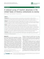
Figure 1 Identification and validation of B cell epitopes from cancer/testis antigen XAGE-1b. (A). ELISA was used to screen candidate
peptides from XAGE-1b for recognition by patients’ sera. Three patients (#1-3) were positive for either XAGE-1b:1-25 or 57-81. Sera were diluted
at 1:25, 1:125, and 1:625 with BSA serving as a control target. The mean OD of 8 HD and the OD of one seronegative patient (#4) are also
shown. The use of sera from NSCLC patients for screening is due to higher frequency of Ab against these shared antigens in NSCLC patients.
Previous work has shown that peptide epitopes identified using one type of sera are equally recognized by sera from other cancer patients. (B).
Western blots confirmed recognition of the full-length XAGE-1b protein. Lane 1, 2, and 3 contained, respectively, lysate from 293 cells transfected
with a control plasmid, a plasmid encoding XAGE-1b (denoted with an arrow), and lysate from LNCaP-CL1 cells (expressing XAGE-1b but at a
much lower level based on real-time PCR, data not shown).
Xie et al. Journal of Translational Medicine 2011, 9:43
/>Page 5 of 11
A
B
C
Fitted values Identity lineFitted values Identity lineFitted values Identity line
0
10
20
30
40
50
60
70
seroMAP
ELISA
Signal to Noise Ratio
Figure 2 Characteristics of the sero-MAP based multiplex assay measuring autoAb plus PSA.(A). seroMAP and ELISA were compared for
measuring autoAb against the prototype NY-ESO-1:1-40 peptide and the XAGE-1b:1-25 peptide. Specific MFI ratios (or OD) from randomly
selected seropositive patients were divided by the mean of 8 HD to represent signal-to-noise ratios of the seroMAP and ELISA approach. (B). MFI
ratios of autoAb against NY-ESO-1:1-40 versus a control obtained by the multiplex A+PSA and the singular assay had a correlation coefficient of
0.98 (n = 40), where the linear equation for NY-ESO-1 autoAb is A+PSA = 0.86*singular NY-ESO-1 + 0.20. (C). Comparison of seroMAP-based A
+PSA and classic ELISA for determining total PSA values (ng/ml) using serum samples of randomly selected 376 subjects. The three fitted linear
regressions for 1:10, 1:20, and 1:50 dilution were A+PSA = 0.89*PSA+0.15, A+PSA = 0.90*PSA+0.30, and A+PSA = 0.90*PSA+0.43, respectively.
Methods of determining PSA levels using seroMAP-based A+PSA and classic ELISA (American Qualex) were described in “Materials and Methods”.
Xie et al. Journal of Translational Medicine 2011, 9:43
/>Page 6 of 11
D
A
B
C
FE
Figure 3 Di stribution of autoAb in patients with BPH/prostatitis and prostate cancer. Histograms depicting the frequency or number of
patients and their specific MFI ratios against NY-ESO-1:1-40 (A), AMACR:341-371 (B), SSX-2,4 (C), p90 autoantigen (D), LEDGF (E), and XAGE-1b
(F) in patients with BPH and/or prostatitis (n = 121) and prostate cancer (n = 131). Mean values of MFI ratios from 3 serum dilutions at 1/10, 1/
20 and 1/50 were normalized against those obtained from HD (n = 124) to minimize experiment-to-experiment variations.
Xie et al. Journal of Translational Medicine 2011, 9:43
/>Page 7 of 11
other PCAA are shown in Figure 3B-F. The Kolmo-
gorov-Smirnov tests for each of the 6 autoAb between
prostate cancer and BPH/prostatitis groups were per-
formed resulting in highly statistically significant differ-
ences between the two groups except for autoAb against
AMACR. All four p-values of LEDGF, p90 autoantigen,
SSX-2, 4 and XAGE-1b were less than 0.001 and the p-
values of NY-ESO-1 and AMACR autoAb were 0.029
and 0.134, respectively.
A combined A+PSA in dex was created as the pre-
dicted probability of prostate cancer based on a logistic
regression model. The classifications were made to pros-
tate cancer if the prob ability was > = 0.5 and no cancer
if the probability was <0.5. PSA alone and A+PSA at
three different dilutions were compared with the mean
and maximal dilution for sensitivity, specificity, accuracy
and area under the curve (AUC). Mean values were
selected as the optimal method to obtain sensitivity and
specificity, and the receiver operating characteristic
(ROC) curve was used to compare the diagnostic power
between PSA alone and the combined A+PSA index for
distinguishing prostate cancer from BPH and prostatitis.
While the addition of any individual autoAb marker
barely improved PSA test (data not shown), the addition
of all 6 autoAb markers to PSA increased the assay sen-
sitivity (success rate of predicting cancer), specificity
(success rate of predicting non-cancer) and prediction
accuracy (Table 2). AUC was also increased substantially
from 0.66 for PSA alone to 0.91 for A+PSA. Figure 4
shows the ROC curves comparing the diagnostic power
between PSA alone and the combined A+PSA index for
distinguishing prostate cancer from nonmalignant BPH
and/or prostatitis cases commonly seen in the clinic.
The 95% bootstrap confidence interval in PSA alone was
[0.59, 0.73], whereas the interval of A+PSA including
the 6 autoAb was [0.88, 0.95]. A significant difference of
AUC between A+PSA and PSA alone was observed (P <
0.001). This pilot study indicated potential benefits of
the A+PSA assay in differenti ating prostate canc er from
non-malignant conditions commonly seen in the clinic.
Discussion
To improve PSA tests, a number of approaches have been
investigated in the past, including isoforms of PSA such as
free PSA and proPSA [22], new cancer biomarkers such as
PCA3 [23], as well as combinatory assays measuring PSA
and other parameters such as AMACR [24,25]. In this
study, a novel assay platform was established that
combines the quantification of autoAb against 6 TAA
with a conventional biomarker, PSA in one reaction under
the seroMAP platform. Even though the presence of
autoAb against PSA reported in some patients [26] could
in theory confound the quantification of PSA under the A
+PSA assay platform, no significant discrepancy was
observed among the more than 300 subjects. This a ssay
platform employed B cell epitopes from previously defined
PCAA and avoided peptides from out-of-frame and non-
coding sequence s, w hich have been observed in a la rge-
scale autoAb signature study [4]. While the NY-ESO-1:1-
40 peptide epitope has been validated by various investiga-
tors, the rest of the peptide epitopes panel is first reported
in this study. Extensive validation is still necessary befo re
moving forward into clinical trials. Serum samples from
prostate cancer patients were collected at the time of diag-
nosisorsurgeryandthusincludedthosewithawide
range of Gleason scores. Since this pilot study was focused
on developing a novel A+PSA platform and comparing A
+PSA with the PSA assay, cohort size was not sufficient to
match samples according to potential clinical co-founders.
However, all samples themselves were handled and stored
according to the same conditions prior to assay in order to
minimize experiment-to-experiment variations.
It was predicted that peptide-based methods might
lose conformational epitopes that could have been
detected using full-length proteins. However, autoAb
against NY-ESO-1 were detected in 7 of 131 or about
5% prostate cancer patients by seroMAP microspheres
conjugated with a single peptide, higher than the
reported frequency of 3.3% (3 out of 92) ag ainst the
same peptide epitope using ELISA or 4.3% (4 out of 92)
against the full-length NY-ESO-1 protein using ELISA
in a previou s study [18]. This result suggested that sero-
MAP-based A+PSA assay against dominan t pept ide epi-
topes might compensate losses of conformational epi-
topes, and cover specific patient populations otherwise
overlooked using ELISA methods coated with full-length
proteins. Considering that sero-MAP based A+PSA
assay is performed entirely in liquid phase with
enhanced kinetics over surface-bound ELISA, we will
investigate whether compensation for conformational
epitopes by A+PSA assay also occurs for other PCAA.
Furthermore, the multiplex A+PSA assay requires less
than 20 μl serum samples for three different dilutions
altogether, much fewer handling steps to be completed
within two and half hours, making it user-friendly to
clinical laboratories.
Table 2 Comparison of A+PSA index and PSA based on mean values at 3 different dilutions
Variables Sensitivity Specificity False positive Accuracy AUC
PSA alone in all patients 52% (68/131) 79% (95/121) 21% (26/121) 65% 0.66
A+PSA in all patients 79% (103/131) 84% (102/121) 16% (19/121) 81% 0.91 P < 0.0001
Xie et al. Journal of Translational Medicine 2011, 9:43
/>Page 8 of 11
In order to deliver a fully functional A+PSA assay to
clinical laboratories, we plan to cross-validate with larger
and broader patient cohorts including sex, age, and racial/
ethnic background matched HD and non-cancer controls.
In this pilot study, the A+PSA index also reduced the false
positive rate of PSA tests, which suggested its potential
implications in aiding in prostate cancer diagnosis. Thus,
patients with lung cancer and colon cancer, two common
cancers for elder men will also be included in the cross-
validation in order to enhance the prostate cancer specifi-
city of the assay. Once an optimized A+PSA assay has
been developed, prospective studies comparing A+PSA
with PSA alone as well as emerging genotype-based tests
such as urine PCA3/PSA mRNA ratio detection and
TMPRSS2-ERG fusion gene [27], will be conducted. Other
areas of potential implication such as differentiating lethal
from indolent prostate cancer will also be investigated in
the future. The versatile nature of the multiplex A+PSA
assa y allows the addition and delet ion of specific peptide
epitopes to the panel in order to define correlations with
the intended clinical implications.
Conclusions
TheA+PSAassayrepresentsthefirstmultiplexassay
that integrates autoAb signatures with a conventional
cancer biomarker PSA in a single reaction. Designed to
be user-friendly to clinical laboratories, the A+PSA
assay has the potential to aid in the diagnosis and prog-
nosis of prostate cancer.
Additional material
Additional file 1: Total PSA values are shown for the HD (n = 124),
BPH/prostatitis (n = 121) and prostate cancer patients (n = 131)
involved in the comparison of A+PSA and PSA alone. There are 1
and 28 patients with PSA equal or above 15 ng/ml (filled triangles) in
the BPH/prostatitis and prostate cancer group, respectively.
Additional file 2: Verification of peptide epitopes by Western blot.
Western blots against 50 ng of purified recombinant C-terminal portion
of LEDGF protein (amino acid 322-530, Abcam Biotechnology,
Cambridge, MA) in lane 3 (A) and AMACR protein (Abcam
Biotechnology) in lane 3 (B). In both cases, 10 and 20 μg of 293 cell
lysates were compared as controls (lanes 1 and 2 of each panel). Serum
samples from prostate cancer patients with LEDGF and AMACR specific
autoAb based on peptide screening were used at 1 to 500 dilutions for
the blot. Molecul ar weight standards (kDa) are shown on the sides. (C).
Western blot against bacterial lysate expressing recombinant SSX-2,4, the
C-terminal half of p90 autoantigen, and NY-ESO-1 (lane 1, 2, and 3
respectively in each panel). The left panel was blotted with Ab against
the polyhistidine tag to locate protein bands corresponding to SSX-2,4,
p90, and NY-ESO-1 (as a positive control). The center and right panel
were blotted with serum samples from prostate cancer patients with
positive reactions against p90 and SSX2,4 peptides (p90 and SSX2,4
proteins are circled), respectively.
Figure 4 An ROC curve comparing the A+PSA index and total PSA alone in differ entiating the same group of prostate cancer and
BPH/prostatitis patients as shown in Figure 3. The distribution of total PSA values in samples used in this study are shown in Figure 1S.
Xie et al. Journal of Translational Medicine 2011, 9:43
/>Page 9 of 11
List of Abbreviations
TAA: tumor-associated antigen; PSA: prostate specific antigen; HD: healthy
donors; NSCLC: non-small cell lung cancer; A+PSA: autoantibody plus PSA;
BPH: benign prostatic hyperplasia; Ab: antibody; PCAA: prostate cancer-
associated antigen; OD: optical density; MFI: mean fluorescent intensity; ROC:
receiver operating characteristic.
Acknowledgements and Funding
This research was supported in part by a grant from the Prevent Cancer
Foundation, by NIHR03CA128086 and NIHR21CA137651 grants, and an NCI
Early Detection Research Network associate membership to GZ. CX is
supported in part by the China Scholarship Council. We thank Jun-ying
Zheng and UCLA college students who took the MED99/MED199 courses,
Michael Mangubat, Munira Rahman, Duminda Suraweera, Christina Wu, Junyi
Xie, and Albert Yang for their contributions. Dr. Eiichi Nakayama (Okayam a
University, Japan) provided XAGE-1b recombinant protein for this study.
Author details
1
Department of Urology, Zhongshan Hospital of Fudan University, No.180
Fenglin Road, Shanghai 200032, China.
2
Department of Radiology, David
Geffen School of Medicine at UCLA, 10833 Le Conte Ave, Los Angeles, CA
90095-1721, USA.
3
Department of Urology, David Geffen School of Medicine
at UCLA, 10833 Le Conte Ave, Los Angeles, CA 90095-1738, USA.
4
Department of Medicine, David Geffen School of Medicine at UCLA, 10833
Le Conte Ave, Los Angeles, CA 90095-1732, USA.
5
Department of
Biostatistics, UCLA School of Public Health, 10833 Le Conte Ave, Los Angeles,
CA 90095-1772, USA.
6
Department of Pathology and Laboratory Medicine,
David Geffen School of Medicine at UCLA, 10833 Le Conte Ave, Los Angeles,
CA 90095-1738, USA.
7
Department of Chemistry and Biochemistry, 607
Charles E. Young Drive East, Los Angeles, CA 90095-1569, USA.
8
Advanced
Medical Analysis, LLC, 1941 Walker Ave, Monrovia, CA 91016, USA.
9
Department of Urology, CHU Henri Mondor, Créteil U955 E907, France.
10
Department of Urology, Okinawa Nambu Tokushukai Hospital, 80 Hokama,
Yaese-cho, Shimajiri-gun, Okinawa 901-0417, Japan.
Authors’ contributions
CX carried out the serological assays, participated in the data analysis and
drafted the results of the manuscr ipt. HK and GL participated in the design
of the study and carried out the statistical analysis. JH and AK participated in
the identification of peptide epitopes. BG helped with the seroMAP-based
assays. JR, DC, AP participated in the overall design of the study and
interpretation of results. ML conjugated all peptides/proteins to
microspheres. RP, LS, AT, GW, and HM collected patients’ samples and
provided their clinical information. GZ conceived of the study, participated
in its design and coordination, and drafted the manuscript. All authors read
and approved the final manuscript.
Competing interests
The authors declare that they have no competing interests.
Received: 9 February 2011 Accepted: 19 April 2011
Published: 19 April 2011
References
1. Sahin U, Tureci O, Pfreundschuh M: Serological identification of human
tumor antigens. Curr Opin Immunol 1997, 9:709-716.
2. Finn OJ: Immune response as a biomarker for cancer detection and a lot
more. N Engl J Med 2005, 353:1288-1290.
3. Jiang Z, Fanger GR, Woda BA, Banner BF, Algate P, Dresser K, Xu J, Chu PG:
Expression of alpha-methylacyl-CoA racemase (P504s) in various
malignant neoplasms and normal tissues: astudy of 761 cases. Hum
Pathol 2003, 34:792-796.
4. Wang X, Yu J, Sreekumar A, Varambally S, Shen R, Giacherio D, Mehra R,
Montie JE, Pienta KJ, Sanda MG, et al: Autoantibody signatures in prostate
cancer. N Engl J Med 2005, 353:1224-1235.
5. Shi FD, Zhang JY, Liu D, Rearden A, Elliot M, Nachtsheim D, Daniels T,
Casiano CA, Heeb MJ, Chan EK, Tan EM: Preferential humoral immune
response in prostate cancer to cellular proteins p90 and p62 in a panel
of tumor-associated antigens. Prostate 2005, 63:252-258.
6. Daniels T, Zhang J, Gutierrez I, Elliot ML, Yamada B, Heeb MJ, Sheets SM,
Wu X, Casiano CA: Antinuclear autoantibodies in prostate cancer:
immunity to LEDGF/p75, a survival protein highly expressed in prostate
tumors and cleaved during apoptosis. Prostate 2005, 62:14-26.
7. Stockert E, Jager E, Chen YT, Scanlan MJ, Gout I, Karbach J, Arand M,
Knuth A, Old LJ: A survey of the humoral immune response of cancer
patients to a panel of human tumor antigens. J Exp Med 1998,
187:1349-1354.
8. Scanlan MJ, Altorki NK, Gure AO, Williamson B, Jungbluth A, Chen YT,
Old LJ: Expression of cancer-testis antigens in lung cancer: definition of
bromodomain testis-specific gene (BRDT) as a new CT gene, CT9. Cancer
Lett 2000, 150:155-164.
9. Egland KA, Kumar V, Duray P, Pastan I: Characterization of overlapping
XAGE-1 transcripts encoding a cancer testis antigen expressed in lung,
breast, and other types of cancers. Mol Cancer Ther 2002, 1:441-450.
10. Johansson M, Denardo DG, Coussens LM: Polarized immune responses
differentially regulate cancer development. Immunol Rev 2008,
222:145-154.
11. Dunn GP, Old LJ, Schreiber RD: The Immunobiology of Cancer
Immunosurveillance and Immunoediting. Immunity 2004, 21:137-148.
12. Bouwhuis MG, Suciu S, Collette S, Aamdal S, Kruit WH, Bastholt L, Stierner U,
Sales F, Patel P, Punt CJ, Hernberg M, Spatz A, ten Hagen TL, Hansson J,
Eggermont AM: Autoimmune antibodies and recurrence-free interval in
melanoma patients treated with adjuvant interferon. J Natl Cancer Inst
2009, 101:869-877.
13. Sittler T, Zhou J, Park J, Yuen NK, Sarantopoulos S, Mollick J, Salgia R,
Giobbie-Hurder A, Dranoff G, Hodi FS: Concerted potent humoral immune
responses to autoantigens are associated with tumor destruction and
favorable clinical outcomes without autoimmunity. Clin Cancer Res
2008,
14:3896-3905.
14.
Sabater L, Titulaer M, Saiz A, Verschuuren J, Gure AO, Graus F: SOX1
antibodies are markers of paraneoplastic Lambert-Eaton myasthenic
syndrome. Neurology 2008, 70:924-928.
15. Zhang J-Y, Casiano CA, Peng X-X, Koziol JA, Chan EKL, Tan EM:
Enhancement of Antibody Detection in Cancer Using Panel of
Recombinant Tumor-associated Antigens. Cancer Epidemiol Biomarkers
Prev 2003, 12:136-143.
16. Stone B, Schummer M, Paley PJ, Thompson L, Stewart J, Ford M,
Crawford M, Urban N, O’Briant K, Nelson BH: Serologic analysis of ovarian
tumor antigens reveals a bias toward antigens encoded on 17q. Int J
Cancer 2003, 104:73-84.
17. Lagarkova MA, Koroleva EP, Kuprash DV, Boitchenko VE, Kashkarova UA,
Nedospasov SA, Shebzukhov YV: Evaluation of humoral response to
tumor antigens using recombinant expression-based serological mini-
arrays (SMARTA). Immunol Lett 2003, 85:71-74.
18. Zeng G, Aldridge ME, Wang Y, Pantuck AJ, Wang AY, Liu YX, Han Y,
Yuan YH, Robbins PF, Dubinett SM, de Kernion JB, Belldegrun AS:
Dominant B cell epitope from NY-ESO-1 recognized by sera from a wide
spectrum of cancer patients: implications as a potential biomarker. Int J
Cancer 2005, 114:268-273.
19. Liu H, Li G, Cumberland WG, Wu T: Testing Statistical Significance of the
Area under a Receiving Operating Characteristics Curve for Repeated
Measures Design with Bootstrapping. Journal of Data Science 2005, 3:22.
20. Dubovsky JA, McNeel DG: Inducible expression of a prostate cancer-testis
antigen, SSX-2, following treatment with a DNA methylation inhibitor.
Prostate 2007, 67:1781-1790.
21. Koizumi F, Noguchi Y, Saika T, Nakagawa K, Sato S, Eldib AM, Nasu Y,
Kumon H, Nakayama E: XAGE-1 mRNA expression in prostate cancer and
antibody response in patients. Microbiol Immunol 2005, 49:471-476.
22. Sokoll LJ, Sanda MG, Feng Z, Kagan J, Mizrahi IA, Broyles DL, Partin AW,
Srivastava S, Thompson IM, Wei JT, Zhang Z, Chan DW: A prospective,
multicenter, National Cancer Institute Early Detection Research Network
study of [-2]proPSA: improving prostate cancer detection and
correlating with cancer aggressiveness. Cancer Epidemiol Biomarkers Prev
2010, 19:1193-1200.
23. Laxman B, Morris DS, Yu J, Siddiqui J, Cao J, Mehra R, Lonigro RJ,
Tsodikov A, Wei JT, Tomlins SA, Chinnaiyan AM: A first-generation
multiplex biomarker analysis of urine for the early detection of prostate
cancer. Cancer Res 2008, 68:645-649.
24. Zehentner BK, Secrist H, Zhang X, Hayes DC, Ostenson R, Goodman G, Xu J,
Kiviat M, Kiviat N, Persing DH, Houghton RL: Detection of alpha-
methylacyl-coenzyme-A racemase transcripts in blood and urine
samples of prostate cancer patients. Mol Diagn Ther 2006, 10:397-403.
Xie et al. Journal of Translational Medicine 2011, 9:43
/>Page 10 of 11
25. Prior C, Guillen-Grima F, Robles JE, Rosell D, Fernandez-Montero JM,
Agirre X, Catena R, Calvo A: Use of a combination of biomarkers in serum
and urine to improve detection of prostate cancer. World J Urol 2010,
28:681-686.
26. Zisman A, Zisman E, Lindner A, Velikanov S, Siegel YI, Mozes E:
Autoantibodies to prostate specific antigen in patients with benign
prostatic hyperplasia. J Urol 1995, 154:1052-1055.
27. Marks LS, Bostwick DG: Prostate Cancer Specificity of PCA3 Gene Testing:
Examples from Clinical Practice. Rev Urol 2008, 10:175-181.
doi:10.1186/1479-5876-9-43
Cite this article as: Xie et al.: A novel multiplex assay combining
autoantibodies plus PSA has potential implications for classification of
prostate cancer from non-malignant cases. Journal of Translational
Medicine 2011 9:43.
Submit your next manuscript to BioMed Central
and take full advantage of:
• Convenient online submission
• Thorough peer review
• No space constraints or color figure charges
• Immediate publication on acceptance
• Inclusion in PubMed, CAS, Scopus and Google Scholar
• Research which is freely available for redistribution
Submit your manuscript at
www.biomedcentral.com/submit
Xie et al. Journal of Translational Medicine 2011, 9:43
/>Page 11 of 11