Báo cáo sinh học: "Cancer stem cell subsets and their relationship" pot
Bạn đang xem bản rút gọn của tài liệu. Xem và tải ngay bản đầy đủ của tài liệu tại đây (350.75 KB, 9 trang )
REVIEW Open Access
Cancer stem cell subsets and their relationships
Hai-Guang Liu, Chong Chen, Han Yang, Yi-Fei Pan
*
and Xiao-Hua Zhang
*
Abstract
Emerging evidence suggests that cancer stem cells account for the initiation and progression of cancer. While
many types of cancer stem cells with specific markers have been isolated and identified, a variety of differences
among them began to be appreciated. Cancer stem cells are hierarchical populations that consist of precancerous
stem cells, primary cancer stem cells, migrating cancer stem cells and chemoradioresistant cancer stem cells,
playing different roles in cancer initiation and progression. Here we propose a new concept “ho rizontal hierarchy
of cancer stem cells” to distinguish them from vertical hierarchy cancer stem cells, cancer transient-amplifying cells
and cancer differentiated cells, and summarize our current understanding of these subsets of cancer stem cells
with the aim to open up novel therapeutic strategies for cancer based on this understanding.
Introduction
Cancer is a kind of abnormal tissue that develops the
ability of unlimited growth and the resistance to various
survival stresses. Recently, accumulating experimental
evidence supports that cancer stem cells account for the
initiation and progression of cancer, which challenges the
classical stochastic model of cancer development [1]. The
cancer stem cell model or intrinsic model posits similar
differentiation hierarchy such as hematopoietic system,
cancer stem cells, cancer transient-amplifying (TA) cells
and cancer differentiated cells, which is defined as verti-
cal hierarchy here. Only cancer stem cells or cancer TA
cells that reacquire self-renewal property can initiate can-
cer and progress into more m alignant disease. However,
in the stochastic model no hierarchy in cancer exists and
every single cancer cell has the capacity of initiation and
progression. Cancer stem cell hypothesis suggests that
targeted therapy to cancer stem cells, not cancer TA cells
and cancer differentiated cells, is the best measure to era-
dicate cancer, because traditional cancer therapies target
the cancer TA cells and cancer differentiation cells, but
omit cancer stem cells, thus leading to frequent cancer
relapse [2].
The essential features of cancer stem cells are self-
renewal, multi-differentiation and tumorigenic capacity
[3]. Ca ncer stem cells are also able to migrate and resist
chemotherapy and radiotherapy. However, cancer stem
cells are in constant evolution and these capacities are
different among different p opulations of cancer stem
cells. Thus we propose a horizontal hierarchy that com-
prises precancerous stem cells, primary cancer stem
cells, migrating cancer stem cells and chemoradioresis-
tant cancer stem cells (Figure 1). Below we will describe
the horizontal hier archy of cancer stem cells and discuss
the relationship among these subsets of cancer stem
cells.
Primary cancer stem cells
Cancer cells with featur es of stem cells were discovered
by Rudolf Virchow in the mid-19th century, who found
that some cancer cells had the histological characteris-
tics, proliferation a nd differen tiation capacity similar to
embryonic cells [4]. In 1937, Jacob Furth and Morton
Kahn transplanted human leukemia cells into mice and
found that the tumorigenesis of leukemia cells was dif-
ferent from each other. In 1960s-1970s, based on
spleen-colony forming tests numerous studies showed
that the tumorigenesis of cancer cells was different not
only in leukemia, but also in many types of solid tumors
[5-8]. Thus it is specul ated that cancer, a new type of
stem cell disease, was initiated from transformed stem
cells and develo ped as a heterogeneity tissue, containing
cancer stem cell subpopulations and differentiate d can-
cer cell subpopulations.
The invention of flow cytometry g reatly helped the
use of s pecific markers to isolate subsets of cells [9]. In
1997, Bonnet et al [10] isolated two groups of leukemia
cells from leukemia patients with specific surface mar-
kers CD34 and CD38, an d found that CD34
+
CD38
-
* Correspondence: ;
Department of Oncology, The First Affiliated Hospital of Wenzhou Medical
College, Wenzhou, 325000, China
Liu et al. Journal of Translational Medicine 2011, 9:50
/>© 2011 Liu et al; licensee BioMed Central Ltd. This is an Ope n Access art icle distributed under the terms of the Creative Commons
Attribution License ( which permits unrestricted use, distribution, and reproduction in
any medium, provided the original work is pro perly cited.
leukemia cells had the capacity of self-renewal and
multi-differentiat ion similar to hematopoietic stem cells,
and developed tumor more quickly than CD34
-
CD38
+
leukemia cells. Thus they concluded that CD34
+
CD38
-
subpopulations were the initiating cells of leukemia.
Thi s was the first experimental evidence of cancer stem
cells. Later, Al-Hajj et al. [11] isolated CD44
+
CD24
-
breast cancer stem cells from breast cancer patients in
2003, thus providing the first experimental evidence of
solid tumor stem cells. After that, more types of solid
tumor stem cells were isolated with specific surface
markers (Table 1 [12-59]).
Interestingly, Xu et al [60] discovered a type of benign
tumor stem cells by isolat ing a type of stem-like cells
from pituitary adenoma with self-renewal, multi-lineage
differentiation and neurospheres formation capacity.
Compared with differentiated daughter cells, pituitary
adenoma stem cells expressed high levels of stem cell-
related proteins, anti-apoptotic proteins and pituitary
progenitor markers, and had a stronger resistance to
chemotherapy. Differentiation of pituitary adenoma
stem cells could respond to hypothalamic hormones and
secret the corresponding pituitary hormones, which
were phenotypes of primary pituitary adenoma. Besides
these capacities, pituitary adenoma stem cells could
form tumors in the continuous xenotransplanation
assays. This was the first experimental evidence of the
existence of benign tumor stem cells.
At present, many types of primary cancer stem cells
with specific surface mark ers have been isolated and the
cancer stem cell hypothesis is widely accepted. However,
many questions remain in the field of cancer stem cells
research. For example, where primary cancer stem cells
initiate from; whether primary cancer stem cells are
same in the same type of cancer among different
patients; and how to distinguish cancer stem cells from
normal stem cells. Below, we will focus on the origin
and the fate of primary cancer stem cells.
Precancerous stem cells
Based on current literature, primary cancer stem cells may
be derived from precancerous stem cells. Chen et al [61]
reported the isolation of a type of precancerous stem cells
from dendritic cell-like leukemic mice and the establish-
ment of this precancerous stem cell line. The precancer-
ousstemcellshadstemcell-like phenotype, unlimited
self-renewal, multi-differentiation and could reconstruct
the hematopoietic system of mice after deadly radiation
treatment. Transplantation of such precancerous stem
cells could form tumor in immune-deficient but not in
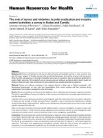
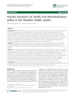
Figure 1 The progression of cancer stem cells and their corresponding pathol ogical process. Transformed normal stem cells
(SCs), progenitors with self-renewal capacity and differentiated cells after reprogramming are the potential origin of precancerous stem cells
(pre-CSCs), whose corresponding pathological process is precancerous condition. Transformation from precancerous stem cells to primary cancer
stem cells (pri-CSCs) is a crucial step of cancer initiation. Upon acquiring migrating capacity, primary cancer stem cells transform to migrating
cancer stem cells (mig-CSCs) and metastasize to distant organs and cause metastatic cancer. In order to escape from chemoradiotherapy, some
of primary cancer stem cells may develop into chemoresistant cancer stem cells (cr-CSCs) and radioresistant cancer stem cells (rr-CSCs). Some
transformation steps are marked with gray arrows to indicate that they are speculative with no direct evidence up to date.
Liu et al. Journal of Translational Medicine 2011, 9:50
/>Page 2 of 9
immune-competent mice. In the evolution of the tumor,
the phenotype and genotype of precancerous stem cells
had developed towards primary cancer stem cells.
Interestingly, Shen et al [62] discovered that the pre-
cancerous stem cells could differentiate into tumo r vas-
culogenic progenitors and generate most of the blood
vessels. Precancerous stem cells sustained the expression
of vascular growth fact or receptor VEGRF-2, which was
under the regulation of hypoxia and various vascular
growth factors such as GM- CSF, Flt3L, and IL-13, to
promote vasculogenesis. In contrast, the expression of
VEGRF-2 was much lowe r in differentiated tumor cells,
indicating that vasculogenesis in precancerous stem cell s
is related to their inherent stem-cell characteristics.
In our opinion, precancerous stem cells have the fol-
lowing characteristics. First, they hide themselves in pre-
cancerous lesions. It is well known that carcinogenesis is
a multi-step process. For instance, colon cancer goes
through mild, moderate and severe dysplasia, adenoma,
carcinoma in situ, to invasive cancer and metastasis
[63]. During this long process of carcinogen esis, precan-
cerous stem cells undergo the transform ation from nor-
mal stem cells to primary cancer stem cells.
Precancerous lesions progress to cancer when precan-
cerous stem cells transform into primary cancer stem
cells [64]. Second, precancerous s tem cell is a mutated
stem cell that highly express stemness factors such as
OCT3/4, SOX2, KLF4 and therefore develops the capa-
cities of self-renewal, multi- differentiation and resistance
to chemoradiotherapy [65]. Third, precancerous stem
cells are subjected to modulation by micro-environment.
They can transform into malignant tumors or benign
disease, mainly depending on their communication with
the micro-environment [61,66].
Based on the three ch aracteristics describ ed above, we
can distinguish precancerous stem cells from primary
cancer stem cells. First i s the location. Preca ncerous
stem cells mainly exist in precancerous lesions, but pri-
mary cancer stem cells exist in primary canc er foci. For
example, ductal carcinoma in situ (DCIS) is generally
considered a type of preca ncerous lesion of breast inva-
sive ductal carcinoma (IDC). The precancerous stem
cells in DCIS stage are confined within the duct, but
develop invasive capacity upon hypoxia or other stimuli,
contributing to the progression of DCIS to IDC. There-
fore, precancerous stem cells develop into primary can-
cer stem cells, and neoplastic ductal is not precanerous
lesion but cancer foci [67]. Second is the genotype and
phenotype. Primary cancer stem cells are derived from
precancerous stem cells and exhibit some genotypes and
Table 1 Cancer stem cells with specific markers
Type of cancer Specific markers References
AML CD34
+
CD38
-
Lin
-
[10]
AML CD123
+
[12,13]
AML CD47
+
[14]
Breast cancer CD24
-
CD44
+
Lin
-
[11,15]
Breast cancer ALDH1
+
[16,17]
Brain tumors CD133
+
[18-20]
Glioblastoma SSEA-1
+
[21]
Glioblastoma A2B5
+
[22]
Prostate cancer a2b1
hi
CD133
+
[23]
Prostate cancer Lin
-
Sca-1
+
CD49f
high
[24]
Bladder cancer ALDH1
+
[25]
Lung cancer SP-C
+
CCA
+
[26]
Lung cancer CD133
+
[27]
Lung cancer ALDH1
+
[28]
Melanoma CD20
+
MCAM
+
[29]
Melanoma CD133
+
ABCG2
+
[30]
Melanoma MDR1
+
[31]
Melanoma ABCG5
+
[32]
Melanoma CD271
+
[33]
Melanoma JARID1B
+
[34]
Colon cancer CD133
+
[35-38]
Colon cancer Lgr5
+
[39]
Colon cancer ALDH1
+
[40]
Colorectal cancer CD44
+
ESA
hi
CD166
+
[41]
Colorectal cancer CD26
+
[42]
Intestinal cancer Lgr5
+
[39]
Intestinal cancer CD133
+
[43]
Pancreatic cancer CD44
+
CD24
+
ESA
+
[44]
Pancreatic cancer CD133
+
[45]
HNSCC CD44
+
[46]
HNSCC ALDH1
+
[47]
B-precursor ALL CD34
+
CD38
+
CD19
+
; CD34
+
CD38
-
CD19
+
[48]
Ovarian cancer CD44
+
CD117
+
[49]
Ovarian cancer CD133
+
[50]
Endometrial
tumors
CD133
+
[51]
Liver cancer CD90
+
[52]
Liver cancer CD133
+
[53]
Liver cancer EpCAM
+
[54]
Renal carcinomas CD105
+
[55]
Medulloblastoma CD15
+
[56,57]
Gastric cancer CD44
+
[58]
Osteosarcoma Oct-4
+
[59]
AML: acute myeloid leukaemia; ALDH: aldehyde dehydrogenase; SP-C:
surfactant protein C; CCA: also known as CC10 or CCSP; MCAM: melanoma cell
adhesion molecule; ABCG: ATP-binding cassette superfamily G member; MDR:
multi-drug resistance protein; ESA: epithelial specific antigen; HNSCC: head
and neck squamous cell carcinoma; ALL: acute lymphocytic leukaemia.
Liu et al. Journal of Translational Medicine 2011, 9:50
/>Page 3 of 9
phenotypes of precancerous stem cells, meanwhile they
have their unique profiles. Castro and colleagues found
that 126 genes were upregulated and 21 genes were down-
regulated in DCIS compared to IDC. Therefore, precan-
cerous stem cells of DCIS exhibit different genotypes in
contrast to primary cancer stem cells of IDC [68]. In addi-
tion, Ma et al. reported that the gene expression profiling
of IDC was inherited from DCIS but developed distinct
gene expression signatures [69]. With regard to epigenetic
alternations, DNA methyl ation is notable. Adenomatous
polyps (APs) is generally considered as precancerous
lesion of adenomatous carcionoma (AdCa). The aberrant
DNA methylation can be completely reversed in APs, but
not in AdCa by a nonsteroidal anti-inflammatory drug cel-
ecoxib [70], suggesting the different epigenetic profilings
between precancerous stem cells in APs and primary can-
cer stem cells in AdCa. Third is the bi-transformation.
Under different micro-environment, precancerous stem
cells can transform into ma lignan t tumors or benig n dis-
ease [61]. Bi-transformation is the most important charac-
teristic to distinguish precancerous stem cells from
primary cancer stem cells. Mammary intraepithelial neo-
plasia outgrowths (MINOs) is a mouse model of DCIS.
The culture of single cells from MINOs expressed bipo-
tential for myoepithelial and luminal differentiation and
formed uniqu e three-dimensional ‘MINOspheres’.When
transplanted in vivo, MINOspheres were able to form
DCIS or IDC under different micro-environment [66].
The next question is the origin of precancerous stem
cells. Several studies suggested that cancer initiating
cells may be responsible for the development of precan-
cerous stem cells. Wang et al [71] reported that a sub-
population of Nkx3-1 positive luminal epithelial cells
was capable of self-renewal in vivo, and such a single
cell was ab le to reconstitute prostate tissue in grafts.
When the tumor suppressor gene Pten was deleted in
Nkx3-1 positive luminal e pithelial cells, the populations
rapidly formed high-grade intraepithelial neoplasm and
carcinoma after androgen mediated regeneration of the
prostate. Therefore, Nkx3-1 positive luminal epithelial
cells were a typ e of prostate stem cells and mutation of
tumor suppressor genes wo uld lead to prostate
carcinogenesis.
Additionally, Barker et al [72] and Zhu et al [43] dis-
covered crypt stem cells as the origin of intestinal can-
cer. They demonstrated that Lgr5 positive or prominin1
positive subpopulations were intesti nal stem cells. Dele-
tion of Apc or activation of endogenous Wnt signaling
in such intestinal stem cells led to their transformation
to abnormal stem cells, resulting in intestinal neoplasm.
However, when the same mutations occurred in transit-
amplifying cells without unlimited self-renewal capacity,
the induced adenomas grew slowly and disappeared
after long observation.
The malignant transformation of normal stem cells
was also discovered in mesenchymal stem ce lls. Røsland
et al [73] showed that after long term culture for 5-106
weeks, 45.8% of bone marrow derived human mesenchy-
mal stem cells underwent sp ontaneous transformation.
They lost differentiati on potential, had increased te lo-
merase activity, escaped senescence, demonstrated
anchorage-independent growth and were capable of
tumorigenesis in vivo.
Moreover, human embryonic stem (hES) cells can
transform into abno rmal stem cells. Werbowetski -Ogil-
vie et al [74] identified two variant hES cell lines (v-
hESC-1 and v-hESC-2) with different features from their
parents. These variants expressed higher levels of pluri-
potency markers Oct4 and SSEA3, less depended on
exogenous growth factors, had decreased diff erentiation
capacity in either hematopoietic or neural conditions,
and had increased frequency of teratoma initiating cells,
however, their teratoma cells did not metastasize to
other organs upon in v ivo transplantation. Therefore,
variant hES cells undergo neoplastic progression and
may be the origin of malignant teratoma stem cells.
Proge nitor cells may be another origin of cancer initi-
ating cells. Jamieson et al. [75] reported that during
blast-crisis of chronic myelogenous leukemia (CML),
granulocyte-macrophage progenitors acquired much
stronger self-renewal property due to the activation of
Wnt/b -catenin pathway, and expressed BCR-ABL pro-
tein and expanded imatinib-resistant CML. Later other
groups confirmed these findings [76,77]. Guibal et al
[78] showed that in a murine model of acute promyelo-
cytic leukemia (APL), a population of committed mye-
loid cells (CD34
+
,c-kit
+
,FcgRIII/II
+
,Gr1
int
)
demonstrated enhanced self-renewal capacity through
the down-regulation of the transcription factor CCAAT/
enhancer binding protein-a(C/EBP- a) and were capable
of efficiently generating l eukemia in recipient mice.
Krivtsov et al [79] reported a more detailed overview of
the transformation from committed progenitor to cancer
stem cells.
Self-renewal is the most esse ntial feature of normal
stem cells and cancer stem cells [80]. Notably, some
mature diff erentiated cells can re-acquire self-renewal
capacity after reprogramming and thus may be addi-
tional origin of tumor initiating cells. Takahashi and
Yamanaka [81] reported that they could reprogramme
mouse fibroblasts into induced pluripotent stem (iPS)
cells by introducing four factors Oct3/4, Sox2, c-Myc
and Klf-4. In vivo transplantation assay demonstrated
that the iPS cells were able to form teratomas and it
was speculated that the two oncogen es c-Myc and Klf-4
might endow iPS cells with the capacity of tumorigen-
esis. More recent studies demonstrated that iPS cells
could be induced from differentiated cells by chemicals
Liu et al. Journal of Translational Medicine 2011, 9:50
/>Page 4 of 9
or proteins without the use of viral vectors [82-89].
These iPS cells with capacity of tumorigenesis might be
another origin of malignant teratoma stem cells.
Taken together, adult stem cells, embryonic stem cells,
progenitors with unlimited self-renewal capacity, and
induced pluripotent stem cells are the potential origins
of cancer initiating cells.
Migrating cancer stem cells
Metastasis is a very important feature of malignant
tumors, accounting for 90% death of tumor pa tients
[90]. Metastasis is a multi-step proc ess that invol ves
progressive growth, vascularization, invasion, detach-
ment, e mbolization, survival in the circulation, arrest,
extravasation, evasion of the host defense and progres-
sive growth [91]. Given its comp licated nature, metasta-
sis is far from being understood completely and many
hypotheses have been proposed to elucidate the underly-
ing mechanisms. In se ed and soil theory it is speculated
that metastasis is closely related to the characteristics of
tumor types and metastatic sites. D ifferent tumor cells
tend to move to their sp ecific distant organs, and differ-
ent distant organs tend to accept specific tumor cells
[91]. In 1980, Hart and Fldier [92] transplanted lung,
ovarian and kidney tissues into subcutaneous and mus-
cle of C57BL/6 mice, and then transplanted B16 mela-
noma cells into these mice after these transplanted
tissue survived. They found t umor formation in the
transplanted lung and ovarian but not kidney tissues.
Importantly, there was no significant difference in the
number of melanoma cells throughout the lung, ovarian
and kidney tissues. This ruled out the influence of
tumor cell numbers and further confirmed that metasta-
sis is related with special distant organs.
According to cancer stem cell hypotheses, cancer stem
cells are ideal seeds of metastasis. Stem cells are indeed
ideal carrier of gene mutations and their accumulation.
First, the initiating cell must b e a cell with extensive
divisions and the mutations will be not lost after several
divisions. Second, the initiating cell must have long life
with strong resistance to different external s tress. In
contrast, a mature differentiated cell is subject to senes-
cence and death and can not be the initiator of cancer.
But not all cancer ste m c ells have the cha racteristic of
migration. Hermann et al [45] reported that CD133
+
CXCR4
+
subsets determined the migrating phenotype of
pancreatic cancer, although both CD133
+
CXCR4
+
and
CD13 3
+
CXCR4
-
panc reatic cancer stem cells could form
pancreatic ca ncer when transplanted into athymic mice.
An inhibitor of CXCR4 could significantly reduce the
metastasis in group CD133
+
CXCR4
+
mice. Furthermore,
removal of CD133
+
CXCR4
+
subset from CD133
+
cancer
stem cells could disrupt the metastasis of pancreatic can-
cer,butdidnotaffecttumorigenesisinprimaryorgan.
Collectively, these data suggest that CD133
+
CXCR4
+
cancer stem cells determine the metastasis and re present
the migrating cancer stem cells of pancreatic cancer.
Furthermore, Yang et al [52] reported that CD90
+
but
not CD90
-
liver cancer cells were able to form tumor.
Notably, CD90
+
CD44
+
subpopulations had stronger capa-
city of tumorigenesis and metastasis than CD90
+
CD44
-
subpopulations, and the proportion of CD90
+
CD44
+
subpopulations in metastasis increased compared to pri-
mary cancer. Therefore, CD90
+
CD44
+
subpopulations
might be the migrating cancer stem cells of liver cancer.
However, current studies on migrating cancer stem
cells are very limited, mainly due to the lack of specific
migrating markers to isolate migrating cancer stem cells
from primary cancer stem cells. It has been established
that epithelial to mesenchymal transition (EMT) is
involved in migration and metastasis, thus providing
some clues on how to isolate migrating subpopulations
from primary cancer stem cells. Mani et al [93] isolat ed
CD44
low
CD24
high
and CD44
high
CD24
low
subpopulations
from five breast cancer tissues and applied serial analysis
of gene expression to reveal that CD44
high
CD24
low
subpopulations expressed high level of mesenchymal
markers N-cadherin, Vimentin, Fibronectin, Zeb2,
Foxc2, Snail, Slug, Twist1 and Twist2, and low level of
E-cadherin. They further transplanted human mammary
epithelial cells constitutively expressing either Snai l or
Twist into i mmune-deficientmiceandfoundthatboth
of them had more e fficiency of tumorigenesis, and the
number of C D44
high
CD24
low
subpopulations is elevated.
Therefore, they concluded that EMT might be responsi-
ble for the generation of migrating cancer stem cells.
Zhang et al. [94] discovered that in three-dimensional
cultu re, epith elial growth factor receptor tyrosine kinase
inhibitor erlotinib inhibited th e motility of inflammatory
breast cancer (IBC) cell line SUM149 and its invasion in
matrigel, accompanied with increased expression of
E-cadherin and reduced expression of vimentin and
b-catenin. Furthermore, they transplanted SUM149 cells
into athymic nude mice and demonstrated that erlotinib
inhibited the growth of tumor and lung metastasis by
regulating the expression of E-cadherin and vimentin.
This study suggests that erlotinib reversed EMT of IBC
to inhibit metastasis. In this aspect, it is important to
note a few of molecule implicated in both EMT and
stemness such as Six1 [95,96] and p21CIP1 [97].
Based on these studies it is a potential approach to
utilize mesenchymal markers to isolate migrating cancer
stem cells from primary cancer stem cells.
Other subsets of cancer stem cells
One significant feature of cancer is its relapse after che-
motherapy and radiation. This is because a few of can-
cer cells evolve with the capacity of resistance to
Liu et al. Journal of Translational Medicine 2011, 9:50
/>Page 5 of 9
chemotherapy and radiation. Whether primary cancer
stem cells can evolve into chemoradioresistant cancer
stem cells is not well known but recent studies provided
indirect evidence for the existence of chemoradioresis-
tant cancer stem cells.
Chemoresistant cancer stem cells
Todaro et al [98-100] reported a subpopulation of human
colon cancer stem cells resistant to the most popular
chemotherapeutic agent oxaliplatin or 5-fluorouracil
(5-FU) at clinically relevant doses. Mechanistically, in this
subpopulation interleukin-4 (IL-4) is produced in an
autocrine manner to induce the expression of the antia-
poptotic proteins cFLIP, Bcl-xL, and PED. The antagonist
of IL-4 combined with oxaliplatin or 5-FU could effec-
tively inhibit the growth of these cancer stem c ells in
vitro and in vivo, and decrease the size of spheroid and
tumor.
ATP-binding cassette superfamily is one type of multi
drug resistant proteins, which can pump chemotherapy
drugs out of the cell and lead to chemoresistance
[101,102]. ABCG2 is a member of this family and repre-
sents a purified marker of cancer stem cells [103]. How-
ever, targeted t herapy with ABCG2 antagonist can only
inhibit partial ly the growth of SP cells and cancer stem
cells. This may be because cancer stem cells express
other drug resistant proteins such as ABCB1 [104].
Despite these reports demonstrating the relationship
between cancer stem cells and chemoresistance, further
studies are crucial to provide direct evidence supporting
the existence of chemoresistant cancer stem cells, which
may help develop alternative strategy for chemotherapy
and targeted therapy.
Radioresistant cancer stem cells
Diehn et al [105] reported that human and mouse breast
cancer stem cells had lower levels of reactive oxygen
species (ROS) than their non-tumorigenic progeny.
Moreover, human cance r stem cells contained higher
levels o f antioxidant defense systems and develop ed less
DNA damage after ionizing radiation, compared with
non-tumor cells. Therefore, the heterogeneity of ROS
levels in cancer stem cell subsets might contribute to
their radioresistance. In addition, in CD133 positive
glioma stem cells the expression of the autophagy-
related proteins LC3, ATG5 and ATG12 was increased
asaresponsetog-radiation [106]. Glioma stem cells
and breast cancer stem cells could also escape from
radio therapy through prefer ential activation of the DNA
damage response [106,107]. However, whether primary
cancer stem cells contain a population of radioresistant
subset remains unclear.
Relationships among cancer stem cell subsets
Up to now, precancerous stem cells, primary cancer stem
cells and migrating cancer stem cells have been proven
to exist in the progression of cancer [45,52,61,62], while
direct experimental evidence for the existence of che-
moradioresistant cancer stem cells is still required.
Based on current literature, precancerous stem cells
may be originated from normal stem cells, progenitors
which acquire unlimited self-renew al, or differen tiated
mature cells after reprogramming. They may exist in
precancerous lesions and are able to transform into
primary cancer stem cells or benign tumor stem cells
depending on the microenvironment. While benign
tumor stem cells may be originated from normal stem
cell s and become the driving force of growth and pro-
gression of benign tumor, it remains unknown whether
benign tumor stem cells can be transformed into pri-
mary cancer stem cells (Figure 1).
Primary cancer stem cells may play the most important
role in the progre ssion of cancer a nd r ecurrence. Thus
the transformation from precancerous stem cells to pri-
mary cancer stem cells is a crucial step in tumorigenesis.
When primary cancer stem cells acquire migrating capa-
city through different mechanisms such as EMT, they
metastasize to distant organs and cause metastatic
cancer. Therefore, migrating cancer stem cells may be
originated from primary cancer stem cells and this transi-
tion may be a key step of metastasis. In order t o escape
from chemoradiotherapy, primary c ancer stem cel ls may
develop into chemoradioresistant subsets, which is an
important reason of chemoradioresistance and cancer
recurrence a fter traditional chemotherapy an d radiation
therapy. Whether chemoradioresistant cancer stem cells
can transform into migrating cancer stem cells is still not
known.
Conclusion
In summary, based on the above discussion we propose
the model shown in Figure 1 to demonstrate the rela-
tionship among the different subsets of cancer stem
cells and their relevance to the pathological process of
tumorigenesis. Undoubtedly, our deeper understanding
of cancer stem cells subsets may help validate this
model and open up novel therapeutic strategies for
cancer. For example, we may attack migrating cancer
stem cells to eliminate cancer metastasis, or eradicate
chemoradioresistant cancer stem cells to overcome the
resistance to chemotherapy and radiation therapy.
Acknowledgements
This work was supported by Social Development Research Project in
Wenzhou (No. Y20090008) and Lucheng (No. S10107)
Liu et al. Journal of Translational Medicine 2011, 9:50
/>Page 6 of 9
Authors’ contributions
LHG, CC, YH, PYF, ZXH all contributed to the development of the concept,
literature review, discussions, and writing of the manuscript. All authors have
read the manuscript and agree to its submission.
Conflicts of interests
The authors declare that they have no competing interests.
Received: 13 December 2010 Accepted: 4 May 2011
Published: 4 May 2011
References
1. La Porta C: Cancer stem cells: lessons from melanoma. Stem Cell Rev 2009,
5(1):61-5.
2. Rajan P, Srinivasan R: Targeting cancer stem cells in cancer prevention
and therapy. Stem Cell Rev 2008, 4(3):211-6.
3. Sales K, Winslet M, Seifalian A: Stem cells and cancer: an overview. Stem
Cell Rev 2007, 3(4):249-55.
4. Huntly B, Gilliland D: Leukaemia stem cells and the evolution of cancer-
stem-cell research. Nat Rev Cancer 2005, 5(4):311-21.
5. Bruce W, Van der Gaag H: A QUANTITATIVE ASSAY FOR THE NUMBER OF
MURINE LYMPHOMA CELLS CAPABLE OF PROLIFERATION IN VIVO. Nature
1963, 199:79-80.
6. Becker A, McCulloch E, Till J: Cytological demonstration of the clonal
nature of spleen colonies derived from transplanted mouse marrow
cells. Nature 1963, 197:452-4.
7. Park C, Bergsagel D, McCulloch E: Mouse myeloma tumor stem cells: a
primary cell culture assay. J Natl Cancer Inst 1971, 46(2):411-22.
8. Hamburger A, Salmon S: Primary bioassay of human tumor stem cells.
Science 1977, 197(4302):461-3.
9. Bonner W, Hulett H, Sweet R, et al: Fluorescence activated cell sorting. Rev
Sci Instrum 1972, 43(3):404-9.
10. Bonnet D, Dick J: Human acute myeloid leukemia is organized as a
hierarchy that originates from a primitive hematopoietic cell. Nat Med
1997, 3(7):730-7.
11. Al-Hajj M, Wicha M, Benito-Hernandez A, et al: Prospective identification of
tumorigenic breast cancer cells. Proc Natl Acad Sci USA 2003,
100(7):3983-8.
12. Jordan C: Unique molecular and cellular features of acute myelogenous
leukemia stem cells. Leukemia 2002, 16(4):559-62.
13. Jin L, Lee EM, Ramshaw HS, et al: Monoclonal antibody-mediated
targeting of CD123, IL-3 receptor alpha chain, eliminates human acute
myeloid leukemic stem cells. Cell Stem Cell 2009, 5(1):31-42.
14. Majeti R, Chao MP, Alizadeh AA, et al: CD47 is an adverse prognostic
factor and therapeutic antibody target on human acute myeloid
leukemia stem cells. Cell 2009, 138(2):286-99.
15.
Bauerschmitz G, Ranki T, Kangasniemi L, et al: Tissue-specific promoters
active in CD44+CD24-/low breast cancer cells. Cancer Res 2008,
68(14):5533-9.
16. Ginestier C, Hur M, Charafe-Jauffret E, et al: ALDH1 is a marker of normal
and malignant human mammary stem cells and a predictor of poor
clinical outcome. Cell Stem Cell 2007, 1(5):555-67.
17. Charafe-Jauffret E, Ginestier C, Iovino F, et al: Aldehyde dehydrogenase 1-
positive cancer stem cells mediate metastasis and poor clinical outcome
in inflammatory breast cancer. Clin Cancer Res 2010, 16(1):45-55.
18. Singh S, Clarke I, Terasaki M, et al: Identification of a cancer stem cell in
human brain tumors. Cancer Res 2003, 63(18):5821-8.
19. Singh S, Hawkins C, Clarke I, et al: Identification of human brain tumour
initiating cells. Nature 2004, 432(7015):396-401.
20. Galli R, Binda E, Orfanelli U, et al: Isolation and characterization of
tumorigenic, stem-like neural precursors from human glioblastoma.
Cancer Res 2004, 64(19):7011-21.
21. Son M, Woolard K, Nam D, et al: SSEA-1 is an enrichment marker for
tumor-initiating cells in human glioblastoma. Cell Stem Cell 2009,
4(5):440-52.
22. Tchoghandjian A, Baeza N, Colin C, et al: A2B5 cells from human
glioblastoma have cancer stem cell properties. Brain Pathol 2010,
20(1):211-21.
23. Collins A, Berry P, Hyde C, et al: Prospective identification of tumorigenic
prostate cancer stem cells. Cancer Res 2005, 65(23):10946-51.
24. Mulholland D, Xin L, Morim A, et al: Lin-Sca-1+CD49fhigh stem/
progenitors are tumor-initiating cells in the Pten-null prostate cancer
model. Cancer Res 2009, 69(22):8555-62.
25. Su Y, Qiu Q, Zhang X, et al: Aldehyde dehydrogenase 1 A1-positive cell
population is enriched in tumor-initiating cells and associated with
progression of bladder cancer. Cancer
Epidemiol Biomarkers Prev 2010,
19(2):327-37.
26. Kim C, Jackson E, Woolfenden A, et al: Identification of bronchioalveolar
stem cells in normal lung and lung cancer. Cell 2005, 121(6):823-35.
27. Eramo A, Lotti F, Sette G, et al: Identification and expansion of the
tumorigenic lung cancer stem cell population. Cell Death Differ 2008,
15(3):504-14.
28. Jiang F, Qiu Q, Khanna A, et al: Aldehyde dehydrogenase 1 is a tumor
stem cell-associated marker in lung cancer. Mol Cancer Res 2009,
7(3):330-8.
29. Fang D, Nguyen T, Leishear K, et al: A tumorigenic subpopulation with
stem cell properties in melanomas. Cancer Res 2005, 65(20):9328-37.
30. Monzani E, Facchetti F, Galmozzi E, et al: Melanoma contains CD133 and
ABCG2 positive cells with enhanced tumourigenic potential. Eur J Cancer
2007, 43(5):935-46.
31. Keshet G, Goldstein I, Itzhaki O, et al: MDR1 expression identifies human
melanoma stem cells. Biochem Biophys Res Commun 2008, 368(4):930-6.
32. Schatton T, Murphy G, Frank N, et al: Identification of cells initiating
human melanomas. Nature 2008, 451(7176):345-9.
33. Boiko AD, Razorenova OV, van de Rijn M, et al: Human melanoma-
initiating cells express neural crest nerve growth factor receptor CD271.
Nature 2010, 466(7302):133-7.
34. Roesch A, Fukunaga-Kalabis M, Schmidt EC, et al: A temporarily distinct
subpopulation of slow-cycling melanoma cells is required for continuous
tumor growth. Cell 2010, 141(4):583-94.
35. O’Brien C, Pollett A, Gallinger S, et al: A human colon cancer cell capable
of initiating tumour growth in immunodeficient mice. Nature 2007,
445(7123):106-10.
36. Ricci-Vitiani L, Lombardi D, Pilozzi E, et al: Identification and expansion of
human colon-cancer-initiating cells.
Nature 2007, 445(7123):111-5.
37.
Shmelkov S, Butler J, Hooper A, et al: CD133 expression is not restricted
to stem cells, and both CD133+ and CD133- metastatic colon cancer
cells initiate tumors. J Clin Invest 2008, 118(6):2111-20.
38. LaBarge M, Bissell M: Is CD133 a marker of metastatic colon cancer stem
cells? J Clin Invest 2008, 118(6):2021-4.
39. Barker N, van Es J, Kuipers J, et al: Identification of stem cells in small
intestine and colon by marker gene Lgr5. Nature 2007, 449(7165):1003-7.
40. Huang E, Hynes M, Zhang T, et al: Aldehyde dehydrogenase 1 is a marker
for normal and malignant human colonic stem cells (SC) and tracks SC
overpopulation during colon tumorigenesis. Cancer Res 2009,
69(8):3382-9.
41. Dalerba P, Dylla S, Park I, et al: Phenotypic characterization of human
colorectal cancer stem cells. Proc Natl Acad Sci USA 2007,
104(24):10158-63.
42. Pang R, Law W, Chu A, et al: A subpopulation of CD26+ cancer stem cells
with metastatic capacity in human colorectal cancer. Cell Stem Cell 2010,
6(6):603-15.
43. Zhu L, Gibson P, Currle D, et al: Prominin 1 marks intestinal stem cells
that are susceptible to neoplastic transformation. Nature 2009,
457(7229):603-7.
44. Li C, Heidt DG, Dalerba P, et al: Identification of pancreatic cancer stem
cells. Cancer Res 2007, 67(3):1030-7.
45. Hermann P, Huber S, Herrler T, et al: Distinct populations of cancer stem
cells determine tumor growth and metastatic activity in human
pancreatic cancer. Cell Stem Cell 2007, 1(3):313-23.
46. Prince M, Sivanandan R, Kaczorowski A, et al: Identification of a
subpopulation of cells with cancer stem cell proper ties in h ead and
neck squamous cell carcinoma. Proc Natl Acad Sci USA 2 007,
104(3):973-8.
47. Chen Y, Chen Y, Hsu H, et al: Aldehyde dehydrogenase 1 is a putative
marker for cancer stem cells in head and neck squamous cancer.
Biochem Biophys Res Commun 2009, 385(3)
:307-13.
48.
Kong Y, Yoshida S, Saito Y, et al: CD34+CD38+CD19+ as well as CD34
+CD38-CD19+ cells are leukemia-initiating cells with self-renewal
capacity in human B-precursor ALL. Leukemia 2008, 22(6):1207-13.
Liu et al. Journal of Translational Medicine 2011, 9:50
/>Page 7 of 9
49. Zhang S, Balch C, Chan M, et al: Identification and characterization of
ovarian cancer-initiating cells from primary human tumors. Cancer Res
2008, 68(11):4311-20.
50. Baba T, Convery P, Matsumura N, et al: Epigenetic regulation of CD133
and tumorigenicity of CD133+ ovarian cancer cells. Oncogene 2009,
28(2):209-18.
51. Rutella S, Bonanno G, Procoli A, et al: Cells with characteristics of cancer
stem/progenitor cells express the CD133 antigen in human endometrial
tumors. Clin Cancer Res 2009, 15(13):4299-311.
52. Yang Z, Ho D, Ng M, et al: Significance of CD90+ cancer stem cells in
human liver cancer. Cancer Cell 2008, 13(2):153-66.
53. Suetsugu A, Nagaki M, Aoki H, et al: Characterization of CD133+
hepatocellular carcinoma cells as cancer stem/progenitor cells. Biochem
Biophys Res Commun 2006, 351(4) :820-4.
54. Terris B, Cavard C, Perret C: EpCAM, a new marker for cancer stem cells in
hepatocellular carcinoma. J Hepatol 2010, 52(2):280-1.
55. Bussolati B, Bruno S, Grange C, et al: Identification of a tumor-initiating
stem cell population in human renal carcinomas. FASEB J 2008,
22(10):3696-705.
56. Read T, Fogarty M, Markant S, et al: Identification of CD15 as a marker for
tumor-propagating cells in a mouse model of medulloblastoma. Cancer
Cell 2009, 15(2):135-47.
57. Ward R, Lee L, Graham K, et al: Multipotent CD15+ cancer stem cells in
patched-1-deficient mouse medulloblastoma. Cancer Res 2009,
69(11):4682-90.
58. Takaishi S, Okumura T, Tu S, et al: Identification of Gastric Cancer Stem
Cells Using the Cell Surface Marker CD44. Stem Cells 2009,
27(5):1006-1020.
59. Levings P, McGarry S, Currie T, et al: Expression of an exogenous human
Oct-4 promoter identifies tumor-initiating cells in osteosarcoma. Cancer
Res 2009, 69(14):5648-55.
60. Xu Q, Yuan X, Tunici P,
et al: Isolation
of tumour stem-like cells from
benign tumours. Br J Cancer 2009, 101(2):303-11.
61. Chen L, Shen R, Ye Y, et al: Precancerous stem cells have the potential
for both beni gn and malignant differentiation. PLoS ONE 2007, 2(3):
e293.
62. Shen R, Ye Y, Chen L, et al: Precancerous stem cells can serve as tumor
vasculogenic progenitors. PLoS ONE 2008, 3(2):e1652.
63. Fearon ER, Vogelstein B: A genetic model for colorectal tumorigenesis.
Cell 1990, 61(5):759-67.
64. Grady WM, Carethers JM: Genomic and epigenetic instability in colorectal
cancer pathogenesis. Gastroenterology 2008, 135(4):1079-99.
65. Bae KM, Su Z, Frye C, et al: Expression of pluripotent stem cell
reprogramming factors by prostate tumor initiating cells. J Urol 2010,
183(5):2045-53.
66. Damonte P, Hodgson J, Chen J, et al: Mammary carcinoma behavior is
programmed in the precancer stem cell. Breast Cancer Res 2008, 10(3):
R50.
67. Espina V, Liotta LA: What is the malignant nature of human ductal
carcinoma in situ?”. Nat Rev Cancer 2011, 11(1):68-75.
68. Castro NP, Osório CA, Torres C, et al: Evidence that molecular changes in
cells occur before morphological alterations during the progression of
breast ductal carcinoma. Breast Cancer Res 2008, 10(5):R87.
69. Ma XJ, Dahiya S, Richardson E, et al: Gene expression profiling of the
tumor microenvironment during breast cancer progression. Breast Cancer
Res 2009, 11(1):R7.
70. Shen R, Tao L, Xu Y, et al: Reversibility of aberrant global DNA and
estrogen receptor-alpha gene methylation distinguishes colorectal
precancer from cancer. Int J Clin Exp Pathol 2009, 2(1):21-33.
71. Wang X, Kruithof-de Julio M, Economides K, et al: A luminal epithelial stem
cell that is a cell of origin for prostate cancer.
Nature 2009,
461(7263):495-500.
72.
Barker N, Ridgway R, van Es J, et al: Crypt stem cells as the cells-of-origin
of intestinal cancer. Nature 2009, 457(7229):608-11.
73. Røsland G, Svendsen A, Torsvik A, et al: Long-term cultures of bone
marrow-derived human mesenchymal stem cells frequently undergo
spontaneous malignant transformation. Cancer Res 2009, 69(13):5331-9.
74. Werbowetski-Ogilvie T, Bossé M, Stewart M, et al: Characterization of
human embryonic stem cells with features of neoplastic progression.
Nat Biotechnol 2009, 27(1):91-7.
75. Jamieson C, Ailles L, Dylla S, et al: Granulocyte-macrophage progenitors as
candidate leukemic stem cells in blast-crisis CML. N Engl J Med 2004,
351(7):657-67.
76. Minami Y, Stuart S, Ikawa T, et al: BCR-ABL-transformed GMP as myeloid
leukemic stem cells. Proc Natl Acad Sci USA 2008, 105(46):17967-72.
77. Stuart S, Minami Y, Wang J: The CML stem cell: evolution of the
progenitor. Cell Cycle 2009, 8(9):1338-43.
78. Guibal F, Alberich-Jorda M, Hirai H, et al: Identification of a myeloid
committed progenitor as the cancer-initiating cell in acute
promyelocytic leukemia. Blood 2009, 114(27):5415-25.
79. Krivtsov A, Feng Z, Armstrong S: Transformation from committed
progenitor to leukemia stem cells. Ann N Y Acad Sci 2009, 1176:144-9.
80. Dick J: Looking ahead in cancer stem cell research. Nat Biotechnol 2009,
27(1):44-6.
81. Takahashi K, Yamanaka S: Induction of pluripotent stem cells from mouse
embryonic and adult fibroblast cultures by defined factors. Cell 2006,
126(4):663-76.
82. Okita K, Nakagawa M, Hyenjong H, et al: Generation of mouse induced
pluripotent stem cells without viral vectors. Science 2008,
322(5903):949-53.
83. Stadtfeld M, Nagaya M, Utikal J, et al: Induced pluripotent stem cells
generated without viral integration. Science 2008,
322(5903):945-9.
84.
Yu JY, Hu KJ, Smuga-Otto K, et al: Human Induced Pluripotent Stem Cells
Free of Vector and Transgene Sequences. Science 2009,
324(5928):797-801.
85. Kaji K, Norrby K, Paca A, et al: Virus-free induction of pluripotency and
subsequent excision of reprogramming factors. Nature 2009,
458(7239):771-5.
86. Lyssiotis C, Foreman R, Staerk J, et al: Reprogramming of murine
fibroblasts to induced pluripotent stem cells with chemical
complementation of Klf4. Proc Natl Acad Sci USA 2009, 106(22):8912-7.
87. Zhou H, Wu S, Joo J, et al: Generation of induced pluripotent stem cells
using recombinant proteins. Cell Stem Cell 2009, 4(5):381-4.
88. Kim D, Kim C, Moon J, et al: Generation of human induced pluripotent
stem cells by direct delivery of reprogramming proteins. Cell Stem Cell
2009, 4(6):472-6.
89. Li W, Wei W, Zhu S, et al: Generation of rat and human induced
pluripotent stem cells by combining genetic reprogramming and
chemical inhibitors. Cell Stem Cell 2009, 4(1):16-9.
90. Weigelt B, Peterse J, van’t Veer L: Breast cancer metastasis: markers and
models. Nat Rev Cancer 2005, 5(8):591-602.
91. Fidler I: The pathogenesis of cancer metastasis: the ‘seed and soil’
hypothesis revisited. Nat Rev Cancer 2003, 3(6):453-8.
92. Hart I, Fidler I: Role of organ selectivity in the determination of
metastatic patterns of B16 melanoma. Cancer Res 1980, 40(7) :2281-7.
93. Mani S, Guo W, Liao M, et al: The epithelial-mesenchymal transition
generates cells with properties of stem cells. Cell 2008, 133(4):704-15.
94. Zhang D, LaFortune T, Krishnamurthy S, et al: Epidermal growth factor
receptor tyrosine kinase inhibitor reverses mesenchymal to epithelial
phenotype and inhibits metastasis in inflammatory breast cancer. Clin
Cancer Res 2009,
15(21):6639-48.
95.
McCoy E, Iwanaga R, Jedlicka P, et al: Six1 expands the mouse mammary
epithelial stem/progenitor cell pool and induces mammary tumors that
undergo epithelial-mesenchymal transition. J Clin Invest 2009,
119(9):2663-77.
96. Radisky D: Defining a role for the homeoprotein Six1 in EMT and
mammary tumorigenesis. J Clin Invest 2009, 119(9):2528-31.
97. Liu M, Casimiro M, Wang C, et al: p21CIP1 attenuates Ras- and c-Myc-
dependent breast tumor epithelial mesenchymal transition and cancer
stem cell-like gene expression in vivo. Proc Natl Acad Sci USA 2009,
106(45):19035-9.
98. Todaro M, Alea M, Di Stefano A, et al: Colon cancer stem cells dictate
tumor growth and resist cell death by production of interleukin-4. Cell
Stem Cell 2007, 1(4):389-402.
99. Todaro M, Perez Alea M, Scopelliti A, et al: IL-4-mediated drug resistance
in colon cancer stem cells. Cell Cycle 2008, 7(3) :309-13.
100. Francipane M, Alea M, Lombardo Y, et al: Crucial role of interleukin-4 in
the survival of colon cancer stem cells. Cancer Res 2008, 68(11):4022-5.
101. Eckford P, Sharom F: ABC efflux pump-based resistance to chemotherapy
drugs. Chem Rev 2009, 109(7):2989-3011.
Liu et al. Journal of Translational Medicine 2011, 9:50
/>Page 8 of 9
102. Fletcher J, Haber M, Henderson M, et al: ABC transporters in cancer: more
than just drug efflux pumps. Nat Rev Cancer 2010, 10(2):147-56.
103. Ding X, Wu J, Jiang C: ABCG2: a potential marker of stem cells and novel
target in stem cell and cancer therapy. Life Sci 2010, 86(17-18):631-7.
104. Shukla S, Wu C, Ambudkar S: Development of inhibitors of ATP-binding
cassette drug transporters: present status and challenges. Expert Opin
Drug Metab Toxicol 2008, 4(2):205-23.
105. Diehn M, Cho R, Lobo N, et al: Association of reactive oxygen species
levels and radioresistance in cancer stem cells. Nature 2009,
458(7239):780-3.
106. Lomonaco S, Finniss S, Xiang C, et al: The induction of autophagy by
gamma-radiation contributes to the radioresistance of glioma stem cells.
Int J Cancer 2009, 125(3) :717-22.
107. Phillips T, McBride W, Pajonk F: The response of CD24(-/low)/CD44+
breast cancer-initiating cells to radiation. J Natl Cancer Inst 2006,
98(24):1777-85.
doi:10.1186/1479-5876-9-50
Cite this article as: Liu et al.: Cancer stem cell subsets and their
relationships. Journal of Translational Medicine 2011 9:50.
Submit your next manuscript to BioMed Central
and take full advantage of:
• Convenient online submission
• Thorough peer review
• No space constraints or color figure charges
• Immediate publication on acceptance
• Inclusion in PubMed, CAS, Scopus and Google Scholar
• Research which is freely available for redistribution
Submit your manuscript at
www.biomedcentral.com/submit
Liu et al. Journal of Translational Medicine 2011, 9:50
/>Page 9 of 9