Báo cáo sinh học: " Cytoreductive surgery plus hyperthermic intraperitoneal chemotherapy improves survival of gastric cancer with peritoneal carcinomatosis: evidence from an " pdf
Bạn đang xem bản rút gọn của tài liệu. Xem và tải ngay bản đầy đủ của tài liệu tại đây (741.18 KB, 9 trang )
RESEARCH Open Access
Cytoreductive surgery plus hyperthermic
intraperitoneal chemotherapy improves survival
of gastric cancer with peritoneal carcinomatosis:
evidence from an experimental study
Li Tang
1†
, Lie-Jun Mei
1†
, Xiao-Jun Yang
1
, Chao-Qun Huang
1
, Yun-Feng Zhou
1
, Yutaka Yonemura
2
and Yan Li
1*
Abstract
Background: Cytoreductive surgery (CRS) plus hyperthermic intraperitoneal chemotherapy (HIPEC) has been
considered as a promising treatmen t modality for gastric cancer with peritoneal carcinomatosis (PC). However,
there have also been many debates regarding the efficacy and safety of this new approach. Results from
experimental animal model study could help provide reliable information. This study was to investigate the safety
and efficacy of CRS + HIPEC to treat gastric cancer with PC in a rabbit model.
Methods: VX2 tumor cells were injected into the gastric submucosa of 42 male New Zealand rabbits using a
laparotomic implantation technique, to construct rabbit model of gastric cancer with PC. The rabbits were
randomized into control group (n = 14), CRS alone group (n = 14) and CRS + HIPEC group (n = 14). The control
group was observed for natural course of disease progression. Treatments were started on day 9 after tumor cells
inoculation, including maximal removal of tumor nodules in CRS alone group, and maximal CRS plus heperthermic
intraperitoneal chemoperfusion with docetaxel (10 mg/rabbit) and carboplatin (40 mg/rabbit) at 42.0 ± 0.5°C for 30
min in CRS + HIPEC group. The primary endpoint was overall survival (OS). The secondary endpoints were bod y
weight, biochemistry, major organ functions and serious adverse events (SAE).
Results: Rabbit model of gastric cancer with PC was successfully established in all animals. The clinicopathological
features of the model were similar to human gastric PC. The median OS was 24.0 d (95% confidence interval 21.8 -
26.2 d ) in the control group, 25.0 d (95% CI 21.3 - 28.7 d ) in CRS group, and 40.0 d (95% CI 34.6 - 45.4 d ) in CRS
+ HIPEC group (P = 0.00, log rank test). Compared with CRS only or control group, CRS + HIPEC could extend the
OS by at least 15 d (60%). At the baseline, on the day of surgery and on day 8 after surgery, the peripheral blood
cells counts, liver and kidney functions, and biochemistry parameters were all comparable. SAE occurred in 0
animal in control group, 2 animals in CRS alone group including 1 animal death due to anesthesia overdose and
another death due to postoperative hemorrhage, and 3 animals in CRS + HIPEC group including 1 animal death
due to anesthesia overdose, and 2 animal deaths due to diarrhea 23 and 27 d after operation.
Conclusions: In this rabbit model of gastric cancer with PC, CRS alone could not bring benefit while CRS + HIPEC
with docetaxel and carboplatin could significantly prolong the survival with acceptable safety.
* Correspondence:
† Contributed equally
1
Department of Oncology, Zhongnan Hospital of Wuhan University; Hubei
Key Laboratory of Tumor Biological Behaviors & Hubei Clinical Cancer Study
Center, Wuhan, 430071, P.R. China
Full list of author information is available at the end of the article
Tang et al. Journal of Translational Medicine 2011, 9:53
/>© 2011 Tan g et al; licensee BioMed Central Ltd. This is an Open A ccess article distributed under the terms of the Creative Commons
Attribution License ( which permits unre stricted use, distribution, and reproduction in
any medium, provided the original work is properly cited.
Background
The loco-regional progression of gastric cancer usually
results in peritoneal ca rcinomatosis (PC), characterized
by the presence of tumor nodules of various size, num-
ber, and distribution on the peritoneal surface as well as
malignant ascites, with very poor prognosis [1-5].
Patients with gastric PC face a dismal outcome, with a
median survival of about 6 months [6].
Current treatments for such PC are systemic che-
motherapy, best support care and palliative therapy. In
order to tackle this problem, a new treatment modality
called cytoreductive surgery (CRS) plus hyperthermic
intraperitoneal chemotherapy (HIPEC) has been devel-
oped over the past 3 decades, taking advantages of sur-
gery to reduce visible tumor burden, and regional
hyperthermic chemotherapy to eradicate m icrometas-
tases [7-10]. Although many clinical studies h ave been
performed to test and confirm the efficacy of this com-
bined treatment approach, there is a lack of high quality
evidence from phase III randomized prospective studies.
In order to more objectively evaluate such treatment, it
is necessary to study this treatment modality under
experimental conditions, in which most of the con-
founding factors could be well contr olled. In this
respect, suitable animal models of PC are indispensable
platforms. Small animal models of PC have been estab-
lished, including mouse models and rat models [11-18].
In most of these animal models, cancer cells are injected
directly into the peritoneum, which will result in wide-
spread PC in due time. Such models have been used to
test various treatment modalities, including CRS and
HIPEC, either alone or in combi nation, producing valu-
able information on the validity of d ifferent therapies.
The small body size and delicate hemodynamic condi-
tions are limiting factors for complex surgical interven-
tions. Large animal models of PC might be mo re
suitable for extensive surgical treatment. Therefore, it is
necessary to establish large animal model of PC from
gastric cancer for experimental studies to test extensive
CRS and HIPEC.
In our previous study [19], we have established a
stable rabbit model of gastric cancer with PC by inject-
ing VX2 cancer cells into the submucosal layer of the
stomach. The model is char acterized by typical ulcera-
tive gastric cancer with progressive PC, making it more
suitable for surgical interventional studies to evaluate
CRS and HIPEC against gastric PC.
This rabbit model of gastric cancer with PC has pro-
vided us with suitable platform to evaluate different
therapeutic approaches against PC. This study was
designed to evaluate the efficacy and safety of CRS +
HIPEC for th e treatment of this large animal model of
gastric PC, so as to provide support to clinical
application.
Methods
Animals
Forty two male New Zealand white rabbits, body weight
between 1.8-2.9 kg (Median 2.0 kg), were obtained from
Animal Biosafety Level 3 Laboratory at the Animal
Experimental Center of Wuhan University (Animal
Study Certificate SCXK 00002826). The animals were
individually housed and allowed free access to standard
laboratory food and water as well as 12 h of light and
dark cycle per day. The animal study protocol was
approved by the Animal Welfare Committee of the
Center.
Construction of rabbit model of VX2 gastric carcinoma
with PC
Rabbit VX2 carcinoma was used to establish gastric can-
cer with PC in this study. The animals were anesthe-
tized by ear vein injection of 1% pentobarbital sodium
(30 mg/kg). The abdominal skin was cleaned and disin-
fect ed. Tumor cells were inject ed into the stomach sub-
mucosa layer to construct rabbit models of PC as
described previously [19]. Briefly , a midline incision of 3
cm long was made beginning 2 c m below the xyphoid
and the upper abdomen was open. The stomach was
exposed, 0.1 ml of tumor cells (5 × 10
10
vial cells/L) was
injected into the submucosal layer of the stomach,
through the serosal layer and the muscle layer, the injec-
tion site was pressed for 1 min to keep the injected
tumor cells in place, and the abdomen was closed with
a double layer 3-0 vicryl interrupted suture. After tumor
inoculation, Penicillin G at the dose of 100,000 IU/d
was intramuscularly injected to each animal for 3 d.
Randomization and treatment
When animal model construction has been confirmed
successful on day 9 after operation, these rabbits were
randomized into 3 groups according to a computer gen-
erated randomize number, 14 animals in each group
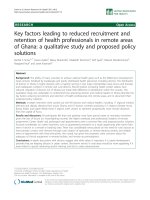
(Figure 1).
The control group was observed for natural course of
disease progression without any intervention.
For CRS alone group, CRS was performed on d 9 after
tumor cells inoculation. Rabbits were given 1% pento-
barbital sodium (30 mg/kg) intravenously for anesthes ia.
The abdominal skin was cleaned and disinfected. The
abdominal exploration was performed through a midline
incision of 8 cm long beginning 1 cm below the
xyphoid. Once the abdominal wall was open, detailed
evaluation of the PC was conducted in diff erent regions
including the parietal peritoneum, visceral peritoneum,
the omentum, stomach, liver, spleen, small intestine,
colon, bladder and other pelvic tissues. Thereafter, max-
imal CRS was performed including a routine omentect-
omy, and optimal removal of tumor nodules.
Tang et al. Journal of Translational Medicine 2011, 9:53
/>Page 2 of 9
Unresectable tumors were cauterized. The gastric tumor
itself, however, was not removed but treated by injection
of absolute alcohol. After completion of CRS the
abdominal wall was closed in 2 layers using 3 -0 Vicryl
constinuous sutures.
For CRS + HIPEC group, maximal CRS was performed
ond9inthesamefashionasintheCRSalonegroup,
which was immediately followed by HIPEC just before
the closure of abdominal cavity. Open HIPEC was per-
formed, as this open technique was believed t o provide
optimal thermal homogeneity and spatial diffusion
[20,21], with 250 mL of heated saline containing 10 mg
of docetaxel (Wanle Pharmaceutical Co., Ltd. Shenzhen,
China.) and 40 mg of carboplatin (Qilu Pharmaceutical
Co., Ltd. Shandong, China.) for each animal. The abdom-
inal cavity was rinsed twice with 250 mL of normal saline
preheated to 42.0°C and perfusion tube was placed in pel-
vic cavity just before HIPEC. T he perfusion equipment
consisted of a miniature heat exchanger and a roller
pump, allowing perfusion with a variable dynamic flow of
6 - 12 ml/min. An inflow catheter was inserted into the
upper abdomen between the hepatic and diaphragmatic
surface and an outflow catheter was placed at the pelvic
floor. The perfusion solution was heated to 42.0 ± 0.5°C
and infused into the peritoneal cavity at a rate of 10 ml/
min through the inflow tube introduced from the auto-
matic perfusion pump. The perfusion in the peritoneal
cavity was stirred manually to make equal spatial distri-
bution. The temperature of the perfusion solution in
peritoneal space was kept at 42.0 ± 0.5°C and monitored
using a thermometer on real time. The total HIPEC time
was 30 min, after which the perfusion solution in the
abdominal cavity was removed.
Twenty min before surgery, 100 ml of 0.9% NaCl solu-
tion with 1 g of ceftriaxone powder, 2 ml of 10% potas-
sium chloride solution and 20 ml of 50% glucose
solution was infused intravenously for rehydration,
energy support and infection control in both the CRS
alone group and the CRS + HIPEC group. Such treat-
ment was continued for 3 d.
Animal observation and disease course monitoring
The general status of the animals was daily recorded in a
standard form. For pathological studies, euthanasia was
performed on the rabbits by overdose injection of 1%
pentobarbital sodium through the ear vein. Post mortem
pathological examinations included gross pathology such
as tumor size and distributions; local t umor features of
gastric cancer such as ulcer formation, obstruction and
perforation; special features of peritoneal carcinomatosis
such as bloody ascites, discrete or confluent tumor
nodules on the peritoneum, omentum cake and intestinal
obstructions; metastases to major organs such as the
liver, adrenal glands, pancreas and the lungs.
For laboratory studies, 5 ml of blood was harvested
from ear vein on the day before tumor cells inoculation
as the baseline, on the day of surgery, and on d 8 after
surgery. The samples were used for routine peripheral
blood test, liver and kidney functions tests and biochem-
ical tests.
Statistical Analysis
The primary endpoint was overall survival (OS) in each
group, defined a s the time interval form animal model
construction to animal death due to any cause, includ-
ing cancer progress. The secondary endpoints were
body weight, biochemistry, major organ functions and
serious adverse events (SAE), which was defined as
severe local and/or systemic infection or death due to
the procedure.
In our previous study to construct this animal model,
we learned that the median survival of this gastric PC
model is about 3 weeks [19]. T herefore, we calculated
the sample size of this study based on t his information.
This trial was designed to detect at least a 30% absolute
difference in OS. With a statistical pow er of 90% to
detect such difference at 5% significance level, at least
12 animals were required in each group. Taking into
consideration of unexpected events during the perfor-
mance of the study, we enlarged the sample size to 1 4
animals in each group. Categorized variables in the two
D-5
D-1
D9
D12
D15
D18
D n
42
rabbits
Survival
Pathology
study
General status monitoring
Blood profile
Biochemistry
Operation
Control
(n=14)
CRS alone
(n=14)
CRS+HIPEC
(n=14)
Blood profile
biochemistry
Blood profile
biochemistry
D0
PC model
construction
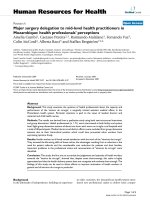
Figure 1 The study protocol. After construction of PC model of gastric cancer, 42 New Zealand white rabbits were randomized into 3 groups
with 14 rabbits per group, and the effects of CRS and CRS + HIPEC were investigated. D, day; PC, peritoneal carcinomatosis; CRS, cytoreductive
surgery; HIPEC, hyperthermic intraperitoneal chemotherapy.
Tang et al. Journal of Translational Medicine 2011, 9:53
/>Page 3 of 9
groups were compared by chi square test or Fisher’ s
exact test. The numerical data were directly recorded,
and the category data were recorded into different cate-
gories. The Kaplan-Meier method was used t o compare
the survival, with log r ank test. Data were analyzed
using the Statistical Package for Social Sciences (SPSS
Inc., Chicago, Illinois, USA), version 13.0 with 2-sided P
< 0.05 as statistically significant.
Results
Histopathological characteristics of PC
Rabbit gastric cancer PC model was established in all
animals (100%, 42/42). Nine days after tumor cells
inoculation, many small, hard and transparent tumor
nodules developed on the greater omentum, and typical
ulcerative cancer about 0.5 cm in diameter formed on
the antrum of the stomach. No ascites was observed. No
obvious PC was found in other regions. There were no
differe nces in the PC severity among three groups. This
could be equivalent to clinical stage I peritoneal carcino-
matosis by Gilly criteria [6].
Typical ulcerative cancer with PC was observed in
post mortem pathological examinations of rabbits in
control group. The stomach wall was totally invaded by
the tumor to create cancer ulcer encased by confluent
nodules on the greater omentum, forming a big tumor
block. The abdominal wall and diaphragm were totally
invaded by the tumor. Many tumor nodules formed on
the intestinal wall, the mesentery and the retroperito-
neum. Bloody ascites could be more than 100 ml. All
the features are similar to the clinicopathologic charac-
teristics of gastric cancer with PC in patients.
Body weight changes
The body weight of each animal was recorded every 3 d.
No significant differences were found in initial body
weight of 3 groups before the treatment. Perioperative
body weight decreased in all groups because of the over-
night fasting. In the control group, the body weight
recovered once food intake was resumed but again
decreased progressively till the study endpoint. In the 2
treatment groups, postoperati ve body weight decreased
considerably during the first 3 d after surgery and then
decrease became gentle along with the increased food
intake in the following 5 d in 2 treatment groups.
Thereafter, body weight decreased progressively again
till the study endpoint in CRS alone group, while body
weight could be maintained or slightly increased for the
following 20 d in CRS + HIPEC group and decreased
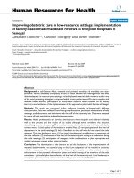
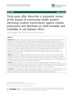
slowly till the study endpoint (Figure 2).
Blood profile changes
At the baseline, on the day of surgery and on day 8 after
surg ery, the peripheral blood cells counts , liver and kid-
ney function tests, and biochem istry parameters were all
comparable (Table 1).
Body Weight Changes Curve
0.8
1
1.2
1.4
1.6
1.8
2
2.2
2.4
2.6
2.8
0 3 6 9 12 15 18 21 24 27 30 33 36 39 42 45 48 51 54 57
Survival (days)
Mean body weight (kg)
Control
CRS alone
CRS+HIPEC
Figure 2 Body weight changes in 3 groups of rabbits. Compared with control and CRS groups, CRS + HIPEC group experienced slower body
weight loss, although the differences among the 3 groups did not reach statistical significance.
Tang et al. Journal of Translational Medicine 2011, 9:53
/>Page 4 of 9
Table 1 Blood routine tests and biochemical test results
Range (median)
Parameters Control (n = 14) CRS (n = 14) CRS+HIPEC (n = 14) P
Peripheral blood tests
A 139~156 (149) 129~139 (131) 124~141 (131) NS
HGB (G/L) B 112~130 (128) 102~137 (121) 104~30 (126) NS
C 78~135 (117) 62~123 (100) 80~103 (92) NS
A 6.55~7.30 (6.77) 6.19~6.80 (6.77) 5.98~6.48 (6.29) NS
RBC (× 10
9
/L) B 5.19~6.18 (5.76) 5.26~6.54 (5.60) 4.87~6.37 (6.14) NS
C 4.27~6.14 (5.26) 2.26~5.76 (5.30) 3.97~4.70 (4.34) NS
A 4.0~10.1 (6.5) 7.4~9.9 (8.8) 4.2~8.7 (4.7) NS
WBC (× 10
9
/L) B 6.5~11.3 (8.0) 4.8~10.3 (9.1) 7.1~9.4 (9.2) NS
C 7.7~18.2 (9.2) 3.2~8.3(4.9) 10.0~8.3 (9.2) NS
A 1.3~3.8 (2.7) 2.2~5.9 (4.5) 2.9~3.8 (3.2) NS
Neu (× 10
9
/L) B 1.8~4.3 (3.8) 1.4~8.4 (3.6) 2.6~3.8 (3.1) NS
C 1.1~4.8 (3.0) 0.7~4.1 (2.0) 4.2~4.8 (4.5) NS
A 169~385 (320) 158~410 (267) 94~415 (319) NS
PLT (× 10
9
/L) B 68~434 (231) 103~398 (232) 232~682 (360) NS
C 302~663 (324) 12~59 (27) 36~426 (231) NS
Liver function tests
A 15~24 (22) 34~44 (35) 15~24 (22) NS
AST (U/L) B 28~91 (75) 65~72 (66) 36~8 (69) NS
C 27~30 (29) 14~22 (20) 15~19 (17) NS
A 28~43 (36) 65~72 (66) 36~81 (69) NS
ALT (U/L) B 12~41 (26) 16~38 (26) 19~89 (29) NS
C 42~131 (50) 54~90 (85) 71~95 (83) NS
A 60.7~77.0 (66.1) 62.8~66.5 (66.0) 58.4~66.3 (63.8) NS
TP (g/L) B 58 .0~69.9 (62.3) 56.5~69.6 (62.8) 53.2~71.5 (59.9) NS
C 61.1~65.6 (62.8) 46.3~63.7 (55.8) 50.2~57.8 (54.0) NS
A 37.3~41.2 (39.6) 36.6~41.8 (40.6) 32.0~41.2 (39.6) NS
ALB (g/L) B 35.0~41.4 (37.1) 31.3~39.8 (35.8) 32.5~38.4 (34.9) NS
C 34.6~38.9 (36.3) 26.4~37.4 (30.9) 30.7~32.8 (31.8) NS
A 23.4~35.8 (26.6) 24.8~26.3 (25.7) 26.4~32.6 (26.6) NS
GLB (g/L) B 22.2~28.5 (25.5) 24.4~32.2 (27.3) 19.7~33.1 (25.2) NS
C 23.9~29.3 (26.5) 19.9~26.3 (24.9) 19.5~25.0 (22.3) NS
A 126~190 (159) 127~248 (147) 159~186 (172) NS
ALP (U/L) B 78~145 (98) 45~177 (86) 51~133 (89) NS
C 73~118 (80) 58~114 (79) 52~114 (83) NS
Renal function tests
A 6.24~15.08 (6.95) 6.47~7.68 (7.47) 7.28~8.44 (8.16) NS
BUN (mmol/L) B 8.83~14.77 (12.24) 0.59~16.64 (10.18) 6.82~14.94 (7.92) NS
C 5.45~6.45 (6.27) 5.33~7.07 (6.61) 4.83~6.45 (5.64) NS
A 81.0~121.2 (86.5) 80.6~99.2 (85.4) 81.0~95.3 (83.7) NS
Cr (μmol/L) B 75.0~99.0 (94.9) 70.4~107.4 (88.8) 85.8~97.8 (91.6) NS
C 67.3~85.6 (75.7) 60.3~69.9 (62.2) 65.8~66.9 (66.4) NS
Electrolytes
A 4.10~18.97 (4.66) 3.52~4.72 (4.21) 3.99~10.97 (4.30) NS
K+ (mmol/L) B 7.34~27.13 (10.44) 4.44~11.09 (6.29) 4.34~12.29 (4.74) NS
C 5.14~5.91 (5.18) 4.98~6.08 (5.14) 5.31~6.32 (5.82) NS
A 139.1~148.7 (145.3) 142.8~148.8 (144.2) 142.2~145.3 (144.7) NS
Na+ (mmol/L) B 124.5~146.4 (140.4) 137.3~148.5 (141.65) 133.6~146.4 (141.1) NS
C 133.2~138.7 (133.5) 132.9~138.9 (135.3) 133.8~134.3 (134.1) NS
A 99.8~121.2 (102.6) 101.9~107.2 (103.5) 100.5~110.2 (102.1) NS
Tang et al. Journal of Translational Medicine 2011, 9:53
/>Page 5 of 9
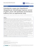
Survival
The animals in the control group did not receive any
active surgical treatment, and only observed for natural
history of dis ease progression. For animals in both CRS
and CRS+HIPEC groups, complete cytoreduction was
achieved either by surgical resection or cauterization for
the peritoneal carcinomatosis, leaving no observable
tumor nodules in the peritoneal cavity. The gastric
tumor itself, however, was not removed but treated by
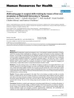
injection of absolute alcohol. The median OS was 24.0
d (95% CI 21.8 - 26.2 d) in the control group, 25.0 d
(95% CI 21.3 - 28.7 d) in CRS group, and 40.0 d (95%
CI 34.6 - 45.4 d) i n C RS + HIPEC group (P =0.00,log
rank test). Compared with CRS only or control grou p,
CRS+HIPECcouldextendOSbyatleast15d(60%)
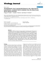
(Figure 3).
Postmortem pathological examinations
Euthanasia was performed on the moribund rabbits by
overdose injection of 1% pentobarbital sodium through
the ear vein. Detailed information on postmort em
pathological examinations was listed in Table 2.
Severe Adverse Events
SAE occurred in 0 animal in control group, 2 animals in
CRS alone group including 1 death due to anesthesia
overdose (OS = 9 d) and another death due to post-
operative hemorrhage (OS = 10 d), and 3 animals in
CRS + HIPEC group including 1 death due to anesthesia
overdose (OS = 9 d), and 2 deaths due to diarrhea 23
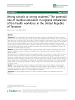
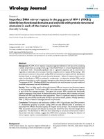
and 27 d after operation. A direct comparison in gross
pathology on d 27 of a rabbit in CRS group (Figure 4A)
and a rabbit in CRS + HIPE C group (Figure 4B) showed
significant differences in PC severity.
Discussion
This study has provided new evidence to support CRS +
HIPEC to trea t gastri c PC. Compared with control
group and CRS alone group, the CRS + HIPEC group
could have an additional survival gain of at least 15 d
Table 1 Blood routine tests and biochemical test results (Continued)
Cl
-
(mmol/L) B 94.6~103.9 (96.6) 96.6~107.7 (101.1) 97.0~106.6 (100.8) NS
C 100.0~104.1 (103.7) 102.2~106.6 (104.7) 103.8~104.2 (104.0) NS
A 3.11~3.77 (3.39) 3.05~3.18 (3.12) 3.18~3.69 (3.63) NS
Ca++ (mmol/L) B 2.80~4.04 (3.70) 3.41~3.96 (3.71) 3.32~3.96 (3.66) NS
C 3.71~4.10 (3.80) 3.19~3.56 (3.45) 3.48~3.52 (3.50) NS
RBC: red blood cells; WBC: white blood cells; HGB: hemoglobin; Neu: neutrophils count; PLT: platelets counts; ALT: alanine transaminase; AST: aspartate
aminotransferase ; A: At baseline; B: On the day of surgery; C: On d 8 after surgery; TP: total protein; ALB: albumin; GLB: globulin; BUN: blood urea nitrogen; Cr:
creatinine.
Figure 3 Kaplan-Meier survival curves for control, CRS alone, and CRS + HIPEC groups. Compared with CRS only or control group, CRS +
HIPEC could extend OS by at least 15 d (60%). (P = 0.00, log rank test)
Tang et al. Journal of Translational Medicine 2011, 9:53
/>Page 6 of 9
(60%). In addition to such significant survival benefit,
other improvements have also been observed, including
body weight, PC severities, ascites, liver and kidney
functions, and blood electrolytes.
This study also suggests that in established gastric PC,
simply performing CRS may not bring survival benefit.
The animals in the CRS group had a median survival of
25 d, which is not statistically different from 24 d in the
control group.
PC has been increasingly recognized as an important
clinical problem and increasing efforts have been
devoted to investigating the mechanism and coping stra-
tegies against this disease. Clinical trials in selected
gastric or colorectal PC patients have provided evidence
in favor of CRS + HIPEC for these patients, and the
only phase III prospective randomized trial in colorectal
PC patients reported a median survival advantage of
70% gain in overall survival (22.4 months in the CRS +
HIPEC group VS 12.6 months with standard palliative
care alone) [22]. The e ncouraging results by Yonemura
[8] and Glehen [9] in gastric PC provided more compel-
ling evidence to suppor t this combined treatment mod-
ality. Nevertheless, controversies regarding the
usefulness and value of such approach remain [23,24]. It
seems unlikely that this issue will be resolved shortly in
randomized clinical trials. Therefore, it is necessary to
Table 2 Results of post mortem pathological study in 3 groups*
Control (n = 14) CRS (n = 12)* CRS+HIPEC (n = 13)§ P
Ulcerative gastric cancer 100% 100% 100% NA
Pyloric obstruction 100% 100% 100% NA
Gastric perforation 28.6% 8.3% 8.3% P = 0.246
Greater omentum cake 100% removed removed NA
Liver metastases 100% 96.7% 38.5% P = 0.000
Pulmonary metastases 0.0% 8.3% 53.8% P = 0.000
Cancerous diaphragm 100% 100% 53.8% P = 0.000
Upper abdominal wall cancer 100% 100% 84.6% P = 0.128
Small intestine & mesentery seeding 100% 100% 61.5% P = 0.002
Adrenal gland metastases 100% 100% 23.5% P = 0.000
Kidney capsule invasion 100% 75.0% 30.8% P = 0.000
Retroperitoneum metastases 100% 100% 76.9% P = 0.038
Pelvic seeding 100% 100% 69.2% P = 0.009
Urine retention 57.1% 75.0% 61.5% P = 0.641
Bloody ascites 100% 100% 38.5% P = 0.000
* Two animals were excluded in CRS group, including 1 death due to anesthesia overdose on d 9 and another death due to postoperative hemorrhage on d 10.
§ One animal in CRS+HIPEC group was excluded due to anesthesia overdose death on d 9.
NA: not applicable.
4A
4B
Figure 4 On day 27, post mortem pathological examinations of a rabbit in CRS group (2A) and a rabbit in CRS + HIPEC group (2B).In
CRS group, widespread PC recurrence was evident even after sytoreductive surgery. In the CRS + HIPEC group, hyperthermic chemoperfusion
significantly retarded PC recurrence.
Tang et al. Journal of Translational Medicine 2011, 9:53
/>Page 7 of 9
study the treatment modality under experimental condi-
tions, in which most of the confounding factors could
be well controlled for more objective evaluation of
HIPEC.
In recent years, increasing number of animal m odels
of PC have been intensively studied, including nude
mouse model of gastric cancer PC c onstructed by
implanting human gastric cancer cells [25-28]; mouse
colon cancer PC model constructed by injecting colon
cancer cells into the abdominal cavity of Balb/C mice
[29]; rat colon carcinoma PC models constructed
through injecti ng CC531 colon carcinoma cells into the
abdominal cavity of Wag/Rij rats [15,30-33] or i njecting
syngeneic colon adenocarcinoma cells (DHD/K12/TRb)
into the abdominal cavity of athymic BD IX/HansHsd
rats [14,18,34,35]; murine xenograft PC model of appen-
diceal mucinous adenocarcinoma constructed by
impl anting human appendiceal neoplasms into the peri-
toneal cavity of homozygous nude mice [36]; mouse
ovarian cancer PC model constructed through injecting
human serous or epithelial ovarian cancer cel ls into the
abdominal cavity of mice [37-39] or injecting murine
ovarian surface epithelial cells (ID8 cells) under ovarian
bursa of C57BL6 mice [39].
Compared with the small animal PC models, our rab-
bit model of gastric PC is the first large animal PC
model, more suitable for complex surgical interventional
studies such as CRS + HIPEC. In addition, this model
reproduces the whole pathological process from the pri-
mary gastric cancer to the development of PC, resem-
bling the complete clinico-pathological features of
human gastric PC.
To our knowledge, there have been 3 reports in the
literature on the efficacy of CRS + HIPEC in experimen-
tal animal models of PC. Klaver et al [34] used the rat
colonic carcinoma PC model to test whether the addi-
tion of HIPEC to CRS is essential for survival benefit.
The rats were randomized into 3 treatment groups of
20 rats each, CRS alone, CRS + HIPEC (mitomycin 15
mg/m
2
at 42.0°C for 90 min) and CRS + HIPEC (mito-
mycin C 35 mg/m
2
at 42.0°C for 90 min). The CRS +
HIPE C achieved a significant survival gain of over 120%
(the median surviva l of 43, 75 and 97 d, P <0.01).Pelz
et al [15] used similar rat colonic carcinoma PC model
to investigate HIPEC. After 10 d of tumor cells inocula-
tion, the rats were randomized into 3 groups of 6 ani-
mals each, control, HIPEC (mitomycin C 15 mg/m
2
at
40.5 - 41.5°C for 90 min), and normathermic intraperi-
toneal chemotherapy (mitomycin C 10 mg/m
2
i.p.).
Although the study did not report the overall survival,
the HIPEC group did have significantly smaller tumor
weight, fewer tumor nodules, decreased cancer index
and better clinical complete response rate, compared
with control or normathermic ip mitomycin treatment
alone. In a similar study on rat colon can cer PC model,
Raue et al [36] found that only CRS + HIPEC with
MMC 15 mg/m
2
at 41.2 - 42.3°C for 60 min could
result in significant reduction in tumor w eigh and PC
index. Again this study did not report on the overall
survival.
Conclusions
In summary, this study on the first large animal model
of gastric PC has proved that CRS + HIPEC could
indeed bring survival benefit with acceptable safety, pro-
viding evidence to support this combined strategy to
treat selected patients of gastric cancer with PC.
Acknowledgements
Supported by the grants supporting New Strategies to Treat Peritoneal
Carcinomatosis from Hubei Sciences and Technology Bureau (2008 BCC011,
2060402-542), the Science Fund for Creative Research Groups of the National
Natural Science Foundation of China (No. 20621502, 20921062), and the
National University Student Innovation Training Project of China (081048646).
Author details
1
Department of Oncology, Zhongnan Hospital of Wuhan University; Hubei
Key Laboratory of Tumor Biological Behaviors & Hubei Clinical Cancer Study
Center, Wuhan, 430071, P.R. China.
2
NPO Organization to Support Peritoneal
Dissemination Treatment, Kishiwada, Osaka, Japan.
Authors’ contributions
YLI conceived, designed and partly conducted the study. LT, LJM, CQH and
XJY conducted the study and drafted the manuscript. YFZ and YY provided
technical support. All authors have read the approved the final manuscript.
Competing interests
The authors declare that they have no competing interests.
Received: 18 January 2011 Accepted: 7 May 2011 Published: 7 May 2011
References
1. Blair SL, Chu DZ, Schwarz RE: Outcome of palliative operations for
malignant bowel obstruction in patients with peritoneal carcinomatosis
from nongynecological cancer. Ann Surg Oncol 2001, 8:632-637.
2. Al-Shammaa HAH, Li Y, Yonemura Y: Current status and future strategies
of cytoreductive surgery plus intraperitoneal hyperthemic for peritoneal
carcinomatosis. World J Gastroenterol 2008, 14:1159-1166.
3. Fizazi K, Doubre H, Le Chevalier T, Riviere A, Viala J, Daniel C, Robert L,
Barthélemy P, Fandi A, Ruffié P: Combination of raltitrexed and oxaliplatin
is an active regimen in malignant mesothelioma: Results of a phase II
study. J Clin Oncol 2003, 21:349-354.
4. Gómez Portilla A, Cendoya I, López de Tejada I, Olabarria I, Martínez de
Lecea C, Magrach L, Gil A, Echebarría J, Valdovinos M, Larrabide I:
Peritoneal carcinomatosis of colorectal origin. Current treatment. Review
and update. Rev Esp Enferm Dig 2005, 97:716-737.
5. Yonemura Y, Bando E, Kawamura T, Ito H, Endo Y, Miura M, Kiyosaki K,
Sasaki T: Cytoreduction and intraperitoneal chemotherapy for
carcinomatosis from gastric cancer. Cancer Treat Res 2007, 134:357-373.
6. Sadeghi B, Arvieux C, Glehen O, Beaujard AC, Rivoire M, Baulieux J,
Fontaumard E, Brachet A, Caillot JL, Faure JL, Porcheron J, Peix JL,
Francois Y, Vignal J, Gilly FN: Peritoneal carcinomatosis from non-
gynecologic malignancies: results of the EVOCAPE 1 multicentric
prospective study. Cancer 2000, 88:358-363.
7. Yang XJ, Li Y, al-shammaa HAH, Yang GL, Liu SP, Lu YL, Zhang JW,
Yonemura Y: Cytoreductive surgery plus hyperthermic intraperitoneal
chemotherapy improves survival in selected patients with peritoneal
carcinomatosis from abdominal and pelvic malignancies: results of 21
cases. Ann Surg Oncol 2009, 16:345-351.
Tang et al. Journal of Translational Medicine 2011, 9:53
/>Page 8 of 9
8. Yonemura Y, Kawamura T, Bandou E, Takahashi S, Sawa T, Matsuki :
Treatment of peritoneal dissemination from gastric cancer by
peritonectomy and chemohyperthermic peritoneal perfusion. Br J Surg
2005, 92:370-375.
9. Glehen O, Schreiber V, Cotte E, Sayag-Beaujard AC, Osinsky D, Freyer G,
Francois Y, Vignal J, Gilly FN: Cytoreductive surgery and intraperitoneal
chemohyperthermia for peritoneal carcinomatosis arising from gastric
cancer. Arch Surg 2004, 139:20-26.
10. Mori T, Fujiwara Y, Sugita Y, Azama T, Ishii T, Taniguchi K, Yamazaki K,
Takiguchi S, Yasuda T, Yano M, Monden M: Application of molecular
diagnosis for detection of peritoneal micrometastasis and evaluation of
preoperative chemotherapy in advanced gastric carcinoma. Ann Surg
Oncol 2004, 11:14-20.
11. Flatmark K, Reed W, Halvorsen T, Sørensen O, Wiig JN, Larsen SG,
Fodstad Ø, Giercksky KE: Pseudomyxoma peritonei–two novel orthotopic
mouse models portray the PMCA-I histopathologic subtype. BMC Cancer
2007, 7:116.
12. Braumann C, Jacobi CA, Rogalla S, Menenakos C, Fuehrer K, Trefzer U,
Hofmann M: The tumor suppressive reagent taurolidine inhibits growth
of malignant melanoma - a mouse model. J Surg Res 2007, 143:327-378.
13. Pelz JOW, Doerfer J, Decker M, Dimmler A, Hohenberger W, Meyer T:
Hyperthermic intraperitoneal chemoperfusion (HIPEC) decrease wound
strength of colonic anastomosis in a rat model. Int J Colorectal Dis 2007,
22:941-947.
14. Otto J, Jansen PL, Lucas S, Schumpelick V, Jansen M: Reduction of
peritoneal carcinomatosis by intraperitoneal administration of
phospholipids in rats. BMC Cancer 2007, 7:104.
15. Pelz JOW, Doerfer J, Dimmler A, Hohenberger W, Meyer T: Histological
response of peritoneal carcinomatosis after hyperthermic intraperitoneal
chemoperfusion (HIPEC) in experimental investigations. BMC Cancer 2006,
6:162.
16. Pelz JOW, Doerfer J, Hohenberger W, Meyer T: A new survival model for
hyperthermic intraperitoneal chemotherapy (HIPEC) in tumor-bearing
rats in the treatment of peritoneal carcinomatosis. BMC Cancer 2005, 5:56.
17. Braumann C, Stuhldreier B, Bobrich E, Menenakos C, Rogalla S, Jacobi CA:
High doses of taurolidine inhibit advanced intraperitoneal tumor growth
in rats. J Surg Res 2005, 129:129-135.
18. Monneuse O, Mestrallet JP, Quash G, Gilly FN, Glehen O: Intraperitoneal
treatment with dimethylthioampal DIMATE combined with surgical
debulking is effective for experimental peritoneal carcinomatosis in a rat
model. J Gastrointest Surg 2005, 9:769-774.
19. Mei LJ, Yang XJ, Tang L, al-shammaa HAH, Yonemura Y, Li Y: Establishment
and identification of a rabbit model of peritoneal carcinomatosis from
gastric cancer. BMC Cancer 2010, 10:124.
20. Yang XJ, Li Y, Yonemura Y: Cytoreductive surgery plus hyperthemic
intraperitoneal chemotherapy to treat gastric cancer with ascites and/or
peritoneal carcinomatosis: results from a Chinese center. J Surg Oncol
2010, 101:457-464.
21. Stewart JH IV, Shen P, Levine EA: Intraperitoneal hyperthermic
chemotherapy for peritoneal surface malignancy: Current status and
future directions.
Ann Surg Oncol 2005, 12:765-777.
22. Verwaal VJ, van Ruth S, de Bree E, van Slooten GW, van Tinteren H, Boot H,
Zoetmulder FAN: Randomized trial of cytoreduction and hyperthermic
intraperitoneal chemotherapy versus systemic chemotherapy and
palliative surgery in patients with peritoneal carcinomatosis of colorectal
cancer. J Clin OncoL 2003, 21:3737-3743.
23. Khatri VP: Cytoreductive surgery and hyperthermic intraperitoneal
chemotherapy for colorectal cancer: a panacea or just an obstacle
course for the patient? J Clin Oncol 2010, 28:5-7.
24. Elias D, Delperro JR, Sideris L, Benhamou E, Pocard M, Baton O,
Giovannini M, Lasser P: Treatment of peritoneal carcinomatosis from
colorectal cancer: impact of complete cytoreductive surgery and
difficulties in conducting randomized trials. Ann Surg Oncol 2004,
11:518-521.
25. Jansen M, Treutner KH, Jansen PL, Zuber S, Otto J, Tietze L, Schumpelick V:
Inhibition of gastric cancer cell adhesion in nude mice by inraperitoneal
phospholipids. World J Surg 2005, 29:708-714.
26. Buchhorn HM, Seidl C, Beck R, Saur D, Apostolidis C, Morgenstern A,
Schwaiger M, Senekowitsch-Schmidtke R: Non-invasive visualisation of the
development of peritoneal carcinomatosis and tumour regression after
213
Bi-radioimmunotherapy using bioluminescence imaging. Eur J Nucl
Med Mol Imaging 2007, 34:841-849.
27. Piso P, Aselmann H, von Wasielewski R, Dahlke MH, Klempnauer J,
Schlitt HJ: Prevention of peritoneal carcinomatosis from human gastric
cancer cells by adjuvant-type intraperitoneal immunotherapy in a SCID
mouse model. Eur Surg Res 2003, 35:470-476.
28. McCarty MF, Takeda A, Stoeltzing O, Liu W, Fan F, Reinmuth N, Akagi M,
Bucana C, Mansfield PF, Ryan A, Ellis LM: ZD6126 inhibits orthotopic
growth and peritoneal carcinomatosis in a mouse model of human
gastric cancer. Br J Cancer 2004, 90:705-711.
29. Kulu Y, Dorfman JD, Kuruppu D, Fuchs BC, Goodwin JM, Fujii T, Kuroda T,
Lanuti M, Tanabe KK: Comparison of intravenous versus intraperitoneal
administration of oncolytic herpes simplex virus 1 for peritoneal
carcinomatosis in mice. Cancer Gene Ther 2009, 16:291-297.
30. Hribaschek A, Kuhn R, Pross M, Meyer F, Fahlke J, Ridwelski K, Boltze C,
Lippert H: Intraperitoneal versus intravenous CPT-11 given intra- and
postoperatively for peritoneal carcinomatosis in a rat model. Surg Today
2006, 36:57-62.
31. Aarts F, Koppe MJ, Hendriks T, vanEerd JEM, Oyen WJG, Boerman OC,
Bleichrodt RP: Timing of Adjuvant Radioimmunotherapy after
Cytoreductive Surgery in Experimental Peritoneal Carcinomatosis of
Colorectal Origin. Ann Surg Oncol 2006, 14:533-540.
32. Aarts F, Bleichrodt RP, de Man B, Lomme R, Boerman OC, Hendriks T: The
effects of adjuvant experimental radioimmunotherapy and hyperthermic
intraperitoneal chemotherapy on intestinal and abdominal healing after
cytoreductive surgery for peritoneal carcinomatosis in the rat. Ann Surg
Oncol 2008, 15:3299-3307.
33. Klaver YLB, Hendriks T, Lomme RMLM, Rutten HJT, Bleichrodt RP, de
Hingh IHJT: Introperative hyperthemic intraperitoneal chemotherapy
after cytoreductive surgery for pertitoneal carcinomatosis in an
experimental model. Br J Surg 2010, 97:1874-1880.
34. Hartmann J, Kilian M, Atanassov V, Braumann C, Ordemann J, Jacobi CA:
First surgical tumour reduction of peritoneal surface malignancy in a
rat’s model. Clin Exp Metastasis 2008, 25:445-449.
35. Raue W, Kilian M, Braumann C, Atanassow V, Makareinis A, Caldenas S,
Schwenk W, Hartmann J: Multmodel approach for treatment of
peritoneal surface malignancises in a tumer-bearing rat model. Int J
Colorectol Dis 2010, 25:245-250.
36. Mavanur AA, Parimi V, Malley MO, Nikiforova M, Bartlett DL, Davison JM:
Establishment and characterization of a murine xenograft model of
appendiceal mucinous adenocarcinoma. Int J Exp Path 2010, 91:357-367.
37. Wei BR, Hoover SB, Ross MM, Zhou WD, Meani F, Edwards JB, Spehalski EI,
Risinger JI, Alvord WG, Quiñones OA, Belluco C, Martella L, Campagnutta E,
Ravaggi A, Dai RM, Goldsmith PK, Woolard KD, Pecorelli S, Liotta LA,
Petricoin EF, Simpson RM: Serum S100A6 concentration predicts
peritoneal tumor burden in mice with epithelial ovarian cancer and is
associated with advanced stage in patients. PLoS ONE 2009, 4:e7670.
38. Sheth RA, Upadhyay R, Stangenberg L, Sheth R, Weissleder R, Mahmood U:
Improved detection of ovarian cancer metastases by intraoperative
quantitative fluorescence protease imaging in a pre-clinical modal. Gyn
Oncol 2009, 112:616-22.
39. Greenaway J, Moorehead R, Shaw P, Petrik J: Epithelial-stromal interaction
increases cell proliferation, survival and tumorigenicity in a mouse
model of human epithelial ovarian cancer. Gyn Oncol 2008, 108:385-394.
doi:10.1186/1479-5876-9-53
Cite this article as: Tang et al.: Cytoreductive surgery plus hyperthermic
intraperitoneal chemotherapy improves survival of gastric cancer with
peritoneal carcinomatosis: evidence from an experimental study. Journal
of Translational Medicine 2011 9:53.
Tang et al. Journal of Translational Medicine 2011, 9:53
/>Page 9 of 9