Báo cáo sinh học: "SULFs in human neoplasia: implication as progression and prognosis factors" potx
Bạn đang xem bản rút gọn của tài liệu. Xem và tải ngay bản đầy đủ của tài liệu tại đây (426.59 KB, 9 trang )
RESEARCH Open Access
SULFs in human neoplasia: implication as
progression and prognosis factors
Caroline Bret
1,2,3
, Jérôme Moreaux
1
, Jean-François Schved
2,3
, Dirk Hose
4,5
and Bernard Klein
1,3*
Abstract
Background: The sulfation pattern of heparan sulfate chains influences signaling events mediated by heparan
sulfate proteoglycans located on cell surface. SULF1 and SULF2 are two endosulfatases able to cleave specific 6-O
sulfate groups within the heparan chains. Their action can modulate signaling processes, many of which with key
relevance for cancer development and expansion. SULF1 has been associated with tumor suppressor effects in
various models of cancer, whereas SULF2 dysregulation was in relation with protumorigenic actions. However,
other observations argue for contradictory effects of these sulfatases in cancer, suggesting the complexity of their
action in the tumor microenvironment.
Methods: We compared the expression of the genes encoding SULF1, SULF2 and heparan sulfate proteoglycans in
a large panel of cancer samples to their normal tissue counterparts using publicly available gene expression data,
including the data obtained from two cohorts of newly-diagnosed multiple myeloma patients, the Oncomine
Cancer Microarray database, the Amazonia data base and the ITTACA database. We also analysed prognosis data in
relation with these databases.
Results: We demonstrated that SULF2 expression in primary multiple myeloma cells was associated with a poor
prognosis in two independent large cohorts of patients. It remained an independent predictor when considered
together with conventional multiple myeloma prognosis factors. Besides, we observed an over-representation of
SULF2 gene expression in skin cancer, colorectal carcinoma, testicular teratoma and liver cancer compared to their
normal tissue counterpart. We found that SULF2 was significantly over-expressed in high grade uveal melanoma
compared to low grade and in patients presenting colorectal carcinoma compared to benign colon adenoma.
We observed that, in addition to previous observations, SULF1 gene expression was increased in T prolymphocytic
leukemia, acute myeloid leukemia and in renal carcinoma compared to corresponding normal tissues. Furthermore,
we found that high SULF1 expression was associated with a poor prognosis in lung adenocarcinoma.
Finally, SULF1 and SULF2 were simultaneously overexpressed in 6 cancer types: brain, breast, head and neck, renal,
skin and testicular cancers.
Conclusions: SULF1 and SULF2 are overexpressed in various human cancer types and can be associated to progression
and prognosis. Targeting SULF1 and/or SULF2 could be interesting strategies to develop novel cancer therapies.
Background
Heparan sulfate proteoglycans (HSPGs) are negatively-
charged proteins located at a high cell density on var-
ious cell types or released into the extracellular m atrix.
As HSPGs b ind a large diversity of molecules: growth
factors (GF), cytokines, chemokines, morphogens, matrix
ligands and cell surface molecules, they are involved in
cell signaling as co-receptors [1]. The complexity of the
heparan sulfate (HS) chains is based on modifications as
epimerisation, de-acetylation and sulfation. These phe-
nomenons strongly influence the ligand binding proper-
ties of HSPGs and define the concept of “HS code”. The
sulfation pattern in glucosamines and uronic acids is
dynamically regulated duri ng many cellular p rocesses,
generating diversity of the chains and thus d iversity of
binding. Such mechanisms are regulated by sulfotrans-
ferases involved in the biosynthesis of HS. Another class
of enzymes is also implicated at the extracellular level:
* Correspondence:
1
INSERM U847, Institut de Recherche en Biothérapie, CHRU de Montpellier,
France
Full list of author information is available at the end of the article
Bret et al. Journal of Translational Medicine 2011, 9:72
/>© 2011 Bret et al; licensee BioMed Central Ltd. This is an Open Access article distributed under the terms of the Creative Commons
Attribution License (http://creativecommons.o rg/licenses/by/2.0), which permits unrestricted use, distribution, and reproduction in
any medium, provided the original work is properly cited.
the sulfatases sulfatase 1 (SULF1) and sulfatase 2
(SULF2). Initially cloned in 2002 [2], these secreted
enzymes display endo glucosamine 6-sulfatase activity.
The expression of the genes encoding these enzymes is
developmentally regulated. In murine model, simulta -
neous disruption of both SU LF1 and SULF2 leads to
perinatal lethality and developmental defects underlying
overlapping and essential roles during development [3].
However, SULF1-deficient mice did not present any
abnormal phenotype whereas SULF2-knock-out mice
displayed a small but significant reduction in litter size
and body weight, and a hydrocephalus at birth resulting
in a life span shorter than 2 weeks [4].
Owing to the involvement of HSPGs as coreceptors of
cell communication molecules, the role of these HSPG
modifying enzymes in human tumorigenesis is activ ely
investigated. Despite similar substra te specificity, SULF1
has mainly tumor suppressor functions whereas SULF2
presents tumor promoting functions. In this article, we
focused on recent and challenging data describing the
implication of SULF1 and SULF2 in human neoplasia.
Methods
Databases
SULF1 and SULF2 gene expression levels in normal or
malignant human tissues or cell lines were obtained
from the Oncomine Cancer Microarray database (http://
www.oncomine.org) [5], the Amazonia database (http://
amazonia.montp.inserm.fr/) [6] and the ITTACA d ata-
base (Integrated Tumor Transcriptome Array and Clini-
cal data Analysis) developed by the Institute Curie
Bioinformatics group and the Institute Curie, CNRS
UMR144 ( [7]. Gene
expression data only obtained from a single study using
the same methodology were compared. All data were
log transformed, median centered per array and the
standard deviation was normalized to one per array.
Primary myeloma cells
Multiple Myeloma cells (MMC) were purifi ed from 206
patients with newly-diagnosed MM after written
informed consent was given at the University hospitals
of Heidelberg (Germany) or Montpellier (France). The
study was approved by the ethics boards of Heidelberg
University and Montpellier University. After Ficoll-den-
sity gradient centrifugation, plasma cells were purified
using anti-CD138 MACS microbeads (Miltenyi Biotech,
Bergisch Gladbach, Germany). Microarray experiments
were performed in DNA microarray platform of the
Institute of Research in Biotherapy at the Montpellier
University Hospital (France) />en/index.php?page=Plateau&IdEquipe=6. The .CEL files
and MAS5 files have been deposited in the ArrayExpress
public database, under accession number E-MTAB-362.
We also used Affymetrix data of a cohort of 345 puri-
fied MMC from previously untreated patients from the
Arkansas Cancer Rese arch Center (ACRC, Little Rock,
AR).Thesedataarepubliclyavailablevia the online
Gene Expression Omnibus (Gene Expression Profile of
Multiple Myeloma, accession number GSE2658, http://
www.ncbi.nlm.nih.gov/geo/).
Statistical analysis
Statistical comparisons were d one with Student t-tests.
The event free or overall survival of subgroups of
patients was compared with the log-ran k test and survi-
val curves computed with the Kaplan-Meier method.
The prognostic values of parameters were compared
with univariate or multivariate Cox analysis. Statistical
tests were performed with the software package SPSS
12.0 (SPSS, Chicago, IL).
Results and disc ussion
Tumor suppressor functions of SULF1
Expression of SULF1 mRNA can be detec ted in seve ral
normal human tissues, as observed by Morimoto-
Tomita et al. [2] in a panel of 24 ti ssue types, the high-
est levels being found in testes, stomach, skeletal mus-
cle, lung, and kidney. SULF1 down-regulation has been
described in human primary tumorous samples and/or
cell lines in ovarian cancer [8-10], hepatocellular carci-
noma [11], breast cancer [12], gastric cancer [12], kidney
cancer [12], prostatic stromal cells from benign prostatic
hyperplasia samples [13] and head and neck squamous
cell carcinoma (SCCHN) cell lines [14]. This low expres-
sion level is mostly explained by epigenetic silencing
mediated by hypermethylation of the promoter of the
gene encoding SULF1 [9,12].
Considering that HSPG sulfation pattern drives in part
cell communication molecule binding [15-17], a loss of
SULF1 expression is expected to disrupt the effects of
these cell communication molecules during malignan-
cies. It has been observed that this down-regulation
results in increased sulfation of HS chains and could
produce the stabilization of ternary receptor complexes,
leading to an increased in GF signalling, as described for
heparin-binding epidermal growth factor-like growth
factor (HB-EGF), fibroblast growth factor 2 (FGF2) or
amphiregulin in ovarian cancer [ 8], SCCHN cell lines
[14], hepatocellular carcinoma [18] or in breast cancer
[19]. This modulation of GF effects can affect major
events including proliferation of can cer cells. A forced
expression of SULF1 induced growth inhibitio n of neck
squamous cell carcinoma cell lines in vitro[14]. A
marked reduction of the growth of myeloma or breast
cancer cell lines was observed in severe combined
immunodeficient (SCID) mice when injected cell lines
were transfected with SULF1 cDNA [20,21]. Forced
Bret et al. Journal of Translational Medicine 2011, 9:72
/>Page 2 of 9
expression of SULF1 also significantly delayed the
growth of hepatocellular carcinoma cell lines xenogr afts
in nude mice [22].
These different models also argued the role of SULF1
as an inhibitor of motility, invasion and angiogenesis
and as a protein linked to drug-induced apoptosis.
Hepatocyte growth factor (HGF)-mediated motility and
invasion were attenuated in SCCHN cell lines displaying
an overexpression of this sulfatase [14]. Xenografts
derived from SULF1-expressing carcinoma cells pre-
sented a significantly reduced ability of vascular HS to
promote a stable complex between FGF2 and its specific
receptor with an inhibition of angiogenesis as a result.
The down-regulation of SULF 1 in human umbilical vein
endothelial cells (HUVECs) could increase vascular
endothelial growth factor (VEGF)-induced angiogenic
response [21]. In hepatocellular carcinoma (HCC),
SULF1 enhanced the induction of apoptosis by the his-
tone deacetylase (HDAC) inhibitors in vitro[22]. The
doxorubicin and apicidin-induced apoptosis was signifi-
cantly increased of in HCC cell lines expr essi ng SULF1.
In addition, the anti-tumor effects of these drugs were
enhanced in vivo when a xenograft was established from
SULF1-expressing HCC [23]. SCCHN-transfected cell
lines displayed significant growth inhibition concomitant
with an increased sensitivity to staurosporine- and cis-
platin-induced apoptosis [14].
Altogether, these data suggest that the widespread
SULF1 down-regulation in cancer might be an impor-
tant contributor to the carcinogenesis process.
SULF2, a protumorigenic endosulfatase
The implication of SULF2 in cancer was less studied
than that of SULF1. However, most of the studies docu-
mented a protumorigenic role of SULF2 at the opposite
of that of SULF1. Lemjabbar-Alaoui et al. [24] observed
an induction of SULF2 expression in human lung adeno-
carcinoma and squamous cell carcinoma with a mean
increase of 3-fold compared to normal lung. They could
obtain a loss of the transformed phenotype of lung carci-
noma cell lines when silencing SULF2 expression with
short-hairpin RNA (sh-RNA). The knock-out of SULF2
in these ce ll lines also resulted in a decreased tumor for-
mation when grafted to nude mice. Besides, SULF2 was
shown to modulate the bioavailability of wingless-type
MMTV integration site family (Wnt) ligands, a critical
canonical cascade reactivated in several tumors [25]. An
up-regulation of SULF2 mRNA was also observed in
human or murine breast cancers compared to normal
breast tissues [26]. SULF2 was up-regulated in primary
HCC samples, as well as in HCC cell lines [11]. It
resulted in an activation of mitogen-activated protein
kinase (MAPK) and v-akt murine thymoma viral onco-
gene homolog 1 (Akt) pathways with an increased cell
growth in vitro and in vivo.Inmultiplemyeloma(MM),
we had previoulsy reported an overexpression of SULF2
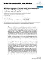
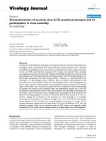
gene in primary myeloma cells of newly-diagnosed mye-
loma compared to normal bone marrow plasma cells
[27]. In this study, we demonstrate for the first time that
SULF2 expression in primary multiple myeloma cells
(MMCs) ("absent” versus “present” Affymetrix call) was
associated with a poor prognosis in two independent
large cohorts of myeloma patients at diagnosis (206
patients in the cohort of Heidelberg-Montpellier and 250
patients in the cohort of Little-Rock previously described
[28], Figure 1A and 1B). Patients with SULF2
absent
MMCs had a significant increased overall survival c om-
pared with patients with SULF2
present
MMCs (p =0.007
in the Heidelberg-Montpellier cohort and p = 0.03 in the
Little-Rock cohort), after high-dose therapy and stem
cell transplantation. In a Cox proportional hazard model
(Table 1), the absence or the presence of SULF2 (p=
0.007, hazard ratio = 4.08) and ISS stage (p = 0.001,
hazard ratio = 1.73) were independently predictive for
overall survival (p = 0.02 and p = 0.001, respect ively). If
SULF2 expression was tested together with classical
prognostic factors, i.e., serum albumin and serum beta 2
microglobulin (b2M), SULF2 expression (p=0.03)and
b2M (p=0.0001) remained independent prognosti c fac-
tors. SULF2 expression was an independent prognostic
factor of spiked MMSET expression, that is an indicator
of t(4;14) translocation [29] (p = 0.023 and p = 0.028
respectively), of the myeloma high risk score (HRS) [30]
(p = 0. 01 and p = 0.002 resp ectively), of the growth pro-
liferation index [31] (p = 0.01 and p = 0.0001 respec-
tively), of the IFM score [32] (p = 0.01 and p = 0.000 1
respectively) and of CD200 expression [33] (p = 0.02 and
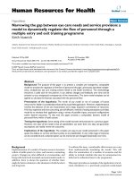
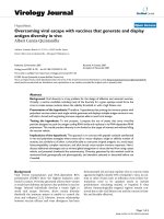
p = 0.05 respectively). Investigating the SULF2 expres-
sion in the 7 groups of the molecular classification [34]
of M M, SULF2 was significantly overexpressed in the
hyperdiploid group and significantly underexpressed in
the groups of patients characterized by Cyclin D1 or
MAF translocations (Figure 2). We analyzed the correl a-
tion between SULF1 or SULF2 expression and HS pro-
teoglycans expression in our cohort of myeloma patients
(syndecan 1-4, glypican 1-6, CD44 isoforms containing
the alternatively spliced exon v3, agrin, betaglycan, perle-
can, serglycin and testican 1-3)[27]. No significant corre-
lation was found between the expression of the SULFs
and of their potential HS proteoglycan targets in MM.
When we analyzed the c orrelation be tween the e xpres-
sion of the sulfatases and of the genes encoding the
transporters and the enzymes involved in HS and chon-
droïtine sulfate biosynthesis pathway [27], we did not
found any correlation for SULF2 but we observed a cor-
relation between SULF1 express ion and GALK1 (galacto-
kinase 1) and SLC2A9 (solute carrier family 2, facilitated
glucose transporter member 9) expression.
Bret et al. Journal of Translational Medicine 2011, 9:72
/>Page 3 of 9
In HCC model, sh-RNA targeting SULF2 induced an
inhibition of HCC cell lines proliferation and migration
in vitro. In nude mice, SULF2 could significantly pro-
mote HCC xenograft growth. Besides, forced expression
of this enzyme increased glypican-3 expression level,
this membrane-anchored HSPG being involv ed in Wnt
pathway, FGF signaling and cell proliferation [35].
Moreover, in patients with HCC, high levels of SULF2
were associated with a worse prognosis [11]. In human
pancreatic carcinoma, the SULFs are up-regulated and it
has been observed that the silencing of SULF2 could
lead to an inhibit ion of Wnt signalling and of cell
growth [36]. In order to explore the pathogenesis of
glioblastoma, Johansson et al. generated a mouse glioma
model using a recombinant Moloney murine leukemia
virus encoding the platelet-derived growth factor B-
chain and intra-cerebrally injected in newborn mice
[37]. Using expression profiling, they identified markers
of gliomagenesis, SULF2 appearing among the candidate
cancer-causing genes.
In addition to its contribution during tumor growth
development, SULF2 could be implicated in resistance
to cancer treatment, as rec ently suggested by Moussay
et al. [38]. A comparison of gene e xpression profiles of
sensitive and resistant clones of chronic lymphocytic
leukemia obtained from patients treated by fludarabine
was performed. Together with v-myc myelocytomatosis
viral oncogene homolog (MYC), SULF2 transcripts were
significantly over-represented in cells of patients resis-
tant to fludarabine.
Recently, SULF2 gene expression was investigated in a
large panel of cancer samples, using the ONCOMINE
microarray database ( 4.3
research edition) [39]. Rosen et al. demonstrated an
overexpression of SULF2 in several cancers including
brain, breast, tongue and renal carcinomas [39]. In
addition to these observations, we found that ot her can-
cer types displayed an over-representation of SULF2
gene expression compared to their tissue counterpart:
skin (p = 2.26E-4 and p=1E-3[40]), colorectal carci-
noma (p = 0.02 [41]), testicular teratoma (p = 6E-3 [42])
and liver canc er (p=1.9E-4an d p=2E-3[43]). Using
the ITTACA database (Integrated Tumor Transcriptome
Array and Clinical data Analysis, ie.
fr/ittaca/)[7] and the AMAZONIA database [6], we
searched to identify if SULF2 expression could be asso-
ciated with tumor progression in these cancer types.
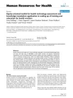
Interestingly, we found that SULF2 was significantly
over-expressed in high grade uveal melanoma compared
to low grade (p = 0.03, Figure 3A). Furthermore, SULF2
was also overexpressed in patients presenting colorectal
carcinoma compared to benign colon adenoma (p =
0.001, Figure 3B).
These different data lend support for a protumorigenic
effect of SULF2 overexpressed by many tumor cell types.
Challenging observations concerning SULF1 and SULF2 in
cancer
Using the ONCOMINE microarray database, Rosen et
al. shown that, in contrast to the down-regulation of
SULF1 reported in various tumor models, SULF1 gene
expression was increased in a large range of cancers
compared to their corresponding normal tissues [39].
SULF1 was clearly over-expressed in adrenal carcinoma,
brain cancer, breast carcinoma, colon adenocarcinoma,
skin carcinoma, esophageal and gastric cancers, head
and neck cancers, lung cancer, mesothelioma, pancreatic
cancer, sarcoma and germ line/testicular canc er [39]. In
addition, we found that other cancer types displayed an
over-representation of SULF1 gene expression: T pro-
lymphocytic leukemia (p = 0.01 [44]), acute myeloid leu-
kemia (p = 0.004 [45]) and renal carcinoma (p < 0.001
A
days
Cumulated survival
HM series, n = 206
OS, p=0.00724
0 400 800 1200 1600 2000 2400 2800
0
0.2
0.4
0.6
0.8
1
1.2
SULF2
absent
SULF2
present
LR-TT2 series, n = 250
OS, p=0.0361
da
y
s
Cumulated survival
0 400 800 1200 1600 2000 2400 2800
0
0.2
0.4
0.6
0.8
1
1.2
SULF2
presen
t
SULF2
absent
B
Figure 1 Overall survival (OS) related to SULF2 gene expression in two independent multiple myeloma patient series. Data are Kaplan-
Meier curves of patients displaying an “absent call” versus patients displaying a “present call”. A. Cohort of 206 patients (HM) from Montpellier
(France) and Heidelberg (Germany). B. Cohort of 250 patients (LR-TT2) of Little-Rock.
Bret et al. Journal of Translational Medicine 2011, 9:72
/>Page 4 of 9
[46]). These data challenge the above concept of SULF1
as a tumor suppressor effector. Using the ITTACA data-
base, we aimed to identify if SULF1 expression could be
associated with tumor progression or bad prognosis in
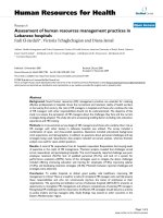
cancers. Indeed, we found that hi gh SULF1 expression
was associated with a poor prognosis in lung adenocar-
cinoma (Figure 4) [47]. Although SULF1 was overex-
pressed in breast cancer compared to its normal
counterpart [39,48,49] , we did not found any significant
association between SU LF1 expression and survival in
breast cancer using data from two independent studies
(data not shown).
Some studies have brought so me explanations abo ut
this contradictory contribution to carcinogenesis. In
pancreatic cancer cells, the expression of SULF1 in
xenograft models was associated with a markedly
reduced growth potential, but with an increase in the
basal invasiveness of these cells [50]. Recently, Sahota
and Dhoot [51] demonstrated in quail model the possi-
bility of alternative splicing of SULF1 gene, generating a
novel shorter isoform called SULF1B.Whilethepre-
viously described SULF1 (SULF1A) enhanced Wnt sig-
naling, SULF1B inhibited Wnt signaling and promoted
angiogenesis. Such splicing has n ot been yet described
in human tissues but could be of i nterest, in particular
in cancer development. In mutiple myeloma, we pre-
viously observed an overexpression of SULF1 by bone
marrow stromal cells, whereas primary malignant
plasma cells did not express the gene encoding for this
sulfatase. Besides, SULF1 was expressed by some human
myeloma cell lines (HMCLs), emphasizing that these
HMCLs can express environment genes, making it pos-
sible to escape from environment dependence [27].
Whereas SULF2 is considered as being associated with
protumorigenic effects, as reviewed above, a few challen-
ging studies argue for a tumor suppressor effect of this
protein. In contrast with our report that SULF2 expres-
sion in primary malignant plasma cells is associated
Table 1 Univariate and multivariate proportional hazards
analyses linking SULF2 expression to prognosis in HM
cohort
HM cohort (OS)
Pronostic variable Proportional hazard ratio P-value
Univariate
Cox analysis
SULF2
ISS
4.08
1.73
0.007
0.001
Multivariate
Cox analysis
SULF2
ISS
3.65
1.70
0.028
0.001
Univariate
Cox analysis
SULF2
b2M
Alb
4.08
1.10
1.60
0.007
0.0001
0.04
Multivariate
Cox analysis
SULF2
b2M
Alb
3.49
1.10
1.35
0.03
0.0001
0.24
Univariate
Cox analysis
SULF2
HRS
4.08
2.30
0.007
0.002
Multivariate
Cox analysis
SULF2
HRS
4.11
2.31
0.01
0.002
Univariate
Cox analysis
SULF2
MS group
4.08
2.14
0.007
0.001
Multivariate
Cox analysis
SULF2
MS group
3.84
1.97
0.023
0.028
Univariate
Cox analysis
SULF2
IFM score
4.08
3.09
0.007
0.0001
Multivariate
Cox analysis
SULF2
IFM score
4.29
3.22
0.014
0.0001
Univariate
Cox analysis
SULF2
GPI
4.08
2.21
0.007
0.0001
Multivariate
Cox analysis
SULF2
GPI
4.47
2.25
0.011
0.0001
Univariate
Cox
analysis
SULF2
MYEOV
4.08
3.16
0.007
0.05
Multivariate
Cox analysis
SULF2
MYEOV
3.71
2.76
0.026
0.08
Univariate
Cox analysis
SULF2
CD200
4.08
2.05
0.007
0.03
Multivariate
Cox analysis
SULF2
CD200
3.86
1.03
0.02
0.05
Univariate analyses were done to screen for prognostic variables linked to
SULF2 expression using Cox proportional hazards regression. The Cox model
was also used for multivariate analysis to identify the most significant
variables related to survival (OS): ISS (international staging system), b2M (beta-
2 microglobulin), Alb (Albumin), HRS (High Risk Score), MS group (MMSET
group), IFM score (IFM score), GPI (Growth Proliferation Index), MYEOV and
CD200. P-values are in bold and italic when a significant result was obtained
(p < 0.05).
MM molecular classification
g
rou
ps
Ύ
Ύ
Ύ
SULF2 express
i
on
(
Affymetr
i
x s
i
gna
l)
PR
LB
MS
HY
CD1
CD2 MF
30000
20000
10000
0
Figure 2 SULF2 exp ression in the 7 groups of the molecular
classification of multiple myeloma. The expression of SULF2 in
LR-TT2 cohort was investigated in the 7 groups of the molecular
classification of multiple myeloma. PR: proliferation, LB: low bone
disease, MS: MMSET, HY: hyperdiploid, CD1: Cyclin D1, CD2: Cyclin
D2, MF: MAF.
Bret et al. Journal of Translational Medicine 2011, 9:72
/>Page 5 of 9
with poor overall survival [27], Dai et al. [20] observed
that a forced expression of SULF2 reduced the growth
of myeloma cell lines in SCID mice. Thus, they con-
cluded to a similar action of SULF1 and SULF2 on mye-
loma cells expansion through the modification of HS
sulfation pattern and its consequence in medullar
microenvironment.
In addition to this in vivo observation, two studies
demonstrated that SULF2 is induced by p53 tumor sup-
pressor. Adamsen et al. [52] firstly suggested that
SULF2 was a putative p53 target gene in colon cancer
cells treated by 5-fluorouracil. Inducible p53 knockdown
cell lines of multiple c ancer types were generated by
Chau et al. [53] and their gene expressio n pro files were
compared to the initial cell lines. This method led to
the identification of downstream targets of p53. SULF2
was found to be a direct transcriptional target of p53
tha t could bind to the SULF2 pr omoter, in particular in
the context of DNA-damaged-induc ed senescence, in
accordance with the observation of Adamsen.
Interestingly, SULF1 was overexpressed in 6/7 cancer
types characterized by SULF2 overexpression compared
to normal tissue counterparts (Table 2). Several HS pro-
teoglycans have been identified so far - syndecan 1-4,
glypican 1-6, CD44 isoforms containing the alternatively
spliced exon v3, agrin, betaglycan, perlecan, serglycin
and testican 1-3 - and their gene expression could be
evaluated by microarrays [27]. In cancer samples dis-
playing an overexpression of SULF1 and/or SULF2 com-
pared to their normal counterparts, we systematically
observed on overexpression of at least one HS proteo-
glycans (Table 2). The functional consequences of the
SULF2 express
i
on
(
Affymetr
i
x un
i
t
)
Uveal melanoma
Low grade
(n=14)
High grade
(n=11)
p=0.03
A
500
1000
1500
2000
2500
3000
3500
4000
B
Normal
colon
(n=8)
Colon
adenoma
(n=15)
Colorectal
carcinoma
(n=15)
Inflammator
y
bowel
disease
(
n=15
)
SULF2 expression (Affymetrix unit)
p=0.02
p=0.001
p=0.009
0
200
400
600
800
1000
1200
1400
Figure 3 Association between SULF2 expression and progression in various cancers.A.SULF2 gene expression in uveal melanoma [55]. B.
SULF2 gene expression in samples of normal colon, adenoma, colorectal carcinoma and inflammatory bowel disease [41]. P values are indicated
in each panel.
SULF1
lo
w
SULF1
high
Cumu
l
ate
d
surv
i
va
l
p=0.04
da
y
s
0 20 40 60 80 100 120
0
0,2
0,4
0,6
0,8
1,0
Figure 4 Over all su rvival (OS) related to SULF1 gene expression in
a lung adenocarcinoma patient cohort. Data are Kaplan-Meier curves
of patients displaying a low SULF1 expression (n = 64) versus pa tients
displaying a high SULF1 exp ression (n = 63, median cutoff) [47].
Bret et al. Journal of Translational Medicine 2011, 9:72
/>Page 6 of 9
presence of the two forms of extracellular sulfatases i n
human cancer have not been described and could be of
interest.
Conclusions
The secretion of SULF1 and SULF2 raises the possibility
for cancer cells to remodel the extra-cellular matrix in
their environment, thereby affecting their development
and/or the neighbour ing host cells. A strong parallelism
can be proposed with heparanase, an enzyme able to
cleave HS chains, generating bioactive fragments and
leading to protumorigenic effects in various models of
cancer and metastatic processes [54]. However, if hepar-
anase is clearly associated to protumorigenic effects,
contradictory observations have been made concerning
SULF1 and SULF2 contribution in human neoplasia, as
we have discussed in this article. These differences
could be explained by the various components of
tumour microenvironment that can be targeted by
SULF1 and SULF2. In addition, most of studies have
explored the expression of these sulfatases by cancer
cells but, as secreted enzymes, their production by other
cell types in cancer stroma could have major effects on
signaling mediated by HSPGs. Besides, the possibility of
splicing variants could partially explain the different
consequences of the surexpression of these proteins in
neoplasia. Finally, targeting SULF1 and/or SULF2 could
be interesting strategies to develop novel cancer
therapies.
List of abbreviations used
Akt: v-akt murine thymoma viral oncogene homolog 1; b2M: beta 2
microglobulin; FGF: fibroblast growth factor; GF: growth factor; GPI: growth
proliferation index; HB-EGF: heparin-binding epidermal growth factor-like
growth factor; HCC: hepatocellular carcinoma; HDAC: histone deacetylase;
HGF: hepatocyte growth factor; HMCL: human myeloma cell line; HRS: high
risk score; HS: heparan sulphate; HSPG: heparan sulfate protéoglycane;
HUVEC: human umbilical vein endothelial cells; MAPK: mitogen-activated
protein kinase; MM: multiple myeloma; MS: MMSET group; MYC: v-myc
myelocytomatosis viral oncogene homolog; OS: overall survival; SCCHN:
head and neck squamous cell carcinoma; SCID: severe combined
immunodéficiente; sh-RNA: short-hairpin RNA; SULF1: sulfatase 1; SULF2:
sulfatase 2; VEGF: vascular endothelial growth factor; Wnt: wingless-type
MMTV integration site family.
Acknowledgements
This work was supported by grants from the Ligue Nationale Contre le
Cancer (équipe labellisée 2009), Paris, France, from INCA (n°RPT09001FFA)
and from MSCNET European strep (N°E06005FF), the Hopp-Foundation. No
financial interest/relationships with financial interest relating to the topic of
this article have been declared.
Table 2 Expression of genes encoding SULF1, SULF2 and heparan sulfate proteoglycans in human cancer samples in
comparison with their normal counterpart
Gene overexpressed in cancer samples in comparison to their normal tissue counterpart
Cancer
sample type
Datasets SULF1 SULF2 Syndecan
1-4
Glypican
1-6
CD44 isoforms
containing the
alternatively spliced
exon v3
Agrin Betaglycan Perlecan Serglycin Testican
1-3
Leukemia 33
Yes No No No No No No Yes Yes Yes
Adrenal
cancer
2
Yes No No No No No No No No No
Brain cancer 23
Yes Yes Yes Yes Yes Yes Yes Yes Yes No
Breast cancer 44
Yes Yes Yes No Yes No No No No Yes
Colon cancer 12 Yes No No No
Yes No No No No No
Esophageal
cancer
4
Yes No Yes Yes Yes Yes Yes Yes Yes No
Gastric cancer 5
Yes No No No No No No Yes No Yes
Head & Neck
cancer
5
Yes Yes Yes Yes Yes No No Yes Yes No
Liver cancer 4 No
Yes No No No No No No No No
Lung cancer 16
Yes No No No No Yes No No No Yes
Mesothelioma 3
Yes No No No No No No No No No
Pancreatic
cancer
6
Yes No Yes No No No No Yes Yes Yes
Renal 11
Yes Yes No No Yes Yes No Yes No No
Sarcoma 11
Yes No No No No No No No No No
Skin cancer 1
Yes Yes No No No No No No No No
Testicular
cancer
1
Yes Yes Yes Yes No Yes No No Yes No
Expression data were obtained from the Oncomine Cancer Microarray database. Genes which were overexpressed in cancer cell samples in comparison with their
normal counterpart are indicated in this table.
Bret et al. Journal of Translational Medicine 2011, 9:72
/>Page 7 of 9
Author details
1
INSERM U847, Institut de Recherche en Biothérapie, CHRU de Montpellier,
France.
2
Laboratoire Central d’Hématologie, CHRU de Montpellier, France.
3
UFR de Médecine, Université de Montpellier, France.
4
Medizinische Kl inik
und Poliklinik V, Heidelberg, Germany.
5
Nationales Centrum für
Tumorerkrankungen, INF350, Heidelberg, Germany.
Authors’ contributions
CB designed the study, supported data analysis and wrote the paper.
JM was involved in the study design and supported data analysis.
JFS and DH participated in the design of the study.
BK is the senior investigator who designed research and wrote the paper.
All authors read and approved the final manuscript.
Competing interests
The authors declare that they have no competing interests.
Received: 29 October 2010 Accepted: 21 May 2011
Published: 21 May 2011
References
1. Sasisekharan R, Venkataraman G: Heparin and heparan sulfate:
biosynthesis, structure and function. Curr Opin Chem Biol 2000, 4:626-631.
2. Morimoto-Tomita M, Uchimura K, Werb Z, Hemmerich S, Rosen SD: Cloning
and characterization of two extracellular heparin-degrading
endosulfatases in mice and humans. J Biol Chem 2002, 277:49175-49185.
3. Holst CR, Bou-Reslan H, Gore BB, Wong K, Grant D, Chalasani S, Carano RA,
Frantz GD, Tessier-Lavigne M, Bolon B, et al: Secreted sulfatases Sulf1 and
Sulf2 have overlapping yet essential roles in mouse neonatal survival.
PLoS One 2007, 2:e575.
4. Kalus I, Salmen B, Viebahn C, von Figura K, Schmitz D, D’Hooge R, Dierks T:
Differential involvement of the extracellular 6-O-endosulfatases Sulf1
and Sulf2 in brain development and neuronal and behavioural plasticity.
J Cell Mol Med 2009, 13:4505-4521.
5. Rhodes DR, Yu J, Shanker K, Deshpande N, Varambally R, Ghosh D,
Barrette T, Pandey A, Chinnaiyan AM: ONCOMINE: a cancer microarray
database and integrated data-mining platform. Neoplasia 2004, 6:1-6.
6. Assou S, Le Carrour T, Tondeur S, Strom S, Gabelle A, Marty S, Nadal L,
Pantesco V, Reme T, Hugnot JP, et al: A meta-analysis of human
embryonic stem cells transcriptome integrated into a web-based
expression atlas. Stem Cells 2007, 25:961-973.
7. Elfilali A, Lair S, Verbeke C, La Rosa P, Radvanyi F, Barillot E: ITTACA: a new
database for integrated tumor transcriptome array and clinical data
analysis. Nucleic Acids Res 2006, 34:D613-616.
8. Lai J, Chien J, Staub J, Avula R, Greene EL, Matthews TA, Smith DI,
Kaufmann SH, Roberts LR, Shridhar V: Loss of HSulf-1 up-regulates
heparin-binding growth factor signaling in cancer. J Biol Chem 2003,
278:23107-23117.
9. Staub J, Chien J, Pan Y, Qian X, Narita K, Aletti G, Scheerer M, Roberts LR,
Molina J, Shridhar V: Epigenetic silencing of HSulf-1 in ovarian cancer:
implications in chemoresistance. Oncogene 2007, 26:4969-4978.
10. Liu P, Khurana A, Rattan R, He X, Kalloger S, Dowdy S, Gilks B, Shridhar V:
Regulation of HSulf-1 expression by variant hepatic nuclear factor 1 in
ovarian cancer. Cancer Res 2009, 69:4843-4850.
11. Lai JP, Thompson JR, Sandhu DS, Roberts LR: Heparin-degrading sulfatases
in hepatocellular carcinoma: roles in pathogenesis and therapy targets.
Future Oncol 2008, 4:803-814.
12. Chen Z, Fan JQ, Li J, Li QS, Yan Z, Jia XK, Liu WD, Wei LJ, Zhang FZ, Gao H,
et al: Promoter hypermethylation correlates with the Hsulf-1 silencing in
human breast and gastric cancer. Int J Cancer 2009, 124:739-744.
13. Zhao H, Ramos CF, Brooks JD, Peehl DM: Distinctive gene expression of
prostatic stromal cells cultured from diseased versus normal tissues.
J Cell Physiol 2007, 210:111-121.
14. Lai JP, Chien J, Strome SE, Staub J, Montoya DP, Greene EL, Smith DI,
Roberts LR, Shridhar V: HSulf-1
modulates HGF-mediated tumor cell
invasion and signaling in head and neck squamous carcinoma.
Oncogene 2004, 23:1439-1447.
15. Ai X, Kitazawa T, Do AT, Kusche-Gullberg M, Labosky PA, Emerson CP Jr:
SULF1 and SULF2 regulate heparan sulfate-mediated GDNF signaling for
esophageal innervation. Development 2007, 134:3327-3338.
16. Langsdorf A, Do AT, Kusche-Gullberg M, Emerson CP Jr, Ai X: Sulfs are
regulators of growth factor signaling for satellite cell differentiation and
muscle regeneration. Dev Biol 2007, 311:464-477.
17. Lamanna WC, Frese MA, Balleininger M, Dierks T: Sulf loss influences N-, 2-
O-, and 6-O-sulfation of multiple heparan sulfate proteoglycans and
modulates fibroblast growth factor signaling. J Biol Chem 2008,
283:27724-27735.
18. Lai JP, Chien JR, Moser DR, Staub JK, Aderca I, Montoya DP, Matthews TA,
Nagorney DM, Cunningham JM, Smith DI, et al: hSulf1 Sulfatase promotes
apoptosis of hepatocellular cancer cells by decreasing heparin-binding
growth factor signaling. Gastroenterology 2004, 126:231-248.
19. Narita K, Chien J, Mullany SA, Staub J, Qian X, Lingle WL, Shridhar V: Loss of
HSulf-1 expression enhances autocrine signaling mediated by
amphiregulin in breast cancer. J Biol Chem 2007, 282:14413-14420.
20. Dai Y, Yang Y, MacLeod V, Yue X, Rapraeger AC, Shriver Z, Venkataraman G,
Sasisekharan R, Sanderson RD: HSulf-1 and HSulf-2 are potent inhibitors
of myeloma tumor growth in vivo. J Biol Chem 2005, 280:40066-40073.
21. Narita K, Staub J, Chien J, Meyer K, Bauer M, Friedl A, Ramakrishnan S,
Shridhar V: HSulf-1 inhibits angiogenesis and tumorigenesis in vivo.
Cancer Res 2006, 66:6025-6032.
22. Lai JP, Yu C, Moser CD, Aderca I, Han T, Garvey TD, Murphy LM, Garrity-
Park MM, Shridhar V, Adjei AA, Roberts LR: SULF1 inhibits tumor growth
and potentiates the effects of histone deacetylase inhibitors in
hepatocellular carcinoma. Gastroenterology 2006, 130:2130-2144.
23. Lai JP, Sandhu DS, Moser CD, Cazanave SC, Oseini AM, Shire AM, Shridhar V,
Sanderson SO, Roberts LR: Additive effect of apicidin and doxorubicin in
sulfatase 1 expressing hepatocellular carcinoma in vitro and in vivo.
J Hepatol 2009, 50:1112-1121.
24. Lemjabbar-Alaoui H, van Zante A, Singer MS, Xue Q, Wang YQ, Tsay D,
He B, Jablons DM, Rosen SD: Sulf-2, a heparan sulfate endosulfatase,
promotes human lung carcinogenesis. Oncogene 29:635-646.
25. Sethi JK, Vidal-Puig A: Wnt signalling and the control of cellular
metabolism. Biochem J 427:1-17.
26. Morimoto-Tomita M, Uchimura K, Bistrup A, Lum DH, Egeblad M,
Boudreau N, Werb Z, Rosen SD: Sulf-2, a proangiogenic heparan sulfate
endosulfatase, is upregulated in breast cancer. Neoplasia 2005,
7:1001-1010.
27. Bret C, Hose D, Reme T, Sprynski AC, Mahtouk K, Schved JF, Quittet P,
Rossi JF, Goldschmidt H, Klein B: Expression of genes encoding for
proteins involved in heparan sulphate and chondroitin sulphate chain
synthesis and modification in normal and malignant plasma cells. Br J
Haematol
2009, 145:350-368.
28.
Zhan F, Huang Y, Colla S, Stewart JP, Hanamura I, Gupta S, Epstein J,
Yaccoby S, Sawyer J, Burington B, et al: The molecular classification of
multiple myeloma. Blood 2006, 108:2020-2028.
29. Sprynski AC, Hose D, Caillot L, Reme T, Shaughnessy JD Jr, Barlogie B,
Seckinger A, Moreaux J, Hundemer M, Jourdan M, et al: The role of IGF-1
as a major growth factor for myeloma cell lines and the prognostic
relevance of the expression of its receptor. Blood 2009, 113:4614-4626.
30. Shaughnessy JD Jr, Zhan F, Burington BE, Huang Y, Colla S, Hanamura I,
Stewart JP, Kordsmeier B, Randolph C, Williams DR, et al: A validated gene
expression model of high-risk multiple myeloma is defined by
deregulated expression of genes mapping to chromosome 110.1182/
blood-2006-07-038430. Blood 2007, 109:2276-2284.
31. Hose D, Reme T, Hielscher T, Moreaux J, Messner T, Seckinger A, Benner A,
Shaughnessy JD Jr, Barlogie B, Zhou Y, et al: Proliferation is a central
independent prognostic factor and target for personalized and risk-
adapted treatment in multiple myeloma. Haematologica 2011, 96:87-95.
32. Decaux O, Lode L, Magrangeas F, Charbonnel C, Gouraud W, Jezequel P,
Attal M, Harousseau JL, Moreau P, Bataille R, et al: Prediction of survival in
multiple myeloma based on gene expression profiles reveals cell cycle
and chromosomal instability signatures in high-risk patients and
hyperdiploid signatures in low-risk patients: a study of the Intergroupe
Francophone du Myelome. J Clin Oncol 2008, 26:4798-4805.
33. Moreaux J, Hose D, Reme T, Jourdan E, Hundemer M, Legouffe E, Moine P,
Bourin P, Moos M, Corre J, et al: CD200 is a new prognostic factor in
multiple myeloma. Blood 2006, 108:4194-4197.
34. Zhan F, Huang Y, Colla S, Stewart JP, Hanamura I, Gupta S, Epstein J,
Yaccoby S, Sawyer J, Burington B, et al: The molecular classification of
multiple myeloma 10.1182/blood-2005-11-013458. Blood 2006,
108:2020-2028.
Bret et al. Journal of Translational Medicine 2011, 9:72
/>Page 8 of 9
35. Lai JP, Sandhu DS, Yu C, Han T, Moser CD, Jackson KK, Guerrero RB,
Aderca I, Isomoto H, Garrity-Park MM, et al: Sulfatase 2 up-regulates
glypican 3, promotes fibroblast growth factor signaling, and decreases
survival in hepatocellular carcinoma. Hepatology 2008, 47:1211-1222.
36. Nawroth R, van Zante A, Cervantes S, McManus M, Hebrok M, Rosen SD:
Extracellular sulfatases, elements of the Wnt signaling pathway,
positively regulate growth and tumorigenicity of human pancreatic
cancer cells. PLoS One 2007, 2:e392.
37. Johansson FK, Goransson H, Westermark B: Expression analysis of genes
involved in brain tumor progression driven by retroviral insertional
mutagenesis in mice. Oncogene 2005, 24:3896-3905.
38. Moussay E, Palissot V, Vallar L, Poirel HA, Wenner T, El-Khoury V, Aouali N,
van Moer K, Leners B, Bernardin F, et al: Determination of genes and
microRNAs involved in the resistance to fludarabine in vivo in chronic
lymphocytic leukemia. Mol Cancer 9:115.
39. Rosen SD, Lemjabbar-Alaoui H: Sulf-2: an extracellular modulator of cell
signaling and a cancer target candidate. Expert Opin Ther Targets 2010,
14:935-949.
40. Riker AI, Enkemann SA, Fodstad O, Liu S, Ren S, Morris C, Xi Y, Howell P,
Metge B, Samant RS, et al: The gene expression profiles of primary and
metastatic melanoma yields a transition point of tumor progression and
metastasis. BMC Med Genomics 2008, 1:13.
41. Galamb O: mRNA expression analysis and classification of colonic biopsy
samples using oligonucleotide and cDNA microarray techniques. Orv
Hetil 2008, 149 :1373-1377.
42. Skotheim RI, Lind GE, Monni O, Nesland JM, Abeler VM, Fossa SD, Duale N,
Brunborg G, Kallioniemi O, Andrews PW, Lothe RA: Differentiation of
human embryonal carcinomas in vitro and in vivo reveals expression
profiles relevant to normal development. Cancer Res 2005, 65:5588-5598.
43. Wurmbach E, Chen YB, Khitrov G, Zhang W, Roayaie S, Schwartz M, Fiel I,
Thung S, Mazzaferro V, Bruix J, et al: Genome-wide molecular profiles of
HCV-induced dysplasia and hepatocellular carcinoma. Hepatology 2007,
45:938-947.
44. Durig J, Bug S, Klein-Hitpass L, Boes T, Jons T, Martin-Subero JI, Harder L,
Baudis M, Duhrsen U, Siebert R: Combined single nucleotide
polymorphism-based genomic mapping and global gene expression
profiling identifies novel chromosomal imbalances, mechanisms and
candidate genes important in the pathogenesis of T-cell prolymphocytic
leukemia with inv(14)(q11q32). Leukemia 2007, 21:2153-2163.
45. Stegmaier K, Ross KN, Colavito SA, O’Malley S, Stockwell BR, Golub TR: Gene
expression-based high-throughput screening(GE-HTS) and application to
leukemia differentiation. Nat Genet 2004, 36:257-263.
46. Gumz ML, Zou H, Kreinest PA, Childs AC, Belmonte LS, LeGrand SN, Wu KJ,
Luxon BA, Sinha M, Parker AS, et al: Secreted frizzled-related protein 1
loss contributes to tumor phenotype of clear cell renal cell carcinoma.
Clin Cancer Res 2007, 13:4740-4749.
47. Bhattacharjee A, Richards WG, Staunton J, Li C, Monti S, Vasa P, Ladd C,
Beheshti J, Bueno R, Gillette M, et al: Classification of human lung
carcinomas by mRNA expression profiling reveals distinct
adenocarcinoma subclasses. Proc Natl Acad Sci USA 2001, 98:13790-13795.
48. Huang E, Cheng SH, Dressman H, Pittman J, Tsou MH, Horng CF, Bild A,
Iversen ES, Liao M, Chen CM, et al: Gene expression predictors of breast
cancer outcomes. Lancet 2003, 361:1590-1596.
49. Sorlie T, Tibshirani R, Parker J, Hastie T, Marron JS, Nobel A, Deng S,
Johnsen H, Pesich R, Geisler S, et al: Repeated observation of breast
tumor subtypes in independent gene expression data sets. Proc Natl
Acad Sci USA 2003, 100:8418-8423.
50. Abiatari I, Kleeff J, Li J, Felix K, Buchler MW, Friess H: Hsulf-1 regulates
growth and invasion of pancreatic cancer cells. J Clin Pathol 2006,
59:1052-1058.
51. Sahota AP, Dhoot GK: A novel SULF1 splice variant inhibits Wnt
signalling but enhances angiogenesis by opposing SULF1 activity. Exp
Cell Res 2009, 315:2752-2764.
52. Adamsen BL, Kravik KL, Clausen OP, De Angelis PM: Apoptosis, cell cycle
progression and gene expression in TP53-depleted HCT116 colon cancer
cells in response to short-term 5-fluorouracil treatment. Int J Oncol 2007,
31:1491-1500.
53. Chau BN, Diaz RL, Saunders MA, Cheng C, Chang AN, Warrener P,
Bradshaw J, Linsley PS, Cleary MA: Identification of SULF2 as a novel
transcriptional target of p53 by use of integrated genomic analyses.
Cancer Res 2009, 69:1368-1374.
54. Vlodavsky I, Elkin M, Abboud-Jarrous G, Levi-Adam F, Fuks L, Shafat I, Ilan N:
Heparanase: one molecule with multiple functions in cancer
progression. Connect Tissue Res 2008, 49:207-210.
55. Onken MD, Worley LA, Ehlers JP, Harbour JW: Gene expression profiling in
uveal melanoma reveals two molecular classes and predicts metastatic
death. Cancer Res 2004, 64:7205-7209.
doi:10.1186/1479-5876-9-72
Cite this article as: Bret et al.: SULFs in human neoplasia: implication as
progression and prognosis factors. Journal of Translational Medicine 2011
9:72.
Submit your next manuscript to BioMed Central
and take full advantage of:
• Convenient online submission
• Thorough peer review
• No space constraints or color figure charges
• Immediate publication on acceptance
• Inclusion in PubMed, CAS, Scopus and Google Scholar
• Research which is freely available for redistribution
Submit your manuscript at
www.biomedcentral.com/submit
Bret et al. Journal of Translational Medicine 2011, 9:72
/>Page 9 of 9