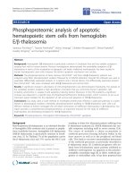
Báo cáo sinh học: " Phosphoproteomic analysis of apoptotic hematopoietic stem cells from hemoglobin E/b-thalassemia" pot
Bạn đang xem bản rút gọn của tài liệu. Xem và tải ngay bản đầy đủ của tài liệu tại đây (888.35 KB, 10 trang )
RESEA R C H Open Access
Phosphoproteomic analysis of apoptotic
hematopoietic stem cells from hemoglobin
E/b-thalassemia
Saranyoo Ponnikorn
1†
, Tasanee Panichakul
2†
, Kitima Sresanga
1
, Chokdee Wongborisuth
3
, Sittiruk Roytrakul
4
,
Suradej Hongeng
5*
and Sumalee Tungpradabkul
1*
Abstract
Background: Hemoglobin E/b-thalassemia is particularly common in Southeast Asia and has variable symptoms
ranging from mild to severe anemia. Previous investigations demonstrated the remarkable symptoms of b-
thalassemia in terms of the acceleration of apoptotic cell death. Ineffective erythropoiesis has been studied in
human hematopoietic stem cells, however the distinct apoptotic mechanism was unclear.
Methods: The phosphoproteome of bone marrow HSCs/CD34
+
cells from HbE/b-thalassemic patients was
analyzed using IMAC phosphoprotein isolation followed by LC-MS/MS detection. Decyder MS software was used to
quantitate differentially expressed proteins in 3 patients and 2 normal donors. The differentially expressed proteins
from HSCs/CD34
+
cells were compared with HbE/b-thalassemia and normal HSCs.
Results: A significant change in abundance of 229 phosphoproteins was demonstrated. Importantly, the analysis of
the candidate proteins revealed a high abundance of proteins that are commonly found in apoptotic cells
including cytochrome C, caspase 6 and apoptosis inducing factors. Moreover, in the HSCs patients a significant
increase was observed in a specific type of phosphoserine/threonine binding protein, which is known to act as an
important signal mediator for the regulation of cell survival and apoptosis in HbE/b-thalassemia.
Conclusions: Our study used a novel method to investigate proteins that influence a particular pathway in a given
disease or physiological condition. Ultimately, phosphoproteome profiling in HbE/b-thalassemic stem cells is an
effective me thod to further investigate the cell death mechanism of ineffective erythropoiesis in b-thalassemia. Our
report provides a comprehensive phosphoproteome, an important resource for the study of ineffective
erythropoiesis and developing therapies for HbE/b-thalassemia.
Keywords: Phosphoproteome, Hemoglobin E/β-thalassemia, HSCs/CD34
+
, apoptosis
Background
Beta-thalassemia (b-thalassemia) is an inherited disorder
of hemoglobin (Hb) synthesis and is present in popula-
tions worldwide however, hemoglobin E/b-thalassemia
(HbE/b-thalassemia) is par ticularly common in South-
east Asia [1]. In Thailand, 7% of the population carries
the b-thalassemia trait and 17% carrie s the Hb E trait
resulting in thirty-five thousand pa tients suffering from
b-thalassemia syndrome [2]. HbE/b-thalassemia has vari-
able symptoms including mild to severe anemia [3,4].
The excess of insoluble a chains accumulates in
erythroid precursors forming inclusion bodies in the
bone marrow as well as in the peripheral red blood
cells. This leads to excessive intramedullary destruction
of the eryt hroid precursors as previously described in
b-thalassemia major [5]. Erythroid progenitor cells iso-
lated from b-thalassemia major patients exhibited an
ineffective erythropoiesis via apoptosis at t he polychro-
matophilic normoblast stage [6] in either erythroid pro-
genitor or erythroblast cells. It has been proposed that
* Correspondence: ;
† Contributed equally
1
Department of Biochemistry, Faculty of Science, Mahidol University,
Bangkok, Thailand
5
Department of Pediatrics, Faculty of Medicine Ramathibodi Hospital,
Mahidol University, Bangkok. Thailand
Full list of author information is available at the end of the article
Ponnikorn et al. Journal of Translational Medicine 2011, 9:96
/>© 2011 Ponnikorn et al; licensee BioMed Central Lt d. This is an Open Access article distributed under the terms of the Creative
Commons Attribution Licen se ( es/by/2.0), which permits unrestricted use, distribution, and
reprodu ction in any mediu m, provided the original work is properly cited.
the precipitation of excess a-globinchainsinmarrow
erythroid precursors as well as an excess of free iron
could lead to oxidative stress a nd potentially to ineffec-
tive erythropoiesis [7,8]. However, the mechanism
responsible for induction of apoptosis and the relevant
signaling pathways in hematopoietic stem cells are still
poorly understood. Hematopoietic stem cells (HSCs)
contain specific markers on their surface such as CD34
and CD133 allowing for the isolation of relatively pure
populations of these cells for in vitro study of the cellu-
lar processes in thalassemic stem cells. While previous
attempts have focused on the cell culture system, in this
study the phosphoproteome of the primary cells from
the immediate collection of HSCs from patien t blood or
bone marrow was analyzed.
Mass spectrometry-based proteomics is an alternative
method for the analysis of protein profiles and provides an
extensive approach to analyze expressed proteins and dis-
cover potential biomarkers for diseases. The development
of MS based technology has provided the opportunity to
investigate cellular signaling mechanisms in terms of the
phosphoproteome, the phosphopeptides or phosphopro-
teins present within a cell [9]. Protein phosphorylatio n is
present on more than 30% of the proteome and regulates
signal transduction pathways under normal conditions as
well as in disorders such as diabetes, neurodegenerative
diseases, autoimmune diseases and several forms of
cancers [10]. Phosphorylatio n and dephosphorylation of
specific amino acid residues, including serine, tyrosine and
threonine, by specific kinases and phosphatases is a major
form of post-translational modification in eukaryotic cellu-
lar machinery and can be regulated to occur at a particular
time or in response to other stimuli. The levels of protein
phosphorylation vary widely and specific sites may be
phosphorylated from less than 1% to greater than 90%
[11]. The global identification and characterization of
phosphorylation is critical to the elucidation of signal
transduction pathways, the understanding of the mechan-
ism of disease progression, and the development of thera-
peutic applications [12]. The phosphoproteome of HSCs
in HbE/b-thalassemic patients has not been previously
investigated and analysis of changes in phosphorylation
patterns may help in the understanding of the mechanisms
that participate in apoptotic cell death leading to the inef-
fective erythopoiesis in bone marrow cells. Here, we
described the first phosphoproteome analysis of HSCs
from HbE/b-thalassemic patients using IMAC phospho-
protein isolation directly coupled with LC MS/MS analy-
sis. Compared to healthy donors, HSCs from HbE/
b-thalassemic bone marrow patients expressed high levels
of several phosphoproteins, suggesting a role for these
proteins in disease. These proteins were identified and
categorized with regard to both the intrinsic and extrinsic
pathway apoptotic pathways. Our results suggest that the
14-3-3 proteins might play a role in regulating apoptosis
of HSCs in HbE/b-thalassemia.
Experimental design an d methods
Samples of bone marrow
Bone marrow samples wer e obtained from three HbE/b-
thalassemic patients admitted to Ramathibodi Hospital,
Bangkok, Thailand and two normal donors. All patients
were children 3-12 years-old and had symptoms of severe
anemia and hepatosplenomegaly. Hemoglobin analysis
showed no expression of the b-hemoglobin band with the
Hb-E trait. Twenty milliliters of marrow was aspirated
from the posterior iliac crest into syringes containing 0.1
ml of heparin and then shipped to the laboratory for stem
cell isolation. Marrow collection was approved by the Ethi-
cal Committee of Research on Human beings at Ramathi-
bodi Hospital, Faculty of Medicine, Mahidol Uni versity,
Bangkok, Thailand and normal donors gave informed con-
sent. All patients had not received blood transfusion
within three months before they participated in the study.
Isolation and culture of hematopoietic stem cells (HSCs)
Bone marrow samples (BM) from Hb E/b-thalassemic
patients and healthy donors were collected for HSCs iso-
lation. Briefly, mononucl ear cells (MNCs) from BM were
separate d by using Isoprep™ solution (Lexis, Sweden).
The MNCs were used to isolate the HSCs with a CD 34
isolation kit with magnetic microbead selection (Mini-
MACS columns, Miltenyi Biotech, Germany). The
method of isolation was performed as described in the
manufacturer’s protocol. 1-2 × 10
8
cellsofMNCswere
reacted with 100 μl of anti-CD 34 antibody coated with
micro beads and 100 μl of anti-FcR antibody, at 4°C, for
30 min. After washing with PBS/2 mM EDTA/0.5%BSA
buffer, cells were applied to a wet magnetic column.
Next, buffer was added onto the column to remove non-
binding cells. This s tep was repeat ed 4-5 times and CD
34
+
cells were then flushed from the column. The iso-
lated HSCs/CD34
+
cells were harvested for phosphopro-
teomic analysis and cultivation of erythroid cells. The
cultivation of erythroid cells derived from HSCs/CD34
+
cells was performed according to a procedure previously
described [13] with some modifications. HSCs/CD34
+
cells were cultured in StemlineII medium (Sigma-
Aldrich Corporation, Missouri , USA) supplemented with
100 ng/ml stem cell factor (SCF) (PeproTech, Rocky Hill,
NJ, USA), 5 ng/ml IL-3 (R& D Systems, Inc., MN, USA),
10 μM hydroco rtisone (Sigma-A ldrich), 100 μg/ml trans-
ferrin (Sigma-Aldrich), 100 μg/ml Humulin
®
N(Lilly
PharmaFertigung UND Distribution Giessen, Germany),
0.18 μg/ml ferrous sulfate (Sigma-Aldrich), 0.16 mM
monothioglycerol (Sigma -Aldrich), and 4 IU/ml erythro-
poietin (EPO; CilagAG International, Zug, Switzerland).
All cultures were incubated at 37°C with 5% CO
2
.After
Ponnikorn et al. Journal of Translational Medicine 2011, 9:96
/>Page 2 of 10
culturing for 4 and 7 days, cells were collected for identi-
fication of erythroid markers and cell morphology. Cell
surface membrane markers were analyzed to confirm cell
types of HSCs and erythroid cells. Cell surface markers
were detected with mouse anti-CD 34, 71, 45, and glyco-
phorin A antibodies conjugated with fluorescent dye fol-
lowed by analysis using flow cytometry (Beckman
Coulter, USA). Cells were stained with Giemsa to visua-
lize the morphology of erythroid cells.
Phosphoprotein analysis
Phosphoproteins of HSCs/CD34
+
cells isolated from HbE/
b-th alassemia patients and normal donors were analyzed
in parallel by liquid chromatography in line with tandem
MS mass spectrometry (Bruker, Germany). Briefly, freshly
isolated CD34
+
cells were washed three times with phos-
phate-buffer saline (PBS), dissolved in lysis buffer contain-
ing 0.15 M NaCl, 5 mM EDTA, 1% Triton X100, 10 mM
Tris-Cl, pH 7.4, 10 mM b-glycerophosphate, 25 mM NaF,
1mMNa
3
VO
4
, 100 mM PMSF and complete protease
inhibitor cocktail (Sigma-Aldrich), and then sonicated for
30 s. After centrifugation at 8,000 rpm for 15 min, the
supernatant was collected and cell lysates were stored at
-80°C until used. The phosphoproteins were enriched by
immobilized metal affinity column (IMAC) (Pierce,
Thermo Scientific, USA) according to the manufacturer’s
protocol. Phosphoproteins were reduced with 10 mM
DTT, alkylated with 55 mM iodoacetamide, and digested
with sequencing grade trysin (Promega, Germany) for
16 h at 37°C. Tryptic peptides were then concentrated by
SpeedVac centrifugation and stored at -80°C prior to use.
The iodoacetamide modified phosphopeptides were dis-
solved in 250 mM acetic acid/30% acetronitrile and pro-
tein concentration was determined by Lowry method [14].
Phosphope ptide sample s were injected into a Ultimate
3000 LC System (Dionex, USA) coupled to ESI-Ion Trap
MS (HCT Ultra PTM Discovery System, Bruker, Ger-
many) with electrospray at a flow rate of 300 nl/min to a
nanocolumn (Acclaim PepMap 100 C18, 3 μm, 100 A,
75 μm id × 150 mm). A Solvent gradient (Solvent A: H
2
O,
0.1% Formic acid; Solvent B: 20% H
2
O, 80% Acetronitrile,
0.1% Formic acid) was used with the following parameters:
10% - 70% B at 0-13 min, 90% B at 13-15 min and 10% B
at 15-20 min. The resolution in MS step is 0.6 and the
mass accuracy is 0.15 u (m/z).
Proteins quantitation and identification
DeCyder MS Differential Analaysis software (GE Health-
care, USA) [15] was used for the differential quantitation
of proteins and peptides based on MS signal intensities
of individual LC-MS analyses . To evaluate the average
abundance ratio of peptides from patients and normal
donors quantitation of phosphopeptides, based on the
peptide signal intensit ies, was performed using the
Pepdetect module allowing for automated detection of
peptides and assignment of charge states. Peptides were
matched across different sign al intensity maps between
the patients and normal donors using the Pepmatch
module. The relative abundances of peptides were
expressed as
2
log intensities with mass tolerance set to
0.5 amu and the retention time tolerance set to 2 min.
All
2
log intensities of patients were normalized with the
ion intensity distribution of normal donors. An average
abundance ratio > 2 fold was determined to be an over-
expressed protein with a significant standard t-test
p-value < 0.05.
The MS/MS data from DeCyderMS analysis w as
searched against the SwissProt database (SwissProt 05
516,603 sequences; 181,919,312 residues) for protein
identification using Mascot software (Matrix Science,
London, UK) [16]. Database interrogation was: taxon-
omy (Human or Eukaryote); enzyme (trypsin); variable
modifications (carbamidomethyl, oxidation of methio-
nine residues, phospho ST and phospho Y); mass values
(monoisotopic); protein mass (unrestricted); peptide
mass tolerance (2 Da); fragment mass toleranc e (± 2
Da), peptide charge state (1+, 2+ and 3+) and three
missed cleavages. Proteins considered as identified pro-
teins had at least two peptides with an individual mascot
score corresponding to p < 0.05 and p<0.1,respec-
tively. A mowse score of 20 or more was c onsidered
acceptable for proteins under MASCOT identification.
Bioinformatic analysis
The phosphorylation sites of phosphopeptides from MS
spectra were compared with the NetPHOS [17]http://
www.cbs.dtu.dk/services/NetPhos/ phosphorylation site
prediction for specific residues p-Tyr, p-Ser and p-Thr.
The preferential phosphorylated residues were deter-
mined and the score with more than the threshold of all
possible phosphorylation motifs was considered. The
matching residues of selected peptides were reported and
compared with the Phosphosite Plus database http://
www.phosphositeplus.org to search for phosphorylation
residues that have been previously investigated. Gene
ontology and cellular pathway identification were ana-
lyzed using PANTHER and
UniprotKB databases.
Results
HSCs/CD34
+
cells were isolated from 5 bone marrow
samples belonging to 3 Hb E/b-thalassemic patients and
2 normal healthy donors. Flow cytometric analysis of
confirmed that the isolated cells highly expressed the cell
surface marker CD34 and had low levels of CD45 and
were negative for glycophorin A. The p urity of the iso-
lated CD34
+
cells was 92% (data not shown). CD34
+
cells
from patients and donors were cultured and cell viability
Ponnikorn et al. Journal of Translational Medicine 2011, 9:96
/>Page 3 of 10
was determined after 4 a nd 7 days of cultivation. The
growth rate of thalassemic cells was reduced compared
to normal cells (Figure 1). Giemsa staining revealed that
CD34
+
cells from patients and donors had similar mor-
phological character istics to bl ast cells with a lar ge
nucleus and 2-3 nucleoli. The characteristics of apoptotic
cells, including membrane blebbling and nuclear frag-
mentation, were only found in thalassemic cells at days 4
and 7 (Figure 2).
After LC-MS/MS analysis of the isolated phosphopep-
tides, all MS/MS spectra were searched against human
protein database. A total of 347 peptides from CD34
+
cells
were detected and found to correspond to 229 proteins
which w ere encoded by 226 genes (additional file 1). Of t he
347 unique peptide identifications, 204 phosphopeptides
(58.79%) with 306 phosphorylation sites were identified.
The specific amino acid residues of pSer:pThr:pTy r were
represented as 67:31:2. The biological characterization of
phosphoproteome in CD34
+
cells could be classified
according to biological process, molecular function and
cellular localization (Figure 3). The subcellular protein
localization for identified phosphoproteins was available
and included several cellular compartments. As expected
in an investigation of the proteome, we identified an abun-
dance of cytosol (33%), nucleus (24%) and membrane
proteins (23%). The gene ontology analysis of our phospho-
proteome revealed several proteins involved in basic mole-
cular functions such as transcription/translation factors
(32%), DNA/RNA binding (19%) and catalytic activity
(14%). In addition to the biological characterization, the
proteins could be identified specifically in metabolic pro-
cesses (35%) and signal transduction (16%). Moreover,
these proteins were categorized by PANTHER cellular
pathway classification together with Uniprot gene ontology
to match with 5 cellular pathways by various relationships
such as protein interactions, modifications and regulation
of expressions. However, this does not mean that all pro-
tein interactions are directly involved with a certain cellular
pathway, but it enables identification of those biochemical
pathways that are possibly altered in the HbE/b-thalasse-
mic stem c ell. In this study, five selected signaling pathways
were represented in Table 1 with protein expression ratios
between patients and normal donors greater than 2:1.
These proteins were categorized as hematopoietic related
proteins and other cellular pathway proteins (Table 1 and
additional file 2).
Interestingly, Several proteins that were over-expressed
in patients were identified as apoptotic proteins. Of
these, five proteins were characterized as common in the
apoptotic pathway including cytochrome C, apoptosis
inducing factor 1 (AIFM1), caspase 6, tumor necrosis fac-
tor ligand 6 (TNFL 6) and tumor necrosis factor receptor
super family 12A (TNFR 12A or TWEAK). These pro-
teins were involved in both mitochondrial dependent
apoptosis (intrinsic mechanism) and death receptor
mediated apoptosis (extrinsic mechanism). Previous evi-
dence suggests that the pathology and disease progres-
sion of b-thalassemia relates to the accumulation of
reactive oxygen species (ROS), which can generate DNA
adducts in the nucleus and induce the DNA damage
pathway. In this study, six proteins encoded by four
genes were identified and found to be responsible for
DNA damage and responsive mechanisms such a s the
p53 pathway. Interestingly, we found the isoforms sigma,
zeta/delta and gamma of the 14-3-3 protein were over-
expresse d in patients more than 2 fold as compared with
normal donors. These 14-3-3 proteins are involved in
cellular pathways including apoptosis and phosphatidyl
inositol 3 kinase activity by PANTHER pathway analysis.
A schematic diagra m of the apoptotic pathway including
the identified phosphoproteins in HbE/b-thalassemic
stem cells was proposed (Figure 4).
Discussion
Conventional methods typically have difficulty using
fresh human hematopoietic stem cells for studying cell
signaling in the apoptotic pathway because of the lim-
ited amount of sample from patients and normal
donors. On the other hand, in vitro culture is capable of
sustaining a large number of cells, which can confer
specific advantages and disadvantages. The use of freshly
isolated human hematopoietic st em cells from thalasse-
mic patients and normal donors to specifically investi-
gate apoptotic mechanisms by avoiding the culture
system was achieved in this study using phosphoproteo-
mic technology. The IMAC technique indicated more
than 50% phosphopeptide specificity, which is similar to
that previously described [18 ]. Likewise, several studies
have reported that direct phosphoproteins/peptides
0
1
2
3
4
5
6
7
8
Day 0
Day 4
Day 7
Log number of viable cell
Figure 1 Viability of HSCs/CD34
+
cells from HbE/b-thalassemic
patients. Cell numbers from 0, 4 and 7 day-old cultures were
examined by trypan blue exclusion, numbers of viable cells from
normal donors (grey) and HbE/b-thalassemic patients (black).
Ponnikorn et al. Journal of Translational Medicine 2011, 9:96
/>Page 4 of 10
enrichment with TiO
2
could improve the sensitivity and
efficacy of phosphoproteome study [18,19]. Decyder MS
is an alternative approach to quantitative proteomics
based on LC-MS/MS analysis of peptides without incor-
porating an isotopic label. The matching of peptides
across patients and normal donors requires accurate
mass and reproducible retention times [15] and some
quantitative proteomic techniques have been proposed
[20].
Ineffective erythropoiesis is one important complica-
tion in thalassemic disease that contributes to the abnor-
mal erythroid cell expansion and differentiation in bone
marrow [21,22]. Apoptotic cell death has been described
as the major contributor as seen in b-thalassemic bone
marrow [5] and even in peripheral blood stem cells in
vitro [23]. The bone marrow of patients with b-thalasse-
mia contains five to six times the number of erythroid
precursors as the healthy controls [24]. The acceleration
of apoptosis and ineffective erythropoiesis in erythroid
precursor cells and HSCs/CD3 4
+
has been observed with
apoptotic cells present at 15 times greater levels in b-tha-
lassemic patients compared to normal donors [5,7]. The
acceleration of apoptosis has been characterized in ery-
throid cells at the polychro matophilic and orthochromic
nomoblast stages dur ing erythroid differentiat ion [6].
However, using a thalassemic mouse model of b-thalasse-
mia major and thalassemia intermedia, it was suggested
that apoptosis and hemolysis were not the major causes
for the ineffective erythropoiesis. Results suggested that
an increase in erythroid cell pro liferation but not cell dif-
ferentiation contributed to the ineffective erythopoiesis
[25]. However, it remains unclear whether the expansion
of erythroid cells but not cell diff erentiation can directly
or indirectly contribute to the acceleration of apoptosis
in b-thalassemia. Our study in HbE/b-thalassemic HSCs
has shown that the grow th reduction of erythroid cells
derived from HbE/b-thalassemic HSCs was substantially
due to induction of apoptosis. This was supported by the
observation of characteristic apoptotic cell morphology
and phosphoproteomic analysis of HbE/b-thalassemia.
Our phosphoproteome data has i dentified specific apop-
totic related proteins from HSCs/CD34
+
cells in HbE/b-
thalassemia. We categorized candidate proteins into two
major groups. First is the common apoptotic proteins
associated with the extrinsic and intrinsic cell death path-
way. Another is a non-distinct mechanism in both cell
death pathways that has been addressed in several stu-
dies. Cytochrome C and apoptosis inducing factor 1
(AIFM1) a re the main common components of the
intrinsic and the mitochondria dependent apoptotic
pathway. The activation of v arious death stimuli triggers
the localization of pro-apoptotic proteins Bcl-2 and BH3
family into mitochondria [26,27]. Consequently, the
release of cytochrome C mediates apoptosome formation
and effective caspase cascade activation. Additionally,
AIFM1 is activated by the same response that elicits
A.
B
.
Day 0 Day 7Day 4
Figure 2 Cultures of HSC/CD34
+
from HbE/b-thalassemic patients (A) and normal donors (B).After7days,theHbE/b thlassemic cell
developed to erythroblasts and showed characteristic cell morphology of cells undergoing apoptosis including cell membrane blebbing and
nuclear fragmentation.
Ponnikorn et al. Journal of Translational Medicine 2011, 9:96
/>Page 5 of 10
chromatin condensation in the nucleus [28-30]. In addi-
tion, a death receptor mediated pathway seems to be
implicated in apoptosis during erythropoiesis with Fas-
Fas ligand interactions [31,32]. We have identified addi-
tional factors in the death receptor mediated pathway
including the tumor necrosis factor ligand member 6
(TNFL 6), tumor necrosis factor receptor superfamily
member 12A (TNFRST 12A) and caspase 6. Both TNFL
6 and TNFRSF 12A are involved in FAS mediated apop-
tosis or death receptor medi ated apop tosis [33,34], while
caspase 6 is generally considered as an effective caspase
that is cleaved by caspase-3 after the activation of the
caspase cascade [35]. Caspase 6 has an important role in
the regulation of chromatin condensation through the
cleavage of nuclear laminar [36]. Interestingly, we have
also identified lamin A or nuclear lamin A (LMNA),
which is a component of nuclear laminar proteins, in our
analysis. A second group of apoptotic related proteins
was also over-expressed in patients. For example, FOXA1
(hepatocyte nuclear factor alpha) is a transcription factor
that is involved in embryonic development and regula-
tion of gene expression in differentiated tissues [37].
FOXA1 is involved in regulating apoptosis by inhibiting
expression of the anti-apoptotic protein Bcl-2 [38].
Another protein highly expressed is sphingosine 1 phos-
phate layase 1 (S1P), one type of sphingosine metabolite.
S1P plays an important role in regulation of cell prolif-
eration, survival and death [39] and is generated during
lipid peroxidation by reactive oxygen species (ROS) accu-
mulation inside the cell [40]. Increasing of S1P or endo-
genous sphingosine levels triggers multiple mechanisms
to induce apoptosis through the activation of caspases
[40]. The accumula tion of ROS is a common complica-
tion in b-thalassemia. ROS was reported to induce lipid
peroxidation that contributes to DNA damage [41,42].
Nevertheless, in vitro studies in erythroid cells have
demonstrated that particular pathways are involved directly
or indirectly in apoptosis and ineffective erythropoiesis in
HbE/b-thalassemia [23,43]. However, the upstream signal-
ing events in the progression of b-thalassemia have not
been identified. Our investigation of the phosphoproteome
was performed using a novel procedure wi th freshly iso-
lated HSCs/CD34
+
cells, allowing for analysis of cell signal-
ing. The related cellular pathways could be identified and
A. B.
C.
Figure 3 Pie chart representing the characterization of identified phosphoproteins according to (A) biological processes, (B) molecular
functions and (C) cellular localization.
Ponnikorn et al. Journal of Translational Medicine 2011, 9:96
/>Page 6 of 10
their possible roles in the pathogenesis of b-thalassemia
could be e xamined. Phosphoproteins are known to m ediate
various pathways and interestingly, we identified 14-3-3
isoforms sigma, gamma and zeta/delta that are linked to
three cellular pathways in cluding p53, phosphatidyl inositol
3 (PI3) kinase, and apoptosis. The 14-3-3 proteins are a
family of multifunctional phosphoserine/phosphothreonine
binding molecules and are involved in various cellular pro-
cesses including cell survival, cell cycle progression and
apoptosis [44,45]. Ultimately, the regulation of 14-3-3
Table 1 The selected relative abundance protein expression.
Gene Name Description Expression Ratio *
Apoptosis-related protein
AIFM1_HUMAN Apoptosis-inducing factor 1, mitochondrial 1.125
APC2_HUMAN Adenomatous polyposis coli protein 2 2.315
CASP6_HUMAN Caspase 6 5.559
CTBL1_HUMAN Beta-catenin-like protein 1 1.127
CYC_HUMAN Cytochrome c 9.480
FOXA1_HUMAN Hepatocyte nuclear factor 3-alpha 4.021
LMNA_HUMAN Lamin-A/C 1.120
MAP2_HUMAN Microtubule-associated protein 2 1.07
PRKDC_HUMAN DNA-dependent protein kinase catalytic subunit 3.423
RN5A_HUMAN 2-5A-dependent ribonuclease 1.41
SGPL1_HUMAN Sphingosine-1-phosphate lyase 1 2.305
ST17A_HUMAN Serine/threonine-protein kinase 17A 1.137
TNFL6_HUMAN Tumor necrosis factor ligand superfamily member 6 0.9352
TNR12_HUMAN Tumor necrosis factor receptor superfamily member 12A 1.094
TAU_HUMAN Microtubule-associated protein tau 5.023
p53 signaling pathway
1433S_HUMAN 14-3-3 protein sigma 2.274
1433Z_HUMAN 14-3-3 protein zeta/delta 2.907
1433G_HUMAN 14-3-3 protein gamma 2.299
BC11B_HUMAN B-cell lymphoma/leukemia 11B 1.125
SHSA5_HUMAN Protein shisa-5 1.143
CTBL1_HUMAN Beta-catenin-like protein 1 1.172
Ubiquitin proteasome pathway
HUWE1_HUMAN E3 ubiquitin HUWE1 3.477
SMUF1_HUMAN E3 ubiquitin SMUF1 1.099
CBL_HUMAN E3 ubiquitin CBL 5.746
DCA15_HUMAN DDB1 2.290
OTU7A_HUMAN OTU domain 1.071
HERC3_HUMAN Probable E3 ubiquitin HERC3 4.907
UBQL1_HUMAN Ubiquilin 1.275
PSA7_HUMAN Proteasome subunit alpha type 1.161
WNT signaling pathway
APC2_HUMAN Adenomatous polyposis coli protein 2 2.315
PCDH7_HUMAN Protocadherin-7 2.252
HXA7_HUMAN Homeobox protein Hox-A7 1.602
WNT8B_HUMAN Protein Wnt-8b 1.094
LRP5_HUMAN Low-density lipoprotein receptor-related protein 5 1.263
PI3 Kinase signaling pathway
FOXF2_HUMAN Forkhead box protein F2 2.273
FOXA1_HUMAN Hepatocyte nuclear factor 3-alpha 4.021
1433S_HUMAN 14-3-3 protein sigma 2.274
1433Z_HUMAN 14-3-3 protein zeta/delta 2.907
1433G_HUMAN 14-3-3 protein gamma 2.299
*: Decyder MS analysis at significant t-test p < 0.05 was performed the peptide intensity. The relative expression ratio was derived by the peptide ion intensity of
patient normalized with normal donor.
Ponnikorn et al. Journal of Translational Medicine 2011, 9:96
/>Page 7 of 10
function is controlled by various upstream signal transduc-
tion pathways in particular conditions such as DNA
damage and oxidative stress pro mote phosphoprylation/
dephosphorylation of specific site [46,47]. In t he case of
apoptosis, the cytosolic 14-3-3 protein down regulates the
p53 pathway and the JNK signaling pathway [44,45] but
mediates the mitochondrial dependent apoptotic pathway.
JNK promotes the pro-apoptotic protein, Bax, which is
translocated to the mitochondria through phosphorylation
of 14-3-3, a cytoplasmic anchor of Bax. Phosphorylation of
14-3-3 leads to dissociation of Bax from this protein and
induces the mitochondrial dependent apoptotic pathway
through cytochrome C release followed by effective caspase
activation [48]. Besides the pro-apoptotic proteins, the 14-
3-3 protein also regulates the localization of other apopto-
tic signa ling proteins such as c-Abl. In response to DNA
damage, JNK induces phosphorylation of the 14-3-3 pro-
tein and releases the binding of c-Abl from cytosol to the
nucleus [49]. In the case of hematopoietic stem cell home-
ostasis, 14-3-3 responds to various physiological stimuli
that can contribute to cell survival as well as death. The
14-3-3 protein binds to the FoxO protein, one member of
the forkhead transcription factors. Specifically, FoxO3 and
FoxO4 are down regulated by 14-3-3, which retains FoxO
in the c ytosol under normal physiological conditions. U pon
DNA damage and oxidative stress, JNK can phosphorylate
the 14-3-3 protein and leads to the dissociation of both
FoxO proteins from their partner [48,50]. Consequently,
FoxO3 can localize to the nucleus and regulate expression
of Bim and FasL, w hich play an important role i n apoptotic
cell death in HSCs [51-54]. The 14-3-3 protein was also
found in stem cell disease using the proteomics approach
[55]. The investigation of bone marrow and peripheral
blood blast cells in acute amyloi d leukemia (AML) by 2-DE
revealed the expression of 14-3 -3 related proteins. More-
over, another study in leukemic stem cells identified the
14-3-3 phosphor ylati on site motif in candidate phospho-
peptides. The results from SCANSITE analysis indicate
that most phosphopeptides are phosphorylated by a kinase
14-3-3 module. Therefore, it was proposed that 14-3-3
may regulate the oncogenic pathway in leukemic stem cell
disease [19]. A recent study of 14-3- 3 protein-protein inter-
action by quantitative mass spectrometry in multiple mye-
loma revealed novel 14-3-3 zeta interacting pro teins that
may have various biological functions and may regulate the
activity of apopto tic proteins suc h as Bax, cytochrom e C
and caspase 6 [56]. Therefore, 14-3-3 zeta is likely to play
an important role i n apoptosis.
Conclusions
We have observed the over-expression of the 14-3-3
protein isoforms sigma, gamma and zeta/delta in HbE/
b-thalassemic stem cells. The 14-3-3 protein may be a
critical mediator of the signal ing pathway that regulates
between cell survival and death due to ineffective ery-
thropoiesis in b-thalassemic patients. Finally, this study
Figure 4 Schematic diagram of candidate phosphoproteins implicated in apoptotic pathways. The color pictures represent proteins that
were identified in this study, the grey pictures are proteins that have been previously identified.
Ponnikorn et al. Journal of Translational Medicine 2011, 9:96
/>Page 8 of 10
has shown that a significant amount of common apop-
totic proteins are found in the patients with HbE/b-tha-
lassemia and these proteins correspond to the disease
complications in apoptosisofHSCsinpatients.These
experiments used a novel method to understand the
proteins that influence a particular pathway in a given
disease or physiological condition. Ultimately, our
results demonstr ate that phosphoproteome profiling in
HbE/b-thalassemic stem cells is an effective technique
to investigate the cell death mechanism of ineffective
erythropoiesis in b-thalassemia.
Additional material
Additional file 1: Table containing the 229 proteins with MS
identified phosphorylation site and the comparison of
phosphorylation site using bioinformatic analysis.
Additional file 2: Table indicating the cellular pathway analysis of
identified proteins in this study.
Abbreviations
HSCs/CD34
+
: CD34
+
hematopoietic stem cells; HbE/β-thalassemia:
hemoglobin E beta thalassemia; IMAC: immobilized metal affinity column;
LC-MS/MS: liquid chromatography with tandem mass spectrometry;
PANTHER: Protein analysis through evolutionary relationshi ps.
Acknowledgements
This work was supported by the Research Matching Fund from Faculty of
Science and Faculty of Medicine Ramathibodi Hospital, Mahidol University.
Ponnikorn, S. was supported by Strategic Scholarship Fellowships Frontier
Research Network from Thai Commission on Higher Education. The authors
acknowledge Janthima Jaresitthikunchai and Narumon Phaonakrop, BIOTEC,
NSTDA for MS data interpretation and Decyder program tutorial. Dr. Samart
Pakakasama, Department of Pediatrics, Faculty of Medicine Ramathibodi
Hospital. Department of Pathobiology for other equipments and facilities.
Dr. Laran Jensen, Department of Biochemsitry, Faculty of Science, Mahidol
University and Daniel B. Janiczak, University of Miami in Florida for their
critical reading in this manuscript.
Author details
1
Department of Biochemistry, Faculty of Science, Mahidol University,
Bangkok, Thailand.
2
Faculty of Science and Technology, Suan Dusit Rajabhat
University, Bangkok, Thailand.
3
Research Center, Faculty of Medicine
Ramathobodi Hospital, Mahidol University, Bangkok, Thailand.
4
National
Center for Genetic Engineering and Biotechnology, National Science and
Technology Development Agency, Pathumthani, Thailand.
5
Department of
Pediatrics, Faculty of Medicine Ramathibodi Hospital, Mahidol University,
Bangkok. Thailand.
Authors’ contributions
SP - performed experiments, interpreted results and drafted manuscript, TP -
performed experiments, interpreted results and drafted manuscript, KS -
performed bioinformatic analysis, CW - performed flow cytometry analysis,
SR - designed and interpreted MS data, SH - designed, collected and
interpreted clinical data, ST- designed and interpreted experiment, prepared
final manuscript. All authors read and approved the final manuscr ipt.
Competing interests
The authors declare that they have no competing interests.
Received: 4 March 2011 Accepted: 25 June 2011
Published: 25 June 2011
References
1. Abetz L, Baladi JF, Jones P, Rofail D: The impact of iron overload and its
treatment on quality of life: results from a literature review. Health Qual
Life Outcomes 2006, 4:73.
2. Thai Thalassemia Foundation: Diagnosis of thalassemia carrier (in Thai
language).[ />3. Premawardhena A, Fisher CA, Olivieri NF, de Silva S, Arambepola M, Perera W,
O’Donnell A, Peto TE, Viprakasit V, Merson L, Muraga G, Weatherall DJ:
Hemoglobin E beta thalassaemia in Sri Lanka. Lancet 2005, 366:1467-1470.
4. Winichagoon P, Fucharoen S, Chen P, Wasi P: Genetic factors affecting
clinical severity in beta-thalassemia syndromes. J Pediatr Hematol Oncol
2000, 22:573-580.
5. Yuan J, Angelucci E, Lucarelli G, Aljurf M, Snyder LM, Kiefer CR, Ma L,
Schrier SL: Accelerated programmed cell death (apoptosis) in erythroid
precursors of patients with severe beta-thalassemia (Cooley’s anemia).
Blood 1993, 82(2):374-377.
6. Mathias LA, Fisher TC, Zeng L, Meiselman HJ, Weinberg KI, Hiti AL, Malik P:
Ineffective erythropoiesis in beta-thalassemia major is due to apoptosis
at the polychromatophilic normoblast stage. Exp Hematol 2000,
28(12):1343-1353.
7. Pootrakul P, Sirankapracha P, Hemsorach S, Moungsub W, Kumbunlue R,
Piangitjagum A, Wasi P, Ma L, Schrier SL: A correlation of erythrokinetics,
ineffective erythropoiesis, and erythroid precursor apoptosis in thai
patients with thalassemia. Blood 2000, 96(7):2606-2612.
8. Kong Y, Zhou S, Kihm AJ, Katein AM, Yu X, Gell DA, Mackay JP, Adachi K,
Foster-Brown L, Louden CS, Gow AJ, Weiss MJ: Loss of alpha-hemoglobin-
stabilizing protein impairs erythropoiesis and exacerbates beta-
thalassemia. J Clin Invest 2004, 114(10):1457-1466.
9. Choudhary C, Mann M: Decoding signalling networks by mass
spectrometry-based proteomics. Nat Rev Mol Cell Biol 2010, 11(6):427-439.
10. Mukherji M: Phosphoproteomics in analyzing signaling pathways. Expert
Rev Proteomics 2005, 2(1):117-128.
11. Macek B, Mann M, Olsen J: Global and Site-Specific Quantitative
Phosphoproteomics: Principles and Applications. Annu Rev Pharmacol
Toxicol 2009, 49:199-221.
12. Schmelzle K, White FM: Phosphoproteomic approaches to elucidate
cellular signaling networks. Curr Opin Biotechnol 2006, 17(4):406-414.
13. Panichakul T, Sattabongkot J, Chotivanich K, Sirichaisinthop J, Cui L,
Udomsangpetch R: Production of erythropoietic cells in vitro for
continuous culture of Plasmodium vivax. Int J Parasitol 2007,
37(14):1551-1557.
14. Lowry OH, Rosebrough NJ, Farr AL, Randall RJ: Protein measurement with
the Folin phenol reagent.
J Biol Chem 1951, 193(1):265-275.
15. Johansson C, Samskog J, Sundström L, Wadensten H, Björkesten L,
Flensburg J: Differential expression analysis of Escherichia coli proteins
using a novel software for relative quantitation of LC-MS/MS data.
Proteomics 2006, 6(16):4475-4485.
16. Perkins DN, Pappin DJ, Creasy DM, Cottrell JS: Probability-based protein
identification by searching sequence databases using mass
spectrometry data. Electrophoresis 1999, 20(18):3551-3567.
17. Blom N, Gammeltoft S, Brunak S: Sequence and structure-based
prediction of eukaryotic protein phosphorylation sites. J Mol Biol 1999,
294(5):1351-1362.
18. Carrascal M, Ovelleiro D, Casas V, Gay M, Abian J: Phosphorylation analysis
of primary human T lymphocytes using sequential IMAC and titanium
oxide enrichment. J Proteome Res 2008, 7(12):5167-5176.
19. Ge F, Xiao CL, Yin XF, Lu C, Zeng H, He QY: Phosphoproteomic analysis of
primary human multiple myeloma cells. J Proteomics 2010, 73(7):1381-1390.
20. Schulze W, Usadel B: Quantitation in Mass-Spectrometry-Based
Proteomics. Annu Rev Plant Biol 2010, 61:491-516.
21. Rivella S: Ineffective erythropoiesis and thalassemias. Curr Opin Hematol
2009, 16(3):187-194.
22. Rund D, Rachmilewitz E: Beta-thalassemia. N Engl J Med 2005,
353(11):1135-1146.
23. Lithanatudom P, Leecharoenkiat A, Wannatung T, Svasti S, Fucharoen S,
Smith DR: A mechanism of ineffective erythropoiesis in beta-
thalassemia/Hb E disease. Haematologica 2010, 95(5):716-723.
24. Centis F, Tabellini L, Lucarelli G, Buffi O, Tonucci P, Persini B, Annibali M,
Emiliani R, Iliescu A, Rapa S, Rossi R, Ma L, Angelucci M, Schrier SL: The
importance of erythroid expansion in determining the extent of
Ponnikorn et al. Journal of Translational Medicine 2011, 9:96
/>Page 9 of 10
apoptosis in erythroid precursors in patients with beta-thalassemia
major. Blood 2000, 96(10):3624-3629.
25. Libani I, Guy E, Melchiori L, Schiro R, Ramos P, Breda L, Scholzen T,
Chadburn A, Liu Y, Kernbach M, Baron-Luhr B, Porotto M, Sousa M,
Rachmilewitz EA, Hood JD, Cappellini D, Giardina PJ, Grady RW, Gerdes J,
Rivella S: Decreased differentiation of erythroid cells exacerbates
ineffective erythropoiesis in beta-thalassemia. Blood 2008, 112(3):875-885.
26. Harris MH, Thompson CB: The role of the Bcl-2 family in the regulation of
outer mitochondrial membrane permeability. Cell Death and
Differentiation 2000, 7(12):1182-1191.
27. Opferman JT, Korsmeyer SJ: Apoptosis in the development and
maintenance of the immune system. Nat Immunol 2003, 4(5):410-415.
28. Ow Y, Green DR, Hao Z, Mak T: Cytochrome c: functions beyond
respiration. Nat Rev Mol Cell Biol 2008, 9(7):532-542.
29. Garrido C, Galluzzi L, Brunet M, Puig PE, Didelot C, Kroemer G: Mechanisms
of cytochrome c release from mitochondria. Cell Death and Differentiation
2006, 13(9):1423-1433.
30. Gogvadze V, Orrenius S, Zhivotovsky B: Multiple pathways of cytochrome
c release from mitochondria in apoptosis. Biochim Biophys Acta 2006,
1757(5-6):639-647.
31. De Maria R, Testa U, Luchetti L, Zeuner A, Stassi G, Pelosi E, Riccioni R,
Felli N, Samoggia P, Peschle C: Apoptotic role of Fas/Fas ligand system in
the regulation of erythropoiesis. Blood 1999, 93(3):796-803.
32. Schrier SL, Centis F, Verneris M, Ma L, Angelucci E: The role of oxidant
injury in the pathophysiology of human thalassemias. Redox report:
communications in free radical research 2003, 8(5):241-245.
33. Wiley SR, Winkles JA: TWEAK, a member of the TNF superfamily, is a
multifunctional cytokine that binds the TweakR/Fn14 receptor. Cytokine
Growth Factor Rev 2003, 14(3-4):241-249.
34. Han S, Yoon K, Lee K, Kim K, Jang H, Lee NK, Hwang K, Young Lee S: TNF-
related weak inducer of apoptosis receptor, a TNF receptor superfamily
member, activates NF-kappa B through TNF receptor-associated factors.
Biochemical and biophysical research communications 2003, 305(4):789-796.
35. Inoue S, Browne G, Melino G, Cohen GM: Ordering of caspases in cells
undergoing apoptosis by the intrinsic pathway. Cell Death and
Differentiation 2009, 16(7):1053-1061.
36. Ruchaud S, Korfali N, Villa P, Kottke TJ, Dingwall C, Kaufmann SH,
Earnshaw WC: Caspase-6 gene disruption reveals a requirement for
lamin A cleavage in apoptotic chromatin condensation. EMBO J 2002,
21(8):1967-1977.
37. Friedman JR, Kaestner KH: The Foxa family of transcription factors in
development and metabolism.
Cell Mol Life Sci 2006, 63(19-20):2317-2328.
38. Song L, Wei X, Zhang B, Luo X, Liu J, Feng Y, Xiao X: Role of Foxa1 in
regulation of bcl2 expression during oxidative-stress-induced apoptosis
in A549 type II pneumocytes. Cell Stress and Chaperones 2009,
14(4):417-425.
39. Maceyka M, Payne SG, Milstien S, Spiegel S: Sphingosine kinase,
sphingosine-1-phosphate, and apoptosis. Biochim Biophys Acta 2002,
1585(2-3):193-201.
40. Reiss U, Oskouian B, Zhou J, Gupta V, Sooriyakumaran P, Kelly S, Wang E,
Merrill AH, Saba JD: Sphingosine-phosphate lyase enhances stress-
induced ceramide generation and apoptosis. J Biol Chem 2004,
279(2):1281-1290.
41. Meerang M, Nair J, Sirankapracha P, Thephinlap C, Srichairatanakool S,
Arab K, Kalpravidh R, Vadolas J, Fucharoen S, Bartsch H: Accumulation of
lipid peroxidation-derived DNA lesions in iron-overloaded thalassemic
mouse livers: Comparison with levels in the lymphocytes of thalassemia
patients. Int J Cancer 2009, 125(4):759-766.
42. Meerang M, Nair J, Sirankapracha P, Thephinlap C, Srichairatanakool S,
Fucharoen S, Bartsch H: Increased urinary 1, N6-ethenodeoxyadenosine
and 3, N4-ethenodeoxycytidine excretion in thalassemia patients:
markers for lipid peroxidation-induced DNA damage. Free Radic Biol Med
2008, 44(10):1863-1868.
43. Wannatung T, Lithanatudom P, Leecharoenkiat A, Svasti S, Fucharoen S,
Smith DR: Increased erythropoiesis of β-thalassaemia/Hb E
proerythroblasts is mediated by high basal levels of ERK1/2 activation.
British Journal of Haematology 2009, 146(5):557-568.
44. Tzivion G, Avruch J: 14-3-3 proteins: active cofactors in cellular regulation
by serine/threonine phosphorylation. J Biol Chem 2002, 277(5):3061-3064.
45. Muslin AJ, Xing H: 14-3-3 proteins: regulation of subcellular localization
by molecular interference. Cell Signal 2000, 12(11-12):703-709.
46. Yang J, Hung M: A New Fork for Clinical Application: Targeting Forkhead
Transcription Factors in Cancer. Clinical Cancer Research 2009,
15(3):752-757.
47. Huang H, Tindall DJ: Dynamic FoxO transcription factors. J Cell Sci 2007,
120(15):2479-2487.
48. Tsuruta F, Sunayama J, Mori Y, Hattori S, Shimizu S, Tsujimoto Y, Yoshioka K,
Masuyama N, Gotoh Y: JNK promotes Bax translocation to mitochondria
through phosphorylation of 14-3-3 proteins. EMBO J 2004,
23(8):1889-1899.
49. Yoshida K, Yamaguchi T, Natsume T, Kufe D, Miki Y: JNK phosphorylation
of 14-3-3 proteins regulates nuclear targeting of c-Abl in the apoptotic
response to DNA damage. Nat Cell Biol 2005, 7(3):278-285.
50. Essers MA, Weijzen S, de Vries-Smits AM, Saarloos I, de Ruiter ND, Bos JL,
Burgering BM: FOXO transcription factor activation by oxidative stress
mediated by the small GTPase Ral and JNK.
EMBO J 2004,
23(24):4802-4812.
51. Dijkers PF, Medema RH, Lammers JW, Koenderman L, Coffer PJ: Expression
of the pro-apoptotic Bcl-2 family member Bim is regulated by the
forkhead transcription factor FKHR-L1. Curr Biol 2000, 10(19):1201-1204.
52. Stahl M, Dijkers PF, Kops GJ, Lens SM, Coffer PJ, Burgering BM, Medema RH:
The forkhead transcription factor FoxO regulates transcription of
p27Kip1 and Bim in response to IL-2. J Immunol 2002, 168(10):5024-5031.
53. Modur V, Nagarajan R, Evers BM, Milbrandt J: FOXO proteins regulate
tumor necrosis factor-related apoptosis inducing ligand expression.
Implications for PTEN mutation in prostate cancer. J Biol Chem 2002,
277(49):47928-47937.
54. Brunet A, Bonni A, Zigmond MJ, Lin MZ, Juo P, Hu LS, Anderson MJ,
Arden KC, Blenis J, Greenberg ME: Akt promotes cell survival by
phosphorylating and inhibiting a Forkhead transcription factor. Cell 1999,
96(6):857-868.
55. Hütter G, Letsch A, Nowak D, Poland J, Sinha P, Thiel E, Hofmann WK: High
correlation of the proteome patterns in bone marrow and peripheral
blood blast cells in patients with acute myeloid leukemia. J Transl Med
2009, 7:7.
56. Ge F, Li WL, Bi LJ, Tao SC, Zhang ZP, Zhang XE: Identification of Novel 14-
3-3zeta Interacting Proteins by Quantitative Immunoprecipitation
Combined with Knockdown (QUICK). J Proteome Res 2010,
9(11):5848-5858.
doi:10.1186/1479-5876-9-96
Cite this article as: Ponnikorn et al.: Phosphoproteomic analysis of
apoptotic hematopoietic stem cells from hemoglobin E/b-thalassemia.
Journal of Translational Medicine 2011 9:96.
Submit your next manuscript to BioMed Central
and take full advantage of:
• Convenient online submission
• Thorough peer review
• No space constraints or color figure charges
• Immediate publication on acceptance
• Inclusion in PubMed, CAS, Scopus and Google Scholar
• Research which is freely available for redistribution
Submit your manuscript at
www.biomedcentral.com/submit
Ponnikorn et al. Journal of Translational Medicine 2011, 9:96
/>Page 10 of 10