Báo cáo sinh học: "Protein kinase CK2a is overexpressed in colorectal cancer and modulates cell proliferation and invasion via regulating EMT-related genes" docx
Bạn đang xem bản rút gọn của tài liệu. Xem và tải ngay bản đầy đủ của tài liệu tại đây (5.44 MB, 11 trang )
RESEARCH Open Access
Protein kinase CK2a is overexpressed in
colorectal cancer and modulates cell proliferation
and invasion via regulating EMT-related genes
Jinjin Zou
1
, Hesan Luo
1
, Qin Zeng
1
, Zhongyi Dong
1
, Dehua Wu
1*
and Li Liu
2*
Abstract
Background: Protein kinase CK2 is a highly conserved, ubiquitous protein serine/threonine kinase that
phosphorylates many substrates and has a global role in numerous biological and pathological processes.
Overexpression of the protein kinase CK2a subunit (CK2a) has been associated with the malignant transformation
of several tissues, with not nearly as much focus on the role of CK2a in colorectal cancer (CRC). The aims of this
study are to investigate the function and regulatory mechanism of CK2a in CRC development.
Methods: Expression levels of CK2a were analyzed in 144 patients (104 with CRC and 40 with colorectal adenoma)
by immunohistochemistry. Proliferation, senescence, motility and invasion assays as well as immunofluorescence
staining and western blots were performed to assess the effect of CK2a in CRC.
Results: The immunohistochemical expression of nuclear CK 2a was stronger in tumor tissues than in adenomas
and normal colorectal tissues. Suppression of CK2a by small-interfering RNA or the CK2a activity inhibitor emodin
inhibited proliferation of CRC cells, caused G0/G1 phase arrest, induced cell senescence, elevated the expression of
p53/p21 and decreased the expression of C-myc. We also found that knockdown of CK2a suppressed cell motility
and invasion. Significantly, CK2a inhibition resulted in b-catenin transactivation, decreased the expression levels of
vimentin and the transcription factors snail1 and smad2/3, and increased the expression of E-cadherin, suggesting
that CK2a regulates the epithelial-mesenchymal transition (EMT) proce ss in cancer cells.
Conclusions: Our results indicate that CK2a plays an essential role in the development of CRC, and inhibition of
CK2a may serve as a promising therapeutic strategy for human CRC.
Introduction
Colorectal cancer (CRC) is the second-most common
cause of cancer de ath in the West [1] and its incidence
in China has increased rapidly during the past few dec-
ades [2]. Colorectal cancers can be divided into tumors
exhibiting chromosomal instability and tumors exhibit-
ing microsatellite instability [3,4]. In the last few years,
molecular biology adv ances have led to a growi ng
knowledge of the mechan isms underlying CRC develop-
ment, including the mutational a ctivation of oncogenes
and alteration of several tumor suppressor genes, such
as adenomatous polyposis coli (APC), deleted in color-
ectal cancer (DCC) and p53 [5-8]. However, molecular
markers that indicate the occurrence and development
of CRC are still needed.
Protein kinase CK2 (formerly casein kinase II) has tra-
ditionally been classified as a messenger-independent
protein serine/threonine kinase that is typically found in
tetrameric complexes consisting of two catalytic (a and/
or a’ ) subunits and two regulatory b subunits [9]. To
date, more than 300 CK2 substrates have been identified;
one third of these are implicated in gene expression and
protein synthesis as translational elements [10]. CK2a-
knockout mice are not viable because of defects in heart
and neural tube development [11]. The disruption of
CK2a expression in Saccharomyces cerevisiae and knock-
out of CK2b in mice are lethal events, indicating the
* Correspondence: ;
1
Department of Radiation Oncology, Nanfang Hospital, Southern Medical
University, Guangzhou 510515, Guangdong Province, China
2
Hepatology Unit and Department of Infectious Diseases, Nanfang Hospital,
Southern Medical University, Guangzhou 510515, Guangdong Province,
China
Full list of author information is available at the end of the article
Zou et al. Journal of Translational Medicine 2011, 9:97
/>© 2011 Zou et al; licensee BioMed Central Ltd . This is an Open Access article distributed under the terms of the Creative Co mmons
Attribution License ( which p ermits unrestricted use, distribution, and reproduction in
any medium, provided the original work is properly cited.
importance of CK2 in the maintenance of cell viability
during the normal cell life and embryogenesis [12,13].
CK2a also participates in the regulation of various cell
cycle stages, presumably through phosphorylation of the
proteins associated with cell cycle progression [14].
Furthermore, CK2 involvement has been found in chro-
matin remodeling as well as protein transcription, trans-
lation, and degradation [15-17]. Recent studies suggest
that CK2 creates an environment that is favorable for the
development of the tumor phenotype [18].
In the present study, we assessed CK2a expression in
colorectal cancer, adenoma, and normal colorectal
epithelium and found CK2a involvement in CRC tumori-
genesis. Moreover, the role of CK2a in cell proliferation,
senescence, motility and invasion was examined in CRC
cell lines that were subjected to CK2a knockdown or to
the CK2a activity inhibitor emodin. Further analysis was
conducted to elucidate the mechanisms of CK2a involve-
ment in the occurrence and development of CRC.
Materials and met hods
Patient characteristics
We obtained paraffin-embedded samples of 104 CRCs
and 40 adenomas that were diagnosed on the basis of his-
tological and clinical findings at the Nanfang Hospital
between 2005 and 2007. Prior patient consent and
approval from the Institute Research Ethics Committee
were obtained bef ore we used these clinical materials for
resear ch purposes. The CRC stage was defined according
to the AJCC classification. The clinical characteristics of
the patients with CRC are summarized in detail in
Table 1. The tumors taken from the adenoma group (20
males and 20 females; age, 28 - 73 years [mean: 50.5])
consisted of 3 serrate adenomas, 22 canalicular adeno-
mas, 9 villous adenomas, and 6 tubulovillous adenomas.
Immunohistochemistry
Immunohistochemical staining was performed using a
Dako Envision System (Dako, Carpinteria, CA, USA) fol-
lowing the manufacturer’ s r ecommended protocol.
Briefly, all paraffin sections, 4 μm in thickness, were
heat ed for 1 h at 65°C, deparaffinized with xylene, rehy-
drated through a graded series of ethanol/distilled water
concentrations, submerged in EDTA buffer (pH 8.0),
heated in a microwave for antigen retrieval, treated with
0.3% H
2
O
2
for 15 min to block the endogenous peroxi-
dase, incubated overnight with rabbit monoclonal anti-
CK2a antibody (1:50; Abcam, Cambridge, UK) at 4°C,
washed, incubated with horseradish peroxidase (HRP) at
4°C for 30 min, and visualized with diaminobenzidine
(DAB). For negative controls, the antibody was replaced
by normal goat serum.
Evaluation of staining
The immunohistochemically stained tissue sections were
scored separately by two pathologists who were blinded
to the clinical parameters. For assessment of CK2a,the
entire tissue section was scanned before assigning the
scores. The staining intensity was scored as 0 (negative),
1 (weak), 2 (medium), or 3 (strong). The extent of stain-
ing was scored as 0 (0%), 1 (1 - 25%), 2 (26 - 50%), 3
(51 - 75%), or 4 (76 - 100%), according to the percen-
tages of the posit ive staining areas relative to the entire
carcinoma-involved area or, for the normal samples, the
entire section. The sum of the intensity and extent
scores was used as the final CK2a staining score (0 - 7).
This relatively simple, reproducible scoring method
gives highly concordant results between independent
evaluators and has been used in previous studies
[19,20] . For the purpose of statistical evaluation, tumors
with a final staining score of ≥3wereconsideredtobe
positive for CK2a.
Table 1 Clinicopathological characteristics of the 104
patients and expression of CK2a in CRC.
N (%)
Gender
Male 56 (53.8)
Female 48 (46.2)
Age
≥55 54 (51.9)
<55 50 (48.1)
Tumor location
Colon 53 (51.0)
Rectum 51 (49.0)
T stage
T1-T2 49 (47.1)
T3-T4 55 (52.9)
N stage
Nx-0 55 (52.9)
N1-2 49 (47.1)
M stage
M0 60 (57.7)
M1 44 (42.3)
TNM stage
I-II 30 (28.8)
III-IV 74 (71.2)
Degree of differentiation
Well 35 (33.7)
Moderately 45 (43.3)
Poorly 24 (23.0)
Expression of CK2a
Low expression 43 (41.3)
High expression 61 (58.7)
Zou et al. Journal of Translational Medicine 2011, 9:97
/>Page 2 of 11
Cell lines and culture conditions
The human colore ctal cancer cell lines LoVo, SW480,
HT29, HCT116 and LS174T were maintained in RPMI
1640 (Gibco, Grand Island, NY, USA) supplemented
with 10% fetal bovine serum at 37°C in a 5% CO
2
humi-
dified incubator.
CK2a siRNA
Cells were seeded onto a six-well plate 16 h before
transfection. In each well, 100 pmol of CK2a siRNA
(CSNK2A1 siRNA: 5’-GAUGACUACCAGCUGGUUC-
3’ ) or scramble sequences and 5 μl of Lipofectamine
2000 (Invitrogen, Carlsbad, CA, USA) were added to
Opti-MEM medium and mixed gently. The plate was
incubated for 48 h until it was ready for further assay.
Western blot analysis
Cells and tissues were washed twice with cold phosphate-
buffered saline (PBS) and lysed on ice in RIPA buffer (1 ×
PBS,1%NP40,0.1%SDS,5mMEDTA,0.5%sodium
deoxycholat e, and 1 mM sodium orthovanadate) wit h
protease inhibitors. Whole extract s were resolved on 10%
SDS polyacrylamide gels and electrotransferred to polyvi-
nylidene fluoride (PVDF; Immob ilon P; Millipore, Bed-
ford, MA, USA) membranes, which were then blocked in
5% non-fat dry milk in Tris-buffered saline (TBST) (pH
7.5; 100 mM NaCl, 50 mM Tris, and 0.1% Tween-20)
and immunoblotted w ith rabbit anti-CK2a monoclonal
antibody (1:800; Abcam), mouse anti-E-cadherin (1:500;
Santa Cruz Biotechnology, Santa Cruz, CA, USA), anti-b-
catenin (1:500; Santa Cruz), mouse anti-vimentin (1:500;
Santa Cruz), mouse anti-C-myc (1:200; Santa Cruz),
mouse anti-p53 (1:200; Santa Cruz), mouse anti-p21
(1:200; Santa Cruz), mouse anti-GAPDH monoclonal
antibody (1:1000; San ta Cruz), rabbit anti-snail1 (1:750;
Bioworld Technology, St. Louis Park, MN, USA), or rab-
bit anti-smad2/3 (1:750; Cell Signaling Technology, B ev-
erly, MA, USA) overnight at 4°C, f ollowed by their
respective secondary antibodies conjugated to horserad-
ish peroxidase (HRP). The signals were detected by
enhanced chemiluminescence (ECL; Pierce, Rockford, IL,
USA). The images were analyzed by Image J software.
Immunofluorescence staining
Cells were cultured on coverslips overnight, fixed with
4% paraformaldehyde for 20 min, treated with 0.25%
Trit on X-100 for 10 min, blocked in 10% normal block-
ing serum at room temperature for 10 min, incubated
with mouse monoclonal anti-b-catenin (1:50; Santa
Cruz) at 4°C overnight, washed with PBS three times,
incubated with TRITC (teramethylrho damine-6-thiocar-
bamoyl)-conjugated anti-mouse secondary antibodies
(Invitrogen, Carlsbad, CA, USA) for 30 min at room
temperature, and stained with 4,6-diamidino-2-phenylin-
dole (DAPI; Invitrogen).
In vitro cell growth assay
The cells were prepared at a concentration of 1 × 10
4
cells/ml. Aliquots (100 μl) were dispensed into 96-well
microtiter plates. The cells were incubated for 1, 2, 3, 4,
5, or 6 days, and th e 3-(4,5-dimethylthiazol-2-yl)-2,5-
diphenyltetrazolium bromide (MTT) assay was per-
formed by adding 20 μl of MTT (5 mg/ml; Promega,
Madison, WI, USA) for 4 hours. When the MTT incu-
bation was complete, the supernatants were removed.
Dimethyl sulfoxide (Sigma, St. Louis, MO, USA) wa s
added to each well (150 μl). Fifteen minutes later, the
absorbance (OD) of each well was measured with a
microplate reader set at 570 nm.
Colony formation assay
Approximately 1 × 10
2
cells from each treatment group
were seeded in triplicate wells ( 3 cm in diameter) of a
six-well culture plate, incubated at 37°C for 12 days,
washed twice with PBS, and stained with Giemsa solu-
tion. The number of colonies containing more than 50
cells was counted under a microscope.
Senescence-associated b-galactosidase staining
Cells were seeded in triplicate on 12-well plates, fixed
with 4% paraformaldehyde for 30 min, and stained with
senescence-associated b-galactosidase (SA-b-gal) solu-
tion (Invitrogen). The numbers o f blue-stained (SA-b-
gal-positive) and total cells were manually counted
under a microscope and averaged for three regions per
sample well. The percentage of SA-b-gal-positive cells
was calculated accordingly.
Flow cytometry assay
Cells were harvested at an exponential growth phase,
and single-cell suspensions containing 1 × 10
6
cells were
fixed with 70% alcohol. The cell cycle was monitored
using propidium iodide (PI) staining of nuclei. The
fluorescence of DNA-bound PI in cells was measured
with a FACScan flow cytometer (BD Biosciences), and
the results were analyzed with ModFit 3.0 software
(Verity Software House, Topsham, ME).
Wound migration assay
Monolayers were wounded by scraping with a 200-μl
pipette tip. Scratches were monitored for the percen-
tage of wound closure over the next 24 h. The wound
was measured in 12 places located at preset distances
and averaged. Wound healing was quantified, and sta-
tistical analysis was conducted relative to the control
siRNA.
Zou et al. Journal of Translational Medicine 2011, 9:97
/>Page 3 of 11
Tumor cell invasion assay
Warm serum-free medium was added to the top chamber
of the cell invasion chamber (Chemicon, Temecula, CA,
USA) to rehydrate the ECM layer for 2 h at room tem-
perature. Tumor cells in serum-free medium (300 μl con-
taining 1 × 10
5
cells) were added to the top chamber. The
bottom chamber was prepared with 10% FBS as a che-
moattractant. After 18 h o f incubation, noninvasive cells
were removed with a cotton swab. The cells that had
migrated through and adhered to the lower surface of the
membrane were fixed with methanol, stained with hema-
toxylin,andcountedunderamicroscopeinfiveran-
domly selected fields at × 200 magnifications.
Statistical analysis
All statistical analyses were carried out using the SPSS
statistic al software package, version 13.0 (SPSS, Chicago,
IL, USA). A chi-squared test was used to analyze the
differential expression of CK2a in colorectal cancers,
adenomas and adjacent normal colorectal mucosa. The
Mann-Whitney U-test and Kruskal-Wallis H-test were
used to analyze the relationship between CK2a expres-
sion and gender, age, tumor location, degree of differen-
tiation, T stage, N stage, M stage, and clinical stage.
Paired t-tests, Student’s t-tests, factorial analysis and
one-way ANOVA were used to analyze the findings of
the in vitro cell assay. A P valueoflessthan0.05was
considered statistically significant.
Results
CK2a is overexpressed in colorectal cancer
CK2a protein expression was analyzed in 144 patients
(104 with CRC and 40 with colorectal adenoma). Stain-
ing for CK2a wasnearlynegativeinallofthenormal
colorectal epithelium samples (Figure 1A), and nuclear
staining for CK2a was extremely weak in only 11 nor-
mal colorectal epithelium samples (11 of 86, 12.8%),
positive in 17 of 40 (42.5%) colore ctal adenoma samples
(Figure 1B, C), and positive in 61 of 104 (58.7%) CRC
samples (Figure 1D, E, F). CK2a immunoexpression was
much stronger in CRC than in adenomas, while its
expression was greater in ad enomas than in normal col-
orectal epithelium (c
2
= 42.035, P < 0.05). These data
indicate that CK2a mayhavearoleintheprocessof
CRC tumorigenesis. We also assessed CK2a expression
in 8 normal-CRC tissue pairs by western blot. Similar to
the result in our immunohistochemistry assay, CK2a
expression was significantly higher in colorectal tumor
tissues than in normal colorectal tissues (Figure 2A, B)
(P < 0.01). In addition, CK2a was expressed in five CRC
cell lines (Figure 2C).
CK2a overexpression is correlated with T classification in
colorectal cancer
Next, we investigated t he association between CK2a
expression and the clinicopathological characteristics of
CRC cases and found that CK2a overexpression was
significantly associated with T classifica tion (P =0.002).
The expression of the CK2a protein in CRC in the T3-
T4 stage was signific antly higher than in t he T1-T2
stage. However, no significant correlation was found
between CK2a expression and gender, age, degree of
differentiation, N classification, distant metastasis, or
location (Table 2) (P > 0.05). Because T describes how
far the main (primary) tumor has grown into the wall of
the intestine and w hether it has grown into nearby
are as, we speculated that CK2a may participate in CRC
cell invasion.
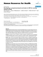
Figure 1 Immunohistochemical detection of CK2a expression in colorectal cancers, adenomas and adjacent normal colorectal mucosa.
Staining was (A) negative in normal colorectal epithelium cells, (B, C) weak to moderate in the nuclei of colorectal adenoma cells, (D, E, F) and
strong in the nuclei of colorectal cancer cells. (E is a close-up of the inset in D [framed in red]). Original magnification: × 200 (D), × 400 (A, B, C, E, F).
Zou et al. Journal of Translational Medicine 2011, 9:97
/>Page 4 of 11
CK2a regulates growth, proliferation and senescence of
CRC cell lines
Because the proce ss of tumorigenesis is closely corre-
lated with eternal proliferation of tumor cells, we deter-
mined whet her CK2a expression plays a role in human
CRC cell growth and prolif eration using siRNA to
knock down CK2a expression or emodin to inhibit
CK2a activity (Figure 3A). The MTT assay showed that
knockdown of CK2a significantly decreased CRC cell
proliferation compared to the control (nonspecific
siRNA) (F = 32.854, P < 0.01 for LoVo cells; F = 32.655,
P < 0.01 for SW480 cells), and treatment with emodin
marked ly reduced proliferation (F = 33.290, P <0.01for
LoVo cells; F = 57.052, P < 0.01 for SW480 cells; Figure
3B). Furthermore, in the colony formation assay, inhibi-
tion of CK2a expression dramatically de creased the
number of CRC colonies (t = 20.252, P <0.01forLoVo
cells; t = 12.034, P < 0.01 for SW480 cells; Figure 3C)
and promoted CRC cell senescence (t = 43.052, P <
0.01;Figure3D).Takentogether,theresultsindicate
that CK2a plays a very important role in human CRC
cell proliferation and senescence. CK2a knockdown or
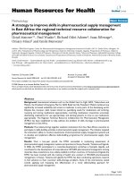
Figure 2 CK2a protein expression in CRC tissues and cell lines. (A) Western blot analysis of CK2a expression in eight pairs of CR C tissues
and adjacent, normal colorectal mucosa tissues. N: normal colorectal mucosa tissue; T: tumor tissue. (B) Quantitative analysis of CK2a protein
expression in eight pairs of CRC tissues and adjacent normal colorectal mucosa tissues. Columns, mean CK2a protein level after normalizing the
data to GAPDH expression; bars, SD. *P < 0.01. (C) Western blot was used to detect CK2a expression in five CRC cell lines. GAPDH expression
was used as a loading control.
Zou et al. Journal of Translational Medicine 2011, 9:97
/>Page 5 of 11
depression visibly inhibited cell proliferation and pro-
moted cell senescence.
After CK2a knockdown, the percentage of G0/G1
phase cells significantly increased (t = -9.577, P <0.01),
and the percent of S phase cells significantly decreased
(t = 8.749, P < 0.01; Figure 4A, B), indicatin g that CK2a
knockdown induced G0/G1 phase arrest. Moreover,
CK2a knockdown increased endogenous p53 and p21
expression and decreased endogenous C-myc expression
(Figure 4C). Thus, it can be inferred that the inhibition
of cell proliferation and cell cycle arrest in CK2a knock-
down cells are associated with alterations in p53, p21
and C-myc expression.
CK2a knockdown inhibits cell migration and invasion
Migration and matrigel invasion assays were performed
to e xamine the effect of CK2a on tumor c ell migration
and invasion, respectively. Knockdown of CK2a greatly
inhibited wound closure (F = 53.517, P <0.01forLoVo
cells; F = 40.319, P < 0.01 for SW480 cells; Figure 5A)
and invasion (t = 5.955, P < 0.01 for LoVo cells; t =
4.339, P < 0.05 for SW480 cells; Figure 5B). Accordingly,
CK2a was positively correlated with CRC cell migration
and invasion ability.
CK2a knockdown reversed nuclear translocation of b-
catenin and altered the expression of E-cadherin and
vimentin, in association with repression of the
transcription factors snail1 and smad2/3 expression
Knockdown of CK2a reversed the cytoplasmic-to-
nuclear transfer of b-catenin resulted by EGF stimuli
(Figure 6A). We also measured the expression levels of
EMT-related genes by analyzing western blots. Cells
transfected with CK2a siRNA had dramatically reduced
levels of endo genous CK2a and increased levels of E-
cadherin, an epithelial marker; there was no effect on
the b-catenin expression level and a decreased level of
vimentin, a mesenchymal marker. In addition, knock-
down of CK2a de creased the expression of the tran-
scription factors snail1 and smad2/3 (Figure 6B). The
results show that CK2a knockdown represses EMT in
CRC. We also treated cells with emodin and found that
CK2a activity, but not protein expression, was affected.
Emodin increased the expression of E-cadherin, had no
effect on the expression of b-catenin, and decreased the
expression of vimentin in a concentration-dependent
manner (Figure 6C). Thus, depression of CK2a activity
can inhibit the expression of EMT-related genes, sug-
gesting that an increase in CK2a protein or activity may
facilitate EMT and thus plays an important role in col-
orectal cancer invasion.
Discussion
In this present study, we assessed CK2a expression in
colorectal cancer, adenoma and normal colorectal
epithelium and found that CK2a was ove rexpressed in
CRC. Consistent with a recent study by Lin et al. [ 21],
our findings convincingly demonstrate that CK2a was
significantly upregulated in CRC. Our study further
showed that CK2a protein expression levels were
increased in both CRC and colorectal adenoma, and
CK2a expression was much higher in CRC than in ade-
noma, suggesting that CK2a maybeinvolvedinthe
progression from adenoma to CRC. In addition, we
found that CK2a overexpression was only associated
with T classification, but there were no significant corre-
lations with other clinical characteristics, possibly due to
our relatively small sample size.
Several studies have shown that the dysregulation of
CK2 enhances tumor cell survival [22,23], but the func-
tion of CK2a in CRC is less well known. In our study,
we assessed the role of CK2a in the biological behavior
of CRC. As in a recent study [21], we found that CK2a
knockdown in hibited cell proliferation and colon forma-
tion in other CRC cell lines. Moreover, for the first
Table 2 Correlation between the clinicopathological
features and expression of the CK2a protein.
CK2a (%)
Characteristics N Low
expression
High
expression
P
Gender 0.646
Male 56 22 (39.3) 34 (60.7)
Female 48 21 (43.8) 27 (56.2)
Age 0.897
≥55 y 54 22 (40.7) 32 (59.3)
<55 y 50 21 (42.0) 29 (58.0)
Tumor location 0.554
Colon 53 21 (39.6) 32 (60.4)
Rectum 51 22 (43.1) 29 (56.9)
T stage 0.002*
T1-T2 49 21 (42.9) 28 (57.1)
T3-T4 55 15 (27.2) 40 (72.7)
N stage 0.515
Nx-0 55 20 (36.4) 35 (63.6)
N1-2 49 23 (46.9) 26 (53.1)
M stage 0.632
M0 60 26 (43.3) 34 (56.7)
M1 44 17 (38.6) 27 (61.4)
TNM stage 0.539
I-II 30 11 (36.7) 19 (63.7)
III-IV 74 32 (43.2) 42 (56.8)
Degree of
differentiation
0.632
Well 35 13 (37.1) 22 (62.9)
Moderately 45 21 (46.7) 24 (53.3)
Poorly 24 9 (37.5) 15 (62.5)
*Statistically significant difference.
Zou et al. Journal of Translational Medicine 2011, 9:97
/>Page 6 of 11
time, we observed that, in CRC, CK2a knockdown
induces G0/G1 phase arrest and promotes cell senes-
cence. Similarly, inhibition of CK2a activity by emodin
induced proliferation repression. In additio n, CK2a
knockdown increased p53/p21 expression and decreased
C-myc expression. Accordingly, our results demonstrate
that CK2a has multiple roles in the biological behavior
of CRC, which is mediated by the regulation of
oncogenes and anti-oncogenes, including C-myc, p53
and p21.
In our study, CK2a was found to have an important
role in the biological behavior of CRC. Therefore, it is
vitally important to i nvestigate the potential regulatory
mechanisms of CK2a. However, the regulatory mechan-
ism of CK2a in contributing to the development of
CRC is still unknown. The progression from normal
Figure 3 Knockdown of CK2a inhibited cell proliferation and promoted cell senescence of CRC cell lines. (A) Western blot analysis of
CK2a protein in lysates of cells transfected with a specific CK2a siRNA or treated with emodin. GAPDH expression was used as a loading
control. (B) MTT assay of the proliferating cells transfected with a CK2a-specific siRNA or a nonspecific siRNA and treated with emodin. Points,
mean of three independent experiments; bars, SD. *P < 0.01 versus LoVo/Mock; #P < 0.01 versus SW480/Mock; †P < 0.01 versus LoVo/DMSO; ‡P
< 0.01 versus SW480/DMSO. (C) The number of colonies formed from cells transfected with CK2a siRNA. Colonies were stained with crystal violet
and counted. Columns, mean of three independent experiments; bars, SD. ++P < 0.01. (D) The number of SA-b-gal-positive cells (green) 48 h
after transfection with CK2a siRNA. Cells were stained with SA-b-gal staining solution. Columns, mean of three independent experiments; bars,
SD. ‡‡P < 0.01.
Zou et al. Journal of Translational Medicine 2011, 9:97
/>Page 7 of 11
intestinal mucosa to adenoma (adenomatous mucosa)
and finally to adenocarcinoma in CRC is closely corre-
lated w ith the EMT process and changes in the expres-
sion of a series of genes, such as E-cadherin, vimentin,
and b-catenin [24,25]. Thus, we further investigated
whether CK2a expression is associated with the EMT
process. Interestingly, in our study, assays of EMT-
related markers found that CK2a knockdown or activity
inhibition can alter the expression of E-cadherin and
vimentin and reverse the EGF-induced cytoplasmic-to-
nuclear translocation of b-catenin. We confirmed that
CK2a modulates the process of EMT, thereby affecting
Figure 4 CK2a inhibition induced G0/G1 phase arrest. (A) LoVo cells were transfected with CK2a-specific siRNA or nonspecific siRNA, stained
with propidium iodide (PI), and monitored by flow cytometry to determine the cell cycle phase distribution. (B) Comparison of the percentage
of cells in each phase of the cell cycle between LoVo cells transfected with CK2a-specific siRNA and nonspecific siRNA. Columns, mean of three
independent experiments; bars, SD. *P < 0.01. (C) CK2a, p53, p21, C-myc and GAPDH expression in cells transfected with CK2a-specific siRNA
was detected by western blot analysis.
Zou et al. Journal of Translational Medicine 2011, 9:97
/>Page 8 of 11
the regulation of cell migration and invasion by colorec-
tal cancer cells. Snail1 and Smad2/3 are important tran-
scriptional regulators of EMT that repress E-cadherin
expression t hrough binding to E-box mo tifs (5’ -
CANNTG-3’) in the promoter [26-28]. In our study, we
found t hat CK2a knockdown decreases the expressions
of snail1 and smad2/3. It is clearly shown that downre-
gulation of snail1 and smad2/3 by CK2a knockdown
facilitates an increase in E-cadherin expression and
EMT repression. Previous studies found that, in Her-2/
neu-driven mammary tumor cells, CK2 may be involved
in EMT repression, which can be induced by green tea
Figure 5 Knockdown of CK2a inhibited cell migration and invasion of CRC cell lines. (A) Monolayers of cells transfected with CK2a-specific
siRNAs were wounded by scraping, and wound closure was followed at 0, 12, and 24 h. The distance of the wound was measured. Columns,
mean of three independent experiments; bars, SD. *P < 0.01. (B) After transfection with CK2a-specific siRNAs for 18 h, cells that migrated
through the filters were counted in five randomly selected fields. Columns, mean of three independent experiments; bars, SD. #P < 0.05.
Zou et al. Journal of Translational Medicine 2011, 9:97
/>Page 9 of 11
polyphenol epigallocatechin-3-gallate (EGCG) [29]. In
untransformed mammary epithelial cells, ectopic expres-
sion of CK2a facilitates the induction of EMT-related
genesexpression,suchasthatofSlugandAhR,which
may thus promote the process of EMT [30]. Here we
show for t he first tim e that, in CRC, CK2a modulates
the EMT process through regulating the location or
expression of EMT-related genes. Recent studies have
indicated that, in breast cancer, p53/p21 and C-myc not
only regulate growth and senescence but are also
involved in regulating the EMT process [31-34]. Thus,
we inferred that, in CRC, alteration of p53/p21 and C-
myc expression by CK2a knockdown may facilitate the
EMT repression observed in our study. These findings
may account in part for the association of CK2a overex-
pression with EMT i n colorectal cancer. Additional stu-
dies are required to clarify the involvement of CK2a in
EMT and the development of colorectal cancer.
Conclusions
Our study demonstrates that CK2a is overexpressed in
CRC and that CK2a expression is much greater in CRC
than in adenoma and is greater in adenoma than in nor-
mal colorectal epithelium. Moreover, it is noteworthy to
observe that, for the first time, overexpression of CK2a
seems to be involved in the carcinogenesis and develop-
ment of CRC through regulation of EMT-related genes.
CK2a may be a promising molecular target for the diag-
nosis and treatment of human CRC.
Acknowledgements
This work was supported by the Natural Science Foundation of Guangdong
Province, China (No. 10151051501000062).
Author details
1
Department of Radiation Oncology, Nanfang Hospital, Southern Medical
University, Guangzhou 510515, Guangdong Province, China.
2
Hepatology
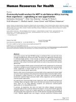
Figure 6 Inhibition of CK2a reversed the nuclear translocation of b-catenin and altered EMT-related genes expression. Reversal of EGF-
induced nuclear translocation of b-catenin occurred in LoVo cells transfected with CK2a-specific siRNA (A), treated with EGF (100 ng/ml) for 2 h,
and stained for immunofluorescence with b-catenin antibody (red) and DAPI (blue). (B) Western blot was used to detect the expression levels of
CK2a, E-cadherin, b-catenin, vimentin and the transcription factors snail1 and smad2/3 in cells transfected with CK2a-specific siRNA. (C) One
week later, in LoVo cells treated with emodin (40 μmol/l, 50 μmol/l and 60 μmol/l), the expressions of E-cadherin, b-catenin and vimentin were
detected by western blot analysis. GAPDH expression was used as a loading control.
Zou et al. Journal of Translational Medicine 2011, 9:97
/>Page 10 of 11
Unit and Department of Infectious Diseas es, Nanfang Hospital, Southern
Medical University, Guangzhou 51051 5, Guangdong Province, China.
Authors’ contributions
JZ, HL, ZD, and QZ designed and performed experiments. JZ and HL
performed the statistical analysis and drafted the manuscript. DW and LL
helped in drafting the manuscript and contributed specific information and
critical analysis throughout the manuscript. All authors read and approved
the final manuscript.
Competing interests
The authors declare that they have no competing interests.
Received: 26 February 2011 Accepted: 25 June 2011
Published: 25 June 2011
References
1. Jemal A, Siegel R, Ward E, Hao Y, Xu J, Murray T, Thun MJ: Cancer statistics,
2008. CA Cancer J Clin 2008, 58:71-96.
2. Sung JJ, Lau JY, Goh KL, Leung WK: Increasing incidence of colorectal
cancer in Asia: implications for screening. Lancet Oncol 2005, 6:871-876.
3. Pino MS, Chung DC: The chromosomal instability pathway in colon
cancer. Gastroenterology 138:2059-2072.
4. Boland CR, Goel A: Microsatellite instability in colorectal cancer.
Gastroenterology 138:2073-2087, e2073.
5. Hadziavdic V, Pavlovic-Calic N, Eminovic I: Microsatellite instability and loss
of heterozygosity of tumor suppressor genes in Bosnian patients with
sporadic colorectal cancer. Bosn J Basic Med Sci 2008, 8:313-321.
6. Gocke CD, Benko FA, Kopreski MS, McGarrity TJ: p53 and APC mutations
are detectable in the plasma and serum of patients with colorectal
cancer (CRC) or adenomas. Ann N Y Acad Sci 2000, 906:44-50.
7. Derks S, Bosch LJ, Niessen HE, Moerkerk PT, van den Bosch SM, Carvalho B,
Mongera S, Voncken JW, Meijer GA, de Bruine AP, et al: Promoter CpG
island hypermethylation- and H3K9me3 and H3K27me3-mediated
epigenetic silencing targets the deleted in colon cancer (DCC) gene in
colorectal carcinogenesis without affecting neighboring genes on
chromosomal region 18q21. Carcinogenesis 2009, 30:1041-1048.
8. Toyota M, Ohe-Toyota M, Ahuja N, Issa JP: Distinct genetic profiles in
colorectal tumors with or without the CpG island methylator phenotype.
Proc Natl Acad Sci USA 2000, 97:710-715.
9. Litchfield DW: Protein kinase CK2: structure, regulation and role in
cellular decisions of life and death. Biochem J 2003, 369:1-15.
10. Meggio F, Pinna LA: One-thousand-and-one substrates of protein kinase
CK2? FASEB J 2003, 17:349-368.
11. Seldin DC, Lou DY, Toselli P, Landesman-Bollag E, Dominguez I: Gene
targeting of CK2 catalytic subunits. Mol Cell Biochem 2008, 316:141-147.
12. Padmanabha R, Chen-Wu JL, Hanna DE, Glover CV: Isolation, sequencing,
and disruption of the yeast CKA2 gene: casein kinase II is essential for
viability in Saccharomyces cerevisiae. Mol Cell Biol 1990, 10:4089-4099.
13. Buchou T, Vernet M, Blond O, Jensen HH, Pointu H, Olsen BB, Cochet C,
Issinger OG, Boldyreff B: Disruption of the regulatory beta subunit of
protein kinase CK2 in mice leads to a cell-autonomous defect and early
embryonic lethality. Mol Cell Biol 2003, 23:908-915.
14. Kulartz M, Hiller E, Kappes F, Pinna LA, Knippers R: Protein kinase CK2
phosphorylates the cell cycle regulatory protein Geminin. Biochem
Biophys Res Commun 2004, 315
:1011-1017.
15. Canton DA, Litchfield DW: The shape of things to come: an emerging
role for protein kinase CK2 in the regulation of cell morphology and the
cytoskeleton. Cell Signal 2006, 18:267-275.
16. Gu L, Husain-Ponnampalam R, Hoffmann-Benning S, Henry RW: The protein
kinase CK2 phosphorylates SNAP190 to negatively regulate SNAPC DNA
binding and human U6 transcription by RNA polymerase III. J Biol Chem
2007, 282:27887-27896.
17. Guo C, Davis AT, Yu S, Tawfic S, Ahmed K: Role of protein kinase CK2 in
phosphorylation nucleosomal proteins in relation to transcriptional
activity. Mol Cell Biochem 1999, 191:135-142.
18. Ruzzene M, Pinna LA: Addiction to protein kinase CK2: a common
denominator of diverse cancer cells? Biochim Biophys Acta 1804:499-504.
19. Wu D, Ding Y, Wang S, Zhang Q, Liu L: Increased expression of high
mobility group box 1 (HMGB1) is associated with progression and poor
prognosis in human nasopharyngeal carcinoma. J Pathol 2008,
216:167-175.
20. Masunaga R, Kohno H, Dhar DK, Ohno S, Shibakita M, Kinugasa S,
Yoshimura H, Tachibana M, Kubota H, Nagasue N: Cyclooxygenase-2
expression correlates with tumor neovascularization and prognosis in
human colorectal carcinoma patients. Clin Cancer Res 2000, 6:4064-4068.
21. Lin KY, Tai C, Hsu JC, Li CF, Fang CL, Lai HC, Hseu YC, Lin YF, Uen YH:
Overexpression of nuclear protein kinase CK2 alpha catalytic subunit
(CK2alpha) as a poor prognosticator in human colorectal cancer. PLoS
One 6:e17193.
22. Ahmad KA, Harris NH, Johnson AD, Lindvall HC, Wang G, Ahmed K: Protein
kinase CK2 modulates apoptosis induced by resveratrol and
epigallocatechin-3-gallate in prostate cancer cells. Mol Cancer Ther 2007,
6:1006-1012.
23. Brown MS, Diallo OT, Hu M, Ehsanian R, Yang X, Arun P, Lu H, Korman V,
Unger G, Ahmed K, et al: CK2 modulation of NF-kappaB, TP53, and the
malignant phenotype in head and neck cancer by anti-CK2
oligonucleotides in vitro or in vivo via sub-50-nm nanocapsules. Clin
Cancer Res 16:2295-2307.
24. Vincan E, Brabletz T, Faux MC, Ramsay RG: A human three-dimensional cell
line model allows the study of dynamic and reversible epithelial-
mesenchymal and mesenchymal-epithelial transition that underpins
colorectal carcinogenesis. Cells Tissues Organs 2007, 185:20-28.
25. Chen X, Halberg RB, Burch RP, Dove WF: Intestinal adenomagenesis
involves core molecular signatures of the epithelial-mesenchymal
transition. J Mol Histol 2008, 39:283-294.
26. Vincent T, Neve EP, Johnson JR, Kukalev A, Rojo F, Albanell J, Pietras K,
Virtanen I, Philipson L, Leopold PL, et al: A SNAIL1-SMAD3/4
transcriptional repressor complex promotes TGF-beta mediated
epithelial-mesenchymal transition. Nat Cell Biol 2009, 11:943-950.
27. Phanish MK, Wahab NA, Colville-Nash P, Hendry BM, Dockrell ME: The
differential role of Smad2 and Smad3 in the regulation of pro-fibrotic
TGFbeta1 responses in human proximal-tubule epithelial cells. Biochem J
2006,
393:601-607.
28. Nawshad A, Medici D, Liu CC, Hay ED: TGFbeta3 inhibits E-cadherin gene
expression in palate medial-edge epithelial cells through a Smad2-
Smad4-LEF1 transcription complex. J Cell Sci 2007, 120:1646-1653.
29. Belguise K, Guo S, Sonenshein GE: Activation of FOXO3a by the green tea
polyphenol epigallocatechin-3-gallate induces estrogen receptor alpha
expression reversing invasive phenotype of breast cancer cells. Cancer
Res 2007, 67:5763-5770.
30. Belguise K, Guo S, Yang S, Rogers AE, Seldin DC, Sherr DH, Sonenshein GE:
Green tea polyphenols reverse cooperation between c-Rel and CK2 that
induces the aryl hydrocarbon receptor, slug, and an invasive phenotype.
Cancer Res 2007, 67:11742-11750.
31. Kim T, Veronese A, Pichiorri F, Lee TJ, Jeon YJ, Volinia S, Pineau P,
Marchio A, Palatini J, Suh SS, et al: p53 regulates epithelial-mesenchymal
transition through microRNAs targeting ZEB1 and ZEB2. J Exp Med
208:875-883.
32. Schubert J, Brabletz T: p53 spreads out further: suppression of EMT and
stemness by activating miR-200c expression. Cell Res 21:705-707.
33. Cho KB, Cho MK, Lee WY, Kang KW: Overexpression of c-myc induces
epithelial mesenchymal transition in mammary epithelial cells. Cancer
Lett 293:230-239.
34. Liu M, Casimiro MC, Wang C, Shirley LA, Jiao X, Katiyar S, Ju X, Li Z, Yu Z,
Zhou J, et al: p21CIP1 attenuates Ras- and c-Myc-dependent breast
tumor epithelial mesenchymal transition and cancer stem cell-like gene
expression in vivo. Proc Natl Acad Sci USA 2009, 106:19035-19039.
doi:10.1186/1479-5876-9-97
Cite this article as: Zou et al.: Protein kinase CK2a is overexpressed in
colorectal cancer and modulates cell proliferation and invasion via
regulating EMT-related genes. Journal of Translational Medicine 2011 9:97.
Zou et al. Journal of Translational Medicine 2011, 9:97
/>Page 11 of 11