Báo cáo sinh học: " Transcription and translation of human F11R gene are required for an initial step of atherogenesis induced by inflammatory cytokines" doc
Bạn đang xem bản rút gọn của tài liệu. Xem và tải ngay bản đầy đủ của tài liệu tại đây (505.68 KB, 14 trang )
RESEA R C H Open Access
Transcription and translation of human F11R
gene are required for an initial step of
atherogenesis induced by inflammatory cytokines
Bani M Azari
1
, Jonathan D Marmur
1
, Moro O Salifu
2
, Yigal H Ehrlich
3
, Elizabeth Kornecki
2,4
and Anna Babinska
1,2*
Abstract
Background -: The F11 Receptor (F11R; aka JAM-A, JAM-1) is a cell adhesion protein present constitutively on the
membrane surface of circulating platelets and within tight junctions of endothelial cells (ECs). Previous reports
demonstrated that exposure of ECs to pro-inflammatory cytokines causes insertion of F11R molecules into the
luminal surface of ECs, ensuing with homologous interactions between F11R molecules of platelets and ECs, and a
resultant adhesion of platelets to the inflamed ECs. The main new finding of the present report is that the first
step in this chain of events is the de-novo transcription and translation of F11R molecules, induced in ECs by
exposure to inflammatory cytokines.
Methods -: The experimental approach utilized isolated, washed human platelet suspensions and cultured human
venous endothelial cells (HUVEC) and human arterial endothelial cells (HAEC) exposed to the proinflammatory
cytokines TNF-alpha and/or IFN-gamma, for examination of the ability of human platelets to adhere to the
inflamed ECs thru the F11R. Our strategy was based on testing the effects of the following inhibitors on this
activity: general mRNA synthesis inhibitors, inhibitors of the NF-kappaB and JAK/STAT pathways, and small
interfering F11R-mRNA (siRNAs) to specifically silence the F11R gene.
Results -: Treatment of inflamed ECs with the inhibitors actinomycin, parthenolide or with AG-480 resulted in
complete blockade of F11R- mRNA expression, indicating the involvement of NF-kappaB and JAK/STAT pathways in
this induction. Transfection of ECs wi th F11R siRNAs caused compl ete inhibition of the cytokine-induced
upregulation of F11R mRNA and inhibition of detection of the newly- translated F11R molecules in cytokine-
inflamed ECs. The functional consequence of the inhibition of F11R transcription and translation was the significant
blockade of the adhesion of human platelets to inflamed ECs.
Conclusion -: These results prove that de novo synthesis of F11R in ECs is required for the adhesion of platelets to
inflamed ECs. Because platelet adhesion to an inflamed endothelium is crucial for plaque formation in non-
denuded blood vessels, we conclude that the de-novo translation of F11R is a crucial early step in the initiation of
atherogenesis, leading to atherosclerosis, heart attacks and stroke.
Background
The healthy, non-thrombogenic endothelium of the vascu-
lature does not attract nor bind circulating platelets [1-3].
However, following its exposure to proinflammatory cyto-
kines, the non-thrombogenic endothel ium becomes acti-
vated and converts into a prothrombotic endothelium [3],
resulting in a procoagulant state associated with a
predisposition to the adhesion of platelets, atherosclerosis
and thrombosis. The adhesion of platelets to the activated
endothelium was shown to occur in areas highly prone to
atherosclerotic plaque development prior to the detection
of lesions , and prior to the infiltration and adhesion of
monocytes or leukocytes [2,3]. A critical molecule shown
to be involved in the process of platelet adhesion to the
activated endothelium is the F11R protein, first described
by Kornecki et al in 1990 [4]. F11R is the symbol approved
by the Human Gene Nomenclature Committee for the
F11 receptor protein (GenBank Accession # 207907; NBC
* Correspondence:
1
Division of Cardiology, Department of Medicine, State University of New
York, Downstate Medical Center, Brooklyn, New York 11203, USA
Full list of author information is available at the end of the article
Azari et al. Journal of Translational Medicine 2011, 9:98
/>© 2011 Azari et al; licensee BioMed Central Ltd. This is an Open Access article distributed under the terms of the Creative Commons
Attribution License ( which permits unrestricted use, distribution, and reproduction in
any medium, provided the original work is properly cited.
#S56749). In 1995, the amino acid sequences of the
N-terminus and internal domains of the platelet F11R
molecule were detailed [5]. A protein termed JAM,
described in 1998 [6] showed correspondingly-identical
amino acid sequences to those of the F11R protein, and
hence the alias of JAM-A is also provided here. Direct
phosphorylation and dimerization of the F11R protein
[5,7] were shown following the activation of human plate-
lets by physiological agonists. The cloning of the human
F11R gene revealed that this molecule is a cell adhesion
molecule, member of the Ig superfamily [8].
Studies of the adhesion of human platelets to cyto-
kine-inflamed endothelial cells (ECs) [9] determined that
homophilic interactions between the F11R molecules
expressed constitutively on the platelet surface and the
F11R molecules expressed de-novo on the luminal sur-
face of ECs when stimulated by cytokines, exert over
50% of the adhesi ve force between these cells. This
observat ion was evidenced by demonstrating the inhibi -
tion of the adhesion of platelets to cytokine-infla med
ECs by a recombinant, soluble form of the F11R protein,
and by domain-specific F11R peptides with amino acid
sequences stretching in the N-terminal region and the
1st Ig fold of the F11R molecule, respectively [10]. Ana-
lysis of the F 11R gene identified NF-Bbindingsitesin
the promoter region [11], indicating that cytokines, dur-
ing processes of inflammation, can cause up-regulation
of the F11R gene. Yet, both the biochemical and genetic
evidence thus far only suggests the involvement of F11R
in the adhesion of circulating platelets to the cytokine-
inflamed endothelium. In this report we demonstrate
directly, by utilizing small interfering F11R RNAs (siR-
NAs), that F11R plays a critical role in the adhesion of
platelets to the inflamed endothelium, an important
early step in atherogenesis.
Materials and methods
Human endothelial cells and proinflammatory cytokines
Human aortic endothelial cells (HAEC) and human
umbilical vein endothelial cells (HUVEC) (frozen vials of
10
6
cells) were purchased from Cascade Biologics, Inc.,
Portland, OR, and grown in Medium 200 containing 1%
or 2% fetal calf serum (FCS) (Cascade Biologics, Inc.,
Portland, OR). For the experiments detailed below, both
HAEC and HUVEC at 2
nd
passage, were treated with
purified human recombinant TNFa (100 units/ml) (R&D
Systems, Inc., Minneapolis, MN) and/or IFNg (200 units/
ml) (Roche Diagnostics, Mannheim, Germany), main-
tained at 37°C for the indicated periods of time. In a ser-
ies of dose-response experiments in which the
concentrations of TNF-a and IFN-g were varied , a con-
centration of 50 pM TNFa is equivalent to100 units/ml
TNF-a, and a concentration of 5.8 nM IFNg is equivalent
to 200 units/ml IFNg.
Quantification of F11R mRNA in HAEC and HUVEC by
real-time PCR
HAEC and HUVE C endothelial cells were grown to con-
fluence and treated with cytoki nes at various t imes and
doses. The treated cells were washed with 1× PBS, lysed,
the total RNA extracted utilizing RNeasy Mini Kit (Qia-
gen, Valencia, CA, USA), and analyzed by real -time PCR
on three separate experiments conducted in triplicate.
The levels of F11R mRNA were deter mined by us e of an
ABI Prism 7000HT Se quence Detection System (ABI;
AppliedBiosystem, Foster City, CA). The F11R primers
consisted of t he forward primer - 740: CCG TCC TTG
TAA CCC TGA TT, reverse primer - 818: CTC CTT
CAC TTC GGG CAC T A and probe -788: TGG CCT
CGG CTA TAG GCA AAC C. The GAPDH forward pri-
mer - 620: GGA CTC ATG ACC ACA GTC CA, reverse
primer - 738: CCA GTA GAG GCA GGG ATG AT, and
the probe - 675: ACG CCA CAG TTT CCC GGA GG.
Thermal cycles consisted of: 1 cycle at 48°C for 30 min,
10 min at 95°C and 40 cycles for 15 sec at 95 °C, 1 min at
60°C. The probes were dual-labeled with FAM-TAMRA,
obtained fro m ABI. Each mRNA level was express ed as a
ratio to GAPDH. The mRNA levels were calculated using
astandardcurveofRNAisolatedfromnormalhuman
kidney (Stratagene) for the time course and dose curve or
QPCR Human Reference total RNA (Stratagene) utilizing
the ABI Prism 7000 SDS Software (Applied Biosystems).
Statistical analysis for real-time PCR
The RNAs, derived from ECs grown and treated in tissue
culture wel ls, were isolated individual ly. Real time PCR
procedures were performed in triplicate and averaged for
each sampl e in three separate experiments (n = 9). The
data were analyzed by Student’s t-test and by mixed lin-
ear model analysis using SPSS software. Differences were
considered significant at P < 0.05.
Preparation of inhibitors of RNA synthesis, NF-B and JAK
protein kinase
Actinomycin D (Sigma, St. Louis, MO), a known inhibi-
tor of RNA synthesis, was diluted i n DMSO to a 500 μg/
ml (100X) stock solution. Parthenolide (Sigma), an inhi-
bitor of the nuclear factor kappa B, NF-kB signaling [12],
was diluted in chloroform to a 5 0 mM (1000X) stock
solution. The inhibitor of Janus kinase, JAK protein
kinase, the tyrosine kinase inhibitor tyrphostin AG490
[13], (Sigma) was di luted in et hanol to a 5 mM ( 100X)
stock solution. All stock solutions were diluted in culture
media to 1X concentration prior to experimentation.
HAEC and HUVEC were grown to confluence and then
treated with either actomycin D, parthenolid, or AG490,
added in culture media without growth factor supple-
ments for 1 hr at 37°C. Proinflammatory cyto ki nes,
Azari et al. Journal of Translational Medicine 2011, 9:98
/>Page 2 of 14
TNFa and/or IFNg were then applied to the media and
the ECs were further incubated at 37°C for up to 24 hrs.
Silencing of the F11R gene of HAEC and HUVEC
endothelial cells: transfections with small interfering RNAs
(siRNAs)
Transfections were performed using Oligofectamine (Invi-
trogen, Carlsbad, CA) according to the manufacturer’s
instructions. Briefly, 9 × 10
4
HAEC and HUVEC cells
were seeded onto 96 well plates in 200 M media supple-
men ted with LSG S without antibiotics, and the transfec-
tions of ECs were carried-out with either the stealth F11R
siRNA HSS121425 (5’ GGGACUUCGGAGUAAGAAG-
GUGAUUU 3’) (300 nM) or the control, non-targeting
siRNA No. 2 (Dharmacon). Subsequently, the transfected
ECs were incubated in 200 M media containing 1% FBS
followed by the application of cytokines TNFa (100 units/
ml) and/or IFNg (200 units/ml) fo r various periods of
time.
Analysis of F11R in HAEC and HUVEC lysates and cell
culture media
Monolayers of arterial and venous endothelial cells (90 -
95% confluence) were collected and homogenized in lysis
buffer containing 20 mM Tris, 50 mM NaCl, 2 mM
EDTA, 2 mM EGTA, 1% sodium deoxycholate, 1% Triton
X-100, and 0.1% SDS, pH 7.4 supplemented with protease
and phosphatase inhibitors (Sigma-Aldrich) for the pre-
paration of t otal cell lysate material derived from human
arterial and venous endothelial cells. Protein concentration
was quantified by the bicinchoninic acid (BCA) assay. Pro-
cedures utilizing SDS-polyacrylam ide gel electrophoresis
(10%, PAGE) followed by immunoblotting were performed
as described previously [14].
Collection and analysis of F11R in the media from
cultured endothelial cells
The media derived from the arterial and venous, cytokine-
treated and nontreated endothelial cells were collected at
the time of cell harvesting and concentrated 200X using
the centrifugal filter Centricon YM-10. Identification of
the F11R protein within the collected media involved the
resolution of all proteins by SDS-PAGE (10%) followed by
immunoblotting procedures utilizing anti-F11R antibody,
as described previously [10].
Quantitation of immunoblots
Quantitation o f the i mmunoblots was performed using
image J (NIH). Briefly, scanned images of immunoblots
were opened i n image J, the protein bands were selected
using the freeform tool and measured for integrated den-
sity. The values were normalized to tubulin levels by divid-
ing the integrated density o f the specific band by the
integrative density of the tubulin band. ANOVA statistical
analysis was performe d on the normalized values. All
values are the average of three immunoblots ± SEM.
The adhesion of platelets to endothelial cells: labeling of
human platelets by calcein
Platelet rich plasma (PRP) was prepared from 100 mL of
citrated whole blood, by centrifugation at 200 × g for
20 min at 23°C. Calcein (2 μg/mL)(Invitrogen) [15,16] was
added to the PRP, and the PRP was maintained at 30°C for
1 hr in the absence of light. Platelets were isolated from
PRP, washed as detailed previously [10] and resuspended
at final concentrations ranging from 2.5 - 3.5 × 10
8
/mL
Assay s co nducted for measuring the adhesion of plat e-
lets to endothelial cells were performed in the dark due to
the sensitivity of th e calcium probe calcein to light expo-
sure. Initially, HAEC and HUVEC, plated in cell culture
wells, were in cuba ted with 1% FBS/BSA in 200 M media
for 1 hr at 37°C to block nonspecific binding sites. Ali-
quots of freshly-prepared, calcei n-labeled platelets (3.3 ×
10
8
/ml) were added to each of the cell-culture wells, and
plates were incubated at 37°C for 1 hr. Paraformaldhyde
(4%), pH 7.4, was added to each well and incubation con-
tinued at 23°C for 15 min. The addition of paraformalde-
hyde, before washings, did not affect the natural capacity
of the platelets to adhere to endo thelial cells. The plates
were washed 3× wit h pre-w armed growth factor -free 200
M media. Then aliquots (100 μl) of pre-warmed PBS were
added to wells, and wells were read using a Perkin Elmer
plate reader Victor 3, 1420 multilabel counter with fluor-
escein filter, as detailed previously described [9].
Statistical analysis performed for assays involving platelet
adhesion to endothelial cells
To improve normality of distribution, the dependent
variable (number of platelets per endothelial cell) was
transformed by dividing by 10, adding 1 and taking the
natural log. A mixed lin ear model was construc ted that
introduced treatment, cell type a nd the state of platelet
activation (nonactivated vs agonist-activated) (and their
mutual interactions) as fixed factors, with plate as a ran -
dom factor. Since the variance of the dependen t variable
differed substantially according to plate, treatment and
platelet state, variances were estimated separa tely for
each combination of these factors. Due to the unbalanced
nature of the study design, Satterthwaite adjustments
were applied to numerator degrees of freedom. To offset
the i ssue of multiple testing, Tukey-adjustments were
applied to p-values for pair-wise g roup comparisons.
Analysis of model residuals was undertaken to check for
model fit and outliers. SAS Release 9.3 (SAS Institute,
Cary NC) PROC MIXED software was used. Four outly-
ing observations were excluded from analysis. All of the
fixed main effects and their interactions were statistically
significant at the 0.001 lev el, with the exception of the
Azari et al. Journal of Translational Medicine 2011, 9:98
/>Page 3 of 14
cell type main effect (p = 0.783). Discrepancies of means
among the 11 plates were significant (Z = 2.11, p =
0.017). The inter-assay coefficient of variance was 0.7 ±
0.3 (S.E). The intra-assay coefficient of variance for each
condition on the same plate was lower [(range from 0.05
to 0.16 ± .02 (S.E.) (Z > 6. 00, P < 0.0001)] than the inter-
assay coefficient of variance.
Results
Expression of F11R mRNA in human aortic (HAEC) and
umbilical vein (HUVEC) endothelial cells exposed to pro-
inflammatory cytokines: time and dose-response
The e xpression of F11R mRNA was examined both in
arterial HAEC and venous HUVEC following their expo-
sure to the pro-inflammatory cytokines TNFa and IFNg.
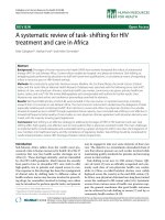
As shown in Figure 1, a time-dependent increase in F11R
mRNA expression was observed following the exposure
of arterial and venous cells to TNFa or IFNg,ortheir
combination. Arterial endothelial cells (top panels)
demonstrated a slow, significant increase in the level of
F11R mRNA at 12 hrs of exposure to e ither TNFa or
IFNg. Although a further increase was observed with
TNFa for a subsequent 12 hr period, further exposure of
cells to INFg resulted in a drop in the F11R mRNA level.
The simultaneous treatment of cells with TNFa and
IFNg resulted in a shortening in response time, with
maximal F11R mRNA levels observed already at 3 hrs of
cytokine-exposure. Similarly, venous endothelial cells
(lower panels) demonstrated a gradual enhancement
(also significant at 12 hrs) of F11R mRNA expression fol-
lowing the application of cytokines, alone or in
combination.
H
AEC
0361224
.
0.0
0.2
0.4
0.6
0.8
1.0
1.2
1.4
.
0361224
.
0.0
0.2
0.4
0.6
0.8
1.0
.
0361224
.
0.0
0.2
0.4
0.6
0.8
1.0
TNF
D
IFN
J
TNF
D
& IFN
J
Time (hrs
)
*
*
**
*
*
*
F11R mRNA levels
Normalized to GAPDH
0481224
0.0
0.1
0.2
0.3
0.4
0.5
0.6
0.7
IFN
J
0481224
.
0.0
0.1
0.2
0.3
0.4
0.5
0.6
0.7
TNF
D
&IFN
J
*
*
*
*
0 4 8 12 24
.
0.0
0.2
0.4
0.6
0.8
TNF
D
*
*
H
UVEC
Time (hrs)
F11R mRNA levels
Normalized to GAPDH
Figure 1 Expression of F11R mRNA in human aortic endothelial cells (HAEC) and umbilical vein endothelial cells (HUVEC) exposed to
proinflammatory cytokines TNFa and/or IFNg: time course. Real-time PCR was performed in cultured HAEC (top panels) treated for 0, 3, 6,
12, and 24 hrs with TNFa (100 u/mL) and/or IFNg (200 u/mL), and in cultured HUVEC (bottom panels) treated for 0,4,8,12, and 24 hrs with TNFa
(100 u/mL) and/or IFNg (200 u/mL). Real-time PCR was performed three times in triplicate for each time point. Values represent the mean ± SEM.
*P < 0.05 indicates the level of significance determined at a specific interval of time of cytokine- treatment of ECs in comparison to the zero
time points.
Azari et al. Journal of Translational Medicine 2011, 9:98
/>Page 4 of 14
Comparison of the F11R mRNA level in untreated vs
cytokine-stimulated endothelial cells indicated that F11R
mRNA levels were higher in arterial than in venous
ECs, with the overall pattern in the response-t ime to
cytokines similar in both cell types.
By varying the concentration of cytokines, the level of
F11R mRNA was observed to increase in both cell
types, in a dose-depend ent manner following a 12 hr
exposure to either TNFa or IFNg .AsshowninFigure
2, significant increases in F11R mRNA levels in arterial
EC in response to TNFa, already were observed at con-
centrations of TNFa as low as 0.5 pM (1 unit/ml), with
maximal responses to TNFa observed at 50 pM (100
units/ml). In HUVECs, significant increases in F11R
mRNA levels in response to TNFa also were observed
at a concentration of TNFa of 0.5 pM, whereas maximal
increases occurred at a concentration of 100 pM TNF-a
(200 units/ml).
Arterial EC exhibited sensitivity to IFNg alre ady at a
concentration of 0.1 nM (3.4 units/ml), with maximal,
significant increases in F11R mRNA l evels in response
to IFNg at 5.8 nM (200 units/ml). However, the treat-
ment of arterial endothelial cells with higher concentra-
tions of TNFa (of 100 or 1000 pM; 200 or 2,000 units/
ml) or IFNg (10 or 100 nM; 344 or 3448 units/ml),
resulted in a drop in the expression of F11R mRNA to
pretreatment levels, as was observed with IFNg (Figure
2, top panels). Similarly, venous endothelial cells demon-
strated significant increases in F11R mRNA level in
response to TNFa at0.5pM(1unit/ml)and0.1nM
IFNg (17 units/ml) with maximal increases occurring at
concentrations of 50 pM TNFa (100 units/ml) and 10
nM IFNg (344.8 units/ml). A ten-fold higher concentra-
tion of IFN g produced a slight decrease in the expres-
sion of F1 1R mRNA in venous endothelial cells, but not
a complete drop, as observed in arterial endothelial cells
at higher concentrations.
A comparison of the concentrations of cytokines
used in this study and the physiological and pathophy-
siological concentrations of cytokines measured in
individuals indicates that serum concentrations of
TNFa, found in normal individuals were about 0.8
pM, whereas pathophysiological concentrations of
TNFa,4-foldhigher(3.2pM),weredetectedinthe
serum of patients (see the link- .
nih.gov/pmc/articles/PMC1533889/table/T1/). As
shown in Figure 2, the concentrations of TNFa that
significantly induced F11R mRNA in both HAEC and
HUVEC were in the same range. Likewise, a concen-
tration of IFNg, of about 0.1 nM, was reported in the
serum of patients (see link above) - a concentration of
IFNg shown to significantly induce F11R mRNA in
both HAEC and HUVEC (see Figure 2).
Inhibition of the expression of F11R-mRNA in inflamed
endothelial cells
We examined whether the observed increases in the level
of F11R mRNA in inflamed endothelial cells resulted from
the de novo expression of F11R by conducting experiments
involving the pretreatment of endothelial cells with
the RNA synthesis inhibitor actinomycin D (5 μg/ml).
Endothe lial cells were pretreated (or not pretreated) with
actinomycin D for a period of 1 hr at 37°C prior to their
exposure to either TNFa or IFNg. Cells that were not pre-
treated with actinomycin (ActD) demonstrated a signifi-
cant increase in the level of F11R mRNA following their
exposure to TNFa, as sho wn in Figure 3a (TFNa),
whereas cells pretreated with ActD were unable to demon-
strate the induced increase in the level of F11R mRNA
induced by TNFa treatment, and a complete inhibition
was observe d (see TNF a & ActD). Pretreatment of cells
with actinomycin D alone did not produce a decrease in
basal levels of F11RmRNA (see ActD) as identical values
to the basal levels measured in untreated cells were
obtained. Similar to the results observed with TNFa,
venous cells treated wi th IFNg (200 u/ml) (as shown i n
Figure 3b, IFNg) demonstrated a significant rise in their
level of F11R mRNA; such an increase in F11R mRNA
level could be completely blocked by the presence of ActD
(see Figure 3b, IFNg &ActD),
Next, a series of experiments utilizing specific inhibi-
tors were examined for the potential involvement of
specific pathways in the up-regulation of the F11R gene.
As shown in Figure 4 (panel a), venous endothelial cells
exposes to TNFa alone demonstrated a significant
increase in mRNA level - how ever, pretreatment of
these cells with parthenolide (50 μM), an inhibitor of
the function of NF-B, prior to their exposure to TNFa
(see TNFa & Part henolide), resul ted in a complete
blockade of their ability to up-r egulate the F11R gene in
response to TNF a. In the presence of the inhibitor,
parthenolide, the level of F 11R mRNA in cel ls exposed
to TNFa remained unchanged (see TNFa & Partheno-
lide) from baseline values measured in cells not exposed
to TNFa (see “untreated”), or cells treated with only the
inhibitor parthenolide (see “Parthenolide”). In contrast,
the blockade by parthenolide of the induction of the
F11R gene by TNFa (as shown in Figure 4, panel a) was
not observed in venous cells exposed to IFNg (see Fig-
ure 4b, IFNg & Parthenolide). Indeed, the presence of
the same concentration of pathenolide did not prevent
IFNg from inducing an increase of F11R mRNA in
HUVEC, and a further rise in the level of F11R mRNA
could be detected in response to IFNg in the presence
of parthenolide. A possibility of cross-regulation of the
IFN-g pathway by TNFa may account for the enha nced
IFN-g responses observed in this study.
Azari et al. Journal of Translational Medicine 2011, 9:98
/>Page 5 of 14
Since the inhibition of the activity of NFB by parthe-
nolide did not b lock the increase in the level of F11R
mRNA induced by IFNg, w e examined whether the
IFNg-induced increase in the level of F11R mRNA could
be blocked by AG490, a known inhibitor of the Jak/Stat
pathway. We observed that the increase in the F11R
mRNA level induced by the exposure of venous cells to
the cytokine IFNg was blocked by the pretreatment of
venous cells with tyrphostin AG-490 (50 μM), the JAK
protein kinase inhibitor, asshowninFigure4(panelc)
(see IFNg & AG-490).
Synthesis and release/shedding of F11R by inflamed
endothelial cells
Previous studies have reported an enhanced presence of a
soluble form of F11R (termed sF11R) in the circulation of
cardiovascular patients [17] possibly due to the state of
inflammation of the diseased blood vessels. As our study
involved the treatment of cultured endothelial cells with
inflammatory cytokines , we examined the possibilit y that
such cytokine-treatment may result in the release/shed-
ding and/or secretion of the F11R protein. Figure 5
shows the results of experiments designed to identify, by
F11R mRNA levels
Normalized to GAPDH
IFNȖ Concentration
(
nM
)
.
0 0.1 1 5.8 10 100
.
0.0
0.2
0.4
0.6
0.8
1.0
1.2
1.4
*
*
*
*
*
TNFĮ Concentration
(
pM
)
HUVEC
00.55 501001000
.
0.0
0.1
0.2
0.3
0.4
0.5
0.6
0.7
*
*
*
*
*
F11R mRNA levels
Normalized to GAPDH
IFNȖ Concentration (nM)TNFĮ Concentration (pM)
.
0 0.5 5 50 100 1000
.
0.0
0.2
0.4
0.6
0.8
1.0
*
*
*
*
*
0 0.1 1 5.8 10 100
.
0.0
0.2
0.4
0.6
0.8
1.0
.
*
HAEC
Figure 2 The expression of F11R mRNA in human endothelial cells (ECs) exposed to proinflamm atory cytokines TNFa and IFNg: dose
response. Endothelial cells, HAEC and HUVEC in culture, were treated with different concentrations of TNFa (0.5 to 1000 pM; 1 to 2000 units)
and IFNg (0.1 - 100 nM; 3.4 - 3448 units/ml) for 12 hrs at 37°C. Real-time PCR was performed three times in triplicate for each time point. Values
represent the mean ± SEM. * P < 0.05. Significant differences in F11R mRNA observed at the indicated concentrations of cytokines in
comparison to levels of F11R mRNA measured in the absence of cytokines.
Azari et al. Journal of Translational Medicine 2011, 9:98
/>Page 6 of 14
use of F11R specific antibody, the level of F11R in the
media and lysates of inflamed endothel ial cells. Figure 5a
demonstrates that the F11R protein was present in the
media collected from untreated venous and arterial
endothelial cells. The arrow points to the immunostained
F11R band calculated as a protein of molecular mass of
37 kDa. Following the treatment of these cells with
TNFa and/or IFNg, the F11R molecule continued to be
detected in the media as a protein of 37 kDa. Analysis of
cell lysates for the presence of F11R indicated that F11R
could be detected in untreated venous a nd arterial cells
(prepared as cell lysates) as a protein of 37 kDa, and fo l-
lowing the treatment of venous and arterial endothelial
cells with TNFa and/or IFNg, F11R continued to be
recognized as a protein of 37 kDa. Results of the quanti-
tation of the level of the F 11R protein in the cell l ysates
and in the media of these endothelial cells are shown in
Figurs 5b and 5c, respectively. As shown in Figure 5b (for
cell lysates), the level of the F11R protein found within
the cell lysates of venous endothelial cells (HUVEC) was
significantly elevated (> 3.5 fold) following their exposure
to TNFa and/or IFNg. In arterial endothelial cell (HAEC)
lysates, a small incremental increase in F 11R was
observed in response to TNFa, although a significant
increase (1.5X) in the F11R level was observed in
response to IFNg, with a further increase of F11R mea-
sured in cell lysates of arterial cells treated with both
TNFa & IFNg (Figure 5b).
The quantitation of the level of the F11R protein,
detected as the 37 kDa protein in the cell culture media
obtained from inflamed venous and arterial endothelial
cells, is shown in Figure 5c. Culture media obtained from
untreated HUVEC demonstrated a low, basal level of
F11R. Following the treatment of HUVEC with either
TNFa or IFNg, t he level of the F11R protein was signifi-
cantly enhanced (2X) in the m edia of the se cells. In the
presence of both TNFa and IFNg, a further doubling in
the F11R level was observed in the media of these cells.
Arterial endothelial cells (HAEC) followed a similar trend
in F11R enhancement in the media in response to cyto-
kines as that observed with media from inflamed venous
endothelial cells. Approximately twice the amount of
F11R was measured in the media of untreated H AEC a s
compared to HUVEC. The treatment of arterial endothe-
lial cells with TNFa resulted in a significant, 2.5-fold
increase in the level of F11R detected in the media, with
approximately a 1.5-fold increase in F11R detected in the
media of IFNg-treated cells. The simultaneous treatment
bothTNFg &IFNg resulted in a 2-fold increase in F11R
F11R mRNA levels
Normalized to GAPDH
a
0
0.5
1
1.5
2
2.5
3
3.5
*
T
NF
D
& ActD
Untreated
TNF
D
ActD
Untreated
IFN
J
IFN
J
& ActD
ActD
0
0.5
1
1.5
2
2.5
3
3.5
*
b
Figure 3 De novo expression of F11R mRNA in inflamed endothelial cells: blockade of F11R mRNA expression in endothelial cells
treated with TNFa and IFNg by the RNA synthesis inhibitor, actinomycin. Confluent monolayers of HUVEC were maintained under
Untreated conditions, or pretreated with the RNA synthesis inhibitor, actinomycin D (ActD) (5 μg/mL), in growth supplement-free media for 1 hr
at 37°C. The response of HUVEC maintained in the presence of ActD alone is shown in histogram labeled ActD. The response of HUVEC treated
with TNFa alone(100 u/ml) is shown in Figure 3a, and the response of HUVEC treated with IFNg alone(200 u/ml) for 24 hrs is shown in Figure
3b. The response of HUVEC pretreated with ActD prior to 24 hr exposure to either TNFa (100 u/mL) or IFNg (200 u/mL), is shown in the
histograms labeled TNFa & ActD (see Figure 3a) or IFNg & ActD (see Figure 3b). The F11R mRNA levels were measured by Real-Time PCR in
triplicate for each condition. Values are the mean ± SEM. * P < 0.05 significant differences in F11R mRNA observed between cells exposed to
TNFa or IFNg alone vs ECs treated (or not treated) with ActD alone or ECs treated with ActD followed by their exposure to either TNFa or IFNg.
Azari et al. Journal of Translational Medicine 2011, 9:98
/>Page 7 of 14
protein in the media of these cells, levels simila r to those
observed with either TNFa or IFNg alone.
Effects of the silencing of the F11R gene: blockade of
F11R protein expression in endothelial cells
To determine directly whether the F11R protein is a cri-
tical molecule involved in the adhesion of pla telets to
endothelial cells, t he expression of the F11R gene was
silenced in inflamed endothelial cells by utilizing small
interfering RNAs, F11R siRNAs. Transfect ed endothelial
cells then were examined for their ability to recruit
freshly-isolated human platelets in platelet-adhesion
experiments. However, prior to this series of experi-
ments, we determined the degree of knockdown o f the
F11R gene due to the transfection of venous and arterial
endothelial cells by F11R siRNA: indeed, we observed
that 82% knockdown of F11R occurred in HUVEC, and
a 72% knockdown of F11R occurred in HAEC.
A comparison of the effects of transfection of
endothelial cells on F11R levels in arterial (HAEC) and
venous (HUVEC) endothelial cells transfected either by
a nonspecific siRNA or a specific F11R siRNA is shown
in Figure 6a. As shown in lane 1, the u tilization of a
nonspecific siRNA in the transfection of TNF a and
IFNg-inflamed arterial endothelial cells(HAEC) did not
block the enhancement of the synthesis of t he F11R
protein which was identified both in the lysate of these
arterial cells as well as in their media (see Figure 6a,
HAEC, lane 1). In contrast, as shown in Lane 2, the
transfection of arterial endothelial cells (HAEC) by the
specifi c-F11R targeting siRN A resulted in the inhibition
of F11R synthesis - the F 11R protein was neither
expressed in lysates nor detected in the media of TNFa
and IFNg-treated arterial endothelial cells (HAEC, see
lane 2). Similar to the results obtained with inflamed
arterial cells transfected with a non-targeting siRNA, the
synthesis of the F11R protein was not blocked following
the transfection of inflamed venous endothelial cells
(HUVEC) by the non-targeting siRNA (see Figure 6a,
HUVEC, lane 3). However, as shown in Lane 4, the
F11R protein was neither expressed in t he lysate nor
detected in the media of TNFa and IFNg-inflamed
venous endothelial cells following the transfection of
HUVEC by the specific-F11R target ing siRNA (HUVEC,
lane 4). Quantitation of the F11R protein (immunos-
tained 37 kDa) revealed that the transfection of inflamed
arterial (HAEC) and inflamed venous (HUVEC)
endothelial cells by specific interfering F11R siRNA
0
0.5
1
1.5
2
2.5
3
3.5
*
Untreated
AG- 490
IFN
J
I
FN
J
&AG- 490
c
F11R mRNA levels
Normalized to GAPDH
TNF
D
&
Parthenolide
Parthenolide
Untreated
TNF
D
0
0.5
1
1.5
2
2.5
3
3.5
*
a
b
Untreated
IFN
J
IFN
J
&
Parthenolide
Parthenolide
0
0.5
1
1.5
2
2.5
3
3.5
*
*
Figure 4 Upregulation of F11R mRNA expression by TNFa and INFg in endothelial cells: inhibition by the NF-kB blocker and JAK protein
kinase inhibitor. Panels (a) and (b). Confluent monolayers of HUVEC were pretreated (or Untreated) for 1 hr at 37°C with the NF-kB inhibitor,
parthenolide (50 μM, final concentration), added to growth supplement-free media. The proinflammatory cytokines, TNFa (100 u/mL) or IFNg (200
u/ml), were added to the media, and the cells were incubated at 37°C for an additional 24 hrs (see TNFa & Parthenolide in Figure 4a, and IFNg &
Parthenolide in Figure 4b). The response of cells exposed only to TNFa alone (100 u/ml) is shown in the histogram displayed in Figure 4a, and the
response of cells exposed only to IFNg alone is shown in Figure 4b. The F11R mRNA levels were measured by Real-time PCR performed in triplicate
for each condition. Values are the mean ± SEM. * P < 0.05 level of significance observed between ECs exposed to TNFa or IFNg alone vs ECs not
exposed to TNFa/INFg or ECs previously treated with parthenolide followed by their exposure to cytokines. Figure 4c demonstrates the
upregulation of F11R mRNA in endothelial cells by IFNg and its inhibition by the JAK protein kinase inhibitor, AG-490. Confluent monolayers of
HUVEC were either Untreated or treated with the JAK protein kinase inhibitor AG-490 (50 μM) alone (AG 490) added to growth supplement-free
media and incubated for 1 hr at 37°C. The response of cells that were exposed to the cytokine IFNg alone is depicted in the histogram IFNg. The
response of cells that were pretreated with AG 490 for 1 hr followed by their exposure to IFNg (200 u/mL) for an additional 24 hrs is depicted in
histogram labeled IFNg & AG-490. The F11R mRNA levels were measured by Real-time PCR performed in three separate experiments, in triplicate,
for each condition. Values are the mean ± SEM. * P < 0.05 significance differences in F11R mRNA in ECs exposed to IFNg alone vs untreated ECs or
ECs treated with AG-490 alone or ECs previously treated with AG-490 followed by their exposure to IFNg
Azari et al. Journal of Translational Medicine 2011, 9:98
/>Page 8 of 14
resulted in a significant inhibition in the synthesis and
release/shedding of the F1 1R protein. As shown in Fig-
ure 6b, almost 100% decrease of F11R occurred in
media of F11R siRNA-transfected HAEC; an 80%
decrease of F11R in the media of F11R siRNA-trans-
fected HUVEC was observed. Furthermore, the targeted
transfection of TNFa and IFNg-treated HAEC and
HUVEC by F11R siRNA resulted in the complete inhibi-
tion of F11R expression in the cell lysates of these
inflamed arterial and v enous endothelial cells as (shown
in Figure 6c).
Effects of the silencing of the F11R gene: inhibition of
platelet adhesion to inflamed endothelial cells
To examine the functional consequences resul ting from
the silencing of the F11R gene and inhibition of F11R
protein expression by specific targeting of the F11R gene
in endothelial cells, we examined whether the transfection
by F11R siRNA altered the ability of c ytokine-inflamed
endothel ial cell s to attract and bind human platelets. In
this investigation, both the adhesion of nonactivated pla-
telets as well as platelets activated by collagen, a potent
platelet agonist, were examined. As shown in Figure 7 for
HUVEC, the transfection of venous endothelial cells by
F11R siRNA resulted in a significant reduction (by 50%)
in the adhesion of non-activated platelets to F11R
siRNA- transfected HUVEC exposed to cytokines TNFa
and IFNg, although the ability of platelets to bind to
inflamed HUVE C transfected wit h the non-targeting
siRNA remained intact. Furthermore, the transfections of
HUVEC by F11R siRNA significantly inhibited the ability
of collagen-activated platelets to bind to the inflamed
F11R arbitrary units corrected to
tubulin levels (cell lysate)
HAEC
HUVEC
Untreated
TNF
α
α
α
α
IFN
γ
γ
γ
γ
TNF
α
α
α
α&IFN
γ
γ
γ
γ
Untreated
TNF
α
α
α
α
IFN
γ
γ
γ
γ
TNF
α
α
α
α&IFN
γ
γ
γ
γ
Ϭ
Ϭ͘Ϯ
Ϭ͘ϰ
Ϭ͘ϲ
Ϭ͘ϴ
ϭ
*
*
*
*
*
b
a
37 kDa
50 kDa
37 kDa
Lysate
Tubulin
Media
HUVECHAEC
Untreated
TNF
α
α
α
α
IFN
γ
γ
γ
γ
TNF
α
α
α
α&IFN
γ
γ
γ
γ
Untreated
TNF
α
α
α
α
IFN
γ
γ
γ
γ
TNF
α
α
α
α&IFN
γ
γ
γ
γ
Ϭ
ϭϬϬϬ
ϮϬϬϬ
ϯϬϬϬ
ϰϬϬϬ
ϱϬϬϬ
ϲϬϬϬ
F11R in arbitrary units/ml (media)
*
*
*
*
*
*
HAEC HUVEC
Untreated
TNF
α
α
α
α
IFN
γ
γ
γ
γ
TNF
α
α
α
α&IFN
γ
γ
γ
γ
Untreated
TNF
α
α
α
α
IFN
γ
γ
γ
γ
TNF
α
α
α
α&IFN
γ
γ
γ
γ
c
Figure 5 F11R protein expression in endothelial cells treated with TNFa and INFb. (a). Immunoblotting: HAEC or HUVEC cells were
treated with TNFa (100 u/mL), IFNg (200 u/mL) or TNFa (100 u/mL) and IFNg (200 u/mL) for 24 hrs. Collected media and cell lysates were
examined for the presence of the F11R protein by SDS-PAGE (10%) followed by immunoblotting utilizing antibodies against F11R and tubulin
(protein loading control, 50 kDa). (b). Quantitation of immunoblots - cell lysates. Enhanced expression of the F11R protein in cytokine-treated
human aortic endothelial cells (HAEC) and umbilical vein endothelial cells (HUVEC). Quantitation of the F11R protein in cell lysates of the TNFa
and/or IFNg-treated HUVEC and HAEC, as detailed in the legend of Figure 5a. Immunoblots derived, following SDS-PAGE, were immunostained
utilizing an F11R antibody. The level of the immunostained F11R protein band, of 37 kDa, was normalized to tubulin, the loading protein control,
of 50 kDa. Values represent the mean ± SEM. * P < 0.05. (c). Quantitation of immunoblots - cell media. Quantitation of the F11R protein
detected in the cell culture media of TNFa and/or IFNg-treated HUVEC and HAEC (as detailed in the legend of Figure 5a), normalized to input
volume. Values represent the mean ± SEM. * P < 0.05.
Azari et al. Journal of Translational Medicine 2011, 9:98
/>Page 9 of 14
HUVEC, although HUVEC transfected wit h the nontar-
geting siRNA demonstrated a high degree o f binding of
platelets. Similarly, both non-activated as well as col-
lagen-activated platelets exhibited a high degree of adhe-
sion to arterial endothelial cells (HAEC) transfected with
the non-targeting siRNA (Figure 7). Howe ver, the silen-
cing of the F11R gene of HAEC by transfection with
F11R siRNA produced significant effects on the ability of
platelets to adhere to these cells. As shown in Figure 7, a
significant blockade of the adhesion of non-activated
platelets as w ell as collagen-activated platelets was
observed following the transfection of the inflamed
HAEC by F11R siRNA.
Discussion
The results reported here provide direct evidence for the
critical role of F11R in the initiation of a therogenesis.
This study demonstrates that inhibition b y specific
siRNA of the de-novo biosynthesis of F11R, induced in
endot helial cells by inflammatory cytokines, significantly
50 kDa
1- non- targeting siRNA
2- F11RsiRNA
3- non- targeting siRNA
4- F11RsiRNA
a
37 kDa
F11R
37 kDa
F11R
lysate
tubulin
media
HAEC HUVEC
1 2
3 4
F11R in arbitrary units/ml
(media)
HAE
C
H
U
VE
C
b
F11R in arbitrary units/ml
(media)
Ϭ
ϭϬϬϬ
ϮϬϬϬ
ϯϬϬϬ
ϰϬϬϬ
ϱϬϬϬ
ŶŽŶͲ
ƚĂƌŐĞƚŝŶŐ
ƐŝZE
&ϭϭZ
ƐŝZE
ŶŽŶͲ
ƚĂƌŐĞƚŝŶŐ
ƐŝZE
&ϭϭZ
ƐŝZE
b
c
Ϭ
Ϭ͘ϭ
Ϭ͘Ϯ
Ϭ͘ϯ
Ϭ͘ϰ
Ϭ͘ϱ
Ϭ͘ϲ
Ϭ͘ϳ
Ϭ͘ϴ
ŶŽŶͲ
ƚĂƌŐĞƚŝŶŐ
ƐŝZE
&ϭϭZ
ƐŝZE
ŶŽŶͲ
ƚĂƌŐĞƚŝŶŐ
ƐŝZE
&ϭϭZ
ƐŝZE
F11R protein normalized
to tubulin levels (lysate)
HAEC HUVEC
Figure 6 Expression of the F11R protein in inflamed endothelial cells: silencing of the F11R gene in HAEC and HUVEC using F11R
siRNA. (a). Immunoblots demonstrate the detection of the F11R protein retained in cells (cell lysates) and released into the media of inflamed
HAEC and HUVEC. Both aortic and umbilical vein endothelial cells were transfected with either the control, non-targeting siRNA or by the
specific F11R targeting siRNA (as detailed in the Material and Methods section). Subsequently, the cells were treated with the proinflammatory
cytokines TNFa (100 u/ml) and IFNg (200 u/ml) for 24 hrs, followed by SDS-PAGE and immunoblotting utilizing F11R antibody (arrows point to
F11R), and tubulin, as the protein loading control, of 50 kDa. Lanes 1 and 3 depict the F11R protein as detected in cytokine-treated HAEC or
HUVEC transfected with the nontargeting siRNA. Lanes 2 and 4 depict the F11R protein as detected in cytokine-treated HAEC and HUVEC
transfected with the specific targeting F11R siRNA.(b). Quantitation of immunoblots of the immunostained F11R protein, detected in the cell
culture media of HAEC and HUVEC endothelial cells transfected with either the non-targeting siRNA or the specific targeting F11R siRNA,
followed by the exposure of transfected HAEC and HUVEC to a combination of the proinflammatory cytokines TNFa (100 u/ml) and IFNg (200 u/
ml) for 24 hrs. The values for F11R were normalized to tubulin levels by dividing the integrated density of the specific band by the integrative
density of the tubulin band. ANOVA statistical analysis was performed on the normalized values. All values are the average of three immunoblots
± SEM. (c). Quantitation of the immunostained F11R protein within the cell lysates of HAEC and HUVEC transfected with either the non-targeting
siRNA or the specific targeting F11R siRNA, and further treated with the proinflammatory cytokines TNFa (100 u/ml) and IFNg (200 u/ml) for 24
hrs. F11R-immunostained protein bands were quantified by normalization to tubulin using image J. The F11R values were normalized to tubulin.
ANOVA was performed on the normalized value (n = 3). Values depict the mean ± SEM, * p < 0.005.
Azari et al. Journal of Translational Medicine 2011, 9:98
/>Page 10 of 14
inhibits the adhesion of human platelets to inflamed
endothelial cells, an adhesion that would lead to produc-
tion of atherosclerotic plaques in non-denuded blood
vessels [3].
Under physiological conditions, the non-activate d,
healthy endothelium expresses low levels of F11R-
mRNA and the F11R/JAM-A protein resides primarily
within the endothelial tight junctions [6]. Under these
conditions, circulating human platelets that constitu-
tively express the F11R protein on their cell surface
4
do
NOT adhere to a non-inflamed end othelium [3]. On the
other hand, when endothelial cells are expo sed to the
proinflammatory cytokines TNFa and/or IFNg,F11R-
mRNA levels rise significantly, followed by increased de-
novo synthesis of the F11R-protein and the insertion of
newly-synthesized F11R molecules into the luminal sur-
face of the endothelium [18]. The present study provides
direct evidence for the progression of this chain of
events by the use of two blockers of mRNA synthesis:
Actinomycin, an overall inhibitor of RNA synthesis, and
F11R-siRNA, a specific inhibitor of the synthesis of
F11R-mRNA. Both of t hese inhibitors blocked the
enhancement of expression of F11R-mRNA and of the
synthesis of the F11R protein in cytokine-stimulated
arterial and venous endothelial cells. Most importantly,
the critical pathophysiological role of the F11R-protein
in the formation of a thrombogenic surface was proven
by demonstrating that the inhibition of the expression
of F11R-mRNA and thus of the increase in F11R protein
in cytokine-exposed endothelial cells prevents the adher-
ence of human platelets to inflamed endothelial cells.
Ozaki et al. [19], were the first to report the changes
in the localization of JAM/F 11R protein in human
umbilical vein endothelial cells that were treated simul-
taneously with the cytokines TNFa and IFNg.Asthis
treatment caused a disappearance of J AM from intercel-
lular junctions, but no change in the total level of the
protein [19], the authors concluded that the exposure of
HAE
C
H
U
VE
C
Non
activated
Collagen
activated
Non
activated
Collagen
activated
Platelets bound / well [x10
5
cells]
0
2
4
6
8
10
12
14
16
non-
targeting
siRNA
F11R
siRNA
non-
targeting
siRNA
F11R
siRNA
non-
targeting
siRNA
F11R
siRNA
non-
targeting
siRNA
F11R
siRNA
*
*
*
*
Figure 7 Blockade of platelet adhesion to inflamed human aortic (HAEC) and h uman umbilical endothelial vein endothelial cell s
(HUVEC) by F11R siRNA: inhibition by silencing of the F11R gene. Transfection of HUVEC and HAEC was conducted by using either the
non-targeting siRNA or the F11R targeting F11R siRNA, as detailed in the Material and Methods section. Following transfection, both HAEC and
HUVEC were pretreated with a combination of cytokines TNFa (100 u/ml) and IFNg (200 u/ml) for 24 hrs. Afterwards, suspensions of either non-
activated or collagen-activated platelets (as detailed in the Material and Methods section) were applied unto monolayers of the inflamed ECs,
and the adhesion of platelets to the cytokine-treated ECs was monitored. The values represent the adjusted means ± SEM for the number of
platelets bound to the ECs/per well from 5 separate experiments. * P < 0.05.
Azari et al. Journal of Translational Medicine 2011, 9:98
/>Page 11 of 14
endothelial cells to cytokines causes a redistribution of
this protein from intercellular junctions to the surface of
the plasma membrane of the inflamed endothelium. Our
present r esults demonstrate that such treatment o f
arterial and venous endothel ial cells with the cytokines
TNFa and IFN g induces de-novo biosynthesis of F11R-
mRNA and of the F11R protein. Taken together, all the
data indicate that the lack of change in overall levels
observed in t he redistribution o f the F11R/JAM protein
in inflamed EC involve the disappearance of F11R/JAM-
A molecules of the intercellular junctions that are
degraded and/or released to the circulation (as discussed
below). These are replaced with newly synthesized mole-
cules of F11R/JAM-A that are inserted into the luminal
side of the plasma membrane, that then acquires a
thrombogenic surface.
As reported here, the biochemical pathway leading to
the upregulation of the F11R gene following exposure of
endothelial cells to the cytokine TNFa involves the NF-
B signaling pathway. Parthenolide, an inhibitor of NF-
B, blocked the TNFa-induced expression of the F11R
gene - results consistent with our findings of NF-B
binding-sites in the promoter region of the F11R gene
[11]. On the other hand, the upregulation of F 11R
mRNA by IFNg was blocked solely by the antagonist
AG-490, a JAK tyrosine kinase inhibitor, indicating the
involvement of the JAK/STAT signaling pathway in the
induction of F11R mRNA and the de-novo expression of
the F11R protein by IFNg. As the analysis of F11R gene
structureindicatesthepresenceoftwopromoterswith
regulatory elements consisting of NF-B, GATA, Inr, ets
sequences, TATA, and sev eral GC and CCA AT boxes
[11],thusitistheparticipation of these regulatory ele-
ments t hat may account for the effects of IFNg on the
induction of F11R mRNA and protein observed here.
An additional important result of the present report is
that exposure of endothelial cells to the inflammatory
cytokines TNFa and IFNg res ults in the release of solu-
ble F11R molecules (sF11R) into the extracellular med-
ium. Thus, the release of F11R appears to be an integral
part of the pathological process induced within the vas-
culature in response to inflammatory cytokines. The
important clinical implications of this process were
reported previously [17,20]. A significant increase in the
level of sF11R was found in the serum of patients with
coronary artery disease (CAD) associated with high risk
of atherosclerosis and heart attack [17]. Furthermore, in
this study the levels of serum-sF11R correlated signifi-
cantly with the clinical severity of the disease [17]. In
other clinical studies, Salifu et al. [20] reported of signif-
icantly enhanced levels of sF11R in the plasma of renal
dis ease patients prone to atherosclerosis, and Ong et al.
[21] have demonstrated enhanced levels of sF11R in the
serum of hypertensive patients. An increase in the level
of the cytokine TNFa was also determined in the circu-
lation of CAD patients and hemodialysis patients [17]
and these levels correlated positively with the circulating
levels of sF11R. We have proposed that in creased levels
of sF11R immunoreactivity in plasma or serum can
serve as markers for the initiation and progression of
atherosclerosis. Similar to the results observed with
HAEC and HUVEC, recent studies [22] have shown that
the exposure of cultured primary or immortalized
human brain microvascular ECs to proinflammatory
cytokines resulted in a decrease of F11R immunostain-
ing at the tight junctions. However, the serum levels of
sF11R were NOT a ltered in patients with multiple
scleros is and ischemic stroke that have demonstrated an
inflamed blood-brain barrier. Haarmann et al. [22], sug-
gest that ECs of the blood-brain barrier are not induced
to release sF11R by inflamma tory stimuli, and that this
resistance serves as a unique prote ction of the CNS
compartment.
Potential mechanisms by which inflammation may
lead to the formation of F11R detected in the plasma or
serum of cardiovascular patients may involve the shed-
ding of endothelial cell membrane-microparticles, as-
well-as the release of soluble fragments of F11R by the
action of circulating extracellular proteases. The occur-
rence of both these types of events have been previously
reported. In early studies reported in 19 86, we have
demo nstrat ed that exposure of human platelets to gran-
ulocytic elastase (released during inflammation) results
in the release of soluble fragments of the platelet fibri-
nogen receptor, a
2
b
3
integrin, and consequently in the
direct binding o f fibrinogen and the aggregation of pla-
telets by fibrinogen [23]. Evidence for the potential
involvement of the disintegrin- metalloproteases in the
proteolytic cleavage of J AM-A was provided by Koenen
et al. [24], who detected a soluble form of the F11R/
JAM molecule with molecular mass of 33kDa in the
conditioned media of inflamed HUVEC in culture,as
well as in-vivo in cytokine-treated mice [24]. The gen-
eration of endothelial-membrane microparticles has
been reported by Combes et al. [25] and by VanWijka
et al. [26]. Thus, the shedding of F11R-containing
microparticles from platelets and endothelial cell mem-
branes, and the action of proteases de grading the pro-
tein in intercellular junctions of EC that disappear
during inflammatory processes, and/or on the surface of
theplasmamembraneofplatelets,mayallrepresent
alternate mechanisms operating during inflammatory
processesthatareresponsiblefortheappearanceof
soluble a nd microparticle-bound F11R molecules in t he
plasma and serum of patients with cardiovascular
diseases.
We previously have shown that significant levels of
the F11R mRNA and protein are expressed in vessels of
Azari et al. Journal of Translational Medicine 2011, 9:98
/>Page 12 of 14
CAD patients exhibiting clinical symptoms of coronary
artery disease associated with atherosclerotic plaques
[18]. The increased expression of F11R at sites of ather-
osclerotic lesions was shown by others to be highest in
unstable atherosclerotic plaques [27], thereby demon-
strating the involvement of F11R in both atherogenesis
and atherothrombosis.
We have previously identified three different types of
cells present in the atherosclerotic plaque express high
levels of F11R. These are platelets, endothelial cells and
smooth muscle cells [4,28]. Accordingly, the pathophy-
siological functioning of the F11R protein was examined
for each cell type, and demonstrated to involve platelet-
endothelial cell adhesive inter actions, platelet aggrega-
tion, and the migration and proliferation of cytokine-sti-
mulated smooth muscle cells. Stellos et al. [29] reported
a role for the F11R in the repair of the injured, inflamed
endothelium, by showing that JAM-A/F11R molecules
expressed on endothelial progenitor cells are required
for the re-endothelialization of the vasculature, yet
another critical role for F11R. Our previous studies uti-
lized two F11R peptide-antagonists to determine that
F11R provides well over 50% of the adhesi ve force oper-
ating between platelets and inflamed EC [9]. The invol-
vement of JAM-A in neointima formation following
wire-injury of carotid arteries was reported by Zernecke
et al. [30]. Interactions between activated platelets,
through their release of the chemokine RANTES, and
its deposition onto endothelial cells were shown to be
dependent on JAM-A [ 30]. The results of the pres ent
study obtained with an experime ntal approach that spe-
cifically silences the F11R gene, provide direct evidence
for t he critical role of F11R in the adhesion of platelets
to the endothelium under inflammatory conditions,
which is an early, initial stage of plaque formation in
atherogenesis. Accordingly, we propose that specific
antagonists of the pathological actions of F11R represent
a new target for the development of novel drugs for the
prevention and treatment of atherosclerosis, heart
attacks, stroke, and other cardiov ascular disorders trig-
gered by inflammatory processes.
Conclusion
We conclude that the transcription and translation of
the human F11R gene are required initia l steps of ather-
ogenesis induced by inflammatory cytokines in the vas-
culature, leading to atherosclerosis, heart attacks and
stroke.
Abbreviations
(BCA): Bicinchoninic acid; (DMSO): dimethyl sulfoxide; (ECs): endothelial cells;
(EDTA) acid: ethylenediaminetetraacetic; (EGTA): ethylene glycol tetraacetic
acid; (F11R): F11 receptor; (FCS): fetal calf serum; (GAPDH): glyceraldehyde-3-
phosphate dehydrogenase; (HAEC): Human Aortic Endothelial Cells; Human
Umbilical Vein Endothelial Cells (HUVEC); (IFNγ): interferon gamma; (JAK/
STAT): Janus kinase/signal transducer and activator of transcription; (JAM-A):
junctional adhesion molecule-A; (LSGS): low serum growth supplement;
(mRNA): messenger ribonucleic acid; (NF-κB): nuclear factor kappa-B; (PBS):
phosphate buffered saline; platelet rich plasma (PRP); (SDS): sodium dodecyl
sulfate; (SDS-PAGE): sodium dodecyl sulfate-polyacrylamide gel
electrophoresis; (siRNA): short interfering RNA; (TNFα): tumor necrosis factor-
alpha; (AG-490): tyrphostin, tyrosine kinase inhibitor.
Author details
1
Division of Cardiology, Department of Medicine, State University of New
York, Downstate Medical Center, Brooklyn, New York 11203, USA.
2
Division of
Nephrology, Department of Medicine, State University of New York,
Downstate Medical Center, Brooklyn, New York 11203, USA.
3
Program in
Neuroscience, College of Staten Island of the City University of New York,
Staten Island, New York 10314, USA.
4
Department of Cell Biology and
Anatomy, State University of New York, Downstate Medical Center, Brooklyn,
New York 11203, USA.
Authors’ contributions
BMA: Participated in design of studies, carried out all experiments and was
involved in the drafting of the manuscript. These studies constitute a partial
requirement for the attainment of her PhD in the Department of Medicine
and Cell Biology/Anatomy.
JDM: Has made significant contributions to the conception and
interpretation of the data.
MOS: Has made significant contributions to this work, has participated in
analysis and interpretation of data, has performed the statistical analysis and
was involved in drafting of the manuscript.
EK: Has been involved in experimenta l design, data analysis, the writing of
the manuscript. Was critically important for the intellectual content of this
work, and has given final approval of the version to be published.
YHE: Has been involved in experimental design, data analysis, the writing of
the manuscript, and critically important for intellectual content of this work.
AB: Has made significant contributions to the conception, design and
supervision of all experiments, performed data analysis and interpretation of
data, supervised and coordinated all studies, and drafted the manuscript. All
of the authors have read and approved the manuscript.
Competing interests
The authors declare that they have no competing interests.
Received: 12 April 2011 Accepted: 26 June 2011
Published: 26 June 2011
References
1. Langer HF, Gawaz M: Platelet-vessel wall interactions in atherosclerotic
disease. Thromb Haemost 2008, 99:480-486.
2. Theilmeier G, Michiels C, Spaepen E, Vreys I, Collen D, Vermylen J,
Hoylaerts MF: Endothelial von Willebrand factor recruits platelets to
atherosclerosis-prone sites in response to hypercholesterolemia. Blood
2002, 99:4486-4493.
3. Massberg S, Brand K, Grüner S, Page S, Müller E, Müller I, Bergmeier W,
Richter T, Lorenz M, Konrad I, Nieswandt B, Gawaz M: A critical role of
platelet adhesion in the initiation of atherosclerotic lesion formation. J
Exp Med 2002, 196:887-896.
4. Kornecki E, Walkowiak B, Naik UP, Ehrlich YH: Activation of human
platelets by a stimulatory monoclonal antibody. J Biol Chem 1990,
265:10042-10048.
5. Naik UP, Ehrlich YH, Kornecki E: Mechanisms of platelet activation by a
stimulatory antibody: cross-linking of a novel platelet receptor for
monoclonal antibody F11 with the Fc gamma RII receptor. Biochem J
1995, 310:155-162.
6. Martìn-Padura I, Lostaglio S, Schneemann M, Williams L, Romano M,
Fruscella P, Panzeri C, Stoppacciaro A, Ruco L, Villa A, Simmons D, Dejana E:
Junctional adhesion molecule, a novel member of the immunoglobulin
superfamily that distributes at intercellular intercellular junctions and
modulates monocyte transmigration. J Cell Biol 1998, 142:117-127.
7. Sobocka MB, Sobocki T, Babinska A, Hartwig JH, Li M, Ehrlich YH, Kornecki E:
Signaling pathways of the F11 receptor (F11R; a.k.a. JAM-1, JAM-A) in
human platelets: F11R dimerization, phosphorylation and complex
Azari et al. Journal of Translational Medicine 2011, 9:98
/>Page 13 of 14
formation with the integrin GPIIIa. J Recept Signal Transduct Res 2004,
24:85-105.
8. Sobocka MB, Sobocki T, Banerjee P, Weiss C, Rushbrook JI, Norin AJ,
Hartwig J, Salifu MO, Markell MS, Babinska A, Ehrlich YH, Kornecki E:
Cloning of the human platelet F11 receptor: a cell adhesion molecule
member of the immunoglobulin superfamily involved in platelet
aggregation. Blood 2000, 95:2600-2609.
9. Babinska A, Kedees MH, Athar H, Ahmed T, Batuman O, Ehrlich YH,
Hussain MM, Kornecki E: F11-receptor (F11R/JAM) mediates platelet
adhesion to endothelial cells: role in inflammatory thrombosis. Thromb
Haemost 2002, 88:843-850.
10. Babinska A, Kedees MH, Athar H, Sobocki T, Sobocka MB, Ahmed T,
Ehrlich YH, Hussain MM, Kornecki E: Two regions of the human platelet
F11-receptor (F11R) are critical for platelet aggregation, potentiation and
adhesion. Thromb Haemost 2002, 87:712-721.
11. Sobocki T, Sobocka MB, Babinska A, Ehrlich YH, Banerjee P, Kornecki E:
Genomic structure, organization and promoter analysis of the human
F11R/F11 receptor/junctional adhesion molecule-1/JAM-A. Gene 2006,
17:128-44.
12. Molestina RE, Miller RD, Lentsch AB, Ramirez JA, Summersgill JT:
Requirement for NF-kB in Transcriptional Activation of Monocyte
Chemotactic Protein 1 by Chlamydia pneumoniae in Human Endothelial
Cells. Infection and Immunity 2000, 68:4282-4288.
13. Fritzenwanger M, Foerster M, Meusel K, Jung C, Figulla HR: Cardiotrophin-1
induces interleukin-6 synthesis in human umbilical vein endothelial
cells. Cytokine 2006, 36:101-106.
14. Kedees MH, Babinska A, Swiatkowska M, Deitch J, Hussain MM, Ehrlich YH,
Kornecki E: Expression of a recombinant protein of the platelet F11
receptor (F11R) (JAM-1/JAM-A) in insect cells: F11R is naturally
phosphorylated in the extracellular domain. Platelets 2005, 16:99-109.
15. Heger M, Salles II, van Vuure W, Hamelers IH, de Kroon AI, Deckmyn H,
Beek JF: On the interaction of fluorophore-encapsulating PEGylated
lecithin liposomes with hamster and human platelets. Microvascular
Research 2009, 78:57-66.
16. Braut-Boucher F, Pichon J, Rat P, Adolphe M, Aubery M, Font J: A non-
isotopic, highly sensitive, fluorimetric, cell-cell adhesion microplate assay
using calcein AM-labeled lymphocytes. J Immunol Methods 1995,
178:41-51.
17. Cavusoglu E, Kornecki E, Sobocka MB, Babinska A, Ehrlich YH, Chopra V,
Yanamadala S, Ruwende C, Salifu MO, Clark LT, Eng C, Pinsky DJ,
Marmur JD: Association of the F11 Receptor/Junctional Adhesion
Molecule-A (F11R/JAM-A) with Human Atherosclerosis. JACC 2007,
50:1768-76.
18. Babinska A, Azari BM, Salifu MO, Liu R, Jiang XC, Sobocka MB, Boo D, Al
Khoury G, Deitch JS, Marmur JD, Ehrlich YH, Kornecki E: The F11 receptor
(F11R/JAM-A) in atherothrombosis: overexpression of F11R in
atherosclerotic plaques. Thromb Haemost 2007, 97:272-281.
19. Ozaki H, Ishii K, Horiuchi H, Arai H, Kawamoto T, Okawa K, Iwamatsu A,
Kita T: Combined treatment of TNF-α and INF-γ causes redistribution of
junctional adhesion molecule in human endothelial cells. J Immunol
1999,
163:553-557.
20. Salifu MO, Kolff Q, Murty P, Haria DM, Zimpa M, Shakeel M, Lee H,
Kornecki E, Babinska A: Relationship between the soluble F11 receptor
and markers of inflammation in hemodialysis patients. J Investig Med
2007, 55:115-119.
21. Ong KL, Leung RY, Babinska A, Salifu MO, Ehrlich YH, Kornecki E, Wong LY,
Tso AW, Cherny SS, Sham PC, Lam TH, Lam KS, Cheung BM: Elevated
plasma level of soluble F11 receptor/junctional adhesion molecule-A
(F11R/JAM-A) in hypertension. Am J Hypertens 2009, 22:500-5.
22. Haarmann A, Deiß A, Prochaska J, Foerch C, Weksler B, Romero I,
Couraud PO, Stoll G, Rieckmann P, Buttmann M: Evaluation of Soluble
Junctional Adhesion Molecule-A as a Biomarker of Human Brain
Endothelial Barrier Breakdown. PLoS One 2010, 5(10):e13568.
23. Kornecki E, Ehrlich YH, De Mars DD, Lenox RH: Exposure of fibrinogen
receptors in human platelets by surface proteolysis with elastase. J Clin
Invest 1986, 77:750-6.
24. Koenen RR, Pruessmeyer J, Soehnlein O, Fraemohs L, Zernecke A,
Schwarz N, Reiss K, Sarabi A, Lindbom L, Hackeng TM, Weber C, Ludwig A:
Regulated release and functional modulation of junctional adhesion
molecule A by disintegrin metalloproteinases. Blood 2009, 113:4799-4809.
25. Combes V, Simon AC, Grau GE, Arnoux D, Camoin L, Sabatier F, Mutin M,
Sanmarco M, Sampol J, Dignat-George F: In vitro generation of
endothelial microparticles and possible prothrombotic activity in
patients with lupus anticoagulant. J Clin Invest 1999, 104:93-102.
26. VanWijka MJ, VanBavelb E, Sturkc A, Nieuwland R: Microparticles in
cardiovascular diseases. Cardiovascular Research 2003, 59:277-287.
27. Slevin M, Elasbali AB, Miguel Turu M, Krupinski J, Badimon L, Gaffney J:
Identification of differential protein expression associated with
development of unstable human carotid plaques. Am J Pathol 2006,
168:1004-1021.
28. Azari BM, Marmur JD, Salifu MO, Cavusoglu E, Ehrlich YH, Kornecki E,
Babinska A: Silencing of the F11R gene reveals a role for F11R/JAM-A in
the migration of inflamed vascular smooth muscle cells and in
atherosclerosis. Atherosclerosis 2010, 212:197-205.
29. Stellos K, Langer H, Gnerlich S, Panagiota V, Paul A, Schönberger T, Ninci E,
Menzel D, Mueller I, Bigalke B, Geisler T, Bültmann A, Lindemann S,
Gawaz M: Junctional adhesion molecule A expressed on human CD34+
cells promotes adhesion on vascular wall and differentiation into
endothelial progenitor cells. Arterioscler Thromb Vasc Biol 2010,
30:1127-1136.
30. Zernecke A, Liehn EA, Fraemohs L, von Hundelshausen P, Koenen RR,
Corada M, Dejana E, Weber C: Importance of junctional adhesion
molecule-A for neointimal lesion formation and infiltration in
atherosclerosis-prone mice. Arterioscler Thromb Vasc Biol 2006, 26:10-13.
doi:10.1186/1479-5876-9-98
Cite this article as: Azari et al.: Transcription and translation of human
F11R gene are required for an initial step of atherogenesis induced by
inflammatory cytokines. Journal of Translational Medicine 2011 9:98.
Submit your next manuscript to BioMed Central
and take full advantage of:
• Convenient online submission
• Thorough peer review
• No space constraints or color figure charges
• Immediate publication on acceptance
• Inclusion in PubMed, CAS, Scopus and Google Scholar
• Research which is freely available for redistribution
Submit your manuscript at
www.biomedcentral.com/submit
Azari et al. Journal of Translational Medicine 2011, 9:98
/>Page 14 of 14
![Báo cáo khoa học: Uptake and metabolism of [3H]anandamide by rabbit platelets Lack of transporter? ppt](https://media.store123doc.com/images/document/14/rc/cg/medium_cgh1394241609.jpg)