Báo cáo sinh học: " Spectratyping analysis of the islet-reactive T cell repertoire in diabetic NOD Igμnull mice after polyclonal B cell reconstitution" potx
Bạn đang xem bản rút gọn của tài liệu. Xem và tải ngay bản đầy đủ của tài liệu tại đây (1.24 MB, 10 trang )
RESEA R C H Open Access
Spectratyping analysis of the islet-reactive T cell
repertoire in diabetic NOD Igμ
null
mice after
polyclonal B cell reconstitution
Allen M Vong, Nazila Daneshjou, Patricia Y Norori, Huiming Sheng, Todd A Braciak, Eli E Sercarz and
Claudia Raja Gabaglia
*
Abstract
Background: Non Obese Diabetic mice lacking B cells (NOD.Igμ
null
mice) do not develop diabetes despite their
susceptible background. Upon reconstitution of B cells using a chimera approach, animals start developing
diabetes at 20 weeks of age.
Methods: We have used the spectratyping technique to follow the T cell receptor (TCR) V beta repe rtoire of NOD.
Igμ
null
mice following B cell reconstitution. This technique provides an unbiased approach to understand the
kinetics of TCR expansion. We have also analyzed the TCR repertoire of reconstituted animals receiving
cyclophosphamide treatment and following tissue transplants to identify common aggressive clonotypes.
Results: We found that B cell reconstitution of NOD.Igμ
null
mice induces a polyclonal TCR repertoire in the
pancreas 10 weeks later, gradually diversifying to encompass most BV families. Interestingly, these clonotypic BV
expansions are mainly confined to the pancreas and are absent from pancreatic lymph nodes or spleens.
Cyclophosphamide-induced diabetes at 10 weeks post-B cell reconstitution reorganized the predominant TCR
repertoires by removing potential regulatory clonotypes (BV1, BV8 and BV11) and increasing the frequency of
others (BV4, BV5S2, BV9, BV16-20). These same clonotypes are more frequently present in neonatal pancreatic
transplants under the kidne y capsule of B-cell reconstituted diabetic NOD.Igμ
null
mice, suggesting their higher
invasiveness. Phenotypic analysis of the pancreas-infiltrating lymphocytes during diabetes onset in B cell
reconstituted animals show a predominance of CD19
+
B cells with a B:T lymphocyte ratio of 4:1. In contrast, in
other lymphoid organs (pancreatic lymph nodes and spleens) analyzed by FACS, the B:T ratio was 1:1.
Lymphocytes infiltrating the pancreas secrete large amounts of IL-6 and are of Th1 phenotype after CD3-CD28
stimulation in vitro.
Conclusions: Diabetes in NOD.Igμ
null
mice appears to be caused by a polyclonal repertoire of T cell accumulation
in pancreas without much lymphoid organ involvement and is dependent on the help by B cells.
Keywords: NOD, NOD.Igμ
null
, diabetes, immunoscope, T cell receptor, B cells, IL-6
Introduction
Type 1 diabetes (T1D) is a T cell mediated disease in
which both CD4 and CD8 lymphocytes i nfiltrate the
islets of Langerhans, causing destruction of insulin-pro-
ducing beta cells and consequently, hyperglycemia.
Many characteri stics of human T1D are shared with the
spontaneous onset of disease in inbred Non Obese
Diabetic (NOD) mice, which is commonly used as a
model of human pathology. In NOD mice, T cell islet
infiltration starts within 3-4 weeks of life, ultimately
producing overt diabetes in 80% of female mice beyond
30 weeks of age. Interestingly, NOD.Igμ
null
mice (which
are B cell deficient) do not become diabetic [1], but
develop disease if reconstituted with B cells [2]. B cell
reconstitution performed early, at 4 weeks of age by a
chimera approach (to bypass the MHC class I-mediated
* Correspondence:
Laboratory of Vaccine Research, Torrey Pines Institute for Molecular Studies.
3550 General Atomics Court. San Diego, 92121, CA, USA
Vong et al. Journal of Translational Medicine 2011, 9:101
/>© 2011 Vong et al; licensee BioMed Central Ltd. This is an Open Access article distributed under the terms of the Creative Commons
Attribution License ( nses/by/2.0), which permits unrestricted use, distr ibution, and reproduction in
any medium, provided the original work is properly cited.
rejection), precipitates disease in 65% of the a nimals
starting at 20 weeks of age.
Prior studies have indicated the ro le of B cell s is to
stimulate the auto-reactive T cell repertoire by providing
enhanced antigen presentation and costimulatory capa-
cities that compensate for natural defects in dendritic
cells and macrophage antigen presenting cell popula-
tions in NOD mice [3,4]. It is known that to cause dis-
ease, the B cells are required to possess the I-A
g7
MHC
class II molecule [5] and that the specificity of the B
cell s is also important, as reconstitution of HEL-specific
transgenic B cells in NOD.Igμ
null
mice did not cause
diabetes [6]. B cell reconstitution has been shown to
restore an autoimmune T cell response to GAD65, an
autoantigen in diabetes, we and others have found to be
important in disease etiology [2,7]. Importantly, NOD.
Igμ
null
mice have been shown to contain a functional
autoimmune T cell repertoire (in the absence of B cells)
capable of causi ng diabetes if transferred into NOD.scid
mice [8].
CDR3 spectratyping or immunoscope analysis is a
highly sensitive technique allowing a non-biased identifi-
cation of the T cell receptor (TCR) repertoire ex-vivo in
target organs, spleen and lymph nodes. Diversity in the
TCR repertoire is the result of random combinations of
V, D and J segments and nucleotide insertions during
recombination. This process results in CDR3 lengths
being generated that are between four and 14 amino
acid residues long. If no T cell expansion is induced
within a particular BV family, a Gaussian distribut ion of
CDR3 length is observed, typical of background and
polyclonal responses.
In th is study, we performed TCR spe ctratype analysis
of V beta (BV) gene expansions at the BV-C beta level
on NOD.Igμ
null
mice in comparison to B cell-reconsti-
tuted NOD.Igμ
null
animals, at different time points post-
reconstitution. This allowed us to identify the expanding
TCR repertoire infiltrating the islets of NOD.Igμ
null
mice that are dependent on B cells. We observed that
without B cell reconstitution, NOD.Igμ
null
mice had no
pancreatic T cell expansion. No T cell receptor PCR
product across the entire BV family repertoire was
detected, despite Gaussian BV distributions (non-
expanded T cells) being observed in pancreatic lymph
nodes and splenocytes of these animals. However , upon
B cell reconstitution, a progressive infiltration and
increase in diversity of the T cell repertoire was detected
in the pancreases, with most of the BV families present
at pre-diabetic and d iabetic stages. A similar expansion
profile of the BV TCR repertoire was also observed in
the pancreas of B cell-reconstitu ted animals treated with
cyclophosphamide (CYP). CYP treatment produced
accelerated diabetes onset, but no disease in age-
matched unreconstituted NOD.Igμ
null
mice. These
results demonstrate that B cells are required for the
generat ion of a pathogenic repertoire of T cells infiltrat-
ing the pancreas that promote diabetes.
Materials and met hods
NOD.Igμ
null
mouse B cell chimeras and blood glucose
measurements
NOD.Igμ
null
mice (kindly provided by Dr. Serreze, Jack-
son Laboratories-Bar Harbor, ME) were bred in the
TPIMS animal facility. All experiments were performed
under approved TPIMS guidelines for animal care and
use. B cell reconstitution of NOD.Igμ
null
mice was per-
formed according to the previously described protocol
of Serreze et al [2]. Briefly, 4 weeks old female NOD.
Igμ
null
mice were sub-lethally irradiated (1200 rads)
prior to i.v. injection with 5 × 10
6
cells from syngeneic
age-matched bone marrow (NOD.Igμ
null
)and3×10
6
purified B cells from spleens of 4 weeks old NOD mice.
Control animals received only NOD.Igμ
null
syngeneic
bone marrow transplant. Animals were grouped at 4 or
5 per cage and blood glucose levels (Accu-Check Com-
pact Plus, Roche Diagnostics) were determined weekly,
starting at 10 weeks post B cell reconstitution. Three
consecutive blood glucose measurements over 200 mg/
dl were the criteria used as positive determination of
diabetes.
Spectratyping analysis
Tissues were processed from animals at different time
points of disease from whole pancreata, pancreatic
lymph nodes and spleen and spectratyped according to
the protocol of Pannetier et al [9]. Total RNA was iso-
lated from pancreatic tissue or cells isolated from
spleen or lymph nodes, w ith a Qiagen RNeasy kit (Hil-
den, Germany). cDNA was generated by reverse-tran-
scription using an oligo-dT primer ((dT)15) and
amplified by PCR using a sense primer f or each BV
segment and an anti-sense primer (Cbeta145) from the
constant region of the beta chain. The generated PCR
products were d enatured in formamide at 92°C and
subjected to analysis on an ABI PRISM 3100 Genetic
Analyzer using GeneMapper v4.0 software (Applied
Biosystems, Foster City, CA). Lengths for each frag-
ment were determined using the Genescan 400HD
ROX size standard (Applied Biosystems). Non-Gaus-
sian peaks representing T cell clonotype expansions
were quantified by dividing the expanded peak area by
the total area of the entire BV expansion spectratype
profile. Only peaks representing 40% or higher of the
total profile area were considered significant expan-
sions in our analysis. When 2 expansions were present,
the area of each peak needed to represent over 30% of
the total area in our analysis, to be considered
significant.
Vong et al. Journal of Translational Medicine 2011, 9:101
/>Page 2 of 10
Pancreatic lymphocyte isolation
Pancreatic lymphocytes were isolated as previously
described [10]. Briefly, after performing total a nimal
body perfusion with 3 0 ml of PBS, pancreata were har-
vested and cut in small pieces in cold high glucose PBS
supplemented with 5% fetal bovin e serum and the tryp-
sin inhibitors, Aprotinin (Sigma) and TCLK (Sigma).
Pancreata were then further digested in warm PBS with
Liberase (Roche) for 20 min at 37°C under gentle agita-
tion and lymphocytes isolated by ficoll gradient before
charact erization of surface markers and phenotypic stu-
dies by flow cytometry.
Flow cytometry and phenotypic studies
In flow cytometry, fluorochrome labeled CD3, CD4,
CD8, CD19 and CD44 (supplied by BDSciences, San
Diego, CA) were used for analysis. For the phenotypic
characterization of cytokine production, in vitro stimula-
tion of lymphocytes isolated from pancreas with anti-
CD3 and anti-CD28 b eads (Invitrogen Dynabeads) was
performed, and 5 day supernatants were a nalyzed for
cytokine content by flow cytometry using the CBA kit
screening for IL-2, IL-4, IL-6, IL-10, IL-17, IFNg and
TNFa (BD Biosciences Th1/Th2/Th17 CBA Kit).
Cyclophosphamide depletion of regulatory T cells
Regulatory T cells were depleted by using a 200 mg/kg
dose of cyclophosphamide as previously described [11].
Briefly, 200 μl of a 20 mg/ml saline solution containing
cyclophosphamide (Cytoxan, Mead Johnson, Princeton,
NJ) was administered i.p. to 14 weeks old NOD.Igμ
null
mice that had been reconstituted wit h B cells. NOD and
unreconstituted age-matched NOD.Igμ
null
animals were
used as controls. Cyclophosphamide treatment causes
depletion of regulatory T cells in the pancreas for up to
9 days following treatment [11].
Neonatal NOD.scid transplant under the kidney capsule
Diabetic NOD.Igμ
null
mice reconstituted with B cells
were kept alive by subcutaneous insertion of insulin pel-
lets (Linplant, Linshin, Scarborough, Canada) for 2-4
weeks prior to receiving neonatal (24 hours old) pan-
cre as transplanted under their kidney capsules. Animals
were sacrificed 40 hours later and the implants were
processed for spectratyping analysis as described above.
Results
Profiles of T1D in NOD.Igμ
null
mice reconstituted with
NOD splenic B cells
We studied the progr ession of diabetes in > 100 NOD.
Igμ
null
mice reconstituted with NOD splenic B cells in
comparison to controls (mice receiving NOD.Igμ
null
bone marrow only and naive unreconstituted NOD.
Igμ
null
animals). In our facilities, we found a 65%
incidence of diabetes among the B cell-reconstituted
animals, similar to tha t observed by other groups using
this model [2]. In NOD.Igμ
null
B cell reconstituted ani-
mals, the typical time frame for diabetes onset occurred
between 18 to 22 weeks. In som e mice disease occurred
as early as 14 weeks and as late as 34 weeks post-recon-
stitution (data not shown). Naïve unreconstituted NOD.
Igμ
null
mice or controls (NOD.Igμ
null
mice receiving
bone marrow only) did not develop disease up to 34
weeks of age. H owever, 10% of these mice kept for
long-term observation did develop diabetes very late in
life, beyond 12 months of age! Therefore, onset of T1D
following B cell reconstitution was roughly equivalent to
that as seen for spontaneous disease in the NOD foun-
der strain. A slight delay in disease onset (4 weeks) is
found in B cell reconstituted mice. Lack of disease in
controls clearly indicated a key role for B cells in the
onset of pathology.
Phenotypic analysis of lymphocyte infiltrate in the
pancreata of B cell-reconstituted NOD.Ig μ
null
mice
Flow cytometry was performed in lymphocytes isolated
from the pancreas by enzyme digestion and ficoll isola-
tion [10]. Because of the low yield of lymphocytes recov-
ered by the isolation technique in younger animals (9
weeks post-reconstitution), only diabetic mice (between
20 and 30 weeks post-reconstitution) were used for flow
cytometric analysis of pancreatic infiltrating lympho-
cytes. Interestingly, we found that CD19
+
B cells repre-
sented the majority of cells infiltrating the pancreases
representing 64 to 74% of total lymphocytic infiltrate.
Only 13-20% of the cells detected were CD3
+
T cells
(Figure 1A). Amongst the CD3
+
T cell compartment,
the c omposition of the CD4
+
lymphocytes ranged from
50 to 70%, and CD8
+
were 20 to 25%. Approximately
half of the CD4
+
cells and 80% of CD8
+
lymphocytes
detected had a memory marker of CD44
high
expression
(Figure 1B). This pattern for the pancreas T cell infiltra-
tion was in stark cont rast to pancreatic lymph nodes
and spleens, where the majority of cells were CD44
low
(Figure 1B). Interestingly, the B cell accumulation
observed in the pancreas was n ot observed in any other
lymphoid organs, including pancreatic lymph nodes and
spleen (Figure 1A).
Next, we determined cytokine s ecretion profile of
mononuclear cells infiltrating the pancreas. Lympho-
cytes isolated from pancreas were in vitro stimulated
with anti-CD3 and anti-CD28 beads for 5 days. Cytokine
production was evaluated by flow cytometry using cyto-
kine bead assays. Upon CD3 and CD28 stimulation,
high levels of IL-6 cytokine (12,124 pg/ml) were fol-
lowed by IFNg (1,757 pg/ml). Low levels of IL-10 (483
pg/ml), TNFa (163 pg/ml), IL-17 (92 pg/ml) and IL-2
(77 pg/ml) were also detected, while IL-4 (0.29 pg/ml)
Vong et al. Journal of Translational Medicine 2011, 9:101
/>Page 3 of 10
A)
B)
C)
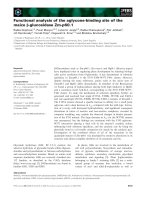
Figure 1 B and Th1 memory l ymphocyt es accumulate in the pancreas of NOD.Igμ
null
mice following B cell reconstitution. A) Flow
cytometry analyses of pancreas infiltrating lymphocytes in diabetic animals (between 20 and 30 weeks post-reconstitution) demonstrated an
accumulation of CD19
+
B cells (74%). T cells accumulate preferentially in pancreatic nodes (73%) and spleen (50%). Data represent 1 of 3
separate experiments with 2-4 animals per group. B) The majority of T cells found infiltrating the pancreas expressed memory marker CD44
high
(80% of CD8
+
and 50% of CD4
+
respectively). C) Pancreas infiltrating lymphocytes were in vitro stimulated with anti-CD3/CD28 beads and 5-day
supernatants were screened by cytokine bead assays (average and SD of 3 animals).
Vong et al. Journal of Translational Medicine 2011, 9:101
/>Page 4 of 10
was just above limits of detection (Figure 1C). The
observed pattern of cytokine production is characteristic
of a Th1 response associated with diabetogenic T cells.
This T cell response is likely the consequence of the
predominance of B cells activating effector T cells infil-
trating the pancreas.
Significant pancreatic TCR expansions are dependent
upon B cell reconstitution in NOD.Igμ
null
mice
Because of the already described role of T cells causing
T1D, pancreata from NOD.Igμ
null
mice were used for
spectratyping analysis and detection of T cell receptor
V beta chain (BV) expansions at different time points
between 5 to 22 weeks of age. Spleen and pancreatic
lymph nodes isolated from t he majority of NOD.Igμ
null
mice presented only Gaussian distributions across
every BV family tested. An example profile is demon-
strated in Figure 2 for BV2, 10, 12, 14 in 14 week-old
NOD.Igμ
null
mice (PLN-pancreatic lymph nodes and
SP-spleens). In the majority of the unreconstituted
NOD.Igμ
null
animals, pancreatic tissue did not generate
any d etectable PCR product for most BV-TCR families,
or presented rare Gaussian expansions (Figure 2).
These results indicated that T cells had not infiltrated
or were not clonally expanded in the pancreas in the
absence of B cells. I n contrast, clonotypic expansions
were observed in the pancreases of NOD.Igμ
null
mice
at 10 w eeks after B cell-reconstitution, indicating a
role for B cells in the recruitment and expansion of
pathogenic T cells ( Figure 2).
Predominant TCR expansion peaks were d etected by
spectratyping in BV2, 10, 12, 14, 18, 19 and 20 in B cell
reconstituted NOD.Igμ
null
mice. Interestingly, this TCR
expansion (non-Gaussian BVs) was specific to the pan-
creas, as pancreatic lymph nodes and spleens from these
animals only produced Gaussian distributions for these
same BV families. Total cell numbers recovered from
pancreatic lymph nodes were unchanged following B
cell reconstitution (data not shown) suggesting that the
T cell autoimmune response precipitating diabetes do
not appear to be expanding in lymphoid organs.
B cell reconstitution of NOD.Igμ
null
mice promotes
progressive expansion of the TCR repertoire in the
pancreas
To follow the progression of T cell infiltration after B
cell reconstitution of NOD.Igμ
null
mice, the animals
were spectratyped at different time points. At early time
points, 9-10 weeks post-B cell reconstitution, the major-
ity of the reconstituted animals accumulated BV2, BV10,
12 and 14 in the pa ncreas (Figure 3A). At intermediate
time points (13-16 weeks post-reconstitution), and even
later pre-diab etic and diabetic stages (19-31 weeks post-
reconstitution), an increase in the number of BV
families was observed. In particular, members of the
BV16 to 20 TCR repertoire were present at late r time
points (Figures 3B and 3C). These results demonstrate
that B cell reconstitution is required before a progressive
T cell infiltrate is found in the pancreas. The initial TCR
repertoire infiltrating the pancreas is less diverse, but
ultimately expands over time during diabetogenesis to
include a much broader TCR repertoire. This finding is
consistent with the spreading and diversification of the
pathogenic T cell repertoire [12,13].
B cell reconstituted NOD.Igμ
null
mice develop accelerated
diabetes following cyclophosphamide-treatment
To better under stand t he functionality of the TCR
expanded repertoire promot ed by B cell reconstitution
in NOD.Igμ
null
mice, we made use of the cyclophospha-
mide-accelerated diabe tes model. Cyclophosphamide
(CYP) has been shown to deplete the subset of T cells
with regulatory function and accelerate diabetes in NOD
mice [11]. We tested whether 14 week-old ureconsti-
tuted NOD.Igμ
null
mice could also develop accelerated
disease. Interestingly, we found these animals were resis-
tant to CYP-accelerated diabetes. However, in B-cell
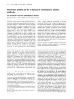
Figure 2 Representative comparative spectratype analysis for
BV families found in spleens, pancreatic lymph nodes and
pancreas from untreated and B-cell reconstituted NOD.Igμ
null
mice. Splenocytes (SP) and Pancreatic lymph nodes (PLN) were
analyzed from naïve NOD.Igμ
null
and B cell-reconstituted mice (NOD.
Igμ
null
+ B cells). Gaussian profiles for BV2, BV10, BV12 and BV14
families were found in spleens and lymph nodes. Pancreata (PN) of
naïve NOD.Igμ
null
animals had no expansions for these clonotypes,
but non-gaussian expansions were detected in high frequency
following B cell reconstitution.
Vong et al. Journal of Translational Medicine 2011, 9:101
/>Page 5 of 10
reconstituted animals, C YP treatment produced earlier
sickness with increased percentages of afflicted animals,
compared to age-matched NOD controls (data not
shown). We spectratyped the T cell repertoire in the
pancreata following CYP-treatment (Figure 4), and
found a decrease and/or loss of BV1, BV8 and BV11
TCR expansions. These families are normally present at
this time point in B cell reconstituted untreated NOD.
Igμ
null
mice, indicating their potential regulatory func-
tion. Furthermore, i ncreased expansions in BV4, BV5.2
and BV9 repertoires were found after CYP treatment, as
well as additional expansions of the BV16 to BV20
A)
B)
C)
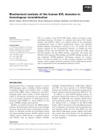
Figure 3 Policlonal BV r epertoire expansions are found in the pancreata following B cell reconstitution in NOD.Igμ
null
mice. A)
Spectratype analysis of BV-BC (Vbeta-Cbeta) expansions for pancreas-infiltrating T cells 10 weeks post-B cell reconstitution demonstrate a
polyclonal profile of induced clonotypes, with BV2, BV10, BV12 and BV14 being present on over 60% of the animals, followed next in appearance
by BV8S3 and BV11, present in 50% of the mice. B) As disease progresses, a higher diversity of clonotypes is observed, particularly for the
appearance of BV16, BV17, BV18, BV19 and BV20 in 13-16 weeks post-reconstitution and later C) at pre-diabetic stages (19-31 weeks post-
reconstitution).
Vong et al. Journal of Translational Medicine 2011, 9:101
/>Page 6 of 10
subsets of T cells, in comparison to age-matched B-cell
reconstituted NOD.Igμ
null
animals. These expansions
include BV families directed against antigens proposed
as targets of autoimmune response in diabetes patho-
genesis [7,14].
Pancreatic implants into diabetic NOD.Igμ
null
mice reveal
early invasive expansions in select BV clonotypes during
tissue rejection
To search for most aggressive/invasive BV expansions, we
studied the pancreatic-graft rejection model. We reasoned
that the repertoire potentially mediating early-graft rejec-
tion could be as i mportant in initiating T1D. In this
model, neonatal NOD.scid pancreases were implanted
under the kidney capsule of diabetic B cell reconstituted
NOD.Igμ
null
mice. After developing diabetes, mice were
kept alive by subcutaneous administration of insulin pel-
lets for 2-4 weeks to stabilize their glycemic levels prior to
implantation of neonat al NOD.scid pancreas under their
kidney capsule. After 40 hours, implants were removed for
spectratyping analysis of TCR repertoire of inf iltrating T
cells. Earlier studies suggested this time point to be the
best for examining implant infiltrate, before rejection and
fibrosis. Spectratyping analysis of the implants (Figure 5)
revealed polyclonal expansions, with several TCR families
(BV1,2,4,5.2,8.3,10and15)beingpresentinmostof
the implants, including BVs suspected to be of regulatory
phenotype based on the cyclophosphamide experiments
(Figure 4). Except for BV10, present in similar frequency
in the implants and the pancreas, these BV families were
found in higher frequencies in the implants, suggesting
their higher invasiveness.
Discussion
Previous studies examining T cell responses and reper-
toire analysis involved in the autoimmune response of
diabetes have produced conflicting results related to the
identification of the pathogenic T cell repertoire. Some
groups have described polyclonal T cell expansions ar is-
ing very early in the pancreas being responsible for islet
destruction, [15,16], while others have claimed that only
particular clonal expansions are the driving force behind
autoimmune responses in diabetes [17,18].
These variable findings likely reflect the different tech-
niques employed to characterize T c ell responses in the
pancreas during the course of spontaneous disease. Here
we have employed spectratyping analysis to detect T cell
expansions ex-vivo, in a non-biased attempt at examin-
ing the T cell responses in the pancreas following B cell
reconstitution in NOD.Igμ
null
mice. We found that by 9-
10 weeks post-B cell reconstitution, the majority of the
animals present pancreatic TCR expansions (at 13
weeks of life). Of note, these animals do not have clono-
typic expansions in their pancreatic lymph nodes or
spleens, suggesting that clonotypic TCR expansions in
lymphoid organs are not involved in disease induction
(Figure 2). The initial pancreatic T cell infiltration con-
sisted of several clonotypes, including BV2, BV10, and
BV12, clonotypes already described as reactive to insulin
or GAD65 [7,14]. BV12 has been found to b e enriched
in islets of NOD mice when compared to thymus and
spleens [15,19]. We also found a BV15 exp ansion that is
a possible candidate for BDC-10.1, a chromogranin A-
reactive BV15 T cell [20], which had been previously
charact erized with a high diabeto genic capacity [14]. As
disease progressed, an even larger TCR repertoire infil-
trating the organ was observed. This finding is consi s-
tent with spreading of the T cell response [21].
Considering the ever-growing list of islet antigens
described as being targets of autoimmune response in
T1D this polyclonality is expected [17,22]. We found
that during the pre-diabetic and diabet ic stages,
Figure 4 Spectratyping profile of B cell-reconstituted NOD.Igμ
null
mice following cyclophosphamide treatment. Spectratype analysis of
BV-BC expansions for pancreas-infiltrating T cells at 10 weeks post-B cell reconstitution of NOD.Igμ
null
mice are shown, following treatment with
cyclophosphamide (black bars) in comparison to 10 weeks old age-matched of NOD.Igμ
null
mice reconstituted with B cells (white bars), and
diabetic NOD.Igμ
null
mice reconstituted with B cells (grey bars).
Vong et al. Journal of Translational Medicine 2011, 9:101
/>Page 7 of 10
additional expansions in BV16, 17, 18, 19 and 20
families were commonly detected, but these were not
predominant in early infiltrates (Figure 3A). It is possi-
blesomeoftheseexpansionscouldcomprisealready
described pathogenic clones. A BV16 GAD65-reactive
clone (11H11) has been found in the islets of pre-dia-
betic NOD, w ith the distinct promiscuous capacity of
recognizing different GAD65 peptides using a single
TCR [23].
In attempts to find commonalities in clonotype expan-
sions in different pathological states of islet infiltration
in the B cell reconstituted NOD.Igμ
null
model, we also
examined the cyclophosphamide-accelerated diabetes
and the rejection of pancreatic implants in diabetic
NOD.Igμ
null
B cell reconstituted mice. Following cyclo-
phosphamide treatment, known to eliminate regulatory
T c ells from the pancreas [11], we found the pancreas-
infiltrating repertoire to be quite distinct from that of
age-matched non-diabetic NOD.Igμ
null
Bcellreconsti-
tuted mice, demonstrating that some regulatory compo-
nent is also promoted by B cell reconstitution.
Interestingly, BV1, BV8 and BV11 T cell e xpansions
were greatly reduced or lost, while a new set of BV4,
BV5S2, BV9 and BV16-20 expansions arose, suggesting
their role in pathogenicity. Furthermore, BV8S1 and
BV8S2 are absent in the cyclophosphamide treated
group (a treatment known to destroy regulatory T cells),
but present in 50% of the B cell-reconstituted animals.
BV8S1 h as been previously described as a predominant
clonotype infiltrating the islets of partially diabetes-resis-
tant male NOD mice [24] and, interestingly, is also pre-
sent in the bloo d of T1D patients [25]. This may
indicate that some regulatory component may still be
present, although ineffective, at final diseased stages
post-B cell reconstitution.
In another approach to address the identity of the
pathogenic repertoire, we examined the infiltrating T
cells rejecting new pancreatic implants (Figure 5). Pan-
creatic tissue from neonatal NOD.scids transplanted
under the kidney capsule of diabetic B cell reconstitu ted
NOD.Igμ
null
mice were rejected very fast (within 4 days),
with the peak of T cell infiltration occurring within 2
days after implantation. The spectratype profile of the
BV repertoire from day 2 implants (Figure 5, black bars)
was very similar to that seen in the diabetic pancrea s
(Figure 5 , grey bars) but with over 70% of the implants
presenting BV1, BV2, BV4, BV5S 2, BV8S3, BV10 and
BV15 clonotypes. Interestingly, BV1 and BV8 clonotypes
were decreased by cyclophosphamide treatment (Figure
4, black bars), therefore, wit h potential regulatory func-
tion. These findings indicate that in B cell reconstituted
NOD.Igμ
null
mice, highly invasive clonotypes predomi-
nantly infiltrating transplants are composed of particu-
larly high pathogenic effectors, as well as regulatory T
cells.
The breaking of T cell tolerance and passage through
“Checkpoint 1-End of Ignorance” [26] by B cell reconsti-
tution, may result owing to two different possibilities.
First, homeostatic proliferatio n of pathogenic T cells fol-
lowing sublethal irradiation, could awaken autoimmune
responses. Homeostati c proliferation in an immunodefi-
cient host due to sublethal irradiation or in NOD.scid
recipients, follows a pattern of expansion tha t takes
circa 6 weeks for complete reconstitution [14]. This
Figure 5 Spec tratyping profile of lymph ocytes infiltrating neonatal pancreas implanted under the kidney capsule of diab etic B cell-
reconstituted NOD.Igμ
null
mice. Spectratyping analysis of BV-BC of neonatal NOD.scid pancreas transplanted under the kidney capsule of
diabetic NOD.Igμ
null
mice reconstituted with B cells (black bars). Spectratyping profile of pancreata from diabetic B cell-reconstituted NOD.Igμ
null
(grey bars).
Vong et al. Journal of Translational Medicine 2011, 9:101
/>Page 8 of 10
mechanism has be en shown in the past to generate
autoimmune responses [8,27]. Second, autoimmunity
could be mediated by the expansion of a T cell reper-
toire remodeled by the presence of B cells, through their
unique antigenic display and enhanced proinflammatory
and costimulatory capacities. We argue that homeostatic
proliferation seems less likely, as T cells from control
animals (reconstitu ted with bone marrow following irra-
diation) also go through homeostatic proliferation but
do not develop diabetes! B cells from NOD mice are
known to produce strong inflammatory responses, when
compared to other non-autoimmune strains [28] and
present higher levels of costimulatory molecules [29].
Therefore, our data describing a B:T cell ratio of 4 in
the p ancreases support the second mechanism, and the
role for B cells as an important antigen presenting cells
in NOD.Igμ
null
mice.ItislikelythatBcellshelpinthe
induction/activation of the autoreactive TCR repertoire.
During diabetes promoted after B cell reconstitution
in NOD.Igμ
null
mice, B cells encompass over 64% of the
lymphocyte population infiltrating the pancreas, despite
equal numbers of B and T cells in the other lymphoid
organs analyzed (spleens and pancreatic lymph nodes).
Interestingly, recent study on the cellularity composition
of individual pancreatic islets in female and male NOD
mice at different time points of disease evolution do not
report a h igh accumulation of B cells in the pancreas
when compared to lymphoid organs [30], while another
study identify comparable B:T cell ratios in the spleen
for NOD animals [5]. The accumulation of B cells in
the pancreas of NOD.Igμ
null
reconstituted mice could
bypass the requirement for T cell lymph node recruit-
ment and may help explain why clonotypic T cell
expansions detected in the pancreas are not likewise
present in adjacent pancreatic lymph nodes by spectra-
typing studies. The autoimmune responses m ay be pre-
ferentially localized to the pancreas, induced by larger
numbers of antigen presenting B cells (B:T ratio of 4)
which could be promoting the effector T cell repertoires
unbalancing the regulatory clonotypes. The number of
CD19
+
B cells circulating in blood post-reconstitution in
NOD.Igμ
null
mice varied from 1 to 13%, with no correla-
tion of higher blood B cells and diabetes onset (data not
shown). B cell accumulation in the pancreases but not
in lymphoid organs, suggest that the direct activation of
effector T cells in the target organ by B cells may be the
crucial trigger for disease induction.
B cell accumulation in the pancreas appears to main-
tain CD4 and CD8 lymphocytes in an activated state
(CD44
high
Figure 1) and IL-6 secreted by the mononuc-
lear pancreatic infiltrate could modulate T cell activity.
IL-6 is known to alter phagolysosomal processing, enhan-
cing presentation of cryptic antigenic determinants [31]
and to provide survival signal for T cells [32]. Thus,
reintroduction of B cells appear to provide an ideal envir-
onment for pathogenic T cell activation and survival.
Conclusions
This study demonstrates that a polyclonal repertoire of
pathogenic T cell expansion is dependent upon B cell
reconstitution in NOD.Igμ
null
mice. Diabetes progression
appears to be facilitated by B cell accumulation in the
pancreas. Interestingly, the clonotypic T cell expansion
observed in the pancreas is not observed in other tradi-
tionally involved lymphoid organs, including the pan-
creatic lymph nodes and spleen. The dependence on B
cells for the appearance of the pathogenic repertoire of
T cells infiltrating the pancreas ma y help explain why
current therapies targeting B cells can affect T1D in
NOD mice and humans [33].
Acknowledgements
This paper is dedicated to the memory of Eli Sercarz, who passed away
before the completion of this work. This work was supported by grants to
Eli Sercarz: JDRF, Diabetes National Research Group and R01 AI65937-NIH.
We are very grateful to Dr. D. Serreze, Jackson Laboratory, for the NOD.
Igμ
null
mice and to Dr. V. Kumar (TPIMS) for critical review of the manuscript.
Authors’ contributions
AV, ND and PN performed NOD.Igμ
null
bone marrow and B cell chimera
reconstitutions, blood glucose measurements and spectratype experiments.
FACS and cytokine studies were performed by AV, HS and CG. CG, ES and
TB conceived and designed experiments. CG and TB wrote the manuscript.
Authors have read and approved the manuscript.
Competing interests
The authors declare that they have no competing interests.
Received: 23 March 2011 Accepted: 2 July 2011 Published: 2 July 2011
References
1. Serreze DV, Chapman HD, Varnum DS, Hanson MS, Reifsnyder PC,
Richard SD, Fleming SA, Leiter EH, Shultz LD: B lymphocytes are essential
for the initiation of T cell-mediated autoimmune diabetes: analysis of a
new “speed congenic” stock of NOD.Ig mu null mice. J Exp Med 1996,
184:2049-2053.
2. Serreze DV, Fleming SA, Chapman HD, Richard SD, Leiter EH, Tisch RM: B
lymphocytes are critical antigen-presenting cells for the initiation of T
cell-mediated autoimmune diabetes in nonobese diabetic mice. J
Immunol 1998, 161:3912-3918.
3. Serreze DV, Gaskins HR, Leiter EH: Defects in the differentiation and
function of antigen presenting cells in NOD/Lt mice. J Immunol 1993,
150:2534-2543.
4. Vasquez AC, Feili-Hariri M, Tan RJ, Morel PA: Qualitative and quantitative
abnormalities in splenic dendritic cell populations in NOD mice. Clin Exp
Immunol 2004, 135:209-218.
5. Noorchashm H, Lieu YK, Noorchashm N, Rostami SY, Greeley SA,
Schlachterman A, Song HK, Noto LE, Jevnikar AM, Barker CF, Naji A: I-Ag7-
mediated antigen presentation by B lymphocytes is critical in
overcoming a checkpoint in T cell tolerance to islet beta cells of
nonobese diabetic mice. J Immunol 1999, 163:743-750.
6. Silveira PA, Johnson E, Chapman HD, Bui T, Tisch RM, Serreze DV: The
preferential ability of B lymphocytes to act as diabetogenic APC in NOD
mice depends on expression of self-antigen-specific immunoglobulin
receptors. Eur J Immunol 2002, 32:3657-3666.
7. Quinn A, McInerney M, Huffman D, McInerney B, Mayo S, Haskins K,
Sercarz E: T cells to a dominant epitope of GAD65 express a public CDR3
motif. Int Immunol 2006, 18:967-979.
Vong et al. Journal of Translational Medicine 2011, 9:101
/>Page 9 of 10
8. Chiu PP, Serreze DV, Danska JS: Development and function of
diabetogenic T-cells in B-cell-deficient nonobese diabetic mice. Diabetes
2001, 50:763-770.
9. Pannetier C, Cochet M, Darche S, Casrouge A, Zoller M, Kourilsky P: The
sizes of the CDR3 hypervariable regions of the murine T-cell receptor
beta chains vary as a function of the recombined germ-line segments.
Proc Natl Acad Sci USA 1993, 90:4319-4323.
10. Clough LE, Wang CJ, Schmidt EM, Booth G, Hou TZ, Ryan GA, Walker LS:
Release from regulatory T cell-mediated suppression during the onset of
tissue-specific autoimmunity is associated with elevated IL-21. J Immunol
2008, 180:5393-5401.
11. Brode S, Raine T, Zaccone P, Cooke A: Cyclophosphamide-induced type-1
diabetes in the NOD mouse is associated with a reduction of CD4+CD25
+Foxp3+ regulatory T cells. J Immunol 2006, 177:6603-6612.
12. Lehmann PV, Sercarz EE, Forsthuber T, Dayan CM, Gammon G: Determinant
spreading and the dynamics of the autoimmune T-cell repertoire.
Immunol Today 1993, 14:203-208.
13. Dai YD, Carayanniotis G, Sercarz E: Antigen processing by autoreactive B
cells promotes determinant spreading. Cell Mol Immunol 2005, 2:169-175.
14. Burton AR, Vincent E, Arnold PY, Lennon GP, Smeltzer M, Li CS, Haskins K,
Hutton J, Tisch RM, Sercarz EE, Santamaria P, Workman CJ, Vignali DA: On
the pathogenicity of autoantigen-specific T-cell receptors. Diabetes 2008,
57:1321-1330.
15. Sarukhan A, Gombert JM, Olivi M, Bach JF, Carnaud C, Garchon HJ:
Anchored polymerase chain reaction based analysis of the V beta
repertoire in the non-obese diabetic (NOD) mouse. Eur J Immunol 1994,
24:1750-1756.
16. Sarukhan A, Bedossa P, Garchon HJ, Bach JF, Carnaud C: Molecular analysis
of TCR junctional variability in individual infiltrated islets of non-obese
diabetic mice: evidence for the constitution of largely autonomous T
cell foci within the same pancreas. Int Immunol 1995, 7:139-146.
17. Drexler K, Burtles S, Hurtenbach U: Limited heterogeneity of T-cell
receptor V beta gene expression in the early stage of insulitis in NOD
mice. Immunol Lett 1993, 37:187-196.
18. Galley KA, Danska JS: Peri-islet infiltrates of young non-obese diabetic
mice display restricted TCR beta-chain diversity. J Immunol 1995,
154:2969-2982.
19. Baker FJ, Lee M, Chien YH, Davis MM: Restricted islet-cell reactive T cell
repertoire of early pancreatic islet infiltrates in NOD mice. Proc Natl Acad
Sci USA 2002, 99:9374-9379.
20. Stadinski BD, Delong T, Reisdorph N, Reisdorph R, Powell RL, Armstrong M,
Piganelli JD, Barbour G, Bradley B, Crawford F, Marrack P, Mahata SK,
Kappler JW, Haskins K: Chromogranin A is an autoantigen in type 1
diabetes. Nat Immunol 2010, 11:225-231.
21. Lehmann PV, Forsthuber T, Miller A, Sercarz EE: Spreading of T-cell
autoimmunity to cryptic determinants of an autoantigen. Nature 1992,
358:155-157.
22. Waters SH, O’Neil JJ, Melican DT, Appel MC: Multiple TCR V beta usage by
infiltrates of young NOD mouse islets of Langerhans. A polymerase
chain reaction analysis. Diabetes 1992, 41:308-312.
23. Li L, Wang B, Frelinger JA, Tisch R: T-cell promiscuity in autoimmune
diabetes. Diabetes 2008, 57:2099-2106.
24. Berschick P, Fehsel K, Weltzien HU, Kolb H: Molecular analysis of the T-cell
receptor V beta 5 and V beta 8 repertoire in pancreatic lesions of
autoimmune diabetic NOD mice. J Autoimmun 1993, 6:405-422.
25. Naserke HE, Durinovic-Bello I, Seidel D, Ziegler AG: The T-cell receptor beta
chain CDR3 region of BV8S1/BJ1S5 transcripts in type 1 diabetes.
Immunogenetics 1996, 45:87-96.
26. Andre I, Gonzalez A, Wang B, Katz J, Benoist C, Mathis D: Checkpoints in
the progression of autoimmune disease: lessons from diabetes models.
Proc Natl Acad Sci USA 1996, 93:2260-2263.
27. Baccala R, Theofilopoulos AN: The new paradigm of T-cell homeostatic
proliferation-induced autoimmunity. Trends Immunol 2005, 26:5-8.
28. Hussain S, Salojin KV, Delovitch TL: Hyperresponsiveness, resistance to B-
cell receptor-dependent activation-induced cell death, and accumulation
of hyperactivated B-cells in islets is associated with the onset of insulitis
but not type 1 diabetes. Diabetes 2004, 53:2003-2011.
29. Hussain S, Delovitch TL: Dysregulated B7-1 and B7-2 expression on
nonobese diabetic mouse B cells is associated with increased T cell
costimulation and the development of insulitis. J Immunol 2005,
174:680-687.
30. Young EF, Hess PR, Arnold LW, Tisch R, Frelinger JA: Islet lymphocyte
subsets in male and female NOD mice are qualitatively similar but
quantitatively distinct. Autoimmunity 2009, 42:678-691.
31. Drakesmith H, O’Neil D, Schneider SC, Binks M, Medd P, Sercarz E,
Beverley P, Chain B: In vivo priming of T cells against cryptic
determinants by dendritic cells exposed to interleukin 6 and native
antigen. Proc Natl Acad Sci USA 1998, 95:14903-14908.
32. Teague TK, Schaefer BC, Hildeman D, Bender J, Mitchell T, Kappler JW,
Marrack P: Activation-induced inhibition of interleukin 6-mediated T cell
survival and signal transducer and activator of transcription 1 signaling.
J Exp Med 2000, 191:915-926.
33. O’Neill SK, Liu E, Cambier JC: Change you can B(cell)eive in: recent
progress confirms a critical role for B cells in type 1 diabetes. Curr Opin
Endocrinol Diabetes Obes 2009, 16:293-298.
doi:10.1186/1479-5876-9-101
Cite this article as: Vong et al.: Spectratyping analysis of the islet-
reactive T cell repertoire in diabetic NOD Igμ
null
mice after polyclonal B
cell reconstitution. Journal of Translational Medicine 2011 9:101.
Submit your next manuscript to BioMed Central
and take full advantage of:
• Convenient online submission
• Thorough peer review
• No space constraints or color figure charges
• Immediate publication on acceptance
• Inclusion in PubMed, CAS, Scopus and Google Scholar
• Research which is freely available for redistribution
Submit your manuscript at
www.biomedcentral.com/submit
Vong et al. Journal of Translational Medicine 2011, 9:101
/>Page 10 of 10