Báo cáo sinh học: " Simian immunodeficiency virus (SIV) envelope quasispecies transmission and evolution in infant rhesus macaques after oral challenge with uncloned SIVmac251: increased" pdf
Bạn đang xem bản rút gọn của tài liệu. Xem và tải ngay bản đầy đủ của tài liệu tại đây (2.73 MB, 15 trang )
BioMed Central
Page 1 of 15
(page number not for citation purposes)
Virology Journal
Open Access
Research
Simian immunodeficiency virus (SIV) envelope quasispecies
transmission and evolution in infant rhesus macaques after oral
challenge with uncloned SIVmac251: increased diversity is
associated with neutralizing antibodies and improved survival in
previously immunized animals
Jennifer L Greenier
1
, Koen KA Van Rompay
1
, David Montefiori
2
,
Patricia Earl
3
, Bernard Moss
3
and Marta L Marthas*
1,4
Address:
1
California National Primate Research Center, University of California, Davis, CA 95616, USA,
2
Duke University Medical Center,
Durham, NC 27710, USA,
3
Laboratory of Viral Diseases, National Institutes of Health, Bethesda, MD 20892, USA and
4
Department of Pathology,
Microbiology and Immunology, School of Veterinary Medicine, University of California, Davis, CA 95616, USA
Email: Jennifer L Greenier - ; Koen KA Van Rompay - ;
David Montefiori - ; Patricia Earl - ; Bernard Moss - ;
Marta L Marthas* -
* Corresponding author
pediatricvaccineHIVHMA
Abstract
Background: Oral infection of infant macaques with simian immunodeficiency virus (SIV) is a useful
animal model to test interventions to reduce postnatal HIV transmission via breast-feeding. We previously
demonstrated that immunization of infant rhesus macaques with either modified vaccinia virus Ankara
(MVA) expressing SIV Gag, Pol and Env, or live-attenuated SIVmac1A11 resulted in lower viremia and
longer survival compared to unimmunized controls after oral challenge with virulent SIVmac251 (Van
Rompay et al., J. Virology 77:179–190, 2003). Here we evaluate the impact of these vaccines on oral
transmission and evolution of SIV envelope variants.
Results: Limiting dilution analysis of SIV RNA followed by heteroduplex mobility assays of the V1–V2
envelope (env) region revealed two major env variants in the uncloned SIVmac251 inoculum. Plasma
sampled from all infants 1 week after challenge contained heterogeneous SIV env populations including one
or both of the most common env variants in the virus inoculum; no consistent differences in patterns of
env variants were found between vaccinated and unvaccinated infants. However, SIV env variant
populations diverged in most vaccinated monkeys 3 to 5 months after challenge, in association with the
development of neutralizing antibodies.
Conclusions: These patterns of viral envelope diversity, immune responses and disease course in SIV-
infected infant macaques are similar to observations in HIV-infected children, and underscore the
relevance of this pediatric animal model. The results also support the concept that neonatal immunization
with HIV vaccines might modulate disease progression in infants infected with HIV by breast-feeding.
Published: 14 February 2005
Virology Journal 2005, 2:11 doi:10.1186/1743-422X-2-11
Received: 24 December 2004
Accepted: 14 February 2005
This article is available from: />© 2005 Greenier et al; licensee BioMed Central Ltd.
This is an Open Access article distributed under the terms of the Creative Commons Attribution License ( />),
which permits unrestricted use, distribution, and reproduction in any medium, provided the original work is properly cited.
Virology Journal 2005, 2:11 />Page 2 of 15
(page number not for citation purposes)
Background
The continued need for breast-feeding in developing
countries due to nutritional or socio-economic reasons
poses a considerable risk for postnatal mother-to-child
transmission of HIV, and breastfeeding is estimated to
account for 33–50% of infant HIV infections worldwide
[1-5]. This dilemma underscores the need for a vaccine
that, when administered shortly after birth to the infant,
could protect against HIV transmission via breast-feeding.
The ultimate goal of a neonatal HIV vaccine is to prevent
infection; however, vaccination of newborns of HIV-
infected women early in life may elicit HIV-specific
immune responses that substantially reduce infant disease
progression in the event that breast milk transmission
occurs.
Advances in the understanding of the mechanisms of oral
transmission of HIV variants may aid the development of
an effective infant HIV-1 vaccine. Recent studies have
demonstrated that infants of HIV-infected women can be
infected with single or multiple HIV variants [6,7] shortly
before or during the birth process. However, little is
known regarding the diversity of HIV transmitted by
breastfeeding. These questions are difficult to address in
human studies because the characteristics of HIV variants
in breast-milk at the time of transmission are unknown.
In addition, it is often difficult to obtain virus from infants
at early times after HIV infection. Finally, the presence in
infants of different levels of transplacentally transferred
HIV-specific maternal antibodies with differing anti-viral
properties complicates assessments of HIV variant
transmission.
Longitudinal studies of HIV-infected adults have shown
that the rate of disease progression is inversely related to
the rate of evolution of HIV envelope quasispecies [8,9].
Also, without antiviral treatment, virus-specific immune
responses are directly related to HIV quasispecies evolu-
tion [10]. The reported relationship between HIV enve-
lope variant evolution and disease progression in HIV-
infected infants and children is contradictory. Some stud-
ies have found greater HIV envelope variant evolution in
rapid progressors [11-13] while other investigations have
found that slowly progressing HIV-infected children have
greater HIV quasispecies divergence or diversity over time
[14,15]. However, all of these retrospective studies neces-
sarily evaluated HIV variant evolution in a limited
number of serial blood samples during the first months of
life from a small number of HIV-infected children (two to
six per cohort). More recently, a longitudinal study of 10
perinatally HIV-infected children found that changes in
HIV envelope quasispecies during the first year of life were
associated with a better clinical outcome [7]. A few reports
have described a correlation between nascent HIV-specific
immune responses, the evolution of HIV variants and dis-
ease progression in HIV-infected infants [16,17].
Simian immunodeficiency virus (SIV) infection of infant
macaques is a useful and relevant animal model of pedi-
atric HIV infection for rapidly testing the efficacy of pedi-
atric HIV vaccine and drug interventions [18-20]. This
SIV/infant macaque model was previously used to assess
the efficacy of two vaccines, (i) modified vaccinia virus
Ankara (MVA) expressing SIV Gag, Pol and Env (MVA-
SIVgpe) and (ii) live-attenuated SIVmac1A11, against oral
challenge with virulent uncloned SIVmac251. We
reported an improved clinical outcome (i.e., disease-free
survival) for vaccinated compared with unvaccinated
infants, which was associated with reduced plasma SIV
RNA and sustained SIV-specific humoral immune
responses [21]. Here in this report, we used a heterodu-
plex mobility assay (HMA) to evaluate the genetic diver-
sity in the V1–V2 envelope (env) region of SIV variants
present in the SIVmac251 virus inoculum and compare
the transmission and evolution of the SIV env quasispecies
in plasma following oral inoculation of these vaccinated
and unvaccinated infant macaques. Three major ques-
tions were addressed: (i) Compared to the SIVmac251
virus inoculum, are few SIV envelope variants transmitted
orally?, (ii) Is the lower viremia and better clinical out-
come of vaccinated infants related to the initial genetic
diversity of SIV env quasispecies?, and, (iii) Is the evolu-
tion of SIV envelope quasispecies during the course of
infection associated with the development of SIV neutral-
izing antibody? We demonstrate that while the vaccines
did not modulate oral transmission of viral variants, an
association was found between vaccine-induced
enhanced antiviral immune responses, increased env
diversity, and a slower disease course. These findings in
vaccinated infant macaques are similar to observations in
HIV-infected children with slow disease progression and
support the relevance of the SIV infant macaque model for
developing neonatal vaccine strategies to prevent pediat-
ric HIV infection and AIDS.
Results
Characterization of variants in SIVmac251-5/98 virus stock
HMA analysis revealed that the undiluted SIVmac251-5/
98 virus stock was comprised of a diverse population of
V1–V2 env variants. To determine the most common var-
iant(s) in the virus stock, six independent serial dilution
experiments were conducted. Viral RNA was isolated from
1 ml of virus stock and 10-fold dilution series (undiluted
to 10
-9
) of the RNA were prepared from 6 separate aliq-
uots of virus stock. The resulting RNA was analyzed by RT-
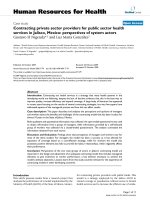
PCR and HMA. Figure 1 shows the results of 4 of these 6
separate virus stock dilution/HMA experiments. The
observation that multiple heteroduplex bands were
observed through the 10
-5
or 10
-6
dilutions of viral RNA
Virology Journal 2005, 2:11 />Page 3 of 15
(page number not for citation purposes)
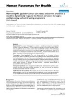
Characterization of variants in SIVmac251-5/98 virus stockFigure 1
Characterization of variants in SIVmac251-5/98 virus stock. HMA analysis of four separate dilution series of viral RNA
from the SIVmac251-5/98 virus stock is shown. The presence of multiple bands in the undiluted samples (lane 1 of each gel)
reveals the virus stock was comprised of a diverse viral population. The last lane of each gel shows the variants in the highest
dilution that yielded an RT-PCR product. Dilution series A shows an example of a dilution experiment that did not result in a
virus stock endpoint (homogenous variant population); the 10
-6
dilution included more than 1 variant, while the next dilutions
(10
-7
–10
-9
) dilution did not yield RT-PCR products, and therefore no variant pattern is shown for those dilutions. This dilution
pattern was observed in 3 of 6 dilution series (other 2 not shown). For the other 3 dilution series (B, C, and F), the variant
(band) remaining in the highest dilution was considered to be the most common variant, and was designated the Virus Stock
Endpoint Variant (VSEV). Dilution series B: no product was amplified from the 10
-7
dilution (lane 8), but a product was ampli-
fied from the 10
-8
dilution (lane 9). Dilution series C: lanes 7 and 8 show the presence of 2 different variants (VSEV-1 and
VSEV-2) in the endpoint dilutions (10
-6
and 10
-7
) of this series. Dilution series F; the 10
-6
dilution in this series harbored an end-
point variant that migrated to the same gel position as VSEV-2 in dilution series C.
Virology Journal 2005, 2:11 />Page 4 of 15
(page number not for citation purposes)
indicates that the undiluted SIVmac251-5/98 stock con-
tains multiple env variants at high frequency. An RT-PCR
endpoint (i.e. dilution to a single variant) was not reached
in 3 of the 6 dilution experiments. An example of this is
shown in dilution series A (Figure 1). In the other 3 dilu-
tion series (Fig. 1, series B, C, and F), the last dilution that
yielded an RT-PCR product consisted of a homogeneous
population of envelope variants represented by one main
variant (homoduplex band). This endpoint variant was
designated the virus stock endpoint variant (VSEV). The
fact that endpoint variants were reached at different dilu-
tions for each dilution series is probably due to the varia-
bility at each step of these independently performed
experiments.
The VSEV in dilution series B and F had different mobili-
ties on the HMA gel (Fig. 1). Dilution series C resulted in
two endpoint variants, one at 10
-6
and the other at 10
-7
;
the positions of these two VSEV corresponded to one of
each of the two VSEV in dilution series B and F. Thus, the
dilution of the virus stock to an RT-PCR endpoint resulted
in 4 independent variants (represented by homoduplex
bands) that migrated to two different positions on the
HMA gels. Based on these positions, the homoduplex
bands that migrated furthest were referred to as VSEV-1
and the variants that migrated a shorter distance were des-
ignated VSEV-2 (Fig. 1). To confirm that the four endpoint
homoduplexes represented only two variants, an HMA
mixture experiment was performed, in which all pairwise
combinations of the virus stock endpoint variants were
mixed prior to HMA [22]. These experiments demon-
strated that the two variants designated VSEV-1 are indeed
similar (i.e., = 1–2% difference in nucleotides with no
insertion/deletion), as their mixtures resulted in the for-
mation of a single homoduplex band on an HMA gel; sim-
ilarly, the two variants referred to as VSEV-2 are similar
(Fig. 2). In contrast, the formation of heteroduplexes and
two main homoduplexes in the mixtures of VSEV-1 and
VSEV-2 demonstrate that these 2 variants are significantly
different from each other (Fig. 2). Thus, VSEV-1 and VSEV-
2 are 2 distinct variants that exist at similar frequencies
and represent the most common variants in the undiluted
SIVmac251-5/98 virus stock. These results are consistent
with observations of the virus stock from which
SIVmac251-5/98 was made [22].
Experimental design of animal experiments and summary
of outcome
Nineteen newborn rhesus macaques were divided into 5
experimental vaccine groups (table 1). Group 1 (n = 5)
consisted of unimmunized control animals. Group 2 (n =
2), group 3 (n = 4) and group 4 (n = 4) were vaccinated
with MVA-SIVgpe at 0 and 3 weeks of age; group 4 had
maternally-derived SIV antibodies (due to immunization
of their mothers with inactivated SIV). Group 5 (n = 4)
was immunized with live-attenuated SIVmac1A11 at 0
and 3 weeks of age. As described elsewhere [21], except for
group 2, all other groups were inoculated orally with
SIVmac251-5/98 at 4 weeks of age; all these animals
became persistently viremic, but the immunized animals
had lower virus levels, enhanced antiviral immune
responses and a delayed disease course in comparison to
the unimmunized animals. Four of the 5 unimmunized
infected animals developed AIDS within 14 weeks of age,
while the fifth animal needed euthanasia at 28 weeks.
Four MVA-SIVgpe-vaccinated SIVmac251-5/98-infected
animals developed AIDS by 19 to 27 weeks of age (2 ani-
mals of groups 3 and 4 each; table 1). The remaining eight
vaccinated SIVmac251-5/98-infected infants, including
all four SIVmac1A11-vaccinated animals, were clinically
stable at the end of the observation period (28 weeks of
age).
Detection of SIV envelope variants in plasma of neonates
early after oral inoculation
The genetic diversity of SIV env variant populations in the
plasma of the infant monkeys one week after oral inocu-
lation with SIVmac251 was analyzed by HMA (Fig. 3).
Each plasma sample was analyzed in replicates (≥ 2) to
assure reproducibility of the gel banding patterns. As indi-
cated by the presence of heteroduplex bands, all infants
were infected with multiple SIV env variants, indicating
that the SIVmac251-5/98 virus stock contained several
variants capable of establishing infection by the oral
route. However, there were differences in HMA banding
patterns. In each group, some animals had several strong
heteroduplex bands; this pattern of variant transmission
was referred to as infection pattern A (e.g. Fig. 3, animal
31319). In contrast, one or two infants in each group were
infected with a genetically more homogenous variant
population, consisting of one major variant (homoduplex
band), while heteroduplex bands were less pronounced.
These monkeys infected with genetically more homogene-
ous viral populations harbored one of two main env vari-
ants, distinguished by different electrophoretic mobilities
of the homoduplexes representing these variants. These
more homogeneous variant populations were referred to
as infection patterns B and C (e.g. Fig. 3, animals 31325
and 31608, respectively). Infection pattern C contained a
homoduplex band that migrated slightly slower than the
homoduplex band characterizing infection pattern B. One
newborn in each vaccine group was infected with a SIV
variant of transmission pattern B. Infection pattern C was
detected in one newborn of each group except the
SIVmac1A11 vaccinates (table 1, group 5). Therefore, no
substantial difference was observed among the different
vaccine groups in viral genetic diversity in plasma col-
lected 1 week after virus inoculation. However, all but one
infant (31540) infected with more homogenous popula-
tions of env variants (infection patterns B or C) had 10- to
Virology Journal 2005, 2:11 />Page 5 of 15
(page number not for citation purposes)
100-fold lower virus levels one week after SIVmac251
challenge than all but one infant (31378) infected with
more heterogeneous populations of SIV variants (trans-
mission pattern A, Table 1). This association of homoge-
neous viral variants with reduced SIV RNA in plasma at 1
week after infection was statistically significant (P ≤ 0.05;
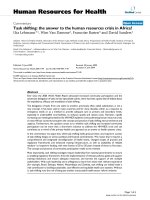
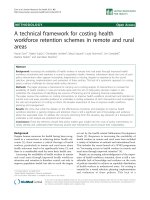
Characterization of the dominant variants in SIVmac251-5/98 virus stockFigure 2
Characterization of the dominant variants in SIVmac251-5/98 virus stock. HMA analysis of all four endpoint variants
shown in Fig. 1 (lanes 1–4) and all possible pairwise mixtures of those variants (lanes 5-10) are shown. Letters B, C, and F refer
to the dilution series shown in Fig. 1. Lane numbers refer to the lane designations of the variants that were mixed in lanes 5–10
(e.g., L1 + L2 indicates that the variants shown in lanes 1 and 2 were mixed). Lane 6 shows that the 2 endpoint variants labeled
VSEV-1 (B 10
-8
and C 10
-7
) are similar variants due to the formation of a single homoduplex and no heteroduplexes when these
2 variants were mixed. Lane 9 indicates that the 2 endpoint variants labeled VSEV-2 (C 10
-6
and F 10
-6
) in Fig. 1 are very similar.
The formation of heteroduplexes and two main homoduplexes in the mixtures shown in lanes 5, 7, 8, and 10 indicate that
VSEV-1 and VSEV-2 do not share the same V1–V2 envelope sequence.
Virology Journal 2005, 2:11 />Page 6 of 15
(page number not for citation purposes)
one-sided Fisher's Exact test) but did not persist. From
week 2 after challenge throughout the duration of the
study, plasma SIV RNA levels showed no correlation with
the initial SIV variant pattern detected in plasma. The rate
of disease progression in these animals was also not asso-
ciated with the initial envelope variant transmission pat-
terns (table 1). Further, there was no correlation between
the presence of the MHC type I alleles Mamu-A*01 or
Mamu-B*01 and the viral variant infection patterns, levels
of SIV RNA in plasma, or disease progression (table 1).
To determine which SIV envelope variant was present in
the highest frequency in each infection pattern, serial end-
point dilution experiments were performed with RNA iso-
lated from plasma collected one week after SIVmac251
challenge, followed by RT-PCR and HMA. Similar to the
methods described above, mixture experiments were then
performed, including with VSEV-1 and VSEV-2. These
experiments demonstrated that 1 week after infection, the
most common variants in animals with the more homog-
enous transmission patterns B and C were similar (i.e.,
less than 1–2 % difference based on the absence of heter-
oduplex bands) to VSEV-1 and VSEV-2, respectively (data
not shown). The most common variants by end-point
dilution in the 11 monkeys with transmission pattern A
and A/C were similar to VSEV-1 (5 animals), or VSEV-2 (5
animals) or both (1 animal).
Table 1: Experimental design and summary of outcome.
Immunization
a
groups and
animal numbers
sex MHC I alleles
b
Variant Pattern
c
Week 1 Plasma
Viral RNA
d
Time of eutha-
nasia (wks)
e
MamuA*01 MamuB*01
Group 1 Unvaccinated +
SIVmac251
31319 M + + A 4.3 × 10
7
13
31321 M +/- - A 1.7 × 10
8
28
31322 F +/- +/- A 1.2 × 10
8
14
31325 M + + B 5.5 × 10
6
12
31608
f
F +/- +/- C 7.5 × 10
5
11
Group 2 MVA-SIVgpe only
31480 M - - na na na
31488 M +/- +/- na na na
Group 3 MVA-SIVgpe +
SIVmac251
31378 M - - A 4.8 × 10
5
28
g
31533 M +/- - A 3.7 × 10
7
26
31540 M +/- - C 2.5 × 10
7
28
g
31542 M - - B 3.3 × 10
5
26
Group 4 MVA-SIVgpe with
Mat. Abs. + SIVmac251
31526 M +/- +/- A 6.9 × 10
7
27
31732 F - +/- A 1.8 × 10
7
19
31833 F +/- - A/C 4.5 × 10
5
28
g
31856 F - +/- B 1.4 × 10
6
28
g
Group 5 SIVmac1A11 +
SIVmac251
31777 F +/- - A 6.8 × 10
7
28
g
31778 F - - B 4.7 × 10
5
28
g
31779 F - - A 2.3 × 10
8
28
g
31780 F +/- - A 9.9 × 10
7
28
g
a
Vaccine administered in 2 doses, at birth and 3 weeks of age. Animals of groups 1, 3, 4 and 5 were challenged orally at 4 weeks of age with
SIVmac251-5/98.
b
The presence of the MHC type I alleles of MamuA*01 and MamuB*01 is indicated as + (present, but unknown whether homozygous or
heterozygous), +/- (heterozygous based on known haplotypes of parents), and - (homozygous for absence of particular allele).
c
Variants in plasma at one week post-challenge with SIVmac251-5/98.
d
Copies of viral RNA per ml one week after challenge with SIVmac251-5/98, as measured by bDNA assay.
e
Age (weeks) at time of euthanasia.
f
Infant 31608 was born to an SIVmac251-infected macaque, and thus had maternal anti-SIV antibodies, but no virus was detected in this infant at 4
weeks of age.
g
indicates that animal was clinically stable at time of experimental euthanasia at 28 weeks of age; all other SIV-infected animals were euthanized due
to life-threatening disease prior to or at 28 weeks of age. The animals of group 2 were not euthanized. na indicates not applicable.
Virology Journal 2005, 2:11 />Page 7 of 15
(page number not for citation purposes)
Greater quasispecies diversity in vaccinated compared to
control infants during chronic SIV infection
HMA was used to analyze the evolution of genetic diver-
sity of V1–V2 env populations in plasma of the monkeys
during the course of infection (1 week after oral
SIVmac251-5/98 challenge until euthanasia) (Fig. 4).
Results from two standard measures of the nucleotide
sequence heterogeneity of V1–V2 env plasma variants
derived from the HMA analyses are shown in Fig. 5: (i)
entropy (E), an estimate of the overall viral RNA sequence
complexity for each sample and, (ii) median mobility
shift (MMS), a measure of the SIV quasispecies sequence
divergence reflected by the degree of base-pair mismatch
after DNA strand re-annealing of strands of envelope var-
iants [8].
The diversity of SIV env quasispecies in plasma varied
among animals at the first sample (1 week after challenge)
as indicated by the gel banding pattern (Fig. 4) and
entropy measures (Fig. 5). Entropy for SIV env popula-
tions was high (> 0.9) for all unvaccinated animals
(Group 1) and for 7 of the 12 vaccinated animals (Fig. 5).
Initial entropy < 0.9 for vaccinated animals was associated
with lower SIV RNA in plasma at 1 week after challenge (P
< 0.05; Fisher's Exact Test). No consistent pattern of
entropy over the 24 week course of infection was
observed; in two of the five controls (31321, 31608) and
three of the 12 vaccinates (31533, 31732, 31780) entropy
decreased near the time of euthanasia. Overall, there was
no association of SIV envelope diversity as measured by
entropy with either viral RNA levels or virus-specific neu-
tralizing antibodies in plasma (see below and Fig. 5).
The sequence divergence of SIV envelope variants in
plasma of each animal over time was estimated by the
MMS, shown in Fig. 5. Four of the five unvaccinated ani-
mals had initial MMS values ≥ 0.5 which decreased at var-
ying rates until the time of euthanasia; the remaining
control animal (31325) had an initial MMS < 0.3 which
did not change significantly over the course of infection
(Fig. 5). Thus, in unvaccinated infants the population of
SIV env variants in plasma exhibited either no sequence
divergence or increasing sequence similarity over time;
this observation is consistent with the absence of sus-
tained SIV-specific immune responses in these animals
([21]; see below). There was no association between MMS
values and SIV RNA plasma levels for these unimmunized
animals.
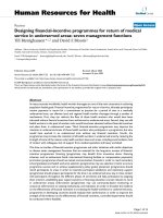
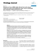
Variant populations present in plasma of infant macaques one week after oral challenge with SIVmac251-5/98Figure 3
Variant populations present in plasma of infant macaques one week after oral challenge with SIVmac251-5/98.
RT-PCR and HMA analysis was performed on replicate samples to confirm reproducibility of the results. Three main transmis-
sion patterns were observed, labeled A (multiple variants; diverse virus population), B and C (one major homoduplex (Ho)
with a few faint heteroduplexes (He); relatively homogenous virus population). One infant (31833) harbored a plasma virus
population that had elements of both transmission patterns A and C. SIV251 V.S. indicates the SIVmac251-5/98 virus stock.
Virology Journal 2005, 2:11 />Page 8 of 15
(page number not for citation purposes)
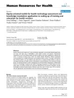
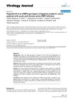
Evolution of plasma variants in SIVmac251-5/98-infected infant macaquesFigure 4
Evolution of plasma variants in SIVmac251-5/98-infected infant macaques. HMA analysis was performed on sequen-
tial plasma RNA samples, and each analysis was done at least twice to assure reproducibility. Virus diversification is evidenced
by the detection of additional minor heteroduplex bands, the disappearance of major heteroduplex bands, and/or the decrease
in density of the homoduplex bands. V.S. indicates the SIVmac251-5/98 virus stock. The lane numbers refer to the number of
weeks after SIVmac251-5/98 inoculation (which was performed at 4 weeks of age). The homoduplex band for week 0 for ani-
mal 31780 (prior to SIVmac251 challenge) represents the vaccine virus SIVmac1A11; viral RNA levels for the other
SIVmac1A11-immunized animals at this time were too low to result in a detectable RT-PCR product.
Virology Journal 2005, 2:11 />Page 9 of 15
(page number not for citation purposes)
For 3 of the 4 vaccinated animals that developed AIDS
within the observation period of 28 weeks (animals
31732, 31533, 31542), we also observed little change or a
decrease of genetic divergence (i.e., as measured by stable
or decreasing MMS values) of plasma env variant
quasispecies. In contrast, diversification in plasma SIV env
variant populations (i.e., a significant increase in MSS val-
ues) was observed by 3 to 5 months of infection in 4 of the
8 vaccinated monkeys (31540, 31833, 31856 and 31778)
that were still relatively healthy at 28 weeks (Fig. 5). This
diversification corresponded to the detection of different
patterns of heteroduplex bands and/or fainter homodu-
plex bands over time (Fig. 4). Although increased diversi-
fication seemed to correlate with improved disease-free
survival, this diversification was not associated with any
obvious changes in plasma virus levels.
SIV neutralizing antibodies in vaccinates correlate with
evolution of SIV quasispecies diversity
The possibility that SIV envelope-specific immune
responses were associated with the observed plasma SIV
RNA levels or evolution of SIV env quasispecies was eval-
uated by measuring levels of plasma antibodies that neu-
tralized the homologous challenge virus, SIVmac251-5/
98, during the course of infection (Fig. 5). SIV neutralizing
antibodies were detected in none of the unvaccinated con-
trol animals, but in all except one (31777) of the 12 vac-
cinated animals within 16 to 20 weeks after infection (Fig.
5). Although the presence of SIV neutralizing antibodies
was associated with increased survival of the vaccinated
animals, no obvious relationship was detected between
the SIV neutralizing antibody levels and either SIV RNA
plasma levels or entropy over time in vaccinated animals.
However, in the 5 animals with increasing sequence diver-
Evolution of viral diversity and SIV neutralizing antibody responseFigure 5
Evolution of viral diversity and SIV neutralizing antibody response. HMA data for each animal (Fig. 4) were further
analyzed by calculating the entropy and the median mobility shift (MMS). Viral RNA levels were measured by bDNA. SIV neu-
tralizing antibodies were determined as described in the Materials and Methods; neutralizing antibody titers below cut-off value
(i.e., < 30) were given a value of 10 for presentation on these graphs. Dashed lines indicate a regression line for entropy, MMS
or neutralizing antibody titer that is significantly different (P < 0.05) from zero (i.e. significantly increasing or decreasing values
from 1 week to 24 weeks pc, with r
2
values ≥ 0.45).
Virology Journal 2005, 2:11 />Page 10 of 15
(page number not for citation purposes)
gence (i.e., increasing MMS values; animals 31540,
31526, 31833, 31856 and 31778), neutralizing antibod-
ies were detected around the time that MMS values
increased, and the neutralizing antibody response was
sustained (i.e., detectable in ≥ 3 plasma samples) in these
5 animals (Fig. 5). In contrast, animals with stable or
declining MMS values had sustained (31533, 31542,
31779), transiently detected (31378, 31732, 31780) or
undetectable (31319, 31321, 31322, 31325, 31608,
31777) anti-SIV neutralizing antibodies. Thus, a sustained
SIV neutralizing antibody response was associated with
increased divergence of SIV envelope variants in plasma
(P = 0.009; Fisher's Exact test).
Discussion
The present study is among the most comprehensive lon-
gitudinal studies describing SIV envelope variation in vivo
following mucosal SIV infection of infant macaques. In
this study, we examined the extent of genetic diversity of
the SIV envelope variant pool in the plasma of infant
macaques that were inoculated orally at 4 weeks of age
with an uncloned, genetically diverse virus stock
SIVmac251-5/98. In addition, this is the first study to eval-
uate whether the transmission and evolution of viral vari-
ants was modulated by two different SIV vaccines, MVA-
SIVgpe and SIVmac1A11, or the presence of maternally-
derived anti-SIV antibodies.
HMA analysis revealed that the animals became infected
with multiple SIV envelope variant populations, but
which predominantly consisted of one of two single enve-
lope variants that were very similar to the two most com-
mon variants in the SIVmac251-5/98 stock. These results
are consistent with reports of mother-to-infant HIV trans-
mission of multiple variants [23-25], single variants
[14,26,27] or both [6,28-31], but inconsistent with stud-
ies reporting vertical transmission of single, minor vari-
ants [10,26,32-34] from the mothers' virus population.
This discrepancy could be explained by differences in the
HIV inoculum regarding dose, virulence and genetic
diversity compared to SIV. In the present study, macaques
were inoculated with a relatively high dose of SIVmac251-
5/98, while infection of human infants is likely to occur
due to exposure to lower amounts of virus. An inherent
limitation of studies of vertical transmission of HIV is that
the exact timing of infection is usually unknown, and
therefore the mothers' population of viral variants at the
time of transmission and the source (e.g., breast-milk)
and dose of virus is unknown. Our observation that oral
exposure of 17 infant macaques to the same dose of the
same virus stock resulted in different transmission pat-
terns further underscores the complexity of studying vari-
ant transmission in humans, and suggests that the
different outcomes observed for vertical transmission of
HIV may not necessarily reflect "selection" of HIV variants
but may be more a stochastic event. In this context, stud-
ies looking at the effect of heterogeneity of viral variants
in the HIV-1 infected mother and the rate of vertical trans-
mission have also shown conflicting results [6,26,35].
Also, the HIV studies mentioned focused on prenatal or
intra-partum transmission, whereas our study modeled
postnatal HIV transmission via breastfeeding by oral inoc-
ulation of 1-month old infant macaques with SIVmac251-
5/98. The route(s) of infection in utero or during birth for
individual infants and source of virus (cell-free or cell-
associated) is usually unknown, and therefore different
mechanisms may be responsible for viral transmission via
these routes [6]. Consistent with this view, others have
reported that more SIV variants were detected in orally
infected newborn macaques than in infants born to SIV-
infected female macaques for which transmission
occurred in utero [36] or during the late breast-feeding
period [37].
Neither of the SIV vaccines used in this experiment (MVA-
SIVgpe and SIVmac1A11), nor the presence of maternal
antibodies in one of the MVA-SIVgpe immunized groups
altered which envelope variants were transmitted because
in each group, some monkeys became infected with more
heterogeneous and others with more homogeneous virus
populations. It is possible that neither MVA-SIVgpe nor
SIVmac1A11 elicited immune responses that effectively
targeted the predominant SIV env variants in the
SIVmac251-5/98 stock, or that anti-envelope immune
responses were elicited against regions of the envelope
other than V1–V2. It is also possible that vaccine-induced
immune mechanisms at the time and/or site(s) of initial
infection were not potent enough to modulate the variant
transmission patterns.
Viral levels in plasma of monkeys with more homogene-
ous populations of SIV env variants tended to be lower
one week after oral inoculation with SIVmac251-5/98.
The higher initial virus levels in infants infected with mul-
tiple variants may reflect higher replication capacities of
diverse variant populations compared to those comprised
of one main variant, especially in the initial target cells
during the first days of infection. We have observed this
previously for adult macaques inoculated intravaginally
[22]. From the second week after SIVmac251-5/98 inocu-
lation onwards, however, there was no correlation
between viral genetic complexity (measured by entropy)
or divergence (measured by MMS) and plasma SIV RNA
levels. Thus, once systemic infection was established, virus
replication attained similar levels regardless of the initial
diversity, and there was no difference in AIDS-free survival
times.
Based on the measurement of MMS values, we observed
little change or a decrease in genetic divergence of plasma
Virology Journal 2005, 2:11 />Page 11 of 15
(page number not for citation purposes)
SIV env variant quasispecies in all unvaccinated and most
vaccinated animals that developed AIDS within the obser-
vation period of 28 weeks. In contrast, diversification in
plasma SIV envelope variant populations was observed in
4 of the 8 vaccinated monkeys that were still relatively
healthy at 28 weeks. This increased divergence of plasma
viral variants at ~3 to 5 months after infection was gener-
ally associated with more sustained levels of SIV-specific
neutralizing antibodies, and also of SIV Gag and Env-spe-
cific antibodies (measured by ELISA, as shown previously
[21]). Similar associations between viral genetic diver-
gence, immune parameters and/or disease progression
have been described in HIV-infected adults and children
[6,8,12,13,15,16,29,38-44], and recently also in juvenile
macaques following intravenous or intra-rectal SIVsm
inoculation [45]. Our studies extend these observations
by demonstrating that this correlation of more sustained
immune responses, enhanced viral divergence and slower
disease progression is also observed in infant macaques
following oral SIV infection. Together, these results sug-
gest that the rate of virus evolution is determined by a
combination of the extent of virus replication (which
induces random mutations due to the error-prone reverse
transcriptase) and selection pressures such as antiviral
immune responses that promote the outgrowth of new
variants. The generation of increasingly divergent viral
variants ("immune escape mutants") reflects attempts of
the immune system, albeit only partially effective, to con-
trol virus replication. In contrast, high viremia and little
evolution of viral envelope variants is associated with
severe immunodeficiency (and thus little immune selec-
tion pressure) and rapid disease progression.
Conclusions
The patterns of SIV env variant transmission and evolution
in infant macaques that were inoculated orally with the
same SIVmac251-5/98 stock reflect the range of results
that is observed in mother-to-infant transmission of HIV,
where the dose and genetic diversity of the virus at the
time of transmission are unknown. While the vaccines
tested here did not modulate oral transmission of viral
variants, an association was found between vaccination
and enhanced antiviral immune responses, increased env
diversity, and a slower disease course. These findings are
similar to observations in HIV-infected children with slow
disease progression and underscore the relevance of the
infant macaque model for developing neonatal vaccine
strategies to prevent pediatric HIV infection and AIDS
[46]. These results also support the concept that neonatal
immunization could prevent rapid disease progression in
infants who become HIV-infected by breast-feeding.
Materials and Methods
Infant immunizations, virus inoculations, and sample
collection
All newborn rhesus macaques (Macaca mulatta) were from
the HIV-2, SIV, type D retrovirus, and simian T-cell lym-
photropic virus type 1-free colony at the California
National Primate Research Center. Newborn monkeys
were hand-reared in a primate nursery, and all animals
were housed in accordance with American Association for
Accreditation of Laboratory Animal Care standards. We
adhered to the "Guide for Care and Use of Laboratory Ani-
mals" [47]. When necessary, animals were immobilized
with 10 mg/kg ketamine hydrochloride (Parke-Davis,
Morris Plains, NJ) injected intramuscularly (IM). EDTA-
anticoagulated blood samples were collected regularly for
monitoring virologic and immunologic parameters as
described previously [21].
Four newborn macaques had maternally derived SIV anti-
bodies, because their mothers had been immunized and
boosted during three or four consecutive pregnancies with
whole-inactivated SIVmac251 plus Montanide ISA 51
adjuvant (Seppic, Fairfield, NJ), administered intramuscu-
larly as previously described [21]. One of two SIV vaccines
was administered to newborn monkeys at birth and 3
weeks of age: Modified Vaccinia virus Ankara expressing
SIVmac239 gag, pol, and env (MVA-SIVgpe) was given to
8 newborn monkeys, including the 4 with maternal
antibodies. SIVmac1A11 was given to 4 newborn mon-
keys. Details about these vaccines are described elsewhere
[21].
At 4 weeks of age, these 17 monkeys were inoculated
orally with 2 doses (24 hours apart) of uncloned virulent
SIVmac251. Ketamine anesthesia was used for each inoc-
ulation. Each dose consisted of 1 ml of undiluted
SIVmac251 of a stock designated by lot number -5/98,
and was administered atraumatically by dispensing virus
slowly into the mouth with a syringe. The SIVmac251-5/
98 virus stock used in this study was derived from a previ-
ous SIVmac251 stock (lot 8/95) that was serially passaged
intravenously in rhesus macaques as described [21]. This
SIVmac251-5/98 stock contained 1 × 10
5
50% tissue cul-
ture infective doses (TCID
50
) and 1.4 × 10
9
copies of RNA
per ml (determined by bDNA assay).
Quantitation of plasma viral RNA
Viral RNA in plasma was quantified using a branched
DNA (bDNA) signal amplification assay specific for SIV,
with conditions as described previously [22].
RNA isolation and RT-PCR
RNA was extracted from plasma samples (100–140 µl)
using a viral RNA isolation kit (Qiagen, Inc., Valencia, CA)
following the manufacturer's protocol. A 590 bp fragment
Virology Journal 2005, 2:11 />Page 12 of 15
(page number not for citation purposes)
encompassing the V1–V2 region of SIV env was then
amplified in a nested RT-PCR assay as previously
described [22].
Analysis of SIV variants by heteroduplex mobility assay
(HMA)
Genetic diversity in viral variant populations was ana-
lyzed using a modification of the HMA methods
described elsewhere [22,48]. In brief, V1–V2 env frag-
ments were generated by RT-PCR as described above, and
the presence of sufficient product was confirmed on a
1.5% agarose gel. The RT-PCR products were then mixed
with 1.5 µl of 10× annealing buffer (1 M NaCl, 100 mM
Tris, 20 mM EDTA), denatured at 94°C for 2 minutes and
placed immediately on wet ice to promote heteroduplex
formation. Samples were then run on non-denaturing 5%
polyacrylamide gels and stained with ethidium bromide
(0.5 µg/ml). Reverse-images of the stained gels were pho-
tographed with a digital imaging system (Alpha Innotech
Corporation, San Leandro, CA). All gel images were color
reversed to enhance visualization of banding patterns
(e.g. black bands on white background). The number of
heteroduplex bands observed is a measure of SIV enve-
lope diversity in each monkey's virus population (i.e. a
large number of bands on a gel corresponds to a large
number of V1–V2 variants in the sample). The RT-PCR
and HMA analysis on plasma samples was performed in
replicates (of at least 2) to assure reproducibility of the gel
banding patterns.
To further characterize SIV envelope variants, we used an
additional form of HMA analysis that assesses the relative
genetic similarity of specific viral variants by comparing
the HMA patterns that result from combinations of these
variants. Mixtures of SIV RNA from plasma samples were
analyzed by HMA to allow estimation of sequence simi-
larity between two different homogeneous virus popula-
tions (i.e. the most common inoculum variants and/or
plasma variants from infected monkeys). This "mixture
analysis" is based on the HMA HIV subtyping protocol
developed by Delwart et al. [49] and was performed as
described previously [22]. Briefly, mixtures of equal vol-
umes of SIV V1–V2 env PCR product amplified from two
different samples were mixed and subjected to HMA anal-
ysis as described above. Mixtures that result in a single
homoduplex band are 98–100% identical [48] in the
nucleotide sequence of the PCR fragment analyzed (V1–
V2 env region). Mixtures that result in the formation of
heteroduplexes are comprised of variant populations with
nucleotide sequences that differ by more than 1–2% or
have an insertion/deletion (i.e., a single codon length
difference will also cause a gel shift) [48]. We have vali-
dated this HMA method for SIVmac251 in a previous
study [22].
Calculation of entropy and median mobility shift
All measures of entropy (E) and median mobility shift
(MMS) were estimated according to methods described by
Delwart et al. [8]. Images from HMA gels were captured
with a CCD camera as binary TIFF files and color reversed
to enhance visualization of banding patterns (e.g. black
bands on white background). Each TIFF gel image file was
then opened using Adobe Photoshop Version 6.0 (Adobe
Inc., San Jose, CA) and edited to ensure that the lightest
inter-lane areas of the gel image had "0" signal intensity
(as read by the NIH Image Program and required for the
Hdent program described below). Digitized gel lanes were
scanned by using the plot profile function of the NIH
Image Program (available at />image). Lane scans within the same gel were of equal
length (i.e. same number of pixels) and were recorded
from positions immediately below the single-stranded
DNA position to immediately below the homoduplex.
The signal intensity at each pixel along the scan was trans-
ferred to a Microsoft Excel (Richmond, Wash.) file.
Because different numbers of pixels per lane were
acquired from different gels, each gel was standardized by
partitioning into 191 divisions, the smallest number of
pixels in the scans under study. This allowed the maxi-
mum distinction of fine banding patterns, while permit-
ting unbiased comparison between gels.
The quasispecies diversity for each sample was estimated
by calculating a normalized Shannon entropy, a measure
of the breadth or spread of the signal distribution in each
HMA gel lane, using the HDent program (available at
/>) as described by Del-
wart et al. [8]. The Shannon entropy (S) is defined as: S =
- Σ (from i = 1 to N)P(i)ln [P(i)], where N is the number
of partitions in a lane, and P(i) is the fraction of the total
signal in partition i. The maximum possible entropy is
ln(N), and we defined the normalized entropy as S/ln(N).
The normalized entropy has a range of 0 to 1, where 0
reflects no diversity (all of the signal is in a single parti-
tion), and 1 reflects maximum entropy, in which the sig-
nal is evenly distributed throughout all partitions in the
lane. Thus, entropy is large for lanes with many, closely
spaced or overlapping bands and small for lanes with only
one band or a few, narrow bands.
Shannon entropy estimates quasispecies genetic diversity
by measuring the pattern of SIV V1–V2 env heteroduplex
distribution in an HMA gel lane rather than the specific
electrophoretic mobility of heteroduplexes. However, the
electrophoretic mobility of heteroduplexes through a
polyacrylamide gel is proportional to the sequence differ-
ences in reannealed DNA strands [38,48,49]. The degree
of SIV quasispecies envelope sequence divergence among
the V1–V2 env variants present in plasma samples was
estimated by calculating a median mobility shift (MMS)
Virology Journal 2005, 2:11 />Page 13 of 15
(page number not for citation purposes)
for each HMA gel lane using the HDent program. The
MMS is a measure of the midpoint of the total signal in an
HMA gel lane that has values between 0 and 1, where 0
corresponds to the bottom of an HMA gel lane (i.e. near-
est homoduplex bands) and 1 corresponds to the top of
the lane. Thus, a MMS score of 1 reflects maximum
sequence diversity (i.e. all heteroduplexes bands have
maximum mobility reduction and no visible homodu-
plexes); a MMS value of 0 reflects maximum sequence
similarity (> 98%) where all signal for a lane is in
homoduplex bands and there are no visible
heteroduplexes.
Assessment of MHC class I alleles
DNA extracted from lymphoid cells (with QIAamp
®
DNA
mini kit, QIAgen, Valencia, CA) was used to screen for the
presence of the rhesus macaque major histocompatibility
complex (MHC) class I alleles Mamu A*01 and Mamu
B*01, using a PCR-based technique [50,51].
Neutralizing antibodies
Neutralizing antibody titers in EDTA-anticoagulated
plasma were measured according to methods described
previously [52], except that CEM-R5 cells (i.e. CEM×174
cells expressing CCR5 by transfection; generously pro-
vided by James Robinson) were used. Neutralizing anti-
body titers were expressed as the reciprocal of the plasma
dilution at which 50% of cells were protected from virus-
induced killing as measured by neutral red uptake. The
virus consisted of SIVmac251-5/98 briefly propagated in
human PBMC.
Statistical analysis
Fisher's exact test, performed with Instat v. 2.03 (Graph-
Pad Software, Inc., San Diego, CA), was used to evaluate
possible association of SIV env V1–V2 variants detected by
HMA in plasma of monkeys one week after oral
SIVmac251 inoculation with the levels of plasma viral
RNA at this same time point. To determine potential lin-
ear associations of Entropy values or MMS values over
time and SIV neutralizing antibody levels over time, linear
regression was performed using Prism v.3.0 (GraphPad
Software Inc., San Diego CA). For all statistical compari-
sons, a P value less than 0.05 was considered significant.
Competing interests
The author(s) declare they have no competing interests.
Authors' contributions
JG carried out the HMA studies and drafted the manu-
script; KVR participated in the design and coordination of
the study, acquisition and analysis of data, and helped
draft the manuscript; DM analyzed and interpreted all
neutralizing antibody assays; PE and BM designed and
provided the MVA constructs, participated in the
experimental design and manuscript writing; MM
designed and coordinated the study, assisted in the data
analyses and helped draft the manuscript.
Acknowledgments
We thank D. Bennett, D. Brandt, I. Bolton, L. Brignolo, K. Christe, L. Hirst,
A. Spinner, W. von Morgenland and the California National Primate
Research Center Colony Services for expert technical assistance; M. Ma for
assistance with image analysis of HMA gels; E. Delwart (Univ. of California,
San Francisco) for useful discussions and suggestions. We thank Shilpa Hat-
tangadi and Lynn Frampton for construction of recombinant MVAs. This
work was supported by Public Health Science grant RR00169 from the
National Center for Research Resources, NIH/NIAID grants AI39109 and
AI46320 (MLM), and Elizabeth Glaser Scientist award #8-97 (MLM) from
the Elizabeth Glaser Pediatric AIDS Foundation.
References
1. Kreiss J: Breastfeeding and vertical transmission of HIV-1. Acta
Paediatr Suppl 1997, 421:113-117.
2. Leroy V, Newell ML, Dabis F, Peckham C, Van de Perre P, Bulterys M,
Kind C, Simonds RJ, Wiktor S, Msellati P: International mulicentre
pooled analysis of late postnatal mother-to-child transmis-
sion of HIV-1 infection. Ghent International Working Group
on Mother-to-Child Transmission of HIV. Lancet 1998,
352:597-600.
3. Nduati R, John G, Mbori-Ngacha D, Richardson B, Overbaugh J,
Mwatha A, Ndinya-Achola J, Bwayo J, Onyango FE, Hughes J, Kreiss J:
Effect of breastfeeding and formula feeding on transmission
of HIV-1. JAMA 2000, 283:1167-1174.
4. Bertolli J, St Louis ME, Simonds RJ, Nieburg P, Kamenga M, Brown C,
Tarande M, Quinn T, Ou CY: Estimating the timing of mother-
to-child transmission of human immunodeficiency virus in a
breast-feeding population in Kinshasa, Zaire. J Infect Dis 1996,
174:722-726.
5. De Cock K, Fowler MG, Mercier E, de Vincenzi I, Saba J, Hoff E, Aln-
wick DJ, Rogers M, Shaffer N: Prevention of mother-to-child
HIV transmission in resource-poor countries. Translating
research into policy and practice. JAMA 2000, 283:1175-1182.
6. Dickover RE, Garratty EM, Plaeger S, Bryson YJ: Perinatal trans-
mission of major, minor, and multiple maternal human
immunodeficiency virus type 1 variants in utero and
intrapartum. J Virol 2001, 75:2194-2203.
7. Essajee SM, Pollack H, Rochford G, Oransky I, Krasinski K,
Borkowsky W: Early changes in quasispecies repertoire in
HIV-infected infants: correlation with disease progression.
AIDS Res Hum Retroviruses 2000, 16:1949-57.
8. Delwart EL, Pan H, Sheppard HW, Wolpert D, Neumann AU, Korber
B, Mullins JI: Slower evolution of human immunodeficiency
virus type 1 quasispecies during progression to AIDS. J Virol
1997, 71:7498-7508.
9. Shankarappa R, Margolick JB, Gange SJ, Rodrigo AG, Upchurch D, Far-
zadegan H, Gupta P, Rinaldo CR, Learn GH, He X, Huang XL, Mullins
JI: Consistent viral evolutionary changes associated with the
progression of human immunodeficiency virus type 1
infection. J Virol 1999, 73:10489-502.
10. Wolinsky SM, Carla MW, Korber BTM, Hutto C, Parks WP, Rosen-
blum LL, Kunstman KJ, Furtado MR, Munoz JL: Selective transmis-
sion of human immunodeficiency virus type 1 variant from
mothers to infants. Science 1992, 255:1134-1137.
11. Strunnikova N, Ray SC, Lancioni C, Nguyen M, Viscidi RP: Evolution
of human immunodeficiency virus type 1 in relation to dis-
ease progression in children. J Hum Virol 1998, 1:224-39.
12. Strunnikova N, Ray S, Livingston R, Rubalcaba E, Viscidi R: Conver-
gent evolution within the V3 loop domain of human immun-
odeficiency virus type 1 in association with disease
progression. J Virol 1995, 69:7548-7558.
13. Hutto C, Zhou Y, He J, Geffin R, Hill M, Scott W, Wood C: Longi-
tudinal studies of viral sequence, viral phenotype, and immu-
nologic parameters of human immunodeficiency virus type 1
infection in perinatally infected twins with discordant dis-
eases courses. J Virol 1996, 70:3589-3598.
Virology Journal 2005, 2:11 />Page 14 of 15
(page number not for citation purposes)
14. Salvatori F, Masiero S, Giaquinto C, Wade CM, Leigh-Brown AJ,
Chieco-Bianchi L, De Rossi A: Evolution of human immunodefi-
ciency virus type 1 in perinatally infected infants with rapid
and slow progression to disease. J Virol 1997, 71:4694-4706.
15. Ganeshan S, Dickover RE, Korber BTM, Bryson YJ, Wolinsky S:
Human immunodeficiency virus type 1 genetic evolution in
children with different rates of development of disease. J Virol
1997, 71:663-677.
16. Halapi E, Leitner T, Jansson M, Scarlatti G, Orlandi P, Plebani A, Rom-
iti L, Albert J, Wigzell H, Rossi P: Correlation between HIV
sequence evolution, specific immune response and clinical
outcome in vertically infected infants. AIDS 1997,
11:1709-1717.
17. Geffin R, Hutto C, Andrew C, Scott GB: A longitudinal assess-
ment of autologous neutralizing antibodies in children peri-
natally infected with human immunodeficiency virus type 1.
Virology 2003, 310:207-215.
18. Marthas ML, Van Rompay KKA, Otsyula M, Miller CJ, Canfield D, Ped-
ersen NC, McChesney MB: Viral factors determine progression
to AIDS in simian immunodeficiency virus-infected newborn
rhesus macaques. J Virol 1995, 69:4198-4205.
19. Van Rompay KKA, Berardi CJ, Dillard-Telm S, Tarara RP, Canfield
DR, Valverde CR, Montefiori DC, Stefano Cole K, Montelaro RC,
Miller CJ, Marthas ML: Passive immunization of newborn rhesus
macaques prevents oral simian immunodeficiency virus
infection. J Infect Dis 1998, 177:1247-1259.
20. Van Rompay KKA, McChesney MB, Aguirre NL, Schmidt KA, Bischof-
berger N, Marthas ML: Two low doses of tenofovir protect new-
born macaques against oral simian immunodeficiency virus
infection. J Infect Dis 2001, 184:429-438.
21. Van Rompay KKA, Greenier JL, Cole KS, Earl P, Moss B, Steckbeck
JD, Pahar B, Rourke T, Montelaro RC, Canfield DR, Tarara RP, Miller
CJ, McChesney MB, Marthas ML: Immunization of newborn rhe-
sus macaques with simian immunodeficiency virus (SIV) vac-
cines prolongs survival after oral challenge with virulent
SIVmac251. J Virol 2003, 77:179-190.
22. Greenier JL, Miller CJ, Lu D, Dailey PJ, Lü FX, Kunstman KJ, Wolinsky
SM, Marthas ML: Route of simian immunodeficiency virus inoc-
ulation determines the complexity but not the identity of
viral variant populations that infect rhesus macaques. J Virol
2001, 75:3753-3765.
23. Lamers SL, Sleasman JW, She JX, Barrie KA, Pomeroy SM, Barrett DJ,
Goodenow MM: Persistence of multiple maternal genotypes
of human immunodeficiency virus type I in infants infected
by vertical transmission. J Clin Invest 1994, 93:380-390.
24. Briant L, Wade CM, Puel J, Leigh Brown AJ, Guyader M: Analysis of
envelope sequence variants suggests multiple mechanisms
of mother-to-child transmission of human immunodefi-
ciency virus type 1. J Virol 1995, 69:3778-3788.
25. Wike CM, Korber BT, Daniels MR, Hutto C, Munoz J, Furtado M,
Parks W, Saah A, Bulterys M, Kurawige JB, Wolinsky SM: HIV-1
sequence variation between isolates from mother-infant
transmission pairs. AIDS Res Hum Retroviruses 1992, 8:1297-1300.
26. Kliks S, Contag CH, Corliss H, Learn G, Rodrigo A, Wara D, Mullins
JI, Levy JA: Genetic analysis of viral variants selected in trans-
mission of human immunodeficiency viruses in newborns.
AIDS Res Hum Retroviruses 2000, 16:1223-1233.
27. Arroyo MA, Tien H, Pagán M, Swanstrom R, Hilyer GV, Cadilla CL,
Meléndez-Guerrero LM: Virologic risk factors for vertical trans-
mission of HIV type 1 in Puerto Rico. AIDS Res Hum Retroviruses
2002, 18:447-460.
28. Pasquier C, Cayrou C, Blancher A, Tourne-Petheil C, Berrebi A, Tri-
coire J, Puel J, Izopet J: Molecular evidence for mother-to-child
transmission of multiple variants by analysis of RNA and
DNA sequences of human immunodeficiency virus type 1. J
Virol 1998, 72:8493-8501.
29. Sutthent R, Foongladda S, Chearskul S, Wanprapa N, Likanonskul S,
Kositanont U, Riengrojpitak S, Sahaphong S, Wasi C: V3 sequence
diversity of HIV-1 subtype E in infected mothers and their
infants. J Acquir Immune Defic Syndr Hum Retrovirol 1998, 18:323-331.
30. Sutthent R, Foongladda S, Chearskul S, Wanaprapa N, Likanonskul S,
Kositanont U, Riengrojpitak S, Sahapong S, Wasi C: Maternal and
viral factors in vertical transmission of HIV-1 subtype E.
Southeast Asian J Trop Med Public Health 1997, 28:689-98.
31. Renjifo B, Chung M, Gilbert P, Mwakagile D, Msamanga G, Fawzi W,
Essex M: In-utero transmission of quasispecies among human
immunodeficiency virus type 1 genotypes. Virology 2003,
307:278-282.
32. Ahmad N, Baroudy BM, Baker RC, Chappey C: Genetic analysis of
human immunodeficiency virus type 1 envelope V3 region
isolates from mothers and infants after perinatal
transmission. J Virol 1995, 69:1001-1012.
33. Contag CH, Ehrnst A, Duda J, Bohlin AB, Lindgren S, Learn GH, Mul-
lins JI: Mother-to-infant transmission of human immunodefi-
ciency virus type 1 involving five envelope sequence
subtypes. J Virol 1997, 71:1292-1300.
34. Mulder-Kampinga G, Kuiken C, Dekker J, Scherpbier H, Boer K,
Goudsmit J: Genomic human immunodeficiency virus type 1
RNA variation in mother and child following intra-uterine
virus transmission. J Gen Virol 1993, 74:1747-1756.
35. Matala E, Crandall KA, Baker RC, Ahmad N: Limited heterogene-
ity of HIV type 1 in infected mothers correlates with lack of
vertical transmission. AIDS Res Hum Retroviruses 2000,
16:1481-1489.
36. Amedee AM, Lacour N, Martin LN, Clements JE, Bohm RB Jr, Davison
B, Harrison R, Murphey-Corb M: Genotypic analysis of infant
macaques infected transplacentally and orally. J Med Primatol
1996, 25:225-235.
37. Amedee AM, Lacour N, Ratterree M: Mother-to-infant transmis-
sion of SIV via breast-feeding in rhesus macaques. J Med
Primatol 2003, 32:187-193.
38. Delwart E, Sheppard H, Walker B, Goudsmit J, Mullins J: Human
immunodeficiency virus type 1 evolution in vivo tracked by
DNA heteroduplex mobility assays. J Virol 1994, 68:6672-6683.
39. Lukashov VV, Kuiken CL, Goudsmit J: Intrahost human immuno-
deficiency virus type 1 evolution is related to length of the
immunocompetent period. J Virol 1995, 69:6911-6916.
40. Mani I, Gilbert P, Sankalé J-L, Eisen G, Mboup S, Kanki PJ: Intrapa-
tient diversity and its correlation with viral setpoint in
human immunodeficiency virus type 1 CRF02_A/G-IbNG
infection. J Virol 2002, 76:10745-10755.
41. Markham RB, Wang W-C, Weisstein AE, Wang Z, Munoz A, Temple-
ton A, Margolick J, Vlahov D, Quinn T, Farzadegan H, Yu X-F: Pat-
terns of HIV-1 evolution in individuals with differing rates of
CD4 T cell decline. Proc Natl Acad Sci USA 1998, 95:12568-12573.
42. Wolinsky SM, Korber BTM, Neumann AU, Daniels M, Kunstman KJ,
Whetsell AJ, Furtado MR, Cao Y, Ho DD, Safrit JT: Adaptive evo-
lution of human immunodeficiency virus-type 1 during the
natural course of infection. Science 1996, 272:537-542.
43. Wolfs TF, de Jong JJ, Van den Berg H, Tijnagel JM, Krone WJ,
Goudsmit J: Evolution of sequences encoding the principal
neutralization epitope of human immunodeficiency virus 1 is
host dependent, rapid, and continuous. Proc Natl Acad Sci USA
1990, 87:9938-9942.
44. Liu SL, Schacker T, Musey L, Shriner D, McElrath MJ, Corey L, Mullins
JI: Divergent patterns of progression to AIDS after infection
from the same source: human immunodeficiency virus type
1 evolution and antiviral responses. J Virol 1997, 71:4284-4295.
45. Rybarczyk BJ, Montefiori D, Johnson PR, West A, Johnston RE, Swan-
strom R: Correlation between env V1/V2 region diversifica-
tion and neutralizing antibodies during primary infection by
simian immunodeficiency virus sm in rhesus macaques. J Virol
2004, 78:3561-3571.
46. Van Rompay KKA, Abel K, Lawson JR, Singh RP, Schmidt KA, Evans
T, Earl P, Harvey D, Franchini G, Tartaglia J, Montefiori D, Hattangadi
S, Moss B, Marthas ML: Attenuated poxvirus-based SIV vaccines
given in infancy partially protect infant and juvenile
macaques against repeated oral challenge with virulent SIV.
J Acquir Immune Defic Syndr 2005, 38:124-134.
47. National Research Council: Guide for the care and use of laboratory
animals Washington, D. C.: National Academy Press; 1996.
48. Delwart E, Shaper E, McCutchan F, Louwagie J, Grez M, Rubsamen-
Waigmann H, Mullins J: Genetic relationships determined by a
heteroduplex mobility assay: analysis of HIV env genes. Sci-
ence 1993, 262:1257-1261.
49. Delwart EL, Herring B, Rodrigo AG, Mullins JI: Genetic subtyping
of human immunodeficiency virus using a heteroduplex
mobility assay. PCR Methods Appl 1995, 4:S202-S216.
50. Evans DT, Knapp LA, Jing P, Mitchen JL, Dykhuizen M, Montefiori DC,
Pauza CD, Watkins DI: Rapid and slow progressors differ by a
single MHC class I haplotype in a family of MHC-defined rhe-
sus macques infected with SIV. Immunol Lett 1999, 66:53-59.
Publish with BioMed Central and every
scientist can read your work free of charge
"BioMed Central will be the most significant development for
disseminating the results of biomedical research in our lifetime."
Sir Paul Nurse, Cancer Research UK
Your research papers will be:
available free of charge to the entire biomedical community
peer reviewed and published immediately upon acceptance
cited in PubMed and archived on PubMed Central
yours — you keep the copyright
Submit your manuscript here:
/>BioMedcentral
Virology Journal 2005, 2:11 />Page 15 of 15
(page number not for citation purposes)
51. Knapp LA, Lehmann E, Piekarczyk MS, Urvater JA, Watkins DI: A
high frequency of Mamu-1*01 in the rhesus macaque
detected by polymerase chain reaction with sequence-spe-
cific primers and direct sequencing. Tissue Antigens 1997,
50:657-661.
52. Montefiori DC, Baba TW, Li A, Bilska M, Ruprecht RM: Neutraliz-
ing and infection-enhancing antibody responses do not cor-
relate with the differential pathogenicity of SIVmac239∆3 in
adult and infant rhesus monkeys. J Immunol 1996,
157:5528-5535.