Báo cáo sinh học: " Soilborne wheat mosaic virus (SBWMV) 19K protein belongs to a class of cysteine rich proteins that suppress RNA silencing" docx
Bạn đang xem bản rút gọn của tài liệu. Xem và tải ngay bản đầy đủ của tài liệu tại đây (6.54 MB, 11 trang )
BioMed Central
Page 1 of 11
(page number not for citation purposes)
Virology Journal
Open Access
Research
Soilborne wheat mosaic virus (SBWMV) 19K protein belongs to a
class of cysteine rich proteins that suppress RNA silencing
Jeannie Te
1
, Ulrich Melcher
2
, Amanda Howard
1
and Jeanmarie Verchot-
Lubicz*
1
Address:
1
Department of Entomology and Plant Pathology, Oklahoma State University, Stillwater, OK 74078, USA and
2
Department of
Biochemistry and Molecular Biology, Oklahoma State University, Stillwater, OK 74078, USA
Email: Jeannie Te - ; Ulrich Melcher - ; Amanda Howard - ; Jeanmarie Verchot-
Lubicz* -
* Corresponding author
Abstract
Amino acid sequence analyses indicate that the Soilborne wheat mosaic virus (SBWMV) 19K protein
is a cysteine-rich protein (CRP) and shares sequence homology with CRPs derived from furo-,
hordei-, peclu- and tobraviruses. Since the hordei- and pecluvirus CRPs were shown to be
pathogenesis factors and/or suppressors of RNA silencing, experiments were conducted to
determine if the SBWMV 19K CRP has similar activities. The SBWMV 19K CRP was introduced
into the Potato virus X (PVX) viral vector and inoculated to tobacco plants. The SBWMV 19K CRP
aggravated PVX-induced symptoms and restored green fluorescent protein (GFP) expression to
GFP silenced tissues. These observations indicate that the SBWMV 19K CRP is a pathogenicity
determinant and a suppressor of RNA silencing.
Background
Viruses survive in their hosts either by evading or counter-
ing host defenses. Viral evasion is a passive mechanism by
which viruses overwhelm host defenses, or invade organs
or cells where the host defenses cannot reach them. The
ability of a virus to counter host defenses requires an
active mechanism to either bypass or disarm the host
machinery. Viruses invading vertebrate hosts produce
virokines and viroceptors which interact with immune
response molecules to inhibit or modulate their anti-viral
activities [1,2]. Recent studies have shown many viruses
infecting a wide range of eukaryotic hosts encode proteins
that suppress the RNA silencing, anti-viral defense
response [3-6]. Silencing suppressors encoded by viruses
limit degradation of viral RNAs by the RNA silencing
machinery. Among plant viruses, some silencing suppres-
sor proteins also affect symptom development and
increase virus titer. The Cucumber mosaic virus (CMV) 2b,
the Tobacco etch virus (TEV) HC-Pro, and the Tomato bushy
stunt virus (TBSV) P19 [7-10] proteins are among the best
studied silencing suppressors that are also pathogenicity
determinants. The TBSV P19 protein was unique because
it affects disease severity in a host specific manner [11,12].
Little is known about the evolution and phylogenetic rela-
tionships of silencing suppressor proteins. In particular,
viruses belonging to the genera Furo-, Hordei-, Tobra-,
Peclu-, Beny-, Carla-, and Pomovirus encode small cysteine-
rich proteins (CRPs) near the 3' ends of their genomes,
and some have been identified as both silencing suppres-
sor proteins and pathogenicity factors. For example, the
Barley stripe mosaic virus (BSMV; a hordeivirus) gamma b
Published: 01 March 2005
Virology Journal 2005, 2:18 doi:10.1186/1743-422X-2-18
Received: 14 December 2004
Accepted: 01 March 2005
This article is available from: />© 2005 Te et al; licensee BioMed Central Ltd.
This is an Open Access article distributed under the terms of the Creative Commons Attribution License ( />),
which permits unrestricted use, distribution, and reproduction in any medium, provided the original work is properly cited.
Virology Journal 2005, 2:18 />Page 2 of 11
(page number not for citation purposes)
protein and the Peanut clump virus (PCV; a pecluvirus) 15K
protein suppress RNA silencing, modulate symptom
severity, and systemic virus accumulation [13-16]. The
Tobacco rattle virus (TRV; a tobravirus) 16K CRP has been
described as a pathogenicity factor and suppresses RNA
silencing [17]. In complementation studies, the Soilborne
wheat mosaic virus (SBWMV; a furovirus) 19K CRP, the
BSMV gamma b protein, and the CMV 2b (which is not a
CRP) protein functionally replaced the 16K CRP of TRV
[15]. Since deletion of the TRV 16K CRP ORF reduced
virus accumulation in plants, functional replacement by
these heterologous viral ORFs indicates that these CRPs
share some common function. Characterizing the func-
tional similarities among these CRPs is crucial to under-
standing their evolutionary relationship. Until now the
phylogenetic relationships among these CRPs are unclear
[18].
This study was undertaken to characterize the SBWMV
19K CRP. SBWMV is a bipartite RNA virus and is the type
member for the genus Furovirus [19]. RNA1 encodes the
viral replicase and putative viral movement protein (MP).
The viral replicase is encoded by a single large open read-
ing frame (ORF) and is phylogenetically related to the
Tobacco mosaic virus (TMV) replicase [20]. The 3' proximal
ORF of RNA1 encodes a 37K MP that shares sequence sim-
ilarity with the dianthovirus MP [21,22]. SBWMV RNA2
encodes four proteins. The 5' proximal ORF of RNA2
encodes a 25K protein from a nonAUG start codon [23]
and its role in virus infection is unknown. The coat pro-
tein (CP) ORF has an opal translational termination
codon and readthrough of this codon produces a large
84K protein [23]. The CP readthrough domain (RT) is
required for plasmodiophorid transmission of the virus
[24]. The 3' proximal ORF of RNA2 encodes a 19K CRP.
To gain insight into the role of the SBWMV 19K CRP in
virus infection, amino acid sequence comparisons were
conducted to determine the relatedness of the SBWMV
19K CRP to other viral CRPs. The Potato virus X (PVX)
infectious clone was used to express the SBWMV CRP and
to study its role in virus pathogenicity and suppressing
RNA silencing.
Results
SBWMV 19K protein is a conserved CRP
The Pfam Protein Families Database reports a family of
CRPs with similar sequences which includes proteins
from BSMV, PSLV, PCV and SBWMV (Pfam 04521.5).
Since there are viruses not included in the Pfam report
that encode CRPs, this study was undertaken to determine
if there is a larger CRP family containing related viral pro-
teins. Further examination in this study reveals that the
CRPs encoded by all known hordei-, peclu- and furovi-
ruses share significant sequence similarity (Fig. 1). Efforts
to find similarity between these proteins and CRPs
encoded by pomo-, beny- and potyviruses were not suc-
cessful. Whether these other plant viral CRPs are also sup-
pressors of silencing can not be concluded at this point for
two reasons: insufficient study and only weak sequence
similarity relationships. Sequences of CRPs that affect
virus replication and are encoded by members of other
virus genera were also determined to be unrelated [25].
The SBWMV 19K protein is a CRP because it contains nine
Cys residues [20]. Seven of these Cys residues are con-
served in all furovirus proteins and are located in the N-
terminal half of the protein. Five of these residues are
within the block of sequences designated as protein fam-
ily Pfam04521.5 and three of the conserved Cys residues
are also conserved in the hordeiviral and pecluviral pro-
teins. A selection from this alignment was corrected for
several misplacements of short peptide sequences and is
shown in Figure 1. The alignment represents the entire
length of these proteins, although the termini are aligned
with less confidence than the core. Examination of the
tobraviral CRP sequences revealed sufficient similarity to
justify their alignment with the Pfam04521.5 sequences.
The alignment resulted in a significance score between 6
and 7, suggesting that the tobraviral proteins belong to
this family.
The multiple sequence alignment of 33 CRPs from furo-,
tobra-, peclu-, and hordeiviruses (Fig. 1) revealed three
absolutely conserved residues: Cys70, Cys112, and
His116 (numbering based on the aligned sequences).
Gly113 was conserved in all viruses (except TRV-CAN)
and is contained within a Cys-Gly-Xaa-Xaa-His motif in
which one of the two Xaa residues is Lys or Arg. There is a
Cys residue at position 7, 8 or 9 which is conserved in all
except PCV and IPCV (pecluvirus) amino acid sequences.
Alignment of the N-terminus is not exact since the PCV
and IPCV proteins are N-terminally truncated. Within the
N-terminal half, there are additional positions containing
Cys residues that are conserved for some but not all
viruses. For example, Cys9 is conserved among hordei-,
tobra-, and some furoviruses; Cys at position 32 and 33 is
conserved among all but pecluviruses; Cys36 is conserved
among hordei- and furoviruses; Cys45 is conserved
among furo and tobraviruses; Cys76 is conserved among
furo and tobraviruses (except for SCSV; the pecluvirus
PCV, but not IPCV, also has Cys76); Cys80 is conserved
among all viruses except PeRSV and PEBV. Lys at position
52 and Arg at position 54 or 55 (Lys-Xaa-Arg or Lys-Xaa-
Xaa-Arg) are conserved among all except PSLV. Gly at
position 77 is conserved among all except tobraviruses.
The secondary structure prediction derived from the mul-
tiple sequence alignment is a long helical region extend-
ing from or slightly beyond the Cys-Gly-Xaa-Xaa-His
Virology Journal 2005, 2:18 />Page 3 of 11
(page number not for citation purposes)
motif to within 20 residues of the C-terminus. The furovi-
ral proteins have spacings of conserved Leu residues from
positions 89 to 106 consistent with a leucine zipper struc-
ture (which was not apparent in the original Pfam
04521.5). The N-terminal halves of the aligned amino
acid sequences, containing most of the Cys residues, have
a mixture of extended, helical and loop predicted
structures.
The pecluviruses PCV and IPCV, and the hordeiviruses
BSMV, LyRSV, and PSLV proteins contain a Ser-Lys-Leu
sequence at the C-terminus. This tripeptide was shown for
PCV to be a peroxisomal targeting signal [16]. This signal
is not present in CRPs of furo- or tobraviruses.
SBWMV 19K CRP aggravates PVX-induced symptoms
The tobravirus and hordeivirus CRPs have been described
as pathogenicity determinants that regulate symptom
severity in infected plants [15]. Since the SBWMV 19K
protein is a similar CRP, experiments were conducted to
determine if it also has an effect on symptom expression.
The SBWMV 19K ORF was inserted into the PVX genome
and PVX.19K infectious transcripts were used to inoculate
N. benthamiana, N. clevelandii, C. quinoa, and C.
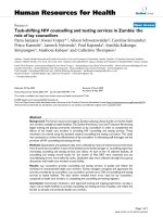
Amino acid sequence alignment of the CRPs encoded by furo-, peclu-, tobra- and hordeivirusesFigure 1
Amino acid sequence alignment of the CRPs encoded by furo-, peclu-, tobra- and hordeiviruses. The positions of amino acids
are numbered above the alignment. The secondary structure prediction is shown directly above the alignment. Cys and His
residues are bold uppercase letters. The leucines of leucine zippers are in bold face. The placement of residues that differ from
Pfam are underlined. Vertical bars at the bottom represent where the Pfam family starts and stops. The genus for each virus is
indicated on the right of the sequence. Abbreviations and accession numbers for the 33 aligned viruses are used (those dis-
played are underlined): LyRSV
, Lychnis ringspot virus gi_1107721; CWMV-2, Chinese wheat mosaic virus gi_14270345; CWMV,
gi_9635448; OGSV Oat golden stripe virus, gi_9635452; SBWMV-NE88 gi_9632360; SBWMV-NE gi_1449160; SBWMV OKl-1
,
gi_1085914; SBWMV-NY, gi_21630062; SBCMV-Ozz, Soilborne cereal mosaic virus gi_12053756; SBCMV-Fra, gi_9635249;
SBCMV-O
, gi_6580881; SBCMV-G, gi_6580877; SBCMV-C, gi_6580873; JSBWMV, Japanese soilborne wheat mosaic virus
gi_7634693; SCSV
, Sorghum chlorotic spot virus gi_21427644; PSLV, Poa semilatent virus gi_321642; BSMV-PV43, Barley stripe
mosaic virus gi_19744921; BSMV-RUS, gi_94465; BSMV-JT, gi_808712; BSMV-ND18, gi_1589671; PCV
, Peanut clump virus
gi_20178597; IPCV
, Indian peanut clump virus gi_30018260; TRV-PpK20, Tobacco rattle virus, gi_20522121; TRV-ORY
gi_2852339; TRV-Pp085 gi_42733086; TRV-PSG, gi_112699; TRV-PLB, gi_465018; TRV-CAN, gi_1857116; TRV-FL,
gi_3033549; TRV-RSTK, gi_6983830; TRV-TCM
, gi_112701; PepRSV, Pepper ringspot virus, gi_20178602; PEBV, Pea early brown-
ing virus, gi_9632342.
10 30 50 70 90
| | | | |
Struct — EEEEEEEEE EE EEE EEHHHHHHHHHHH EEEE EE HHH
PCV mpks
effree rkrrval lge dav-CklngvCgy-sCgmpp-avekvsvpadteedvymli peclu
IPCV maks
dffree rkrrvai lge qav-Ckvngvpgy-sCgmpp-aveqvfvpvdneeeaymlv peclu
BSMV mmatfsCvCCgtstt styCgkrCerkHvyse trnkr-lelykkyll
epqkCalngivgHsCgmpC-siaeeaCdql hordei
LyRSV masspnvkvCtmCCivfdse lefCspkCetragfks erkrraelfakHnl taktCglnkfpae-sCgmya-niaeHqlpdgttt hordei
PSLV mstdlCsvCgnvkdvstfvesqedgkfCsakClrkatfrr vrkqlaeeylkHdl ipvsCqlnsfpgy-HCgmis-alemd-psgk
hordei
CWMV mtt gtHsCekCangfsnviC vskyrtsvykslgl vpvkCrlpadCgv-nCgmpa-afvlvkgHpe furo
SBWMV mstv gfHtCasCvdgpksikC vskyrisvyktlgl dvvkCrlpadCgv-nCgmpa-afv
leqgHpk
furo
SBCMV-O msaC afHsCdkCvdgpknvvC vskyrHsvykvlgl svvkCrlpadCgv-nCgmpa-afvledgHpr
furo
SCSV mtvs tiHsCerClegrtslrC enkyrlsvyqsrqveksayaCkis-qfgv-pCgmpa-qfeldgetlk
furo
TRV-TCM mtCv lk-gCvnevtvlgHetCsigHanklrkqvadmvg vtrrCaen-nCgwfvCviin-dft tobra
PeRSV mtkCa lp-eCeentqkn-qmtCsmkHankynrylaskfd vkrkCeCk-nCgwfpaisvqpdy tobra
PEBV mkCa vs-tCeveaqsn-kftCsmkCankynrHlaekys ikrkCeCv-nCgwypaievradf tobra
|- C k C
110 130 150 170 190 210
| | | | | |
Struct HHHHHHHHHHHHHHHHHH HHHHHHHHHHHHHHHHHHHHHHHH HH
PCV fpyeqfCgekHfklyeslk-dvsdd elklrrLerqretLlasfqqKlkr ydekiall s ekfknlrskl peclu
IPCV fpydgCCgekHyklynsla-disdd dlklqCLerqretLltnfqkKlkd ydsaiall s ekfkklrskm peclu
BSMV pivsrfCgqkHadlydsll-krseq elllefLqkkmqeLklsHivKmak lesevnai rksvassfedsvg Cddsssvskl hordei
LyRSV ltiddyCgskHy yqggl-lavms
d teLkiraaaLkleHqrAtav akgiklak e laalrnsskl hordei
PSLV pvvmnfCgqkHealalalk-akdga
klrleyLerrfyqMkdvyarRldr iaenlkeernrlttsgtitvkrdgeeskqlevsvpmt tadffklskl hordei
CWMV lsmdgfCgekHrgyvvsga-wrmaqlqtLnaeldkLeareesLrsqirgLnea ikastapvyapiklqklkveassvdekkqtrstdlCavmtsvmtklspdstpkktrve furo
SBWMV ltmdgyCgekHrgyvlsga-wrHaqlrsLnaeldaLeareesLraqikaLsag dHCpavlayvpkkltklkaevHdvtgkkqvCitglvdvmdsalvrlapdsppkkissl furo
SBCMV-O ltldgyCgekHkgyvisga-wrHaqlrtLndeldkLekrgefLktqirvLset anantapvyapkkinrmkaevqdvnvkiqdrstalagvmdavalnlspk furo
SCSV vvCdgyCglkHknmaesgs-wrgtllviLqkeleaLqlkeeqLktriaeVtqqHdlvmaetaavlrpdsppkamvttnsrvkyvrrkpaprm furo
TRV-TCM fdvynCCgrsHlekCrkrfearnreiwk-qverirGekasatVkksHksKpsk kkfkerkdfgtpkrflrddvplgidqlfvf tobra
PeRSV vevyfCCgmkHlqkCktd nplkekrlntpkrlfrddvdfglnllfsevC tobra
PEBV ievyfCCgmkHlskviss npkrkerlnspkrlfrddidfgltglfnesC tobra
cons
Cg H -|
Virology Journal 2005, 2:18 />Page 4 of 11
(page number not for citation purposes)
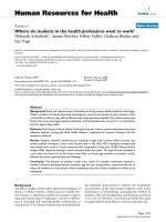
Plants infected with PVXFigure 2
Plants infected with PVX.GFP or PVX.19K at 21 dpi. (A) N. benthamiana plants infected with PVX.GFP (left) and PVX.19K
(right). (B, D) PVX.19K-infected N. benthamiana and N. clevelandii plants, respectively, at 21 dpi show systemic necrosis. (C)
PVX.GFP-infected N. clevelandii plants. (E, F) C. quinoa and C. amaranticolor leaves infected with PVX.19K (left both panels) and
PVX.GFP (right in both panels).
Virology Journal 2005, 2:18 />Page 5 of 11
(page number not for citation purposes)
amaranticolor leaves (Fig. 2). As a control, plants were also
inoculated with PVX.GFP, which has the green fluorescent
protein (GFP) gene inserted into the viral genome. The
spread of PVX.GFP expression was monitored using a
handheld UV lamp to monitor GFP expression and verify
systemic virus accumulation (data not shown).
Symptoms were first observed in plants inoculated with
PVX.GFP and PVX.19K between 10 and 14 dpi. By 21 dpi,
systemic necrosis was evident in N. benthamiana and N.
clevelandii plants inoculated with PVX.19K (Fig. 2A, B and
2D) while PVX.GFP infected plants showed systemic
mosaic symptoms (Fig. 2A and 2C). N. benthamiana
plants infected with PVX.19K were clearly stunted in com-
parison to plants infected with PVX.GFP (Fig. 2A). The
PVX.19K infected N. clevelandii leaves collapsed by 21 dpi
(Fig. 2D).
Immunoblot and northern analyses were conducted to
verify PVX accumulation in the upper leaves of N. bentha-
miana plants. Immunoblot analysis was conducted using
anti-PVX CP serum. High levels of PVX CP was detected in
plants that were systemically infected with PVX.GFP (Fig.
3A lanes 1–4) and PVX.19K (Fig. 3A lanes 5–8). The
SBWMV 19K CRP had no obvious effect on PVX accumu-
lation in upper noninoculated leaves. Viral RNA accumu-
lation was analyzed by northern blot and high levels of
genomic RNA was detected in the upper leaves of
PVX.GFP (Figure 3B lanes 2–4) and PVX.19K (Fig. 3B
lanes 5–8) inoculated plants. Thus, the SBWMV 19K CRP
did not seem to have a deleterious effect on PVX accumu-
lation. RT-PCR was used to verify that the SBWMV 19K
ORF was maintained in the PVX genome in systemically
infected plants. RNA samples taken from the upper leaves
of N. benthamiana plants which were used for northern
analysis, were also used in RT-PCR reactions to verify the
presence of the SBWMV 19K ORF in the PVX genome. In
all samples it appeared that the SBWMV 19K CRP was sta-
bly maintained in the PVX genome (data not shown).
PVX.19K produced large necrotic lesions in the C. quinoa
and C. amaranticolor leaves. Local lesions were detected in
plants inoculated with PVX.GFP or PVX.19K between 5
and 7 dpi. PVX.19K-inoculated C. quinoa plants showed
severe necrotic local lesions (Fig. 2E). The necrotic lesions
gradually merged and the infected tissue eventually col-
lapsed. PVX.19K-inoculated C. amaranticolor leaves
showed enlarged chlorotic lesions advancing to necrotic
lesions over time (Fig. 2F). PVX.GFP-inoculated C. quinoa
leaves showed small chlorotic and necrotic local lesions
while PVX.GFP-inoculated C. amaranticolor leaves showed
mild flecks (Fig. 2F). Association of PVX.GFP with the
local lesions was verified using a hand held UV lamp (data
not shown).
SBWMV 19K CRP is a suppressor of RNA silencing
In this study we employed a widely used "reversal of
silencing assay" to determine if the SBWMV 19K CRP is a
suppressor of RNA silencing in plants [28]. In this assay,
GFP-expression in the 16C transgenic N. benthamiana
plants (Fig. 4B) was silenced by infiltrating young leaves
with a suspension of Agrobacterium expressing GFP. The
progression of GFP silencing was viewed first locally and
then systemically using a hand held UV lamp. Within two
weeks, the spread of GFP silencing was viewed systemi-
cally (Fig. 4C) and by three weeks, the only visible fluores-
cence is red fluorescence due to chlorophyll (Fig. 4D). At
this time, the silenced plants were inoculated with
PVX.19K. As PVX.19K viruses spread locally and then sys-
temically, there was no change in GFP expression in the
inoculated leaves or in the upper leaves (Fig. 4E). How-
ever, GFP expression was observed in the emerging leaves
(Fig. 4F – H). The SBWMV 19K CRP prevented RNA
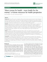
Immunoblot and northern analyses of the PVX infected N. benthamiana plantsFigure 3
Immunoblot and northern analyses of the PVX infected N.
benthamiana plants. (A) Immunoblot analysis conducted
using PVX CP antiserum show similar levels of PVX.GFP
virus (lanes 1–4) and PVX.19K virus (lanes 5–8). Lane 9 con-
tains extract of non inoculated plants. (B) Northern analysis
of RNA isolated from a healthy plant (lane 1), upper noninoc-
ulated leaves of PVX.GFP infected plants (lanes 2 – 4) and
upper noninoculated leaves of PVX.19K infected plants (lanes
5 – 8). Blots were probed with a GFP sequence probe. The
bottom image is the ethidium bromide stained gel showing
ribosomal RNAs. Abbrev.: g, genomic RNA.
Virology Journal 2005, 2:18 />Page 6 of 11
(page number not for citation purposes)
silencing only in emerging leaves where RNA silencing
had not developed prior to virus infection. As a control,
plants were also inoculated with PVX.GUS following infil-
tration with Agrobacterium. There was no evidence of GFP
expression in the inoculated, mature, or new emerging
leaves. The silencing phenotype was unaffected by
PVX.GUS.
Northern analyses was conducted to confirm RNA silenc-
ing in the upper leaves of Agrobacterium-infiltrated leaves
and in the plants inoculated with PVX.GUS (Fig. 4I and
4J). GFP specific RNAs were detected in transgenic plants
(Figure 4I lanes 4–7) and emerging leaves of plants
injected with Agrobacterium and inoculated with PVX.19K
(Figure 4J lanes 1–4). GFP specific RNAs were not
detected in untreated nontransgenic plants (Figure 4I
lanes 1–3) or in plants that were injected with Agrobacte-
rium and inoculated with PVX.GUS (lanes 4–8). RNA
samples collected from non silenced and silenced plants
were also tested by Northern analysis to confirm the sys-
temic accumulation of PVX.GUS or PVX.19K (data not
shown). Since, GFP expression was restored in plants sys-
temically infected with PVX.19K but remained silenced in
plants inoculated with PVX.GUS, it is likely that the
SBWMV 19K ORF is a suppressor of RNA silencing.
Discussion
Many viruses encode proteins that suppress RNA silencing
but the phylogenetic relatedness of these proteins is
poorly understood. In this study, one class of viral CRPs,
which were described as suppressors of RNA silencing
and/or viral pathogenicity determinants, were shown to
be phylogenetically related. These CRPs have a conserved
Cys-Gly-Xaa-Xaa-His motif in which one of the two Xaa
residues is Lys or Arg. The N-terminus has several con-
served Cys residues that likely comprise a zinc finger
motif. In fact, the ability of the gamma b protein of BSMV
to bind Zn(II) was recently demonstrated [25].
Prior to 1999, SBWMV, BNYVV, PCV, and PMTV belonged
to the genus Furovirus. As sequence data from different
furoviruses have become available, it became clear that
there are significant differences in the genome organiza-
tion of these viruses, and therefore furovirus classification
was revised in 1999 [19]. The genus Furovirus now consists
of viruses similar in genome organization to SBWMV [29].
These viruses are bipartite and have a single MP that is
phylogenetically related to the tobamovirus and diantho-
virus MPs [20,22]. BNYVV, PCV, and PMTV were reclassi-
fied into the genera Benyvirus, Pecluvirus, and Pomovirus,
respectively, for two reasons [19,29]. First, the MPs of
these viruses are phylogenetically distinct from SBWMV.
BNYVV, PCV, and PMTV each possess a cluster of three
slightly overlapping ORFs known as the "triple gene
block", which has been shown for BNYVV [30] to mediate
viral cell-to-cell movement. Second, benyviruses and
pomoviruses differ from furoviruses in the number of
genome segments. BNYVV has four or five genome seg-
ments while PMTV has three genome segments [31].
Pecluviruses like furoviruses have two genome segments,
thus the primary difference between these virus genera is
the MP ORFs [32]. This is significant because the initial
amino acid sequence comparisons of CRPs from furo-,
hordei-, tobra-, and carlaviruses included BNYVV as the
type-member of the genus Furovirus and concluded that
these small CRPs were unrelated [33]. Reclassification of
the BNYVV as a member of the genus Benyvirus and inclu-
sion of new members into the genus Furovirus led us to
reexamine the relatedness of the viral CRPs. Based on the
most recently defined taxonomic structure, the current
amino acid sequence comparison presented in Figure 1
indicates that the CRPs derived from viruses of the genera
Furo-, Hordei-, Peclu-, and Tobravirus are phylogenetically
related. On the other hand, these proteins are so different
from CRPs encoded by Pomo-, Beny- and Carlaviruses that
the latter ones could not be included in the alignment (Fig
1).
The present study shows that the SBWMV 19K CRP, when
expressed from the PVX genome, functions as a pathogen-
esis factor and a suppressor of RNA silencing. The SBWMV
19K CRP, when it was expressed from the PVX genome,
induced systemic necrosis on Nicotiana benthamiana, N.
clevelandii, C. quinoa, and C. amaranticolor. These symp-
toms are distinct from the symptoms associated with PVX
infection in these hosts, and from symptoms induced by
SBWMV in its natural hosts. In systemic hosts, both PVX
and SBWMV typically cause mosaic symptoms that range
from mild to severe. In C. quinoa and C. amaranticolor
both PVX and SBWMV cause mild chlorosis. Severe necro-
sis and ultimate collapse of the tissue has been reported
for other unrelated viral proteins that are pathogenicity
factors and suppressors of RNA silencing. This include the
Poa semilatent virus (PSLV) gamma b, TBSV P19, Tobacco
etch virus HC-Pro, and the Rice yellow mottle virus P1
proteins[7,11,14,34].
When we introduced the SBWMV 19K ORF into the TBSV
vector and inoculated it to N. benthamiama, N. tabacum, C.
quinoa, and C. amaranticolor (data not shown) plants, the
SBWMV 19K CRP did not have any effect on symptomol-
ogy (data not shown). However, it was reported
previously that protein expression levels from the TBSV
vector might be too low to test the effects of heterologous
proteins on symptom severity [35]. Since an antibody to
the SBWMV 19K CRP is unavailable, the levels of protein
expression from PVX or TBSV vectors could not be ana-
lyzed to determine if gene dosage or protein expression
levels contribute to symptom severity.
Virology Journal 2005, 2:18 />Page 7 of 11
(page number not for citation purposes)
Evidence for RNA silencing suppression by the SBWMV 19K CRPFigure 4
Evidence for RNA silencing suppression by the SBWMV 19K CRP. (A) nontransgenic N. benthamiana under a UV lamp exhibits
red fluorescence due to chlorophyll. (B) GFP-transgenic N. benthamiana (line 16C) exhibits green fluorescence under a UV
lamp. (C) GFP was systemically silenced in the 16C transgenic N. benthamiana following infiltration with Agrobacterium. Here in
the upper most leaves GFP silencing is vein centric. Systemic GFP silencing is detected initially within 2 weeks. (D) Within 3
weeks, GFP expression is completely silenced in the upper leaves. (E) GFP silenced plant inoculated with PVX.GUS. Emerging
tissues of the infected plant remain silenced. (F, G, and H) GFP expression was observed in the emerging tissues of plants
that were inoculated with PVX.19K. (I) Northern analyses of total RNAs from nontransgenic tissues (lanes 1, 2) and GFP
transgenic tissues (lanes 4 – 7) probed with a labeled GFP sequence probe. Lane 3 is blank. Lanes under the northern blot
show ribosomal RNAs on an ethidium bromide stained gel. (J) Northern analysis of total RNAs from 16C plants infiltrated
with Agrobacterium containing GFP constructs and probed with a labeled GFP sequence probe. Lanes 1–4 are RNA samples
taken from plants that were also inoculated with PVX.19K. Lanes 5–8 are RNA samples taken from plants inoculated with
PVX.GUS. Lanes under the northern blot show ribosomal RNAs.
Virology Journal 2005, 2:18 />Page 8 of 11
(page number not for citation purposes)
In a related study, the SBWMV 19K and the BSMV gamma
b CRPs could substitute for the TRV 16K CRP within the
TRV genome, promoting virus replication and systemic
accumulation [15]. The ability of the SBWMV 19K and the
BSMV gamma b CRPs to induce severe symptoms when
expressed from the PVX genome is reminiscent of phe-
nomena described in relation to viral synergisms. The best
studied viral synergism is between Tobacco etch virus (TEV)
and PVX in which the TEV HC-Pro protein enhances accu-
mulation and disease severity of PVX [34]. HC-Pro pro-
motes infection of PVX by suppressing the anti-viral RNA
silencing defense mechanism that would normally act on
PVX to reduce virus infection. HC-Pro has the ability to
increase PVX accumulation in the same way the SBWMV
19K CRP and the BSMV gamma b proteins were shown
previously to enhance accumulation of TRV in infected
plants [15].
Conclusion
The phylogenetic relatedness of the hordei-, peclu-, and
furovirus CRPs is further substantiated by evidence that
these proteins are all capable of suppressing RNA silenc-
ing in emerging leaves. This was demonstrated in the
present and related studies using the same reversal of
silencing assay used in this study. The SBWMV 19K CRP,
the BSMV and PSLV gamma b CRPs, and the PCV 15K
CRPs were each unable to change GFP expression in leaves
where GFP was silenced prior to virus infection. However
in each case, GFP expression occurred in newly emerging
leaves [14,16]. Thus, members of this family of CRPs sim-
ilarly act on the RNA silencing machinery to block spread
of the silencing signal into newly emerging leaves. In each
case, the silencing suppressor activities of these CRPs have
been compared to CMV and potyviruses in preventing
onset of RNA silencing in new growth [14,16]. While
there is no evidence that the hordei-, peclu- and furovirus
CRPs are related to the CMV or potyvirus silencing sup-
pressors, it seems that the mode of action might be con-
served among diverse viruses.
Methods
Amino acid sequence comparisons
Related protein sequences were identified and retrieved
from the NCBI data bank using PSI-BLAST. A PSI-BLAST
search was launched with the amino acid sequence of the
19K CRP of Chinese wheat mosaic virus (CWMV, a furovi-
rus). A similar search began with the amino acid sequence
of BSMV gamma b, a sequence recovered in the CWMV
search. Both searches converged at the second iteration
and retrieved the same set of 22 sequences. This set con-
tained CRPs derived from furo-, peclu- and hordeiviruses
and contained the conserved P18 PFAM domain ("protein
family"URL reference />[36].
A preliminary alignment of the retrieved proteins
sequences was performed using the multiple sequence
alignment mode of ClustalX. These twenty two furovirus
and hordeivirus sequences were aligned using ClustalX
alignments suggested in the BLAST outputs and PFAM.
The tobraviral CRPs were not recovered by the above pro-
cedure, but upon manual inspection, appeared to have
Cys residues in a linear arrangement that was similar to
the set of 22 proteins. Eleven tobraviral protein sequences,
exclusive members of a conserved domain in the Con-
served Domain database />Structure/cdd/cdd.shtml were aligned using ClustalX [37].
This tobraviral amino acid sequence alignment and the
alignment of the 22 amino acid sequences sequences were
assembled by ClustalX in profile mode, followed by man-
ual adjustment. Amino acid sequences of aligned furo-
and hordeiviral proteins were aligned with tobraviral
amino acid sequences in profile mode of ClustalX (a total
of thirty three sequences were aligned). A total of 33
amino acid sequences were aligned. In all cases, adjust-
ments to the alignments were made using Se-Al [38]. Sig-
nificance scores for the alignment of the two groups of
protein sequences were calculated as previously described,
using a structural conservation matrix, SCM2, for scoring
[39].
Plasmids and bacterial strain
All plasmids were used to transform Escherichia coli strain
JM109 [40]. The plasmids pPVX.GFP is an infectious viral
clone and contains a bacteriophage T7 promoter [39]. The
pPVX.GFP plasmid contains the PVX genome and the GFP
adjacent to a duplicated CP subgenomic promoter. The
plasmid pHST2-14 contains the TBSV genome and a
mutation in the TBSV P19 ORF eliminating expression of
a protein that suppresses RNA silencing [10,42]. The plas-
mid pTBSV.GFP contains GFP inserted into the TBSV
genome replacing the viral CP ORF [10].
The SBWMV 19K CRP ORF was inserted into the PVX.GFP
genome, replacing the GFP ORF. The 19K CRP ORF was
reverse transcribed and PCR amplified from purified
SBWMV RNA using a forward primer (GCG GGG ATC
GAT ATG TCT ACT GTT GGT TTC CAC) containing added
sequences encoding a ClaI restriction site (underlined)
and a reverse primer (CGC GTC GAC
TCA CAA AGA GGA
TAT CTT CTT TGG C) containing sequences encoding a
SalI restriction site (underlined). PCR products and
pPVX.GFP plasmids were digested with ClaI and SalI and
then were ligated to prepare pPVX.19K.
In vitro transcription and plant inoculations
In vitro transcription reactions contained: 0.25 µg of line-
arized DNA, 5 µl of 5X T7 transcription buffer, 1.0 µl of
0.1 M DTT, 0.5 µl of SUPERase·In™ ribonuclease inhibi-
tor (20 U/ µl) (Ambion, Austin, TX), 2.5 µl of an NTP
Virology Journal 2005, 2:18 />Page 9 of 11
(page number not for citation purposes)
mixture containing 5 mM ATP, CTP, UTP, and GTP (Phar-
macia-Pfizer, Mississauga, Ontario, Canada), 0.7 µl of T7
polymerase (Ambion), and nuclease-free water to a final
volume of 25 µl. The reactions were incubated for one and
a half hour at 37°C [10].
Nicotiana benthamiana, N. clevelandii, Chenopodium quinoa,
and C. amaranticolor plants were inoculated with infec-
tious transcripts to study disease severity. Four plants, two
leaves per plant, were inoculated in each experiment.
Experiments were repeated at least three times. Ten µl of
undiluted PVX.GFP or PVX.19K transcripts were rub-inoc-
ulated to each plant.
The transgenic N. benthamiana line 16C was used to study
RNA silencing. This line is homozygous for the GFP trans-
gene at a single locus [44]. Plants were inoculated with
transcripts following infiltration with Agrobacterium (see
below).
Agrobacterium infiltration of leaves
Agrobacterium tumefaciens strain C58C1 (pCH32) carrying
a binary plasmid expressing GFP from a Cauliflower mosaic
virus (CaMV) 35S promoter was used to silence GFP
expression in N. benthamiana line 16C. Agrobacterium cul-
tures were grown overnight at 28°C in 5 ml of L-broth
medium containing 5 µg/ml of tetracycline and 50 µg/ml
of kanamycin. This 5 ml culture was used to inoculate 50
ml L-broth and grown overnight in medium containing 5
µg/ml tetracycline, 50 µg/ml kanamycin, 10 mM MES,
and 20 µM acetosyringone. Cultures of Agrobacterium con-
taining GFP were pelleted by centrifugation and resus-
pended in a solution containing 10 mM MgCl
2
, 10 mM
MES, and 150 µM acetosyringone. The final concentration
of Agrobacterium was 0.5 OD
600
. The suspension was left
at room temperature for 2–3 hours and then loaded into
a 2 ml syringe. The syringe was used to infiltrate the sus-
pension into the underside of the leaf.
Visualization of GFP
A hand-held model B-100 BLAK-RAY long wave ultravio-
let lamp (Ultraviolet Products, Upland, CA) was used to
monitor GFP expression in 16C plants infiltrated with
Agrobacterium and in PVX.GFP inoculated plants. GFP flu-
orescence was recorded with a Sony Digital Still Camera
model DSC-F717 (Sony Corporation of America, New
York City, New York). In all plants analyzed, GFP expres-
sion was monitored every 3 days for up to 21 days post
inoculation (dpi) or post infiltration with Agrobacterium.
Immunoblot analysis
Immunoblot analyses were conducted according to [40].
Total protein from uninfected and infected N. benthami-
ana leaves was extracted in 1:10 (w/v) grinding buffer
(100 mM Tris-HCl pH 7.50, 10 mM KCl, 5 mM MgCl
2
,
400 mM sucrose, 10% glycerol, and 10 mM β-mercap-
toethanol). Extracts were centrifuged at 10,000 g for 10
min. Equal volumes of supernatants and protein loading
buffer (2 % SDS, 0.1 M dithiothreitol, 50 mM Tris-HCl pH
6.8, 0.1% bromophenol blue, and 10 % glycerol) were
mixed and boiled for 5 min. SDS-PAGE was carried out for
1 h at 200 V using 30 µl of each sample and 12.5% SDS -
PAGE and the Biorad Mini-Protean 3 system (Biorad Lab-
oratories, Hercules, CA). Proteins were transferred to
PVDF membranes (Amersham Biosciences Corp., Piscata-
way, NJ) at 4°C overnight using protein transfer buffer
(39 mM glycine, 48 mM Tris base, 0.037% SDS, and 20%
methanol, pH 8.3) and a BioRad Trans-Blot system (Bio-
Rad Laboratories). Immunoblot analyses were conducted
using the ECL-Plus Western Blotting Detection Kit (Amer-
sham Biosciences Corp.). PVX CP antiserum (1:200)
(Agdia, Elkhart, IN) was used.
Northern analysis
Northern analyses were conducted according to [40]. For
analyses of PVX infected plants and GFP expressing trans-
genic plants, a radiolabeled DNA probe was prepared
using Rediprime II Random Prime Labeling System
(Amersham Biosciences Corp.). Labeling was conducted
using PCR products corresponding to either the GFP or
PVX CP ORFs.
For detection of TBSV.GFP and TBSV.19K in infected plant
extracts, a DNA probe was labeled with digoxigenin
(DIG). TBSV.GFP plasmids were digested with NcoI and
SalI and a 614 nt fragment was gel eluted and labeled
using Dig High Prime kit (Roche Applied Science Inc.
Indianapolis, IN). The CSPD DIG Luminescence Detec-
tion Kit (Roche Applied Science Inc.) was used for chemi-
luminescence detection of DIG-labeled probes. Special
thanks to Wenping Qui at Southwest Missouri State
University for assistance with studies using TBSV to
express the SBWMV 19k.
The p26SBE-2 plasmid was obtained from Kay Scheets at
Oklahoma State University and contains the 26S ribos-
omal RNA gene in pBluescript. This plasmid was used to
prepare a DNA probe for membrane detection of rRNA
[45]. The p26SBE-2 plasmid was digested with BamHI and
EcoRI and a 1 kb fragment corresponding to the 26S rRNA
was recovered and labeled using the Dig High Prime DNA
labeling system (Roche Applied Science Inc.).
Competing interests
The author(s) declare that they have no competing
interests.
Authors' contributions
Jeannie Te did all cloning, plant inoculation experiments,
gene silencing experiments. Ulrich Melcher did the amino
Virology Journal 2005, 2:18 />Page 10 of 11
(page number not for citation purposes)
acid sequence alignments and phylogenetic comparisons.
Amanda Howard did some gene silencing experiments,
photography. Jeanmarie Verchot-Lubicz conceived the
study, did some molecular analysis, and wrote the paper.
Special thanks to Wenpiny Qiu at Southwest Missouri
State University for assistance with studies using TBSV to
express the SBWMV 19k.
Acknowledgements
Support for this project was provided by the Oklahoma Wheat Research
Foundation, the USDA NRI Program Award OKLO-2470, and the Okla-
homa Agriculture Experiment Station under the project H-2371.
References
1. Dower SK: Cytokines, virokines and the evolution of
immunity. Nat Immunol 2000, 1(5):367-368.
2. McFadden G, Lalani A, Everett H, Nash P, Xu X: Virus-encoded
receptors for cytokines and chemokines. Semin Cell Dev Biol
1998, 9(3):359-368.
3. Bucher E, Hemmes H, de Haan P, Goldbach R, Prins M: The influ-
enza A virus NS1 protein binds small interfering RNAs and
suppresses RNA silencing in plants. J Gen Virol 2004, 85(Pt
4):983-991.
4. Li WX, Li H, Lu R, Li F, Dus M, Atkinson P, Brydon EW, Johnson KL,
Garcia-Sastre A, Ball LA, Palese P, Ding SW: Interferon antagonist
proteins of influenza and vaccinia viruses are suppressors of
RNA silencing. Proc Natl Acad Sci U S A 2004, 101(5):1350-1355.
5. Kasschau KD, Carrington JC: A counterdefensive strategy of
plant viruses: suppression of posttranscriptional gene
silencing. Cell 1998, 95(4):461-470.
6. Li H, Li WX, Ding SW: Induction and suppression of RNA
silencing by an animal virus. Science 2002, 296(5571):1319-1321.
7. Voinnet O, Pinto YM, Baulcombe DC: Suppression of gene silenc-
ing: a general strategy used by diverse DNA and RNA viruses
of plants. Proc Natl Acad Sci U S A 1999, 96(24):14147-14152.
8. Anandalakshmi R, Pruss GJ, Ge X, Marathe R, Mallory AC, Smith TH,
Vance VB: A viral suppressor of gene silencing in plants. Proc
Natl Acad Sci U S A 1998, 95(22):13079-13084.
9. Lucy AP, Guo HS, Li WX, Ding SW: Suppression of post-tran-
scriptional gene silencing by a plant viral protein localized in
the nucleus. Embo J 2000, 19(7):1672-1680.
10. Qiu W, Park JW, Scholthof HB: Tombusvirus P19-mediated sup-
pression of virus-induced gene silencing is controlled by
genetic and dosage features that influence pathogenicity. Mol
Plant Microbe Interact 2002, 15(3):269-280.
11. Scholthof HB, Scholthof KB, Jackson AO: Identification of tomato
bushy stunt virus host-specific symptom determinants by
expression of individual genes from a potato virus X vector.
Plant Cell 1995, 7(8):1157-1172.
12. Scholthof HB, Scholthof KB, Kikkert M, Jackson AO: Tomato bushy
stunt virus spread is regulated by two nested genes that func-
tion in cell-to-cell movement and host-dependent systemic
invasion. Virology 1995, 213(2):425-438.
13. Donald RG, Jackson AO: The barley stripe mosaic virus gamma
b gene encodes a multifunctional cysteine-rich protein that
affects pathogenesis. Plant Cell 1994, 6(11):1593-1606.
14. Yelina NE, Savenkov EI, Solovyev AG, Morozov SY, Valkonen JP:
Long-distance movement, virulence, and RNA silencing sup-
pression controlled by a single protein in hordei- and potyvi-
ruses: complementary functions between virus families. J Virol
2002, 76(24):12981-12991.
15. Liu H, Reavy B, Swanson M, MacFarlane SA: Functional replace-
ment of the tobacco rattle virus cysteine-rich protein by
pathogenicity proteins from unrelated plant viruses. Virology
2002, 298(2):232-239.
16. Dunoyer P, Pfeffer S, Fritsch C, Hemmer O, Voinnet O, Richards KE:
Identification, subcellular localization and some properties
of a cysteine-rich suppressor of gene silencing encoded by
peanut clump virus. Plant J 2002, 29(5):555-567.
17. Reavy B, Dawson S, Canto T, MacFarlane SA: Heterologous
expression of plant virus genes that suppress post-transcrip-
tional gene silencing results in suppression of RNA interfer-
ence in Drosophila cells. BMC Biotechnol 2004, 4(1):18.
18. Koonin EV, Boyko VP, Dolja VV: Small cysteine-rich proteins of
different groups of plant RNA viruses are related to different
families of nucleic acid-binding proteins. Virology 1991,
181(1):395-398.
19. Mayo MA: Developments in plant virus taxonomy since the
publication of the 6th ICTV report. Arch Virol 1999, 144:1659–
1666.
20. Shirako Y, Wilson TM: Complete nucleotide sequence and
organization of the bipartite RNA genome of soil-borne
wheat mosaic virus. Virology 1993, 195(1):16-32.
21. An H, Melcher U, Doss P, Payton M, Guenzi AC, Verchot-Lubicz J:
Evidence that the 37 kDa protein of Soil-borne wheat mosaic
virus is a virus movement protein. J Gen Virol 2003, 84(Pt
11):3153-3163.
22. Melcher U: The '30K' superfamily of viral movement proteins.
J Gen Virol 2000, 81(Pt 1):257-266.
23. Shirako Y: Non-AUG translation initiation in a plant RNA
virus: a forty-amino-acid extension is added to the N termi-
nus of the soil-borne wheat mosaic virus capsid protein. J Virol
1998, 72(2):1677-1682.
24. Tamada T, Schmitt C, Saito M, Guilley H, Richards K, Jonard G: High
resolution analysis of the readthrough domain of beet
necrotic yellow vein virus readthrough protein: a KTER
motif is important for efficient transmission of the virus by
Polymyxa betae. J Gen Virol 1996, 77 ( Pt 7):1359-1367.
25. Bragg JN, Lawrence DM, Jackson AO: The N-terminal 85 amino
acids of the barley stripe mosaic virus gammab pathogenesis
protein contain three zinc-binding motifs. J Virol 2004,
78(14):7379-7391.
26. Roth BM, Pruss GJ, Vance VB: Plant viral suppressors of RNA
silencing. Virus Res 2004, 102(1):97-108.
27. Torrance L, Mayo MA: Proposed re-classification of furoviruses.
Arch Virol 1997, 142(2):435-439.
28. Gilmer D, Bouzoubaa S, Hehn A, Guilley H, Richards K, Jonard G:
Efficient cell-to-cell movement of beet necrotic yellow vein
virus requires 3' proximal genes located on RNA 2. Virology
1992, 189(1):40-47.
29. Herzog E, Guilley H, Manohar SK, Dollet M, Richards K, Fritsch C,
Jonard G: Complete nucleotide sequence of peanut clump
virus RNA 1 and relationships with other fungus-transmitted
rod-shaped viruses. J Gen Virol 1994, 75 ( Pt 11):3147-3155.
30. Kashiwazaki S, Scott KP, Reavy B, Harrison BD: Sequence analysis
and gene content of potato mop-top virus RNA 3: further
evidence of heterogeneity in the genome organization of
furoviruses. Virology 1995, 206(1):701-706.
31. Koonin EV, Mushegian AR, Ryabov EV, Dolja VV: Diverse groups of
plant RNA and DNA viruses share related movement pro-
teins that may possess chaperone-like activity. J Gen Virol 1991,
72 ( Pt 12):2895-2903.
32. Pruss G, Ge X, Shi XM, Carrington JC, Bowman Vance V: Plant viral
synergism: the potyviral genome encodes a broad-range
pathogenicity enhancer that transactivates replication of
heterologous viruses. Plant Cell 1997, 9(6):859-868.
33. Qiu W, Scholthof KB: Satellite panicum mosaic virus capsid
protein elicits symptoms on a nonhost plant and interferes
with a suppressor of virus-induced gene silencing. Mol Plant
Microbe Interact 2004, 17(3):263-271.
34. Bateman A, Coin L, Durbin R, Finn RD, Hollich V, Griffiths-Jones S,
Khanna A, Marshall M, Moxon S, Sonnhammer EL, Studholme DJ,
Yeats C, Eddy SR: The Pfam protein families database. Nucleic
Acids Res 2004, 32 (Database issue):D138-41.
35. Marchler-Bauer A, Bryant SH: CD-Search: protein domain anno-
tations on the fly. Nucleic Acids Res 2004, 32(Web Server
issue):W327-31.
36. Rambaut A, Grassly NC, Nee S, Harvey PH: Bi-De: an application
for simulating phylogenetic processes. Comput Appl Biosci 1996,
12(6):469-471.
37. Melcher U: HIV-1 proteinase as structural model of intercel-
lular transport proteins of plant viruses. J Theor Biol 1993,
162(1):61-74.
38. Sambrook J, Fritsch EF, Maniatis T: Molecular cloning: A labora-
tory manual. 2nd edition. Cold Spring Harbor, NY , Cold Spring
Harbor Press; 1989.
Publish with BioMed Central and every
scientist can read your work free of charge
"BioMed Central will be the most significant development for
disseminating the results of biomedical research in our lifetime."
Sir Paul Nurse, Cancer Research UK
Your research papers will be:
available free of charge to the entire biomedical community
peer reviewed and published immediately upon acceptance
cited in PubMed and archived on PubMed Central
yours — you keep the copyright
Submit your manuscript here:
/>BioMedcentral
Virology Journal 2005, 2:18 />Page 11 of 11
(page number not for citation purposes)
39. Baulcombe DC, Chapman S, Santa Cruz S: Jellyfish green fluores-
cent protein as a reporter for virus infections. Plant J 1995,
7(6):1045-1053.
40. Hou H, Qiu W: A novel co-delivery system consisting of a
Tomato bushy stunt virus and a defective interfering RNA
for studying gene silencing. J Virol Methods 2003, 111(1):37-42.
41. Scholthof HB: Rapid delivery of foreign genes into plants by
direct rub-inoculation with intact plasmid DNA of a tomato
bushy stunt virus gene vector. J Virol 1999, 73(9):7823-7829.
42. Scholthof HB, Scholthof KB, Jackson AO: Plant virus gene vectors
for transient expression of foreign proteins in plants. Annu Rev
Phytopathol 1996, 34:299-323.
43. Voinnet O, Baulcombe DC: Systemic signalling in gene
silencing. Nature 1997, 389(6651):553.
44. Scheets K: Maize chlorotic mottle machlomovirus and wheat
streak mosaic rymovirus concentrations increase in the syn-
ergistic disease corn lethal necrosis. Virology 1998,
242(1):28-38.