Báo cáo sinh học: " Ultrastructural studies on dengue virus type 2 infection of cultured human monocytes" doc
Bạn đang xem bản rút gọn của tài liệu. Xem và tải ngay bản đầy đủ của tài liệu tại đây (3.15 MB, 14 trang )
BioMed Central
Page 1 of 14
(page number not for citation purposes)
Virology Journal
Open Access
Research
Ultrastructural studies on dengue virus type 2 infection of cultured
human monocytes
Jesus A Mosquera*
1
, Juan Pablo Hernandez
2
, Nereida Valero
3
,
Luz Marina Espina
3
and German J Añez
3
Address:
1
Seccion de Inmunologia y Biologia Celular, Instituto de Investigaciones Clinicas "Dr. Americo Negrette". Facultad de Medicina,
Universidad del Zulia, Maracaibo, Venezuela,
2
Instituto de Investigaciones Biologicas. Facultad de Medicina, Universidad del Zulia, Maracaibo,
Venezuela and
3
Seccion de Virologia, Instituto de Investigaciones Clinicas "Dr. Americo Negrette". Facultad de Medicina, Universidad del Zulia,
Maracaibo, Venezuela
Email: Jesus A Mosquera* - ; Juan Pablo Hernandez - ; Nereida Valero - ;
Luz Marina Espina - ; German J Añez -
* Corresponding author
Abstract
Background: Early interaction of dengue virus and monocyte/macrophages could be an important
feature for virus dissemination after its initial entry via the mosquito vector. Since ultrastructural
analysis of this interaction has not been reported, dengue type 2 (DEN2) virus-infected human
monocyte cultures were studied at 1, 2, 4 and 6 hours after infection.
Results: Typical dengue particles and fuzzy coated viral particles were 35 to 42 nm and 74 to 85
nm respectively. Viruses were engulfed by phagocytosis and macropicnocytosis leading to huge
vacuoles and phagosomes inside the monocytes. Interaction of monocytes with DEN2 virus
induced apoptosis, characterized by nuclear condensation and fragmentation, cellular shrinkage,
blebbing and budding phenomena and phagocytosis of apoptotic cells by neighboring monocytes.
This finding was confirmed by TUNEL. Ultrastructural features associated to DEN2 virus
replication were not observed.
Conclusion: These data suggest that clearance of the virus by monocytes and cellular death are
the main features during the initial interaction of DEN2 virus and monocytes and this could be
important in the rapid elimination of the virus after infection by mosquito vector.
Background
Monocyte/macrophages are one of the major target of
dengue virus and responsible for virus dissemination after
its initial entry via the mosquito vector [1-3]. A detailed
study of this early virus-monocyte interaction by electron
microscopy has not been performed. Since ultrastructural
study is one of the important analysis in the interaction
virus-cell, we performed electron microscopy studies in
DEN2 virus- infected human monocytes at 1, 2, 4 and 6
hours of culture, in order to get more information regard-
ing to morphological aspects of virus, virus replication,
cellular alterations and apoptosis.
Results and discussion
Virus particles
After 1 hour of culture numerous virus particles were
observed attached to plasma membrane, free in the extra-
cellular space and in cytoplasmic vacuoles inside
Published: 31 March 2005
Virology Journal 2005, 2:26 doi:10.1186/1743-422X-2-26
Received: 05 March 2005
Accepted: 31 March 2005
This article is available from: />© 2005 Mosquera et al; licensee BioMed Central Ltd.
This is an Open Access article distributed under the terms of the Creative Commons Attribution License ( />),
which permits unrestricted use, distribution, and reproduction in any medium, provided the original work is properly cited.
Virology Journal 2005, 2:26 />Page 2 of 14
(page number not for citation purposes)
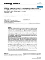
Electron microscope morphological observations of DEN2 virus particlesFigure 1
Electron microscope morphological observations of DEN2 virus particles. A) Typical viral particle in the extracellular environ-
ment (arrow; bar: 200 nm). B) Viral particles engulfed in an intracytoplasmic vacuole (arrow; bar: 50 nm). C) Membrane disrup-
tion of a vesicle containing a virus (arrow; bar: 100 nm). D) Fuzzy coated viral particles occur in the extracellular space
(arrows; bar: 200 nm) E) A fuzzy coated viral particle showing an envelope with projections (arrow; bar: 100 nm). F) Immun-
ofluorescence staining of DEN2 viral antigens at 4 h of culture. A diffuse and patchy pattern of fluorescence was observed in
the cytoplasm (arrows). × 1000.
Virology Journal 2005, 2:26 />Page 3 of 14
(page number not for citation purposes)
monocytes. The predominant viral particles in infected
monocyte cultures were typical viral particles of 35 to 42
nm in diameter (Figures 1A, 1B, 1C). Small number of
fuzzy coated viral particles (74 to 85 nm) showed a core
similar to the usual dengue particles, but they had an
envelope with projections, looking like a fuzzy coat (Fig-
ures 1D, 1E). Typical DEN2 virus particles observed in this
study were similar to those reported in mosquito cell cul-
tures [4]. Similar fuzzy coated virus particles have been
described by Barth et al [4,5] in DEN2 Brazilian virus-
infected C6/36 cell cultures. DEN2 virus used to infect
monocytes was New Guinea C virus strain and isolated
from virus-infected C6/36 cell cultures, suggesting that the
fuzzy coated viral particles are a common feature of DEN2
virus. In addition, fuzzy coated virus particles have also
been detected in other virus infections, but their signifi-
cance remains obscure [6,7]. The presence of DEN2 virus
antigens in the cytoplasm of infected monocytes was also
investigated by direct immunofluorescence. Using a mon-
oclonal antibody against DEN2 virus a diffuse and patchy
patterns of fluorescence were observed in the cytoplasm
(Figure 1F). It was also observed small electron dense
structures (75 to 105 nm) that we called in this report
"dense particles" (Figure 2). In some instances, these
dense particles showed a center similar to dengue virus
nucleocapsid covered by membrane layers and an elec-
tron dense envelope (Figures 2B, 2C). Dense particles
could represent viral particles covered by a homogenous
electron dense material. Since, it was not observe viral rep-
lication ultrastructural features in infected monocyte cul-
tures, the contribution of monocytes to the formation of
this viral envelope is unclear. However, electron dense
material observed on the dense particles could represent a
protein matrix obtained after virus replication on mos-
quito cells. In this regard, a range of variation in one virus
after experimental isolation has been reported in other
virus [6,7]. In general extracellular viral particles were
found as single particles and viral particles forming aggre-
gates were uncommon. Viruses attached to the cell surface
and free in the extracellular space were engulfed by mech-
anisms of phagocytosis or macropicnocytosis via typical
cytoplasmic processes (Figure 3). During phagocytosis or
macropicnocytosis virus particles were engulfed alone or
together with cellular debris, so that, intracytoplasmic
vacuoles and vesicles containing viral particles or large
phagosomes full of an electron dense matrix, cellular
debris and viral particles may soon be found inside the
cells (Figure 4). These data suggest a passive phase leading
to virus inactivation. In this regard, previous reports have
shown that human immunodeficiency virus entering
human macrophages by phagocytosis is noninfectious
[8]. Infection of Kupffer cells by dengue virus resulted in
no viral progeny [9] and only a small proportion of the
monocyte population supports replication of DEN2-virus
[10]. Smooth membrane coated vacuoles containing viral
particles, membrane fragments and moderated electron
dense material were also observed (Figure 1B). In some
instances, cytoplasmic vesicles containing one or more
Electron microscope morphological observations of dense particlesFigure 2
Electron microscope morphological observations of dense particles. A) Dense particles close to the cell surface (arrow; bar:
200 nm). B) Aggregated dense particles in the extracellular space (arrow). Note the nucleocapsid like center and the electron
dense envelopes (bar: 100 nm). C) Dense particles showing a nucleocapsid like center surrounded by membrane layers and an
electron dense material (arrow; bar: 100 nm).
Virology Journal 2005, 2:26 />Page 4 of 14
(page number not for citation purposes)
viral particles showed disruption of the membrane lead-
ing to direct communication of viral particles with the
cytoplasm (Figure 1C), however, no morphological virus-
related structures could be detected free in the cytoplasm.
Features related to viral replication such as virus absorp-
tion by penetrating the cell membrane or by endocytosis
by clathrin-coat vesicles, virion precursors on rough endo-
plasmic reticulum or its cisternae, inside Golgi complex,
cytoplasm free viral cores or viral budding from cell mem-
brane were not observed in monocytes. Previous report
has shown that DEN2 virus can persistently infect trans-
formed lymphoblastoid cells keeping an intact morphol-
ogy without any indication of active viral replication [11].
Our data show no indications of viral replication and the
induction of apoptosis (see below) makes monocytes
unlikely source of persistent dengue virus infection.
Monocyte cultures
As assessed by electron microscopy, monocytes showed
high degree of activation after 1 hour of infection. One of
the most prominent features in DEN2 virus-infected
monocytes was the intense expression of short and long
plasma membrane processes (lamellipods), in most of the
cases engulfing virus particles, cellular debris and apop-
totic cells (Figure 3). Engulfing of extracellular elements
by pseudopods was also observed (Figure 3C). As conse-
quence of this activity, small and huge intracytoplasmic
vacuoles and phagosomes containing cellular debris, virus
particles and myelin like structures in various stages of
digestion were observed (Figures 3 and 4). In some
instances, phagosomes or vacuoles were surrounded by
lysosomes. (Figure 5A). Our data show similar ultrastruc-
tural findings than those obtained from DEN1 virus-
infected Kupffer cells at 1 hour of culture [9], suggesting a
similar cellular response against DEN virus for monocytes
and macrophages. In DEN2 virus- infected monocytes
mitochondria increased in number and size (Figure 5B)
and cytoplasmic structures resembling diverse degrees of
mitochondrial alterations (Figures 5C, 5D) were found.
Mitochondria were observed in association with lyso-
somal granules and vacuoles containing membranous
debris, consistent with mitochondrial digestion by lyso-
somes. Infected monocytes showed extensive prolifera-
tion of endoplasmic reticulum and lysosomal granules
(Figure 5E). Cytoplasmic projections associated with cel-
lular movement (uropods) were also observed (Figure
5F). It was not observed syncytia, however as shown in fig-
ure 6 a curious distribution of monocytes in DEN2 virus-
infected cultures was found. Empty spaces were sur-
rounded by monocytes looking like "acinar" structures. In
some instances, a linear electron dense material occurred
between the empty space and monocytes, suggesting a
previous presence of biological material in the lumen.
These findings could represent a reactive response of
Ultrastructural features of DEN2 virus-infected monocytesFigure 3
Ultrastructural features of DEN2 virus-infected monocytes.
Prominent formation of cellular lamellipods (A) and engulfing
of virus by macropicnocytosis (B) and phagocytosis (C) are
observed 1 hour after infection. Note the presence of virus
(arrows) and cellular debris in the extracellular space. (A and
C bars: 1 µm; B bar: 500 nm).
Virology Journal 2005, 2:26 />Page 5 of 14
(page number not for citation purposes)
Ultrastructural features of DEN2 virus-infected monocytesFigure 4
Ultrastructural features of DEN2 virus-infected monocytes. A) DEN2 virus-infected monocytes after 2 hours of infection.
Observe the presence of virus particles in the extracellular space, on cellular plasma membrane and inside cytoplasmic vacu-
oles (arrows; bar: 1 µm). B) Monocyte showing huge empty vacuoles and vacuoles containing nuclear debris and myelin struc-
tures at 4 hours of culture (arrows; bar: 500 nm). C) Monocyte showing cytoplasmic phagosomes containing cellular debris and
viral particles (arrow; bar: 200 nm). D) A huge vacuole containing numerous viral particles and cellular debris (arrow; bar: 500
nm).
Virology Journal 2005, 2:26 />Page 6 of 14
(page number not for citation purposes)
monocytes around virus particles, cellular debris or virus-
infected cells.
Cellular Death
After 1 hour of infection, electron microscopy revealed
cells with morphological features of apoptosis, however,
previous report has shown apoptosis in Kupffer cells [9]
after 24 hours of DEN-1 virus infection, suggesting differ-
ent susceptibility of monocyte and macrophage to virus-
induced apoptosis or different viral apoptotic effect
depending of DEN virus strain. In this regard, the suscep-
tibility to DEN virus infection depending of the differen-
tiation state of monocytic cells has been reported [12].
Apoptotic cells showed chromatin margination in nuclei,
nuclear fragmentation, condensation and retraction of
cytoplasm and blebbing and budding phenomena (Fig-
ures 7 and 8). Numerous vesicles, some of which
appeared to be releasing to the extracellular space were
observed (Figures 7D and 8E). The budding phenomenon
observed on apoptotic cells led to the formation of apop-
totic bodies containing several types of organelles, includ-
ing nuclear fragments and high number of vesicles. This
could represent a common aspect in virus-induced apop-
tosis, since the formation of vesicular apoptotic bodies
has also been reported in monocytic/macrophage lineage
infected with bovine leukaemia virus [13]. Blebbing of the
plasma membrane was also observed in apoptotic cells.
The surface blebbing has also been described in other viral
infections and related to a role in the direct cell-to-cell
spread of the virus [14] or associated with increased cellu-
lar permeability [15]. Some apoptotic cells showed long
cisternae structures alongside with the plasma membrane
suggesting cytoplasmic splitting (Figure 8G). We have no
explanation for this finding, but it could be due to the
fusion of neighboring cytoplasmic vesicles. Apoptotic
cells also showed bundles of intracellular microfibrils
(Figures 7G and 7H), which resembled the contractile
structures observed in fibroblasts and some glomerular
cells [16]. These structures could be related to the
apoptotic process, since, filamentous material, clumping
of tonofilaments and MyD88 protein association with
fibrillar aggregates containing beta-actin have been associ-
ated with apoptosis and apoptotic bodies formation [17-
19]. Huge phagosomes were observed in the cytoplasm of
apoptotic cells (Figure 7E), and in some instances, vacu-
oles containing few viral particles associated with an elec-
tron dense material were observed (Figures 8E and 8F).
The presence of phagosomes in the cytoplasm of apop-
totic cells suggests previous active phagocytosis. Contra-
rily to non apoptotic cell only scarce number of vacuoles
containing virus and degraded material was observed in
apoptotic cells, suggesting that the absorption of products
of viral degradation could trigger cell death. Several apop-
totic monocytes and apoptotic bodies were ingested by
neighboring healthy monocytes leading to the formation
of huge vacuolar compartments containing different
grades of cellular digestion (Figures 8D and 9). Apoptosis
could avoid the release of viral particles [20] and together
with the phagocytosis and digestion of apoptotic cells rep-
resent mechanisms to prevent viral progeny [9,21,22].
The ultrastructural apoptosis finding was confirmed by
detecting intrachromosomal DNA strand breaks using the
TUNNEL assay. Untreated cultures showed low levels of
TUNEL positive cells compared to higher levels observed
in infected monocyte cultures (Control: 0.9 ± 0.15.
Infected at 1 h: 6.2 ± 1.5; 2 h: 6.4 ± 1.8; 4 h: 7.4 ± 2.3; 6 h:
16.8 ± 3.3; mean ± SE) (Figure 8H). In addition to apop-
tosis, a cellular alteration accompanied by cellular swell-
ing, plasma membrane disruption and karyolysis was
observed (Figure 10). Plasma membrane disruption led to
increased amount of swelled organelles and cellular
debris in the extracellular space and the formation of
"ghost cells" (Figures 10C and 10D), with further engulf-
ing by monocytes (Figure 10E). These lysed cells could
represent nonphagocytized apoptotic cells that have lost
the membrane integrity [23]. Since, noninfected controls
or heat-inactivated DEN2 virus-infected monocytes
showed scarce number of apoptotic cells, apoptosis seems
to be linked to virus infection. We can not rule out the role
of apoptosis inducer proteins in the apoptosis observed in
this study. In this regard, increased production of Tumor
Necrosis Factor has been reported in DEN2 virus-infected
macrophages which could lead to apoptosis. [24].
Conclusion
This in vitro study indicates that the interaction of DEN2
virus with monocytes results in virus engulfment and
apoptosis, suggesting that monocytes may protect against
DEN2 virus infection by eliminating the virus particles
and virus-infected apoptotic cells and this could be
important in the rapid clearance of the initial virus input.
Methods
Preparation of virus stock and virus titration
DEN-2 virus strain New Guinea C was propagated in C6/
36HT mosquito cells that were cultured in Eagle's MEM
medium containing 10% FBS prior to viral monocyte
infection. The virus culture medium was harvested after 5
days of incubation and after removal of cell debris by
centrifugation, the virus supernatant was aliquoted and
stored at -70°C until used. Virus was titrated by plaque
formation assays on VERO cells. Cells were planted at 1 ×
10
6
cells / well in 24-well plates and subsequently, serial
dilutions of virus were added and the mixtures were incu-
bated at 37°C for 7 days. Afterwards, the plaques were vis-
ualized by staining with a dye solution composed of 1%
crystal violet. Virus concentrations are given as plaque-
forming units (PFU) / ml. Virus stock was free of endo-
toxin as determined by limulus amebocyte lysate assay.
Virology Journal 2005, 2:26 />Page 7 of 14
(page number not for citation purposes)
Ultrastructural features of DEN2 virus-infected monocytes at 4 hoursFigure 5
Ultrastructural features of DEN2 virus-infected monocytes at 4 hours. A) Cytoplasmic vacuole containing cellular debris in
close association with lysosomal granules (arrows; bar 200 nm). B) Increased number and size of mitochondria in the cyto-
plasm of monocyte (bar: 1 µm). C) Mitochondrial degeneration: normal mitochondria (1), early step of degeneration (2) and
late step of degeneration (3). Lysosomal granule (large arrow; bar: 500 nm). D) Lysosomes (arrows) in association with mito-
chondria an autophagosome containing probably mitochondrial debris (bar: 200 nm). Intense lysosomal and vesicular accumu-
lation in the cytoplasm (bar: 200 nm). F) Leukocyte locomotion; note the formation of uropods (arrows; bar: 2 µm).
Virology Journal 2005, 2:26 />Page 8 of 14
(page number not for citation purposes)
Ultrastructural features of DEN2 virus-infected monocytesFigure 6
Ultrastructural features of DEN2 virus-infected monocytes. "Acinar" like structure. A and B show empty spaces surrounded by
monocytes. (A bar 1 µm; B bar: 2 µm). C) In some instances, a moderated electron dense material also delimited the empty
space (bar: 1 µm) D) Inset from C shows a lineal electron dense material (large arrow) delimiting the empty space, beyond viral
particles (small arrow) and a monocyte are observed (bar: 200 nm).
Virology Journal 2005, 2:26 />Page 9 of 14
(page number not for citation purposes)
Ultrastructural features of apoptotic cells in DEN2 virus-infected monocytes at 4 hoursFigure 7
Ultrastructural features of apoptotic cells in DEN2 virus-infected monocytes at 4 hours. A) The typical features of apoptosis
are observed in several monocytes (arrows; bar: 2 µm). B) Apoptotic cell showing cellular shrinkage, nuclear condensation and
bundles of microfibrils (arrows; bar: 500 nm). C) Monocyte with dense remnant nucleus and surface blebbing (arrow; bar 500
nm). D) Apoptotic cell showing intense cytoplasmic vacuolization (bar: 500 nm). E) Phagosome in the cytoplasm of apoptotic
cell (arrow; bar: 200 nm). F) Nuclear fragmentation in apoptotic cell (arrow). Note beside a healthy monocyte (bar: 2 µm). G)
Segment of apoptotic cell showing numerous bundles of cytoplasmic fibrils (bar: 200 nm). H) Bundles of microfibrils (arrow) in
the cytoplasm of apoptotic cell (bar: 100 nm).
Virology Journal 2005, 2:26 />Page 10 of 14
(page number not for citation purposes)
Ultrastructural features of apoptotic cells in DEN2 virus-infected monocytesFigure 8
Ultrastructural features of apoptotic cells in DEN2 virus-infected monocytes. A) Apoptotic bodies containing nuclear frag-
ments and several organelles (bar: 2 µm). B) Vesicular apoptotic body formation (bar: 1 µm). C) Vesicular apoptotic body in
the extracellular space (arrow). Note a partial engulfing of the apoptotic body by monocyte processes (bar: 500 nm). D) Mono-
cyte showing an engulfed vesicular apoptotic body (arrow) and intense accumulation of phagosomes containing cellular and
viral material in several degrees of digestion (bar: 2 µm). E) Cytoplasm of apoptotic cell showing intense accumulation of vesi-
cles and releasing of vesicular contents to the extracellular space (black arrows). Note the presence of a vacuole containing
viral particles and electron dense material (white arrow; bar: 200 nm). F) Vacuole containing partial digested viral particles
(arrow) in the cytoplasm of apoptotic cell (bar: 200 nm). G) Cisternae formation alongside the plasma membrane (arrows).
Note a vesicle close to these formations (small arrow; bar: 200 nm). H) TUNEL staining for apoptosis in monocyte cultures
infected for 4 hours with DEN-2 virus. Intense green fluorescence was observed in apoptotic nuclei (arrow). × 400.
Virology Journal 2005, 2:26 />Page 11 of 14
(page number not for citation purposes)
Monocyte cultures
Monocytes were isolated from heparinized peripheral
blood obtained from human healthy volunteers (N = 5)
by density centrifugation over 1.077 Histopaque (Sigma
Chemical Co, St. Louis, MO). Healthy individuals were
informed about the study procedures and their consents
were obtained before enrollment in the investigation fol-
lowing the ethical committee guidelines of the bioethical
Ultrastructural features of apoptotic cells in DEN2 virus-infected monocytesFigure 9
Ultrastructural features of apoptotic cells in DEN2 virus-infected monocytes. Different phases of phagocytosis and digestion of
apoptotic cells. A) Engulfment of apoptotic cell (arrow) by a monocyte (bar: 2 µm). B) A huge phagosome containing a mor-
phological intact apoptotic cell (arrow; bar: 1 µm). C and D show phagosomes (arrows) containing a partial digested apoptotic
cells (C bar: 500 nm; D bar: 1 µm).
Virology Journal 2005, 2:26 />Page 12 of 14
(page number not for citation purposes)
committee of Medical School (Universidad del Zulia,
Maracaibo, Venezuela). Total mononuclear leukocytes
recovered from the interface were washed and resus-
pended in RPMI 1640, 10 % fetal bovine serum and pen-
icillin/streptomycin. Afterwards, 300 µl / well of a cellular
suspension (4 × 10
6
cells / ml) were layered on 8 -well
plastic chamber slides (Nunc, Roskilde, Denmark) or 10
ml on 75 cm
3
tissue culture flasks and incubated for 3
hours at 37°C and 5% CO
2
. Non adherents cells were
washed out with warm medium and adhered cells were
used for experiments.
Infection of monocyte cultures
Monocytes were infected with a virus concentration of 4 ×
10
4
PFU / ml (MOI: 0.08) and incubated for 1, 2, 4 and 6
hours at 37°C and 5% CO
2
. Controls represent mono-
cytes cultured with supplemented medium without virus.
In addition, monocyte cultures were incubated with heat
Ultrastructural features of apoptotic cells in DEN2 virus-infected monocytes at 6 hoursFigure 10
Ultrastructural features of apoptotic cells in DEN2 virus-infected monocytes at 6 hours. A) Swelling of organelles and mem-
brane compartments in an apoptotic cell (bar: 1 µm). B) Release of cellular content from a swelling apoptotic cell. Note numer-
ous viral particles (arrow) probably already present in the extracellular space (bar: 1 µm). C) Advance phase of cellular swelling
(ghost cell) showing disruption of plasma membrane (arrow; bar: 1 µm). D) Ghost cell surrounding by numerous viral particles
(arrow; bar: 500 nm). E) Monocyte engulfing cellular debris (arrow). Note the presence of a phagosome containing partial
digested cellular material (bar: 1 µm).
Virology Journal 2005, 2:26 />Page 13 of 14
(page number not for citation purposes)
inactivated dengue virus (56°C, 30 min.) at 4 × 10
4
PFU /
ml for 6 hours.
Electron microscopy studies
Monocytes planted on 75 cm
3
tissue culture flasks were
incubated for 1, 2, 4 and 6 hours with DEN-2 virus (4 ×
10
4
PFU/ml). Afterwards, cells were detached by
incubation with a solution of 0.01% EDTA and by using a
cell scraper. After centrifugation, infected monocytes and
controls were fixed with 2% glutaraldehyde in 0.1 M
cacodylate buffer, pH 7.3. Cells were postfixed with 1%
osmium tetraoxide, dehydrated in a series of ethanol and
embedded in Epon 812. Samples were cut into ultrathin
sections, stained with uranyl acetate followed by lead cit-
rate and examined in an electron microscopy JEM 1010
(Jeol, Japan).
Direct immunofluorescence for DEN-2 antigens
Experiments were performed in 8-well plastic chamber
slides. Monocytes were infected by incubation with DEN-
2 virus as described above. Monocytes were washed in PBS
and fixed with cold acetone for 5 minutes. Intracellular
viral antigens were detected by a direct immunofluores-
cence assay using a fluorescein-conjugated DEN-2 virus-
specific monoclonal antibody (CDC, Fort Collins, CO.
USA).
TUNNEL assay
The method for nick end -labeling of apoptotic cells was
adapted from that of Gavrieli et al. [25] with a commercial
kit (Pharmigen, San Diego, CA). Adhered monocytes were
treated according to the protocol provided with this kit.
The assay is based on the preferential binding of the FITC-
dUTP by terminal deoxynucleotidyl transferase to 3' OH
ends of the DNA. Positive apoptotic nuclei were assessed
by fluorescence microscopy (Axioskop, Zeiss, Germany).
Competing interests
The authors certify that they have not entered into any
agreement that could interfere with their access to the data
on the research, or upon their ability to analyze the data
independently, to prepare manuscripts, and to publish
them. Authors have not any conflicts of interest.
Authors' contributions
JM designed, coordinated and draft the manuscript. JPH
performed the ultrastructural procedures. NV, LME, GA
performed TUNEL assay, virus isolation, monocytes cul-
tures. All authors read and approved the final manuscript.
Acknowledgements
We thank Dr. Dwane Gubler (Fort Collins, Centre for Disease Control,
Colorado) for the monoclonal antibody anti-dengue virus type 2 that made
the viral immunofluorescence studies reported here possible.
References
1. Gubler DJ: Dengue and dengue hemorrhagic fever. Clin Micro-
biol Rev 1998, 11:480-496.
2. Halstead SB: Pathogenesis of dengue: challenges to molecular
biology. Science 1988, 239:476-481.
3. Halstead SB, O'Rourke EJ, Allinson AC: Dengue virus and mono-
nuclear phagocytes. II. Identity of blood and tissue leuko-
cytes supporting in vitro infection. J Exp Med 1977, 146:218-229.
4. Barth OM: Replication of dengue viruses in mosquito cell cul-
tures. A model from ultrastructural observation. Mem Inst
Oswaldo Cruz 1992, 87:565-574.
5. Barth OM, de Castro-Cortes LM, Lampe E, da Costa M, Farias G,
Filho J: Ultrastructural aspects of viral replication in one fatal
case and several other isolates from a dengue type 2 out-
break in Rio de Janeiro. Mem Inst Oswaldo Cruz 1994, 89:21-24.
6. Brown HR, Goller N, Thormar H, Norrby E: Fuzzy material sur-
rounding measly virus nucleocapsids identified as matrix
protein. Brief report. Arch Virol 1987, 94:163-168.
7. Ohki ST: A potvirus in nature: indistinct populations. Arch Virol
Suppl 1992, 5:217-219.
8. Pauza CD, Price TM: Human immunodeficiency virus infection
of T cells and monocytes proceeds via receptor-mediated
endocytosis. J Cell Biol 1988, 107:959-968.
9. Marianneau P, Steffan AM, Royer C, Drouet MT, Jaeck D, Kirn A,
Deubel V: Infection of primary cultures of human Kupffer cells
by dengue virus: No viral progeny synthesis, but cytokine
production is evident. J Virol 1999, 73:5201-5206.
10. Hotta H, Wiharta AS, Hotta S, Homma M: Dengue type 2 virus
infection in human peripheral blood monocyte cultures.
Microbiol Immunol 1984, 28:1099-1109.
11. Takasaki T, Takada K, Kurane I: Electron microscopic study of
persistent dengue virus infection: analysis using a cell line
persistently infected with Dengue-2 virus. Intervirology 2001,
44:48-54.
12. O'Sullivan MA, Killen HM: The differentiation state of mono-
cytic cells affects their susceptibility to infection and the
effects of infection by dengue virus. J Gen Virol 1994,
75:2387-2392.
13. Domenech A, Goyache J, Llames L, Paya MJ, Suarez G, Gomez-Lucia
E: In vitro infection of cells of the monocytic/macrophage lin-
eage with bovine leukaemia virus. J Gen Virol 2000, 81:109-118.
14. Karimi ST, Schloemer RH, Wilde CE: Accumulation of chlamydial
lipopolysaccharide antigen in the plasma membranes of
infected cells. Infect Immun 1989, 57:1780-1785.
15. Papadimitriou JC, Drachenberg CB, Brenner DS, Newkirk C, Trump
BF, Silverberg SG: "Thanatosomes": a unifying morphogenetic
concept for tumor hyaline globules related to apoptosis. Hum
Pathol 2001, 32:894-895.
16. Fish AJ, Michael AF, Vernier RL, Brown DM: Human glomerular
cells in tissue culture. Lab Invest 1975, 33:330-341.
17. Gebhart W: Cytoid bodies in human. Wien Klin Wochenschr Suppl
1976, 60:1-24.
18. Jaunin F, Burns K, Tschopp J, Martin TE, Fakan S: Ultrastructural
distribution of the death-domain-containing MyD88 protein
in HeLa cells. Exp Cell Res 1998, 243:67-75.
19. Kanerva L: Electron microscopic observations of dyskeratosis,
apoptosis, colloid bodies and fibrillar degeneration after skin
irritation with dithranol. J Cutan Pathol 1990, 17:37-44.
20. Koyama AH, Fukumori T, Fujita M, Irie H, Adachi A: Physiological
significance of apoptosis in animal virus infection. Microbes
Infect 2000, 2:1111-1117.
21. Falasca L, Bergamini A, Serafino A, Balabaud C, Dini L: Human
Kuffer cell recognition and phagocytosis of apoptotic periph-
eral blood lymphocytes. Exp Cell Res 1996, 224:152-162.
22. Persidsky Y, Steffan AM, Gendrault JL, Hurtrel B, Berger S, Roger C,
Stutte HJ, Muchmore G, Aubertin AM, Kirn A: Permissiveness of
Kupffer cell for simian immunodeficiency virus (SIV) and
morphological changes in the liver of rhesus monkeys at dif-
ferent periods of SIV infection. Hepatology 1995, 21:1215-1225.
23. Hockenbery D: Defining apoptosis. Am J Pathol 1995, 146:16-19.
24. Carr JM, Hocking H, Bunting K, Wright PJ, Davidson A, Gamble J, Bur-
rell CJ, Li P: Supernatants from dengue virus type-2 infected
macrophages induce permeability changes in endothelial cell
monolayers. J Med Virol 2003, 69:521-528.
Publish with BioMed Central and every
scientist can read your work free of charge
"BioMed Central will be the most significant development for
disseminating the results of biomedical research in our lifetime."
Sir Paul Nurse, Cancer Research UK
Your research papers will be:
available free of charge to the entire biomedical community
peer reviewed and published immediately upon acceptance
cited in PubMed and archived on PubMed Central
yours — you keep the copyright
Submit your manuscript here:
/>BioMedcentral
Virology Journal 2005, 2:26 />Page 14 of 14
(page number not for citation purposes)
25. Gavriel Y, Sherman Y, Ben-Sasson SA: Identification of pro-
gramed cell death in situ via specific labeling of nuclear DNA
fragmentation. J Cell Biol 1992, 119:493-501.