Báo cáo sinh học: " Quantitative assessment of the effect of uracil-DNA glycosylase on amplicon DNA degradation and RNA amplification in reverse transcription-PCR" pdf
Bạn đang xem bản rút gọn của tài liệu. Xem và tải ngay bản đầy đủ của tài liệu tại đây (308.36 KB, 8 trang )
BioMed Central
Page 1 of 8
(page number not for citation purposes)
Virology Journal
Open Access
Methodology
Quantitative assessment of the effect of uracil-DNA glycosylase on
amplicon DNA degradation and RNA amplification in reverse
transcription-PCR
Steven B Kleiboeker*
Address: Veterinary Medical Diagnostic Laboratory and Department of Veterinary Pathobiology, College of Veterinary Medicine, University of
Missouri, Columbia, Missouri 65211, USA
Email: Steven B Kleiboeker* -
* Corresponding author
Abstract
Although PCR and RT-PCR provided a valuable approach for detection of pathogens, the high level
of sensitivity of these assays also makes them prone to false positive results. In addition to cross-
contamination with true positive samples, false positive results are also possible due to "carry-over"
contamination of samples with amplicon DNA generated by previous reactions. To reduce this
source of false positives, amplicon generated by reactions in which dUTP was substituted for dTTP
can be degraded by uracil DNA glycosylase (UNG). UNG does not degrade RNA but will cleave
contaminating uracil-containing DNA while leaving thymine-containing DNA intact. The availability
of heat-labile UNG makes use of this approach feasible for RT-PCR. In this study, real-time RT-PCR
was used to quantify UNG degradation of amplicon DNA and the effect of UNG on RNA
detection. Using the manufacturers' recommended conditions, complete degradation of DNA was
not observed for samples containing 250 copies of amplicon DNA. Doubling the UNG
concentration resulted in degradation of the two lowest concentrations of DNA tested, but also
resulted in an increase of 1.94 cycles in the C
T
for RNA detection. To improve DNA degradation
while minimizing the effect on RNA detection, a series of time, temperature and enzyme
concentrations were evaluated. Optimal conditions were found to be 0.25 U UNG per 25 µl
reaction with a 20 min, 30°C incubation prior to RT-PCR. Under these conditions, high
concentrations of amplicon DNA could be degraded while the C
T
for RNA detection was increased
by 1.2 cycles.
Background
Molecular techniques have provided a valuable approach
for detection of pathogens in both human and veterinary
medicine [1-5]. As with any diagnostic technique, quality
control of individual steps is critical to ensure the accuracy
of results. When performing diagnostic PCR or reverse
transcription (RT)-PCR, elimination of false positive
results is crucial to ensuring diagnostic accuracy. False
positives can occur due to contamination at any point in
sample preparation and amplification procedures [6]. For
example, cross-contamination between positive and neg-
ative samples may occur during sample collection, nucleic
acid extraction, PCR or RT-PCR reaction assembly or dur-
ing agarose gel electrophoresis analysis. In addition to
contamination by positive samples, false positive results
are also possible due to contamination of samples at any
Published: 11 April 2005
Virology Journal 2005, 2:29 doi:10.1186/1743-422X-2-29
Received: 02 March 2005
Accepted: 11 April 2005
This article is available from: />© 2005 Kleiboeker; licensee BioMed Central Ltd.
This is an Open Access article distributed under the terms of the Creative Commons Attribution License ( />),
which permits unrestricted use, distribution, and reproduction in any medium, provided the original work is properly cited.
Virology Journal 2005, 2:29 />Page 2 of 8
(page number not for citation purposes)
point in the protocol with DNA generated by previous
positive amplification reactions. This source of contami-
nation is of particular concern since a positive amplifica-
tion reaction can generate in excess of 10
11
molecules of
product (amplicon) DNA per reaction. Given that ten or
fewer DNA template molecules can generate a positive
result by PCR or RT-PCR, even minute levels of amplicon
contamination can result in false positive results. Further-
more, the inherent stability of DNA under a variety of
environmental conditions could potentially lead to false
positive results weeks or months after contamination of
reagents or equipment with amplicon DNA.
Uracil-DNA glycosylase (UNG) is a DNA repair enzyme
that will cleave uracil-containing DNA while leaving the
natural, thymine-containing DNA unaffected [7,8]. Dur-
ing PCR, deoxyuridine triphosphate (dUTP) can be substi-
tuted for deoxythymidine triphosphate (dTTP) in the
synthesis of product DNA. Thus to reduce the frequency of
false positive results due to amplicon contamination, one
common recommendation [9-12] has been to substitute
dUTP for dTTP as a source of nucleotides for the PCR reac-
tion. Amplicon DNA that has incorporated dUTP can then
be degraded with uracil-DNA glycosylase prior to subse-
quent amplification reactions, thus preventing these mol-
ecules from producing false positive results by acting as
template. This approach to elimination of carry-over con-
tamination has led several manufacturers of commercial
PCR and RT-PCR reagents to substitute dUTP in the reac-
tion mixture in place of dTTP and in the case of some
manufacturers to include UNG as a standard reagent in
kits. The success of this approach for elimination of con-
taminating amplicon DNA depends on the availability of
a heat-labile UNG enzyme, the heat-inactivation of which
prevents cleavage of product DNA amplified from the tar-
get template. The half-life of heat-labile UNG has been
estimated to be 2 minutes at 40°C [13] thus making the
use of this enzyme feasible for both PCR and RT-PCR
applications since the reverse transcription step is com-
monly performed at 45 – 50°C for 30 min or longer.
Real-time RT-PCR utilizes fluorescence to detect the pres-
ence of amplification products as the reaction occurs. The
cycle at which a positive reaction is first detectible, termed
the cycle threshold (C
T
) is proportionate to the concentra-
tion of template in a sample. Real-time PCR and RT-PCR
amplification products are typically much shorter (e.g. 75
– 150 bases in length) compared to those generated by
standard PCR and RT-PCR assays. In addition to rapid
quantification of template RNA, real-time PCR and RT-
PCR offers significant advantages over standard PCR and
RT-PCR for detection of DNA or RNA in terms of reduced
sample handling, the time required for analysis and ana-
lytical sensitivity. However, most importantly real-time
assays have reduced (though certainly not eliminated) the
opportunities for false positive results due to cross-con-
tamination of samples since real-time assays are con-
ducted in a "closed-tube" system, in which the tubes are
not opened after amplification is complete. Nonetheless,
given the rigorous standards in place for both human and
veterinary diagnostic laboratories and the significant con-
sequences of false positive results, even laboratories using
real-time methods may employ strategies such as UNG
addition prior to RT-PCR to reduce the potential for false
positive results.
While the use of UNG to eliminate amplicon contamina-
tion has been previously reported for RT-PCR assays
[14,15], the effect of UNG on quantitative assay sensitivity
for RNA detection has not been investigated to date. Nor
has a quantitative assessment of the concentrations of
contaminating DNA that can be degraded prior to RT-PCR
been performed. Real-time (quantitative) RT-PCR detec-
tion of Porcine arterivirus (family Arteriviridae, order
Nidovirales) RNA was used for these assessments. This
virus is an important pathogen of swine and is thus fre-
quently the target of diagnostic investigation with RT-PCR
representing the principal assay for pathogen detection in
many laboratories. Some high-value swine herds are free
of this virus thus making the report of false positive results
particularly troublesome since depopulation is a common
method used to eliminate this virus from a herd. In this
study, it was demonstrated that heat-labile UNG had a
concentration, temperature and time-dependent effect on
quantitative RT-PCR sensitivity and DNA degradation.
Conditions were optimized so that minimal effects on tar-
get RNA amplification sensitivity were observed while
maximizing the ability to degrade carry-over amplicon
DNA contamination in a sample.
Results
Effect of UNG concentration on DNA degradation and RT-
PCR amplification of RNA
To assess the effect of UNG on DNA degradation and RNA
detection, reactions were performed under conditions rec-
ommended but the supplier of UNG (Table 1). The tem-
plate for amplification was 10-fold serial dilutions of viral
RNA or amplicon DNA. The amplicon DNA was from a
previous RT-PCR reaction in which dUTP was used in
place of dTTP. An enzyme concentration and temperature-
dependent increase was observed for the C
T
of both DNA
and RNA detection. However, at the enzyme concentra-
tion recommended by the supplier (0.5 U UNG per 25 µl
reaction), complete degradation of amplicon DNA was
not observed at 15°C – 25°C, even in reactions contain-
ing less than 250 copies (Table 1). At double the recom-
mended enzyme concentration (1.0 U UNG per 25 µl
reaction), complete degradation of amplicon DNA was
observed only at 25°C in the two dilutions containing the
lowest concentrations of amplicon DNA. Using UNG
Virology Journal 2005, 2:29 />Page 3 of 8
(page number not for citation purposes)
concentrations recommended by the supplier, increases
in RNA C
T
values were noted ranging from 0.28 cycles, for
0.5 U UNG/reaction and a 15°C incubation, to 1.94
cycles for 1 U UNG/reaction and a 25°C incubation.
To further optimize the effect of UNG on DNA degrada-
tion and minimize the effect of UNG on RNA amplifica-
tion, RT-PCR reactions were performed containing a range
of UNG concentrations with a longer incubation and
higher temperature than recommended by the enzyme
Table 1: Effect of UNG concentration and incubation temperature on DNA degradation and RNA detection
a
0.5 U UNG 1.0 U UNG
15°C 20°C 25°C 15°C 20°C 25°C
Analyte (dil.) Control
c
C
T
incr. C
T
incr. C
T
incr. C
T
incr. C
T
incr. C
T
incr.
RNA (undil.)
b
21.21 21.77 0.56 21.38 0.17 21.81 0.60 22.04 0.83 21.97 0.76 23.34 2.13
RNA (1:10) 24.91 25.11 0.20 25.29 0.38 25.44 0.53 25.10 0.19 26.23 1.32 26.69 1.78
RNA (1:100) 28.32 28.05 -0.27 28.41 0.09 29.23 0.91 28.47 0.15 29.73 1.41 30.13 1.81
RNA (1:1000) 31.16 31.78 0.62 31.59 0.43 32.15 0.99 32.70 1.54 32.70 1.54 33.19 2.03
DNA (1:10
7
)
b
21.20 24.22 3.02 26.04 4.84 27.54 6.34 24.67 3.47 28.09 6.89 30.45 9.25
DNA (1:10
8
) 23.91 28.01 4.10 29.20 5.29 31.46 7.55 28.06 4.15 32.30 8.39 34.98 11.07
DNA (1:10
9
) 27.09 31.71 4.62 32.89 5.80 33.50 6.41 32.00 4.91 36.02 8.93 No C
T
e
-
DNA (1:10
10
) 30.48 34.43 3.95 36.00 5.52 36.24 5.76 36.78 6.30 39.03 8.55 No C
T
-
Mean C
T
increase for RNA 0.28 0.27 0.76
d
0.68 1.26
d
1.94
d
Mean C
T
increase for DNA 3.92 5.36 6.52 4.71 8.19 10.16
a
Incubations were performed before RT-PCR with the indicated concentrations of UNG per 25 µl reaction at the indicated temperatures for 10
min.
b
The undiluted RNA sample and the 1:10
7
dilution of the DNA sample contained 250,000 copies of RNA or DNA, respectively
c
Control reactions did not contain UNG and were not incubated prior to RT-PCR
d
P < 0.05 by paired t-test compared to control values
e
Indicates that amplification was not detected through 40 cycles in each of three replicate reactions
Table 2: Effect of UNG concentration on DNA degradation and RNA detection
a
0.1 U UNG 0.25 U UNG 0.5 U UNG 1.0 U UNG
Analyte (dil.) Control
c
C
T
incr. C
T
incr. C
T
incr. C
T
incr.
RNA (undil.)
b
21.26 22.30 1.04 22.79 1.53 23.09 1.83 23.86 2.60
RNA (1:10) 26.25 26.86 0.61 27.57 1.32 27.95 1.70 27.31 1.06
RNA (1:100) 31.99 32.52 0.53 33.15 1.16 33.47 1.48 34.04 2.05
RNA (1:1000) 35.08 35.25 0.17 36.73 1.65 36.86 1.78 No Ct -
DNA (1:10
7
)
b
21.53 26.56 5.03 35.04 13.51 No Ct - No Ct -
DNA (1:10
8
) 24.53 30.24 5.71 38.34 13.81 No Ct - No Ct -
DNA (1:10
9
) 28.01 33.38 5.37 No Ct
e
- No Ct - No Ct -
DNA (1:10
10
) 30.82 36.44 5.62 No Ct - No Ct - No Ct -
Mean C
T
increase for RNA 0.59
d
1.42
d
1.70
d
1.90
d
Mean C
T
increase for DNA 5.43 13.66 N/A N/A
a
Incubation was performed before RT-PCR at 30°C for 30 min at the indicated enzyme concentrations
b
The undiluted RNA sample and the 1:10
7
dilution of the DNA sample contained 250,000 copies of RNA or DNA, respectively
c
Control reactions did not contain UNG and were not incubated prior to RT-PCR
d
P < 0.05 by paired t-test compared to control values
e
Indicates that amplification was not detected through 40 cycles in each of three replicate reactions
Virology Journal 2005, 2:29 />Page 4 of 8
(page number not for citation purposes)
supplier (Table 2). A concentration-dependent effect was
observed for DNA degradation, with the lowest UNG con-
centration tested (0.1 U per 25 µl reaction) increasing the
C
T
for DNA detection but failing to completely eliminate
DNA contamination even at the lowest concentration
tested. The highest concentrations tested, 0.5 and 1.0
units of UNG per 25 ul reaction, completely eliminated all
detectible uracil-containing DNA, including the highest
concentration of amplicon DNA tested which contained
approximately 250,000 copies in a 25 µl reaction.
An UNG concentration-dependent effect was also
observed for RNA detection by RT-PCR (Table 2). The
lowest UNG concentration tested, 0.1 U per 25 µl
Table 3: Effect of incubation temperature on DNA degradation and RNA detection in the presence of UNG
a
25°C 30°C 35°C
Analyte (dil.) Control
c
C
T
incr. C
T
incr. C
T
incr.
RNA (undil.)
b
21.22 22.40 1.18 23.17 1.95 23.79 2.57
RNA (1:10) 26.15 26.93 0.78 27.54 1.39 28.25 2.10
RNA (1:100) 31.37 32.51 1.14 32.05 0.68 33.79 2.42
RNA (1:1000) 33.62 35.46 1.84 35.24 1.62 37.43 3.81
DNA (1:10
7
)
b
21.48 32.11 10.63 37.51 16.03 38.57 17.09
DNA (1:10
8
) 24.89 34.33 9.44 No Ct
e
- No Ct -
DNA (1:10
9
) 28.22 36.08 7.86 No Ct - No Ct -
DNA (1:10
10
) 31.75 38.48 6.73 No Ct - No Ct -
Mean C
T
increase for RNA 1.24
d
1.41
d
2.73
d
Mean C
T
increase for DNA 8.67 16.03 17.09
a
The UNG concentration was 0.25 units per 25 µl reaction and incubation time before RT-PCR was 30 min at the indicated temperatures.
b
The undiluted RNA sample and the 1:10
7
dilution of the DNA sample contained 250,000 copies of RNA or DNA, respectively
c
Control reactions did not contain UNG and were not incubated prior to RT-PCR
d
P < 0.05 by paired t-test compared to control values
e
Indicates that amplification was not detected through 40 cycles in each of three replicate reactions
Table 4: Effect of incubation time on DNA degradation and RNA detection in the presence of UNG
a
10 min 20 min 30 min
Analyte (dil.) Control
c
C
T
incr. C
T
incr. C
T
incr.
RNA (undil.)
b
21.07 21.79 0.72 22.69 1.62 22.74 1.67
RNA (1:10) 25.80 25.96 0.16 26.83 1.03 27.51 1.71
RNA (1:100) 31.28 31.74 0.46 32.15 0.87 32.78 1.50
RNA (1:1000) 34.81 36.16 1.35 36.01 1.20 35.6 0.79
DNA (1:10
7
)
b
21.46 28.19 6.73 32.71 11.25 35.93 14.47
DNA (1:10
8
) 24.73 31.22 6.49 36.52 11.79 No Ct -
DNA (1:10
9
) 28.08 34.60 6.52 No Ct - No Ct -
DNA (1:10
10
) 31.14 No Ct
e
- No Ct - No Ct -
Mean C
T
increase for RNA 0.67 1.18
d
1.42
d
Mean C
T
increase for DNA 6.58 11.52 14.47
a
The UNG concentration was 0.25 units per 25 µl reaction and incubation before RT-PCR was performed at 30°C for the indicated times.
b
The undiluted RNA sample and the 1:10
7
dilution of the DNA sample contained 250,000 copies of RNA or DNA, respectively
c
Control reactions did not contain UNG and were not incubated prior to RT-PCR
d
P < 0.05 by paired t-test compared to control values
e
Indicates that amplification was not detected through 40 cycles in each of three replicate reactions
Virology Journal 2005, 2:29 />Page 5 of 8
(page number not for citation purposes)
reaction, increased the mean C
T
of RNA detection by 0.59
cycles, while the highest concentration of UNG tested, 1.0
U per 25 µl reaction, increased the mean C
T
of RNA detec-
tion by 1.90 cycles. Intermediate C
T
increases were
observed with 0.25 and 0.5 U UNG per reaction. The
increases in C
T
values for RNA detection following incuba-
tion with UNG correspond to 1.5 – 3.7-fold decreases in
detectible RNA.
Effect of incubation temperature and time on DNA
degradation and RT-PCR amplification of RNA
The effect of temperature on DNA degradation and RNA
amplification in the presence of UNG was assessed at
three incubation temperatures prior to RT-PCR (Table 3).
Incubation at 25°C prior to RT-PCR increased the C
T
for
DNA detection by 8.67 cycles but did not completely
eliminate a positive reaction even at the lowest
concentration of amplicon DNA. Incubation at 30°C and
35°C eliminated positive reactions at the three lowest
concentrations of DNA and increased the C
T
for the high-
est DNA concentration by 16.03 and 17.09 cycles, respec-
tively. A temperature dependent increase in C
T
for RNA
amplification was also noted, with incubation tempera-
tures of 25°C and 30°C resulting in smaller increases in
C
T
values for RNA detection than incubation at 35°C.
Incubation times with UNG of 10, 20, and 30 minutes
were evaluated for DNA degradation and the effect on
RNA amplification by RT-PCR (Table 4). An incubation of
10 min eliminated DNA detection at the lowest concen-
tration and increased the C
T
by a mean of 6.58 cycles for
the other DNA dilutions. Incubation times of 20 and 30
min eliminated progressively more DNA from the reac-
tions. A time-dependent increase in C
T
values was also
observed for RNA detection by RT-PCR, with a 30 min
incubation with UNG resulting in the greatest mean
increase of 1.42 cycles in the C
T
.
Simultaneous detection of RNA amplification and DNA
degradation
To ensure that significant levels of contaminating DNA
could be degraded at the same time that RNA was ampli-
fied and quantified by real-time RT-PCR, viral RNA was
contaminated with amplicon DNA prior to UNG incuba-
tion and RT-PCR amplification (Fig. 1). To accomplish
this, a constant amount of amplicon DNA generated from
a heterologous competitor (which has the viral oligonu-
cleotide primer binding sites but a different recognition
sequence for the dual-labeled TaqMan oligonucleotide
probe) was added to ten-fold serial dilutions of viral RNA.
In reactions which did not contain UNG, approximately
1,000 copies of contaminating DNA were detected in each
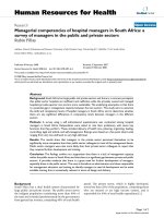
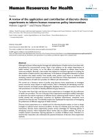
Simultaneous detection of viral RNA and contaminating DNAFigure 1
Simultaneous detection of viral RNA and contaminating DNA. Serial ten-fold dilutions of viral RNA with approximately 1,000
copies of heterologous competitor (amplicon) DNA per reaction were amplified following incubation for 20 min at 30°C either
without (A) or with (B) 0.25 U UNG per reaction. Reactions were performed in triplicate and contained approximately
250,000 copies viral RNA (●), 25,000 copies viral RNA (▼), 2,500 copies viral RNA (■) or 250 copies viral RNA (◆). Each
reaction contained 1,000 copies of amplicon DNA. The fluorescence signal generated by amplicon DNA is indicated by the
open form of the same symbol for each respective reaction. The horizontal line at approximately 0.008 fluorescence units
(dRn) indicates the threshold for a positive reaction. dRn, baseline-corrected normalized fluorescence.
Cycle number
0 5 10 15 20 25 30 35 40
Fluorescence (dRn)
-0.02
0.00
0.02
0.04
0.06
0.08
0.10
0.12
0.14
0.16
0.18
A)
Cycle number
0 5 10 15 20 25 30 35 40
Fluorescence (dRn)
-0.02
0.00
0.02
0.04
0.06
0.08
0.10
0.12
0.14
0.16
0.18
B)
Virology Journal 2005, 2:29 />Page 6 of 8
(page number not for citation purposes)
dilution of viral RNA (Fig. 1A). However, addition of 0.25
U UNG per reaction with incubation at 30°C for 20 min
prior to RT-PCR completely eliminated the signal for con-
taminating DNA in each reaction (Fig. 1B). The amplifica-
tion curves for viral RNA detection were unaffected other
than an increase in C
T
by a mean of 1.2 cycles, a value con-
sistent with results shown above.
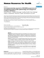
Effect of UNG and incubation prior to RT-PCR on
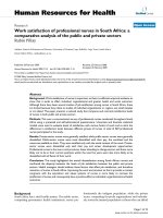
standard curves for RNA quantification
To assess the effect of UNG addition to reaction mixtures
on standard curves used for RNA quantification, reactions
were performed using 10-fold dilutions of heterologous
competitor RNA (Fig. 2). Standard curves were analyzed
for reactions without UNG or containing 0.25 units UNG
and were incubated 30 min at 30°C prior to RT-PCR.
Regression analyses for these curves were compared to a
standard curve using the same RNA dilutions which were
not incubated at 30°C for 20 min prior to RT-PCR. Regres-
sion analysis for all three reaction conditions demon-
strated parallel lines with R
2
values >0.99. The slopes of
the regression lines were essentially equal at approxi-
mately -3.330, which indicate amplification efficiencies
near 100%. Only the y-intercepts differed between the
three reaction conditions, with the values from the reac-
tions containing UNG and incubated at 30°C for 20 min
slightly greater than the standards that did not contain
UNG and were not incubated prior to RT-PCR. The y-
intercept value for the reactions not containing UNG and
incubated at 30°C for 20 min prior to RT-PCR was inter-
mediate of the other two values.
Detection of low viral RNA concentrations in reactions
containing UNG
To determine if UNG addition and incubation prior to RT-
PCR would result in false negative results under the con-
ditions described, 96 replicate samples containing
approximately 20 copies of viral RNA per replicate were
amplified by RT-PCR. Of the 96 replicate samples tested,
positive amplification (defined as C
T
< 40) was not
detected in three samples that contained 0.25 units UNG
and were incubated at 30°C for 30 min prior to RT-PCR
(data not shown). Of 96 control reactions (i.e. no UNG
added and no incubation prior to RT-PCR), amplification
was detected in all but one of the replicates. The mean C
T
increase of samples containing UNG and incubated at
30°C for 30 min prior to RT-PCR was 1.56 cycles, a value
in agreement with results shown above.
Discussion
The techniques of PCR and RT-PCR offer several advan-
tages when compared to traditional viral diagnostic tech-
niques, especially in terms of analytical sensitivity and
time for assay completion. Unfortunately the high level of
sensitivity, which approaches the single molecule level,
also makes this technique prone to false positive results.
Amplicon generated by previous positives reactions in
which dUTP was substituted for dTTP can be degraded by
UNG and thus theoretically eliminated as a source of tem-
plate that would cause false positives in PCR or RT-PCR.
UNG degrades contaminating uracil-containing DNA
while leaving the natural, thymine-containing DNA
intact. The precise, reproducible quantification of real-
time RT-PCR provides a rapid method to assess and opti-
mize the use of UNG to eliminate or reduce the impact of
amplicon DNA in RT-PCR as well as determine the effects
of UNG on RNA detection. The manufacturers' recom-
mendation for the use of heat-labile UNG include addi-
tion of 1 unit enzyme per 50 µl reaction followed by an
incubation time of 10 min at 15 – 25°C. In this study, this
enzyme concentration and the incubation conditions
were not found to eliminate amplicon DNA, presumably
due at least in part to the short length of amplicon DNA
(114 bases). To consistently degrade approximately 500
copies of amplicon DNA, a two-fold increase in UNG con-
centration was necessary. However, this concentration of
UNG resulted in a nearly two cycle increase to reach the
threshold for RNA detection. These results are
corroborated by a recent publication using real-time PCR
for detection of single-copy genes in which it was con-
Effect of UNG on Standard curves for competitor RNAFigure 2
Effect of UNG on Standard curves for competitor RNA. Lin-
ear regression analysis for each standard curve was per-
formed within the analysis software (Stratagene Mx4000
version 4.00). Symbols represent means for samples analyzed
in triplicate. Addition of 0.25 units UNG per reaction fol-
lowed by incubation at 30°C for 20 min prior to RT-PCR
(●), no addition of UNG but incubation at 30°C for 20 min
prior to RT-PCR (❍), no addition of UNG and no incubation
prior to RT-PCR (▼). C
T
, cycle threshold. dRn, baseline-cor-
rected normalized fluorescence.
Starting quantity (copies RNA)
10
0
10
1
10
2
10
3
10
4
10
5
10
6
10
7
10
8
C
T
(dRn)
15
20
25
30
35
40
45
Virology Journal 2005, 2:29 />Page 7 of 8
(page number not for citation purposes)
cluded that manufacturers' recommended conditions
were not adequate to consistently degrade even minimal
(e.g. < 30 copies) amounts of amplicon DNA [16].
To determine optimal conditions for amplicon degrada-
tion with minimal effect on RNA detection by RT-PCR, a
series of time, temperature and enzyme concentrations
were evaluated. From these results, it was shown that
UNG degradation of amplicon DNA in RT-PCR was con-
centration, time and temperature depended, as previously
described for real-time PCR [16]. To allow degradation of
considerable concentrations of carry-over amplicon DNA
while having minimal effects on RNA detection, the opti-
mal concentration of UNG was found to be 0.25 U per 25
µl reaction with a pre-amplification incubation at 30°C
for 20 min. This incubation was performed after the com-
plete reaction had been assembled in the reaction tubes,
just prior to the RT step. These conditions completely
degraded approximately 2,500 copies of carry-over ampli-
con DNA while reducing the detectible concentration of
viral RNA template by 2.2-fold (i.e. C
T
values increased by
a mean of 1.2 cycles). If higher levels of carry-over
contamination are encountered, increasing the incuba-
tion time by an additional 10 min demonstrated degrada-
tion of a 10-fold higher concentration of amplicon DNA
while only slightly increasing the C
T
for RNA detection. It
is interesting to note that, based on analysis of the stand-
ard curves, increases observed in the C
T
for RNA were due
both to the presence of UNG and the incubation at 30°C
prior to RT-PCR.
Given the relatively long time required for the reverse
transcriptase step, a heat-labile UNG that is rapidly and
effectively inactivated at temperatures below that of the
RT step must be used for this approach to be applied to
control false positive reactions in RT-PCR. The commer-
cially available heat-labile UNG used in this study is rap-
idly inactivated at 40°C with a half-life of 2 min [13]. At
the end of the UNG incubation, the samples were held at
55°C to rapidly inactivate UNG just prior to the RT step
which was 50°C for 30 min. For the RT-PCR reagents used
(as well as many other commercially availably RT-PCR
reagents), the manufacture states that the 30 min RT step
can be performed at temperatures of up to 55°C (or
higher for some reagent systems), and reactions analyzed
in preliminary experiments (data not shown) demon-
strated no effect on RT-PCR analytical sensitivity follow-
ing a 2 min incubation at 55°C prior to the RT step.
Results presented herein demonstrated that incubation
with UNG appears to increase the C
T
equally for in vitro
transcribed RNA and viral RNA, thus quantification
through the use of a standard curve can remain accurate
provided that all reactions are performed under the same
conditions. However, a small percentage of weak positive
samples may not be detected due to an increase in C
T
caused by UNG and incubation prior to RT-PCR. To the
reduce the possibility of false negatives with dilute RNA
samples, results from this study suggest that replicates of
two or more reactions should be sufficient, given that the
false negative rate was only increased by 2% for samples
containing UNG and incubated prior to RT-PCR. Also,
two – five additional cycles of amplification may provide
positive amplification of target RNA in samples with very
low concentrations.
Conclusion
Quantitative assessment of the effect of UNG on DNA
degradation and RNA amplification over a range of
enzyme concentrations, temperatures and times demon-
strated that optimization of reaction conditions allows
selection of conditions that maximize carry-over ampli-
con degradation while minimizing the effect on RNA
detection. While this study was performed with real-time
RT-PCR to provide accurate quantification of the effects of
UNG, these findings are potentially useful to both stand-
ard and real-time RT-PCR amplification methods.
Methods
Quantitative (TaqMan) RT-PCR
Amplification reactions were performed using the Qiagen
QuantiTect Probe RT-PCR kit (Qiagen, Inc., Valencia, CA)
with thermocycling and detection performed in a Strata-
gene Mx4000 real-time PCR machine (Stratagene, Inc., La
Jolla, CA). Samples were analyzed in triplicate. The ampli-
fication protocol, oligonucleotide primers and dual-
labeled probe used for 5' exonuclease (TaqMan) amplifi-
cation of the North American PRRSV Ingelvac MLV were
as previously described [17] and amplified a 114-bp frag-
ment. The amplified in the presence of dUTP, the sense
strand and anti-sense strand will contain 36 and 26 uracil
residues, respectively. The dual-labeled probe used for
detection of heterologous competitor RNA (and amplicon
DNA derived from the competitor RNA) was: 5'-HEX-
TGTGCTGCAAGGCGATTAAGTTGGGT-BHQ2-3'. All oli-
gonucleotide primers and dual-labeled probes were syn-
thesized by Integrated DNA Technologies, Inc.
(Coralville, IA). Negative control reactions, in which RNA
extracted from normal (unaffected) swine tissues or serum
was added as template to the RT-PCR reaction mixture,
did not produce a signal for the quantitative RT-PCR
assay.
Preparation of heterologous competitor RNA
Specific oligonucleotide primer binding sites for the
PRRSV real-time RT-PCR assay were incorporated as 5'
extensions in PCR primers and in vitro transcribed heterol-
ogous competitor RNA was prepared and
spectrophotometrically quantified using methods previ-
ously described [18].
Publish with BioMed Central and every
scientist can read your work free of charge
"BioMed Central will be the most significant development for
disseminating the results of biomedical research in our lifetime."
Sir Paul Nurse, Cancer Research UK
Your research papers will be:
available free of charge to the entire biomedical community
peer reviewed and published immediately upon acceptance
cited in PubMed and archived on PubMed Central
yours — you keep the copyright
Submit your manuscript here:
/>BioMedcentral
Virology Journal 2005, 2:29 />Page 8 of 8
(page number not for citation purposes)
Viral RNA extraction
Extraction of Porcine arterivirus RNA from cell culture
stocks of the vaccine strain Ingelvac MLV (derived by serial
passage of U.S. prototype strain VR-2332) was performed
using the Qiagen viral RNA kit (Qiagen, Inc., Valencia,
CA) according to the manufacturer's instructions. Viral
stocks were prepared in MARC-145 cells maintained in
Dulbecco's Modified Eagle medium supplemented with
10% heat-inactivated fetal bovine serum and 2 mM L-
glutamine, 0.25 µg/ml fungizone, and 0.5 mg/ml gen-
tamycin (all supplied by Mediatech, Inc., Herndon, VA).
The viral-infected cell cultures were maintained at 37°C in
a humidified 5% CO
2
incubator for approximately 2 days
until viral cytopathic effect was readily identified through-
out the culture. Extraction of heterologous competitor
RNA after in vitro transcription was performed using the
Qiagen RNeasy kit (Qiagen, Inc., Valencia, CA).
UNG treatment of reactions prior to RT-PCR
The indicated concentrations of heat-labile uracil-DNA
glycosylase (Roche Applied Science, Indianapolis, IN),
purified from the psychrophilic marine organism BMTU
3346 [13] were added to the amplification master mix
prior to dispensing into individual amplification tubes.
Samples were then held in the thermocycler at the indi-
cated temperatures for the indicated times prior to RT-
PCR. At the end of the incubation, the thermocycler was
programmed to ramp the samples at the maximum rate
(2.2°C/sec) to 55°C and hold at this temperature for 5
min prior to the 50°C, 30 min RT step. Control reactions,
which did not contain UNG and were not subjected to
incubation prior to RT-PCR, were placed in the thermocy-
cler at the beginning of the 55°C phase, prior to the 50°C,
30 min RT step.
Competing interests
The author(s) declare that they have no competing
interests.
Acknowledgements
The author wishes to thank Sunny J. Troxell for dedicated and expert tech-
nical contributions.
References
1. Wagar EA: Direct hybridization and amplification applications
for the diagnosis of infectious diseases. J Clin Lab Anal 1996,
10:312-325.
2. Elnifro EM, Ashshi AM, Cooper RJ, Klapper PE: Multiplex PCR:
optimization and application in diagnostic virology. Clin Micro-
biol Rev 2000, 13:559-570.
3. Sellon RK: Update on molecular techniques for diagnostic
testing of infectious disease. Vet Clin North Am Small Anim Pract
2003, 33:677-693.
4. Ivnitski D, O'Neil DJ, Gattuso A, Schlicht R, Calidonna M, Fisher R:
Nucleic acid approaches for detection and identification of
biological warfare and infectious disease agents. Biotechniques
2003, 35:862-869.
5. Yang S, Rothman RE: PCR-based diagnostics for infectious dis-
eases: uses, limitations, and future applications in acute-care
settings. Lancet Infect Dis 2004, 4:337-348.
6. Borst A, Box AT, Fluit AC: False-positive results and contami-
nation in nucleic acid amplification assays: suggestions for a
prevent and destroy strategy. Eur J Clin Microbiol Infect Dis 2004,
23:289-299.
7. Kwok S, Hinguchi R: Avoiding false positives with PCR. Nature
1989, 339:237-238.
8. Longo MC, Berninger MS, Hartley JL: Use of uracil DNA glycosy-
lase to control carry-over contamination in polymerase
chain reactions. Gene 1990, 93:125-128.
9. Pang J, Modlin J, Yolken R: Use of modified nucleotides and
uracil-DNA glycosylase (UNG) for the control of contamina-
tion in the PCR-based amplification of RNA. Mol Cell Probes
1992, 6:251-256.
10. Rys PN, Persing DH: Preventing false positives: quantitative
evaluation of three protocols for inactivation of polymerase
chain reaction amplification products. J Clin Microbiol 1993,
31:2356-2360.
11. Poddar SK, Sawyer MH, Connor JD: Optimized PCR amplifica-
tion of influenza A virus RNA using Tth DNA polymerase,
incorporating uracil N glycosylase (UNG) in a single tube
reaction. J Clin Lab Anal 1997, 11:323-327.
12. Udaykumar , Epstein JS, Hewlett IK: A novel method employing
UNG to avoid carry-over contamination in RNA-PCR. Nucleic
Acids Res 1993, 21:3917-3918.
13. Sobek H, Schmidt M, Frey B, Kaluza K: Heat-labile uracil-DNA
glycosylase: purification and characterization. FEBS Lett 1996,
388:1-4.
14. Hofmann-Lehmann R, Swenerton RK, Liska V, Leutenegger CM, Lutz
H, McClure HM, Ruprecht RM: Sensitive and robust one-tube
real-time reverse transcriptase-polymerase chain reaction
to quantify SIV RNA load: comparison of one- versus two-
enzyme systems. AIDS Res Hum. Retroviruses 2000, 16:1247-5127.
15. Taggart EW, Carroll KC, Byington CL, Crist GA, Hillyard DR: Use of
heat labile UNG in an RT-PCR assay for enterovirus
detection. J Virol Methods 2002, 105:57-65.
16. Pierce KE, Wangh LJ: Effectiveness and limitations of uracil-
DNA glycosylases in sensitive real-time PCR assays. Biotech-
niques 2004, 36:44-46. 48
17. Kleiboeker SB, Schommer SK, Lee S-M, Watkins S, Chittick W, Pol-
son D: Simultaneous detection of North American and Euro-
pean Porcine reproductive and respiratory syndrome virus
using real-time quantitative RT-PCR. J Vet Diagn Invest 2005,
17:165-170.
18. Kleiboeker SB: Applications of competitor RNA in diagnostic
reverse transcription-PCR. J Clin Microbiol 2003, 41:2055-2061.