Báo cáo sinh học: " Adeno-associated virus: from defective virus to effective vector" potx
Bạn đang xem bản rút gọn của tài liệu. Xem và tải ngay bản đầy đủ của tài liệu tại đây (922.9 KB, 17 trang )
BioMed Central
Page 1 of 17
(page number not for citation purposes)
Virology Journal
Open Access
Review
Adeno-associated virus: from defective virus to effective vector
Manuel AFV Gonçalves*
Address: Gene Therapy Section, Department of Molecular Cell Biology, Leiden University Medical Center, Wassenaarseweg 72, 2333 AL Leiden,
the Netherlands
Email: Manuel AFV Gonçalves* -
* Corresponding author
Abstract
The initial discovery of adeno-associated virus (AAV) mixed with adenovirus particles was not a
fortuitous one but rather an expression of AAV biology. Indeed, as it came to be known, in addition
to the unavoidable host cell, AAV typically needs a so-called helper virus such as adenovirus to
replicate. Since the AAV life cycle revolves around another unrelated virus it was dubbed a satellite
virus. However, the structural simplicity plus the defective and non-pathogenic character of this
satellite virus caused recombinant forms to acquire centre-stage prominence in the current
constellation of vectors for human gene therapy. In the present review, issues related to the
development of recombinant AAV (rAAV) vectors, from the general principle to production
methods, tropism modifications and other emerging technologies are discussed. In addition, the
accumulating knowledge regarding the mechanisms of rAAV genome transduction and persistence
is reviewed. The topics on rAAV vectorology are supplemented with information on the parental
virus biology with an emphasis on aspects that directly impact on vector design and performance
such as genome replication, genetic structure, and host cell entry.
Adeno-associated virus biology
Genome structure, DNA replication and virus assembly
The human adeno-associated virus (AAV) was discovered
in 1965 as a contaminant of adenovirus (Ad) preparations
[1]. AAV is one of the smallest viruses with a non-envel-
oped icosahedral capsid of approximately 22 nm (Fig. 1),
the crystal structure of which has been recently deter-
mined to a 3-angstrom resolution [2]. Because a co-infect-
ing helper virus is usually required for a productive
infection to occur, AAV serotypes are ascribed to a separate
genus in the Parvoviridae family designated Dependovirus.
Despite the high seroprevalence of AAV in the human
population (approximately 80% of humans are seroposi-
tive for AAV2) the virus has not been linked to any human
illness. The AAV has a linear single-stranded DNA genome
of approximately 4.7-kilobases (kb). The AAV2 DNA ter-
mini consist of a 145 nucleotide-long inverted terminal
repeat (ITR) that, due to the multipalindromic nature of
its terminal 125 bases, can fold on itself via complemen-
tary Watson-Crick base pairing and form a characteristic
T-shaped hairpin structure (Fig. 2) [3]. According to the
AAV DNA replication model [4] this secondary structure
provides a free 3' hydroxyl group for the initiation of viral
DNA replication via a self-priming strand-displacement
mechanism involving leading-strand synthesis and dou-
ble-stranded replicative intermediates (Fig. 3). The virus
does not encode a polymerase relying instead on cellular
polymerase activities to replicate its DNA [5]. The ITRs
flank the two viral genes rep (replication) and cap (capsid)
encoding nonstructural and structural proteins, respec-
tively. The rep gene, through the use of two promoters
located at map positions 5 (p5) and 19 (p19), and an
Published: 06 May 2005
Virology Journal 2005, 2:43 doi:10.1186/1743-422X-2-43
Received: 08 April 2005
Accepted: 06 May 2005
This article is available from: />© 2005 Gonçalves; licensee BioMed Central Ltd.
This is an Open Access article distributed under the terms of the Creative Commons Attribution License ( />),
which permits unrestricted use, distribution, and reproduction in any medium, provided the original work is properly cited.
Virology Journal 2005, 2:43 />Page 2 of 17
(page number not for citation purposes)
internal splice donor and acceptor site, encode four regu-
latory proteins that are dubbed Rep78, Rep68, Rep52 and
Rep40 on basis of their apparent molecular weights. The
Rep78 and Rep68 proteins participate in the AAV DNA
replication process via their interaction with Rep-binding
element (RBE) and terminal resolution site (trs)
sequences located within the ITRs (Fig. 2). In addition, in
response to environmental cues such as presence or
absence of a helper virus these proteins either positively or
negatively regulate AAV gene expression, respectively [6].
The Rep52 and Rep40 proteins are involved in the gener-
ation and accumulation of single-stranded viral genomes
from double-stranded replicative intermediates [7]. The
resulting single-stranded genomes with plus and minus
polarities are packaged with equal efficiency [8]. The
economy displayed by AAV is staggering and derives not
only from its overlapping genetic organization but also
from the integration of various biochemical activities in
each of its few gene products. For instance, Rep78 and
Rep68 are site-specific DNA binding proteins, as well as
strand- and site-specific endonucleases [9]. They also
exhibit helicase and ATPase activities [10], which are
shared by Rep52 [11] and by Rep40 [12].
The cap gene is transcribed from a single promoter at map
position 40 (p40). Alternative splicing at two acceptor
sites originates two transcripts. The larger transcript
encodes virion protein 1 (VP1), the biggest capsid protein
subunit. The shorter mRNA possesses a noncanonical
start codon (ACG), which is utilized to generate VP2, and
a downstream conventional initiation codon (AUG)
directing the synthesis of VP3. The VP1, VP2 and VP3 pro-
teins differ from each other at their N terminus and have
apparent molecular masses of 87, 72 and 62 kDa, respec-
tively. Together they assemble into a near-spherical pro-
tein shell of 60 subunits with T = 1 icosahedral symmetry.
At the 12 fivefold axes of symmetry lay narrow pores lately
shown to be instrumental for virus infectivity and for
genome packaging [13]. The molar ratio between VP1,
VP2 and VP3 in AAV particles is 1:1:10. This stoichiometry
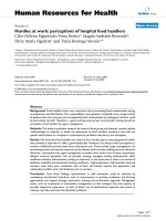
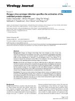
Transmission electron microscopy of AAV2 and Ad5 particles in human cellsFigure 1
Transmission electron microscopy of AAV2 and Ad5 particles in human cells. (A) AAV2 and Ad5 particles in the nucleus of a
HeLa cell at 48 hours after co-infection. Magnification: × 15,000. (B) AAV2 virions in a HeLa cell at 48 hours after co-infection
with Ad5. Magnification: × 40,000.
A
B
AAV
Ad
AAV
500 nm
200 nm
Virology Journal 2005, 2:43 />Page 3 of 17
(page number not for citation purposes)
is thought to reflect the relative abundance of the two cap
gene transcripts and the relative efficiency of translation
initiation at the three start codons for the structural pro-
teins. A conserved phospholipase A
2
(PLA2) motif, ini-
tially identified within the unique N-terminal region of
the parvoviral VP1 proteins [14], was also reported to
have a biological significance in AAV2 infection [15]. Spe-
cifically, although dispensable for capsid assembly, DNA
packaging, and virion internalisation, the VP1-embedded
PLA2 activity seems to play a key role at some stage
between the translocation of the AAV genome from the
endocytic to the nuclear compartment and the initiation
of viral gene expression [15]. Lately, mutational analysis
of amino acid residues involved in AAV2 capsid pore
architecture indicate that conformational changes of the
virion structure during infection lead the VP1 N termini to
protrude through the capsid pores inducing the PLA2
enzymatic activity needed for successful infection [13]. At
the level of virion formation, immunofluorescence data
shows that the VP1 and VP2 proteins are found primarily
in the nuclei of infected cells, whereas VP3 is nearly evenly
distributed between the nucleus and the cytoplasm [16].
However, in the presence of VP1 and/or VP2, VP3 accu-
mulates in the nucleus suggesting transport of the major
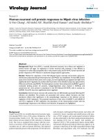
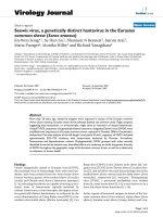
Secondary structure of the AAV2 ITRFigure 2
Secondary structure of the AAV2 ITR. The AAV2 ITR serves as origin of replication and is composed of two arm palindromes
(B-B' and C-C') embedded in a larger stem palindrome (A-A'). The ITR can acquire two configurations (flip and flop). The flip
(depicted) and flop configurations have the B-B' and the C-C' palindrome closest to the 3' end, respectively. The D sequence is
present only once at each end of the genome thus remaining single-stranded. The boxed motif corresponds to the Rep-binding
element (RBE) [119] where the AAV Rep78 and Rep68 proteins bind. The RBE consists of a tetranucleotide repeat with the
consensus sequence 5'-GNGC-3'. The ATP-dependent DNA helicase activities of Rep78 and Rep68 remodel the A-A' region
generating a stem-loop that locates at the summit the terminal resolution site (trs) in a single-stranded form [120,121]. In this
configuration, the strand- and site-specific endonuclease catalytic domain of Rep78 and Rep68 introduces a nick at the trs. The
shaded nucleotides at the apex of the T-shaped structure correspond to an additional RBE (RBE') [121] that stabilizes the asso-
ciation between the two largest Rep proteins and the ITR.
Virology Journal 2005, 2:43 />Page 4 of 17
(page number not for citation purposes)
capsid protein by association with the nuclear localization
signal-bearing proteins VP1 and VP2 [17]. Immunofluo-
rescence results suggest that capsid assembly is confined
to the nucleoli of infected cells. The involvement of nucle-
olar chaperones in this process has been postulated [16].
Fully assembled AAV capsids enter the nucleoplasm in an
AAV Rep-dependent manner. This redistribution of the
structural proteins causes the co-localization of all ingre-
dients necessary for infectious particle formation, i.e., cap-
sids, Rep proteins and viral genomes. Indeed, the AAV
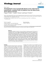
Schematic representation of the AAV DNA replication modelFigure 3
Schematic representation of the AAV DNA replication model. AAV DNA replication is thought to involve a self-priming single-
strand displacement mechanism that is initiated by DNA polymerisation at the 3' hairpin primer of input single-stranded
genomes. This leads to the formation of linear unit-length double-stranded molecules (duplex monomers, DMs) with one cov-
alently closed end. These structures are resolved at the terminal resolution site (trs) by site-specific nicking of the parental
strand opposite the original 3' end position (i.e., at nucleotide 125). The newly generated free 3' hydroxyl groups provide a
substrate for DNA polymerases that unwind and copy the inverted terminal repeat (ITR). Finally, the palindromic linear duplex
termini can renaturate into terminal hairpins putting the 3' hydroxyl groups in position for single-strand displacement synthesis.
Next, single-stranded genomes and new DM replicative forms are made. When nicking does not occur, elongation proceeds
through the covalently closed hairpin structure generating linear double-length double-stranded molecules (duplex dimers,
DDs) with either a head-to-head or a tail-to-tail configuration. The DD replicative intermediates can be resolved to DMs
through the AAV ITR sequences located at the axis of symmetry.
3'-OH
5'
trs
trs
+
Nicking
DM
DD (head-to-head or tail-to-tail)
Dimerization
ITR-primed DNA polymerization
Terminal resolution
ITR renaturation
Single-strand displacement /
elongation
Nicking failed
DD resolution
Parental strands
Daughter strands
AAV ITR
Virology Journal 2005, 2:43 />Page 5 of 17
(page number not for citation purposes)
DNA packaging process is though to take place in distinct
regions of the nucleoplasm [16]. Selective AAV DNA
encapsidation is presumably directed by protein-protein
interactions between pre-formed empty capsids and com-
plexes of Rep78 or Rep68 with the virus genome [18].
Next, the helicase domains of capsid-docked Rep52 and
Rep40 proteins are proposed to act as molecular motors
that unwind and transfer de novo synthesized single-
stranded DNA into empty particles [19] through the pores
located at the fivefold symmetry axes [13].
Host cell infection
AAV2 virions utilize as primary attachment receptor
heparan sulphate proteoglycans [20] while internalisation
is aided by the co-receptors α
v
β
5
integrin heterodimers
[21], fibroblast growth factor receptor type 1 [22] and the
hepatocyte growth factor receptor, c-Met [23]. The use of
ubiquitous heparan sulphate proteoglycans as docking
sites explains in part the well-known broad tropism of this
virus that include, human, non-human primate, canine,
murine and avian cell types. AAV5 and AAV4 also bind to
charged carbohydrate moieties in the form of N- and O-
linked sialic acids, respectively [24]. Expression profiling
of AAV5 permissive and non-permissive cells with cDNA
microarrays led to the identification of platelet-derived
growth factor receptor as another cellular determinant
involved in AAV5 infection [25].
The events and processes that regulate the trafficking of
AAV particles into the nucleus are still not fully under-
stood, however, some findings have been reported. For
instance, infection experiments in HeLa cells expressing a
dominant-negative form of dynamin significantly
reduced AAV2 entry [26,27]. These results indicate that
one route by which this virus can poke through the
plasma membrane involves receptor-mediated endocyto-
sis via the formation of clathrin-coated pits. In addition,
lysomotropic agents and proton pump inhibitors greatly
hamper AAV2 infection suggesting that internalised viri-
ons escape from endosomes and are released in the
cytosol by a low pH-dependent process [27]. In addition,
a powerful new imaging technique based on single-mole-
cule labelling of discrete AAV particles enabled real-time
monitoring of the trajectories of individual virions [28].
In these experiments, it was shown that each endosome
carries a single AAV particle. Moreover, the abrogation of
vectorial motion of virions in nocodazole-treated cells
supported the involvement of microtubule assembly and
motor proteins in active AAV intracellular transportation.
Finally, it has been suggested that AAV particles due to
their very small size can access the nucleus through the
nuclear pore complex (NPC). However, recent research
points to a nuclear entry process that is not dependent on
NPC activity [29,30] whereas the issue of whether AAV
capsids enter nuclei intact or remodelled seems to depend
on the presence or absence, respectively, of co-infecting
helper Ad particles [30].
Lytic and lysogenic pathways
After entry into the host cell nucleus, AAV can follow
either one of two distinct and interchangeable pathways
of its life cycle: the lytic or the lysogenic. The former devel-
ops in cells infected with a helper virus such as Ad or her-
pes simplex virus (HSV) whereas the latter is established
in host cells in the absence of a helper virus. When AAV
infects a human cell alone, its gene expression program is
auto-repressed and latency ensues by preferential integra-
tion of the virus genome into a region of roughly 2-kb on
the long arm (19q13.3-qter) of human chromosome 19
[31,32] designated AAVS1 [33]. Recent research showed
that this locus is in the vicinity of the muscle-specific
genes p85 [34], TNNT1 and TNNI3 [35]. Furthermore, the
AAVS1 sequence lies in a chromosomal region with char-
acteristics of a transcription-competent environment [36].
Interestingly, an insulator within this locus was recently
identified [37]. The targeted integration of the AAV
genome, a phenomenon unique among all known
eukaryotic viruses, enables the provirus DNA to be
perpetuated through host cell division. Moreover, the
level of specificity of this process of AAV biology (a single
preintegration region within the entire human genome)
makes its exploitation highly attractive for achieving the
ultimate goal of safe and stable transgene expression [38].
Even if working models for the targeted DNA integration
mechanism remain sketchy [39,40], the viral components
needed for the site-specific integration reaction have been
identified. They are composed in cis by the AAV ITRs and
in trans by either one of the two largest Rep proteins (i.e.,
Rep78 or Rep68). Recently, another cis-acting sequence
was shown to be necessary for high-level site-specific DNA
integration [41,42]. This sequence overlaps with the
highly regulated p5 promoter and, like the ITR sequence,
harbours an RBE.
Detailed genetic analyses using an AAVS1-containing epi-
some system demonstrated that a 33-bp sequence con-
taining elements related to the RBE and to the trs is
sufficient for targeted DNA integration. Their functional
relevance was demonstrated by the absence of targeted
DNA integration into mutated substrates [39]. In addi-
tion, the AAVS1 region behaves as an origin of replication
in the presence of Rep proteins both in vitro [43] and in
vivo [44]. Finally, the AAVS1-specific RBE and trs are sep-
arated by a spacer element whose sequence and length
affects the efficiency of the site-specific DNA integration
reaction [45]. The human genome has numerous Rep
binding sites. However, database searches have revealed
that an RBE at a proper distance from a trs sequence occurs
only in the AAVS1 locus, which is consistent with the
Virology Journal 2005, 2:43 />Page 6 of 17
(page number not for citation purposes)
specificity of the integration reaction revealed through
biological assays [46]. Moreover, in vitro studies showed
that via their interaction with the RBE sequences present
in the AAV ITRs and in the AAVS1 locus, Rep78 and Rep68
proteins could tether viral to cellular DNA [47]. Although,
as mentioned above, the actual mechanism evolved by
AAV to target its DNA to the AAVS1 locus is currently
unknown, taken together these observations provide at
the molecular level an explanation for the specificity of
the reaction and the requirement for RBE-containing
sequences in cis and either one of the two largest Rep pro-
teins in trans. Remarkably, only recently a study emerged
directly addressing the AAV DNA integration efficiency
and the correlation between random versus targeted inte-
gration levels [48]. Using a tissue culture system, the
authors showed by clonal analyses of target cells and
Southern blot hybridisations that 50% of infected cells
were stably transduced by AAV when a multiplicity of
infection of 100 was used. Raising the dose of virus
increased neither the frequency of infected cells nor the
integration levels. Although multiplicities of infection of
100 and 10 both yielded approximately 80% infected
cells, the frequency of stably transduced cells was below
5% when employing the lower dose. Virtually all integra-
tion events targeted the AAVS1 locus. Finally, for each
multiplicity of infection, the frequency of AAVS1 site dis-
ruption without accompanying DNA insertion was higher
than the frequency of site-specific integration by a factor
of 2.
When a latently infected cell is super-infected with a
helper virus, the AAV gene expression program is activated
leading to the AAV Rep-mediated rescue (i.e., excision) of
the provirus DNA from the host cell chromosome fol-
lowed by replication and packaging of the viral genome.
Finally, upon helper virus-induced cell lysis, the newly
assembled virions are released. The induction of the lytic
phase of the AAV life cycle from a stably integrated provi-
rus can also occur in the absence of a helper virus, though
with a lower efficiency, when the host cell is subjected to
metabolic inhibitors and to DNA damaging agents such as
UV irradiation or genotoxic compounds [49]. Moreover,
in differentiated keratinocytes of an epithelial tissue cul-
ture system modelling skin, AAV2 was shown to initiate
and proceed through a complete replicative cycle in the
absence of helper viruses or genotoxic agents [50]. Taken
together, these phenomena indicate that AAV is not defec-
tive in absolute terms.
Adeno-associated virus vectorology
General principle
Historically, most recombinant AAV (rAAV) vectors have
been based on serotype 2 (AAV2) that constitutes the pro-
totype of the genus [51,52]. Important to those pursuing
the use of rAAV for gene therapy applications is the defec-
tiveness of the parental virus and its presumed non-path-
ogenic nature. The realization that a molecularly cloned
AAV genome could in Ad-infected cells recapitulate the
lytic phase of the AAV life cycle and give rise to infectious
virions enabled not only the detailed genetic analyses of
the virus but provided, in addition, a substrate to generate
rAAV particles [53]. The latter task was facilitated by the
fact that the AAV ITRs contain all cis-acting elements
involved in genome rescue, replication and packaging.
Furthermore, since the AAV ITRs are segregated from the
viral encoding regions, rAAV design can follow the whole-
gene-removal or "gutless" vector rational of, for instance,
retrovirus-based vectors in the sense that the cis-acting ele-
ments involved in genome amplification and packaging
are in linkage with the heterologous sequences of interest,
whereas the virus encoding sequences necessary for
genome replication and virion assembly are provided in
trans (Fig. 4). Typically, rAAV particles are generated by
transfecting producer cells with a plasmid containing a
cloned rAAV genome composed of foreign DNA flanked
by the 145 nucleotide-long AAV ITRs and a construct
expressing in trans the viral rep and cap genes. In the pres-
ence of Ad helper functions, the rAAV genome is subjected
to the wild-type AAV lytic processes by being rescued from
the plasmid backbone, replicated and packaged into pre-
formed AAV capsids as single-stranded molecules.
Production and purification strategies
The Ad helper functions were originally supplied by infec-
tion of rAAV producer cells with a wild-type Ad (Fig. 4).
Subsequent elimination of the helper virus from rAAV
stocks relied on the distinct physical properties of AAV
and Ad virions. In particular, differences in thermostabil-
ity and density between AAV and Ad particles allowed the
specific elimination of helper Ad virions by heat-inactiva-
tion (i.e., half-hour at 56°C) and isopycnic cesium chlo-
ride density ultracentrifugation. The finding that Ad
helper functions are provided by expression of E1A, E1B,
E2A, E4ORF6 and VA RNAs, enabled subsequent Ad-free
production of rAAV vector stocks by incorporating VA
RNAs, E2a and E4ORF6 sequences into a plasmid and
transfecting it together with the rAAV DNA plus rep and
cap templates into Ad E1A- and E1B-expressing cells [54-
56]. During the testing of new packaging plamids for rAAV
production it was also found that reduction of the expres-
sion levels of the two largest AAV Rep proteins leads to an
increase in vector yields [56,57]. Although these methods
improve rAAV production and avoid the need for Ad
infection, they are difficult to scale up due to their
dependence on DNA transfection. The development of
up-scalable transfection-independent methods for rAAV
production have been fiercely pursued by the requirement
for large amounts of highly purified vector particles to per-
form experiments in large animal models and human
clinical trials. One of these transfection-independent
Virology Journal 2005, 2:43 />Page 7 of 17
(page number not for citation purposes)
production strategies involves the generation of packaging
cell lines having the AAV rep and cap genes stably inte-
grated in their genomes. The establishment of effective,
high-titer producer cell lines has proven difficult mainly
due to the inhibitory effects of Rep proteins on cell growth
[58] and the accumulation of low amounts of AAV gene
products relative to a wild-type virus infection. Nonethe-
less, improvements in the control of rep expression
through the development of stringent inducible gene
expression systems can overcome the former hurdle [59]
whereas in situ amplification of integrated rep and cap
templates helps to minimize the latter problem [60,61].
Another transfection-independent approach to produce
rAAV involves the delivery of the viral genes together with
the rAAV DNA and the helper functions via infection of
produced cells with recombinant viruses based on Ad
[60], HSV [62] or baculovirus [63]. In parallel to new
rAAV production platforms, insights into AAV biology are
also leading to significant improvements in the quality
and purity of vectors based on AAV2 as well as on those
based on other serotypes. Specifically, knowledge on AAV
receptor usage has permitted the implementation of up-
scalable affinity column chromatography purification
schemes [64,65]. In addition, a more broadly applicable
column chromatography procedure, based on the ion-
exchange principle, has recently been developed for the
purification of rAAV2, rAAV4 and rAAV5 particles [66].
Tropism modification
An increasingly important area in the development of
AAV as a vector concerns the engineering of altered cell
tropisms to narrow or broaden rAAV-mediated gene deliv-
ery and to increase its efficiency in tissues refractory to
AAV2 infection. Cells can be poorly transduced by proto-
type rAAV2 not only because of low receptor content but
also owing to impaired intracellular virion trafficking and
uncoating [67,68] or single-to-double strand genome
conversion [69-71]. Thus, considering that these processes
depend either directly or indirectly on capsid conforma-
tion, cell targeting strategies determine not only the cell
type(s) with which the vector interacts but also critically
affect the efficiency of the whole gene transfer process.
Several of these approaches rely on the modification by
chemical, immunological or genetic means of the AAV2
capsid structure endowing it with ligands that interact
with specific cell surface molecules [72]. The fact that the
atomic structure of AAV2 has recently been determined
[2] provides a significant boon to those pursuing the
rational design of targeted AAV vectors. Another route to
alter rAAV tropism exploits the natural capsid diversity of
newly isolated serotypes by packaging rAAV2 genomes
into capsids derived from other human or non-human
AAV isolates [73]. To this end, up until now, most
researches employ hybrid trans-complementing
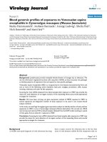
Overview of the initial recombinant AAV production systemFigure 4
Overview of the initial recombinant AAV production system.
The generation of the first infectious clones of AAV permit-
ted functional dissection of the virus genome. This allowed
the construction of plasmids encoding rAAV genomes in
which the minimal complement of wild-type sequences nec-
essary for genome replication and packaging (i.e., the AAV
ITRs) frame a gene of interest (transgene) instead of the AAV
rep and cap genes. When these constructs are transfected
into packaging cells together with a rep and cap expression
plasmid they lead to the production of rAAV particles.
Helper activities required for the activation and support of
the productive phase of the AAV life cycle were originally
introduced by infection of the packaging cells with wild-type
Ad as depicted. Current transfection-based production
methods make use of recombinant DNA encoding the helper
activities instead of Ad infection. Cellular DNA polymerase
activities together with the Rep78 and Rep68 proteins lead
to the accumulation of replicative intermediates both in the
duplex monomer (DM) and duplex dimer (DD) forms. A
fraction of this de novo synthesized DNA is incorporated in
the single-stranded format into preformed empty capsids
most likely through the catalytic activities of the Rep52 and
Rep40 proteins. The resulting infectious rAAV virions are
released from the producer cells together with helper Ad
particles. Sequential heat treatment and buoyant density cen-
trifugation allows the selective elimination of the helper virus
from the final rAAV preparation.
transgene
cap
transgene
+
cap
transgene
Helper Ad elimination
AAV
rep cap
rep
cap
VP1
VP2
VP2
rAAV DNA
packaging DNA
Molecular Cloning
Infection
Assembly
ssDNA packaging
Replication
Rep78, 68, 52 & 40
helper Ad
C
o-transfection
Rep78/68cellular factors
Rep52/40
P
ACKAGING CELL
rAAV
rep
cap
Virology Journal 2005, 2:43 />Page 8 of 17
(page number not for citation purposes)
constructs that encode rep from AAV2 whereas cap is
derived from the serotype displaying the cell tropism of
choice. This pseudotyping approach may also be benefi-
cial in evading neutralizing antibodies to capsid compo-
nents in individuals seropositive for AAV2 or in those in
need of vector readministration. Finally, experiments
published recently using rAAV2 genomes pseudotyped
with coats from AAV6 [74] and AAV8 [75] revealed stun-
ning gene transfer efficiencies when these vectors were
administered alone at high doses or in combination with
a blood vessel permeating agent. The authors could dem-
onstrate transduction of the entire murine striated muscle
system (e.g., diaphragm, heart and skeletal muscles) and
of virtually 100% of the hepatocytes after a single intrave-
nous injection. These body-wide transduction efficiencies
raise both great perspectives as well as caution since they
open new therapeutic avenues for diseases that require
widespread gene delivery (e.g., muscular dystrophies)
while, simultaneously, beg for stringent tissue-specific
transcriptional control to minimize potential deleterious
effects due to transgene expression in non-target tissues.
Moreover, assuming similar avidity of these serotypes for
human tissues, translation of these protocols from mice to
patients will require vastly greater amounts of vector
particles.
Mechanisms of vector DNA persistence
Knowledge on the mechanisms at play following rAAV
transduction is building steadily over recent years mainly
because of its direct relevance to the application of rAAV
in therapeutic gene transfer. DNA vectored through rAAV
can persist long-term in organs such as in the liver and the
striated muscles of mice and dogs. Most importantly, data
showing prolonged and stable expression of an increasing
variety of transgenes in numerous animal models without
notable toxicity is accumulating [76]. It are in fact these
attributes of rAAV-based gene transfer that turns it into
one of the most promising methods for somatic gene ther-
apy providing a rational for the entry of these vectors into
the clinical trial arena. However, at the outset it is impor-
tant to refer that this stability does not arise due to foreign
DNA insertion into the parental virus pre-integration site
since the absence of rep gene products prevents DNA tar-
geting to the AAVS1 locus. Moreover, because rAAV vec-
tors lack viral genes altogether, the molecular fate of the
DNA once in the nucleus is dependent on host cell
activities (though a role for the virion capsomers cannot
be ruled out). These cellular activities, that only recently
have started to be identified, depend on the type as well as
on the physiological status of the target cell. Finally, it is
also of note that the single-stranded nature of AAV
genomes implies that, before transgene expression can
occur, the incoming rAAV DNA needs to be converted into
a transcriptionally functional double-stranded template.
A recent study indicates that free (i.e., unpackaged) single-
stranded rAAV genomes have a very transient presence in
the target cell [67] either because the majority is recog-
nized by host enzymes as damaged DNA and degraded or
because, under certain conditions, single-to-double
strand conversion occurs readily following uncoating.
There are two pathways by which rAAV DNA can be con-
verted from the single- to the double-stranded form each
of them with its own set of supporting experimental data.
One possible route develops through de novo second-
strand DNA synthesis from the hairpin at the 3' end of the
genome (Fig. 2). Initial studies revealed that this step
could be greatly enhanced by Ad E4ORF6 expression, UV
irradiation or treatment of target cells with genotoxic
chemicals [69,70]. Furthermore, a direct correlation
between double-stranded template accumulation and
gene expression was found. More recently, the phosphor-
ylation status of a cellular protein named FKBP52 was
shown to modulate the convertion of single-stranded
rAAV DNA into double-stranded molecules both in tissue
culture [77] and in murine hepatocytes [78]. FKBP52
phosphorylation by the epidermal growth factor receptor
protein tyrosine kinase enables the molecule to bind the
single-stranded AAV ITR D-sequence (Fig. 2). This binding
activity correlates strongly with second-strand DNA syn-
thesis inhibition. Conversely, in its dephosphorylated
state, due to T-cell protein tyrosine phosphatase activity,
FKBP52 does not bind vector genomes allowing synthesis
of the complementary strand to occur with a subsequent
increase in transgene expression levels.
As said before, single-stranded AAV genomes with sense
(plus) and anti-sense (minus) orientations are packaged
equally well. Therefore, another possible route involved
in the generation of double-stranded DNA forms in target
cells comprises the annealing of single-stranded mole-
cules with opposing polarities. Evidence for the existence
of this DNA synthesis-independent pathway came from
experiments using rAAV genomes that were site-specifi-
cally methylated [71]. In these experiments restriction
enzymes were used as probes to evaluate whether modi-
fied rAAV genomes extracted from murine livers were fully
methylated (representing annealing products) or hemi-
methylated (corresponding to second-strand synthesis
products). Thus, seemingly, a contention exits between
advocates of DNA synthesis dependent and independent
models. It is clear, however, that these two pathways are
not necessarily mutually exclusive. In fact, recent experi-
ments in cells under normal physiological conditions
indicate that each of these pathways can contribute to the
generation of transcriptionally active rAAV genomes [67].
For the latter experiments the authors resurrected a tech-
nique deployed to directly demonstrate that AAV is a sin-
gle-stranded virus [8]. Exploiting the differential
thymidine content of complementary polynucleotide
Virology Journal 2005, 2:43 />Page 9 of 17
(page number not for citation purposes)
chains they used incorporation of the thymidine analogue
bromodeoxyuridine (BrdU) to physically separate plus-
from minus-strand containing rAAV particles following
buoyant density centrifugation. Infection of indicator
cells with each vector type led to reporter gene expression
signifying the involvement of second-strand DNA synthe-
sis and precluding an absolute requirement for plus and
minus strand annealing. However, co-infection with both
vector types originated higher numbers of cells expressing
the reporter gene indicating that strand annealing contrib-
utes to the accumulation of double-stranded genomes
[67].
Subsequently, duplex rAAV genomes can, throught intra-
or intermolecular recombination at the ITRs, originate cir-
cular forms or linear concatemers, respectively [71,79].
The circular episomes can also evolve into high-molecu-
lar-weight concatamers in a time-dependent manner [79].
The balance between linear versus circular forms is, at
least in part, regulated by a complex containing DNA-
dependent protein kinase (DNA-PK) [80]. This complex
plays a vital role in the repair of double-stranded chromo-
somal breaks and in V(D)J recombination by non-homol-
ogous end-joining (NHEJ). The absence of the catalytic
subunit of DNA-PK (DNA-PKcs) in severe combined
immunodeficient (SCID) mice (DNA-PKcs-negative)
allowed Song and colleagues to demonstrate its involve-
ment in circular rAAV episome formation in skeletal mus-
cle [80]. Subsequent studies in liver and skeletal muscle of
SCID and normal (DNA-PKcs-positive) mice have
extended the observation that DNA-PK enhances the for-
mation of rAAV circular episomes over linear forms
[81,82]. It has been postulated that free double-stranded
rAAV DNA ends are substrates for the cellular double-
stranded break repair machinery responsible for free-
ended DNA removal through NHEJ ligation [80]. Not-
withstanding their diverse topology and unit numbers, all
these extrachromosomal DNA forms are transcription-
competent templates. Furthermore, they are thought to be
responsible for the stable maintenance of transgene
expression both in skeletal muscles [79] and in the lungs
[83]. In the liver it has been shown that, in addition to the
aforesaid episomal forms, circa 10% of the double-
stranded rAAV genomes can be found inserted in the chro-
mosomal DNA [84].
Backed by the complete mouse genome sequence,
researchers could establish that a significant proportion of
rAAV DNA integration events occur in regions that are
transcriptionally active in murine hepatocytes [85]. In
some instances, sequence micro-homologies and unre-
lated nucleotides are found at the truncated ITR-chromo-
somal DNA junctions. Moreover, rAAV DNA insertion is
consistently associated with host chromosomal deletions.
These characteristics resemble the "fingerprints" following
double-stranded DNA break repair through NHEJ recom-
bination. Thus, taken together, these results point to the
involvement of NHEJ in rAAV DNA integration in addi-
tion to its putative role in the removal of free rAAV DNA
ends, as previously discussed. This interpretation is fur-
ther supported by previous and newly acquired data. For
instance, earlier tissue culture studies revealed a direct cor-
relation between genomic instability due to DNA-damag-
ing agents or genetic defects and stable transduction by
rAAV [86,87]. Other results showed that proteins belong-
ing to the NHEJ complex bind to linear rAAV DNA [88].
More recently, a genetic approach permitted the deliberate
induction of double-stranded chromosomal breaks
within a predefined site [89]. The experimental set up con-
sisted of retrovirus vector-mediated expression of the I-
SceI endonuclease in cells engineered with this enzyme's
18-bp recognition sequence. Following transduction of
these cells with rAAV, the authors could demonstrate
insertion of foreign DNA into I-SceI-induced double-
stranded breaks. Characterization of vector-chromosome
junctions revealed the telltale features observed after rAAV
DNA integration into chromosomal breaks arising spon-
taneously at random sites. It is thus possible to speculate
that rAAV proviral DNA is just another by-product of the
mechanism the cell uses to eliminate free-ended sub-
strates reminiscent of damaged DNA or invading nucleic
acids (e.g., linear retroviral cDNA). As corollary, com-
pared to the integrase-dependent retroviral genome inte-
gration, rAAV DNA insertion is a passive process that relies
instead on pre-existent chromosomal breaks and host cell
enzymes.
Chromosomal DNA integration with current vectors is a
double-edged sword. On the one hand it provides a basis
for permanent genetic correction while, on the other
hand, raises safety issues related to insertional gene-inac-
tivation and proto-oncogene deregulation. It is thus
highly relevant for the clinical deployment of rAAV that
these vectors do not create but instead insert into existing
chromosomal breaks. The latter can be substrates for inac-
curate NHEJ-mediated repair regardless of the presence of
rAAV genomes. Therefore, concerns about insertional
oncogenesis might be less for rAAV- than for retroviral
vector-mediated gene transfer. Additionally, in contrast to
retroviral vectors, rAAV vectors do not display "outward"
promoter activity. Despite this, it is still conceivable that
rAAV DNA insertion can lead to hazardous alteration of
neighbouring gene(s) expression via vector-encoded regu-
latory sequences (e.g., enhancers). Thus, preventive meas-
ures such as judicious choice of transcriptional elements
and use of insulators may turn out to be desirable or even
indispensable in target tissues in which rAAV DNA is
known to integrate at appreciable levels. Adding to the
challenge these genetic elements have to be small enough
Virology Journal 2005, 2:43 />Page 10 of 17
(page number not for citation purposes)
to leave space needed to accommodate the gene of
interest.
Emerging technologies
The small packaging capacity of AAV particles (about 4.7
kb) [90] is considered one of the main limitations of rAAV
vectors since it excludes therapeutically important coding
sequences (e.g., dystrophin cDNA) and potent regulatory
elements (e.g., albumin promoter). As discussed above,
incoming linear rAAV genomes can form concatamers in
target cells through intermolecular recombination at their
free ends. This phenomenon has been successfully
exploited to assemble in target cells large genetic messages
through the joining of two independently transduced
rAAV genomes each of which encompassing a portion of
a large transcriptional unit. mRNA molecules encoding a
functional protein are generated from the rAAV DNA
head-to-tail heterodimers by splicing out the AAV ITR
sequences from the primary transcripts (Fig. 5) [91].
Although this split gene strategy allows expression of
almost double-sized transgenes after rAAV-mediated gene
delivery, its efficiency is consistently lower than that
observed with a single control vector encoding the full-
length transgene. Both vectors have to transduce the same
cell and only heteroconcatamers with a head-to-tail
organization will give rise to a functional full-length gene
product. In addition, there are risks associated with the
integration into host chromosomes of vectors encoding
exclusively regulatory elements or truncated gene prod-
ucts. New work, however, suggests that some of these lim-
itations and concerns can, at least partially, be addressed
[92,93].
Another development in rAAV design is the so-called self-
complementary AAV vectors (scAAV) [94]. The scAAV
approach builds on the ability of AAV to package repli-
cons with half the size of the wild-type DNA in the form
of single-stranded dimeric genomes with an inverted
repeat configuration [95]. In the target cell, these self-
complementary molecules can readily fold back into dou-
ble-stranded forms without the need for de novo DNA syn-
thesis or for the annealing of sense and antisense strands
(Fig. 6). Ultimately, regardless of the mechanism(s) at
play, scAAV lead to enhanced formation of transcription-
competent double-stranded genomes thus improving the
expression kinetics and yields of vector-encoded products.
This scAAV method was subsequently perfected by
mutagenesis of one of the two trs sequences to force the
generation of dimeric over monomeric replicative forms
(Fig. 6) [96]. The main disadvantage of this approach is
the need to limit the size of the transgenes that can be
delivered to approximately half the length of the already
small AAV genome. It is conceivable that this drawback
can be tackled by coupling scAAV with heterodimeriza-
tion strategies. Alternatively, long double-stranded rAAV
genomes can be transferred into target cells via capsids of
larger viruses such as Ad [97-100], baculovirus [101] or
HSV [102]. In some of these hybrid viral vector systems,
integration of the rAAV DNA into the AAVS1 locus on
human chromosome 19 was accomplished by transient
expression of AAV Rep activities in the target cells [38].
Targeted DNA integration is advantageous since it dispels
the insertional oncogenesis concerns discussed above.
Site-specific or targeted DNA integration can also be
achieved through homologous recombination (HR)
between a transduced DNA fragment and an endogenous
gene in the target cell genome. The ability to introduce
precise genetic modifications in germ cells of mice com-
bined with powerful selection markers has revolutionized
mammalian genetics [103]. The same principle can be
applied to achieve correction of defective genes in somatic
human cells. In fact, targeted gene correction is conceptu-
ally an attractive alternative to gene addition since there is
no strict need to transduce the entire gene and associated
regulatory elements but only a fraction of the targeted
gene sequence. In addition, the corrected gene remains in
its chromosomal context thus being subject to the proper
regulatory circuitry. However, gene targeting strategies are
currently not practical mostly due to the inefficiency of
HR after foreign DNA delivery (typical frequencies lie
below 10
-6
). It has been demonstrated that rAAV can be
tailored to introduce precise nucleotide alterations in the
genome of human cells at frequencies approaching 1%
when multiplicities of infection in the order of 10
5
to 10
6
infectious genomes per cell are used [104]. In these exper-
iments, it was observed that for each targeted integration
event 10 non-targeted DNA insertions occurred and that,
in comparison with other methods, the HR process was
less dependent on the extent of homology. More recently,
this technology was successfully used in human mesen-
chymal stem cells to disrupt via HR a mutant COL1A1
allele coding for a dominant-negative type of collagen
causing osteogenesis imperfecta [105].
Clinical trials
Data on safe and long-lasting rAAV-mediated transgene
expression in organs of animal models of human disease
such as lung, liver, central nervous system and eye,
together with improvements in vector production and
purification methods provided the rational for initiating
clinical studies with rAAV vectors. Currently, these clinical
trials are either in phase I or in phase II. The former studies
aim at determining safety and often also maximum toler-
able dose of the therapeutic agent, while the latter entail
the assessment of its efficacy and have higher statistical
significance to detect potential side effects. Ailments being
targeted include Parkinson's disease, Canavan's disease,
α1-antitrypsin deficiency, cystic fibrosis (cystic fibrosis
transmembrane conductance regulator [CFTR] deficiency)
Virology Journal 2005, 2:43 />Page 11 of 17
(page number not for citation purposes)
Diagram of the recombinant AAV split gene principleFigure 5
Diagram of the recombinant AAV split gene principle. An expression unit corresponding to a large gene is roughly divided in
two halves. One of them consists of a promoter (solid box with arrowhead), the 5' half of the gene (open box) and a splice
donor site (SD) while the other encodes a splice acceptor sequence (SA), the 3' portion of the gene (shaded box) and a polya-
denylation signal (solid box). These fragments are independently cloned between two AAV ITRs. Vector stocks are then gener-
ated from the resulting shuttle plasmids and are used to co-transduce target cells. Head-to-tail heterodimerization via
intermolecular recombination between the two vector DNA molecules restores the full-length expression unit and results in
the synthesis of the desired protein after the splicing of the intervening AAV ITR sequences from the primary transcript.
Virology Journal 2005, 2:43 />Page 12 of 17
(page number not for citation purposes)
Diagram of the generation and transduction of a self-complementary AAV vector as compared to that of a conventional recombinant AAVFigure 6
Diagram of the generation and transduction of a self-complementary AAV vector as compared to that of a conventional
recombinant AAV. Left panel: According to the AAV DNA replication scheme, full-length rAAV genomes of both polarities are
generated from duplex monomeric (DM) and duplex dimeric (DD) replicative intermediates and individually packaged in AAV
capsids. In the nucleus of transduced cells the single-stranded genomes can either be a target for degradation or be converted
into transcriptionally active double-stranded templates. The single-to-double strand DNA conversion depends on complemen-
tary chain synthesis or on the recruitment of a complementary genome (i.e., intermolecular hybridization). Right panel:
According to the same replication model, a rAAV genome with roughly half the size of the wild-type AAV DNA and with one
trs mutated, generates DD replicative intermediates with an inverted repeat configuration containing wild-type ITRs at the
extremities and mutated ITRs at the axis of symmetry. Single-stranded molecules derived from these DNA structures are
packaged in AAV capsids. After uncoating in the target cell nucleus, these molecules can readily fold into double-stranded tem-
plates through intramolecular base pairing due to their self-complementary nature (i.e., intramolecular hybridization).
rAAV DNA
amp
ori
trs k.o.
amp
ori
DD
DM
DD
Halved rAAV DNA
DNA Rescue
& Replication
4.6 kb
2.3 kb
An
An
4.6
2.32.3
Degradation
+
-
?
T
ransgene
e
xpression
An
A
n
An
I
ntr
a
m
o
l
ecu
l
a
r
hy
bridization
DNA
s
y
nt
h
es
is
I
nt
e
rm
olecula
r
hy
bridization
Tar
g
et cel
l
G
enome
co
nv
e
rti
o
n
T
r
a
n
sduc
t
io
n
ss
DN
A
p
ackaging
Virology Journal 2005, 2:43 />Page 13 of 17
(page number not for citation purposes)
and hemophilia B (blood clotting factor IX [FIX] defi-
ciency). Cystic fibrosis and hemophilia B are two exam-
ples of which more information is available. In fact, more
than one decade ago, cystic fibrosis patients were the first
human individuals subjected to rAAV administration
[106].
Cystic fibrosis is the most common autosomal recessive
disorder among Caucasians. The CFTR gene encodes a
chloride channel that is essential for the transport of chlo-
ride ions across the membranes of epithelial cells of the
lungs, gastrointestinal tract and sweat glands. The CFTR
aids in the physiological transport of other ions and water.
The pathophysiology of cystic fibrosis in the lung is not
settled [107]. However, it seems uncontroversial that in
the absence of functional CFTR, mucus of high viscosity
and abnormal ionic content covers the airway epithelium
leading to the accumulation of infectious agents. Chronic
inflammation results in lung tissue damage and loss of
respiratory function. Early death ensues.
As said before, all clinical trials are based on preclinical
data retrieved from experiments in animal models. Unfor-
tunately, CFTR knockout mice display primarily intestinal
defects as opposed to the lung deterioration typical of the
human condition. Accordingly, New Zealand white rab-
bits [108] and rhesus monkeys [109] constituted the
major preclinical models for rAAV-mediated CFTR cDNA
transfer. Overall, these studies showed that transduction
with AAV2-based vectors led to prolonged and dose-
dependent CFTR cDNA expression in the respiratory tract
after various modes of administration (e.g., direct bron-
choscopic instillation and aerosol delivery). Importantly,
no overt signs of vector-associated inflammation or toxic-
ity were observed. Equally important, vector DNA was not
detected in the gonads of any of the experimental animals
tested, indicating that the risk of inadvertent germline
transmission is very low. Initial clinical results showed
rAAV2-mediated CFTR delivery to be well tolerated by
human patients as well. It is also known from phase I
dose-escalation studies that the aerosol method permits
the delivery of vector DNA throughout the lung in a dose-
dependent manner. Although vector sequences persisted
for up to 90 days at the highest dose, vector-specific tran-
scripts could not be detected in the samples tested [110].
A follow up placebo-controlled phase II study incorpo-
rated into its design repeated administration of
aerosolized vector particles. In addition to safety monitor-
ing, this trial included the evaluation of proinflammatory
cytokine interleukine-8 (IL-8) levels and pulmonary func-
tion. The treatment was well tolerated and, at days 30 and
14, vector-treated patients showed evidence of improved
lung function and reduced IL-8 concentrations in the spu-
tum, respectively, when compared to placebo-treated
individuals [111]. On the basis of these promising results
new and expanded phase II clinical trials are currently
underway.
In contrast to the mouse model of cystic fibrosis, FIX
knockout mice and naturally occurring FIX-defective
canines with missense and null mutations accurately
mimic hemophilia B in humans. In addition, this X-
linked coagulopathy has other features that turn it into an
attractive target for gene transfer approaches. Firstly, the
limited size of the FIX cDNA (i.e., 2.8 kb) allows the test-
ing of a large variety of gene delivery systems including
those with a small packaging capacity. Secondly, regula-
tion of FIX expression is not needed because the encoded
product has a broad therapeutic index and, importantly,
concentrations above 1% of the physiological level start to
be beneficial (i.e., < 1, 1 to 5, and > 5% correspond to
severe, moderate and mild disease, respectively). Finally,
although the liver is the normal site of FIX production,
synthesis and secretion of a biologically active form of this
protein can also be achieved from ectopic, easily accessi-
ble, tissues such as skeletal muscle. Indeed, sustained
dose-dependent therapeutic levels of canine FIX expres-
sion were attained in hemophilic dogs after both portal
vein [112] and intramuscular [113] injections of rAAV2
particles. Partial phenotypic correction could be
unambiguously established in these studies by measure-
ment of hemostatic parameters such as the whole blood
clotting time (WBCT) and the activated partial thrombo-
plastin time (aPTT) lending support for the testing of
rAAV2 in patients. In 1999, a dose-escalation phase I trial
consisting of three dose cohorts (i.e., 2.0 × 10
11
, 6.0 ×
10
11
, and 1.8 × 10
12
vector genomes per kilogram of body
weight) with three patients each was initiated. The readily
accessible vastus lateralis muscle was chosen as target tissue
for safety reasons. Results from these first parenteral
administrations of rAAV in human subjects showed safe
transfer of FIX without evidence for the formation of
inhibitory antibodies to FIX and for the presence of vector
sequences in semen. Gene transfer was detected by PCR
and Southern blot analyses, whereas immunohistochem-
ical staining of muscle biopsies revealed sustained trans-
gene expression distributed mainly in slow twitch fibers
[114]. However, this trial also showed that the doses
tested were too low to bring about FIX plasma concentra-
tions decisively above 1% of the normal value. It became
apparent that therapeutic doses required numerous injec-
tions with more particles being administered per site. Sev-
eral issues, however, blocked this approach. Firstly, the
number of injections needed rendered the procedure
impractical. Secondly, it was considered that saturation of
the AAV2 receptors and of the capacity of myocytes to
secrete FIX with the correct posttranslational modifica-
tions [115] would curtail the effect of using very high par-
ticle concentrations. Finally, and most importantly, a
correlation was observed between injection of very high
Virology Journal 2005, 2:43 />Page 14 of 17
(page number not for citation purposes)
dosages of rAAV2 into muscle and the development of FIX
neutralizing antibodies [113].
The next phase I trial targeted the liver of individuals with
missense mutations by systemic administration of FIX-
encoding rAAV2. Unfortunately, this trial has been halted.
Low vector doses were well tolerated but did not induce
FIX levels above baseline, whereas high vector doses
achieved only transient FIX expression and induced hepa-
totoxicity and immune responses against the vector and
the transgene product [116]. Hopefully, new develop-
ments in rAAV technologies such as, vectors endowed
with regulatory elements for high-level tissue-specific
expression and higher liver and/or muscle tissue avidities
will increase the therapeutic potency of rAAV-mediated
FIX transfer in humans. Towards this goal, intraportal
administration of an AAV8-based vector directing the syn-
thesis of canine FIX through a liver-specific promoter
achieved stable curative levels of the protein in naùve and
in AAV2-preimmunized hemophilia B dogs (i.e., up to
26% and 16% of normal levels, respectively) [117]. The
results obtained in AAV2-pretreated dogs are particularly
significant if one considers that a significant proportion of
humans have high AAV2 neutralizing antibody titers
[118].
Conclusion
Important strides have recently been made in the optimi-
sation of rAAV technology at the levels of production and
performance. Insights from AAV biology have been instru-
mental in this process and are expected to continue to be
the main catalyst behind the further development and
efficacious deployment of rAAV. Most of the features ini-
tially identified in AAV as being highly desirable in a ther-
apeutic gene carrier such as the seemingly nonpathogenic
nature of the wild-type virus and its ability to infect, non-
dividing, terminally differentiated cells remain valid and
contribute to put rAAV at the forefront of all vector sys-
tems that aim at safe and sustained transgene expression
in vivo. A notable exception of an AAV attribute not
retained by rAAV concerns the loss of AAVS1-targeted
DNA integration.
The number of promising reports documenting rAAV-
mediated stable transgene expression in immunocompe-
tent recipients is steadily increasing. However, the vast
majority of these results have been obtained in inbred
rodent models with relatively little genetic diversity. There
are several indications (e.g., from research on rAAV-medi-
ated FIX transfer) that the results obtained in mice cannot
predict the outcome of experiments carried out in
patients. This underscores the need not only for continu-
ous improvement of the vectors themselves but also for
deepening the knowledge about vector-host interactions
outside the realm of rodent models. The ultimate goal of
this research is to accomplish unequivocal clinical benefit
by the identification of limitations and corresponding
solutions to each particular disease-transgene-vector
trilogy.
Competing interests
The author(s) declare that they have no competing
interests.
Acknowledgements
I am grateful to Drs. Antoine A.F. de Vries, Shoshan-Knaọn Shanzer and
Maria Grazia Pau for their critical comments to this manuscript and to my
lab colleagues for their enthusiasm and help. I thank Dr. Maria Grazia Pau
and Maarten Holkers for making available the images depicted in figure 1
and 2, respectively. I am also thankful to the Fundaỗóo Portuguesa para a
Ciờncia e Tecnologia and the Prinses Beatrix Fonds for neuromuscular dis-
eases for previous (PRAXIS XXI/BD/9157/96) and current grants (MAR04-
0222), respectively.
References
1. Atchison RW, Castro BC, Hammon WM: Adenovirus-associated
defective virus particles. Science 1965, 149:754-756.
2. Xie Q, Bu W, Bhatia S, Hare J, Somasundaram T, Azzi A, Chapman
MS: The atomic structure of adeno-associated virus (AAV-2),
a vector for human gene therapy. Proc Natl Acad Sci USA 2002,
99:10405-10410.
3. Koczot FJ, Carter BJ, Garon CF, Rose JA: Self-complementarity of
terminal sequences within plus or minus strands of adenovi-
rus-associated virus DNA. Proc Natl Acad Sci USA 1973,
70:215-219.
4. Berns KI: Parvovirus replication. Microbiol Rev 1990, 54:316-329.
5. Ni T-H, McDonald WF, Zolotukhin I, Melendy T, Waga S, Stillman B,
Muzyczka N: Cellular proteins required for adeno-associated
virus DNA replication in the absence of adenovirus
coinfection. J Virol 1998, 72:2777-2787.
6. Pereira DJ, McCarty DM, Muzyczka N: The adeno-associated
virus (AAV) Rep protein acts as both a repressor and an acti-
vator to regulate AAV transcription during a productive
infection. J Virol 1997, 71:1079-1088.
7. Chejanovsky N, Carter BJ: Mutagenesis of an AUG codon in the
adeno-associated virus rep gene: effects on viral DNA
replication. Virology 1989, 173:120-128.
8. Berns KI, Adler S: Separation of two types of adeno-associated
virus particles containing complementary polynucleotide
chains. J Virol 1972, 9:394-396.
9. Berns KI, Giraud C: Biology of adeno-associated virus. Curr Top
Microbiol Immunol 1996, 218:1-23.
10. Im DS, Muzyczka N: The AAV origin binding protein Rep68 is
an ATP-dependent site-specific endonuclease with DNA hel-
icase activity. Cell 1990, 61:447-457.
11. Smith RH, Kotin RM: The Rep52 gene product of adeno-associ-
ated virus is a DNA helicase with 3'-to-5' polarity. J Virol 1998,
72:4874-4881.
12. Collaco RF, Kalman-Maltese V, Smith AD, Dignam JD, Trempe JP: A
biochemical characterization of the adeno-associated virus
rep40 helicase. J Biol Chem 2003, 278:34011-34017.
13. Bleker S, Sonntag F, Kleinschmidt JA: Mutational analysis of nar-
row pores at the fivefold symmetry axes of adeno-associated
virus type 2 capsids reveals a dual role in genome packaging
and activation of phospholipase A2 activity. J Virol 2005,
79:2528-2540.
14. Zỏdori Z, Szelei J, Lacoste M-C, Li Y, Gariộpy S, Raymond P, Allaire
M, Nabi IR, Tijssen P: A viral phospholipase A
2
is required for
parvovirus infectivity. Dev Cell 2001, 1:291-302.
15. Girod A, Wobus CE, Zỏdori Z, Ried M, Leike K, Tijssen P, Klein-
schmidt JA, Hallek M: The VP1 capsid protein of adeno-associ-
ated virus type 2 is carrying a phospholipase A2 domain
required for virus infectivity. J Gen Virol 2002, 83:973-978.
Virology Journal 2005, 2:43 />Page 15 of 17
(page number not for citation purposes)
16. Wistuba A, Kern A, Weger S, Grimm D, Kleinschmidt J: Subcellular
compartmentalization of adeno-associated virus type 2
assembly. J Virol 1997, 71:1341-1352.
17. Ruffing M, Zentgraf H, Kleinschmidt JA: Assembly of viruslike par-
ticles by recombinant structural proteins of adeno-associ-
ated virus type 2 in insect cells. J Virol 1992, 66:6922-6930.
18. Im DS, Muzyczka N: Factors that bind to adeno-associated
virus terminal repeats. J Virol 1989, 63:3095-3104.
19. King JA, Dubielzig R, Grimm D, Kleinschmidt JA: DNA helicase-
mediated packaging of adeno-associated virus type 2
genomes into preformed capsids. EMBO J 2001, 20:3282-3291.
20. Summerford C, Samulski RJ: Membrane-associated heparan sul-
fate proteoglycan is a receptor for adeno-associated virus
type 2 virions. J Virol 1998, 72:1438-1445.
21. Summerford C, Bartlett JS, Samulski RJ: αVβ5 integrin: a co-recep-
tor for adeno-associated virus type 2 infection. Nat Med 1999,
5:78-82.
22. Qing K, Mah C, Hansen J, Zhou S, Dwarki V, Srivastava A: Human
fibroblast growth factor 1 is a co-receptor for infection by
adeno-associated virus 2. Nat Med 1999, 5:71-77.
23. Kashiwakura Y, Tamayose K, Iwabuchi K, Hirai Y, Shimada T, Mat-
sumoto K, Nakamura T, Oshimi K, Daida H: Hepatocyte growth
factor receptor is a coreceptor for adeno-associated virus
type 2 infection. J Virol 2005, 79:609-614.
24. Kaludov N, Brown KE, Walters RW, Zabner J, Chiorini JA: Adeno-
associated virus serotype 4 (AAV4) and AAV5 both require
sialic acid binding for hemagglutination and efficient trans-
duction but differ in sialic acid linkage specificity. J Virol 2001,
75:6884-6893.
25. Di Pasquale G, Davidson BL, Stein CS, Martins I, Scudiero D, Monks
A, Chiorini JA: Identification of PDGFR as a receptor for AAV-
5 transduction. Nat Med 2003, 9:1306-1312.
26. Duan D, Li Q, Kao AW, Yue Y, Pessin JE, Engelhardt JF: Dynamin is
required for recombinant adeno-associated virus type 2
infection. J Virol 1999, 73:10371-10376.
27. Bartlett JS, Wilcher R, Samulski RJ: Infectious entry pathway of
adeno-associated virus and adeno-associated virus vectors. J
Virol 2000, 74:2777-2785.
28. Seisenberger G, Ried MU, Endreß T, Büning H, Hallek M, Bräuchle C:
Real-time single-molecule imaging of the infection pathway
of an adeno-associated virus. Science 2001, 294:1929-1932.
29. Hansen J, Qing K, Srivastava A: Infection of purified nuclei by
adeno-associated virus 2. Mol Ther 2001, 4:289-296.
30. Xiao W, Warrington KH Jr, Hearing P, Hughes J, Muzyczka N: Ade-
novirus-facilitated nuclear translocation of adeno-associated
virus type 2. J Virol 2002, 76:11505-11517.
31. Kotin RM, Siniscalco M, Samulski RJ, Zhu XD, Hunter L, Laughlin CA,
McLaughlin S, Muzyczka N, Rocchi M, Berns KI: Site-specific inte-
gration by adeno-associated virus. Proc Natl Acad Sci USA 1990,
87:2211-2215.
32. Samulski RJ, Zhu X, Xiao X, Brook JD, Housman DE, Epstein N,
Hunter LA: Targeted integration of adeno-associated virus
(AAV) into human chromosome 19. EMBO J 1991,
10:3941-3950. erratum 11:1228
33. Kotin RM, Menninger JC, Ward DC, Berns KI: Mapping and direct
visualization of a region-specific viral DNA integration site
on chromosome 19q13-qter. Genomics 1991, 10:831-834.
34. Tan I, Ng CH, Lim L, Leung T: Phosphorylation of a novel myosin
binding subunit of protein phosphatase 1 reveals a conserved
mechanism in the regulation of actin cytoskeleton. J Biol Chem
2001, 276:21209-21216.
35. Dutheil N, Shi F, Dupressoir T, Linden RM: Adeno-associated
virus site-specifically integrates into a muscle-specific DNA
region. Proc Natl Acad Sci USA 2000, 97:4862-4866.
36. Lamartina S, Sporeno E, Fattori E, Toniatti C: Characteristics of
the adeno-associated virus preintegration site in human
chromosome 19: open chromatin conformation and tran-
scription-competent environment. J Virol 2000, 74:7671-7677.
37. Ogata T, Kozuka T, Kanda T: Identification of an insulator in
AAVS1, a preferred region for integration of adeno-associ-
ated virus DNA. J Virol 2003, 77:9000-9007.
38. Owens RA: Second generation adeno-associated virus type 2-
based gene therapy systems with the potential for preferen-
tial integration into AAVS1. Curr Gene Ther 2002, 2:145-159.
39. Linden RM, Winocour E, Berns KI: The recombination signals for
adeno-associated virus site-specific integration. Proc Natl Acad
Sci USA 1996, 93:7966-7972.
40. Young SM Jr, Samulski RJ: Adeno-associated virus (AAV) site-
specific recombination does not require a Rep-dependent
origin of replication within the AAV terminal repeat. Proc Natl
Acad Sci USA 2001, 98:13525-13530.
41. Philpott NJ, Giraud-Wali C, Dupuis C, Gomos J, Hamilton H, Berns
KI, Falck-Pedersen E: Efficient integration of recombinant
adeno-associated virus DNA vectors requires a p5-rep
sequence in cis. J Virol 2002, 76:5411-5421.
42. Philpott NJ, Gomos J, Berns KI, Falck-Pedersen E: A p5 integration
efficiency element mediates Rep-dependent integration into
AAVS1 at chromosome 19. Proc Natl Acad Sci USA 2002,
99:12381-12385.
43. Urcelay E, Ward P, Wiener SM, Safer B, Kotin RM: Asymmetric
replication in vitro from a human sequence element is
dependent on adeno-associated virus Rep protein. J Virol 1995,
69:2038-2046.
44. Young SM Jr, McCarty DM, Degtyareva N, Samulski RJ: Roles of
adeno-associated virus Rep protein and human chromosome
19 in site-specific recombination. J Virol 2000, 74:3953-3966.
45. Meneses P, Berns KI, Winocour E: DNA sequence motifs which
direct adeno-associated virus site-specific integration in a
model system. J Virol 2000, 74:6213-6216.
46. Wonderling RS, Owens RA: Binding sites for adeno-associated
virus Rep proteins within the human genome. J Virol 1997,
71:2528-2534.
47. Weitzman MD, Kyöstiö SRM, Kotin RM, Owens RA: Adeno-associ-
ated virus (AAV) Rep proteins mediate complex formation
between AAV DNA and its integration site in human DNA.
Proc Natl Acad Sci USA 1994, 91:5808-5812.
48. Hamilton H, Gomos J, Berns KI, Falck-Pedersen E: Adeno-associ-
ated virus site-specific integration and AAVS1 disruption. J
Virol 2004, 78:7874-7882.
49. Yalkinoglu AO, Heilbronn R, Burkle A, Schlehofer JR, zur Hausen H:
DNA amplification of adeno-associated virus as a response
to cellular genotoxic stress. Cancer Res 1988, 48:3123-3129.
50. Meyers C, Mane M, Kokorina N, Alam S, Hermonatt PL: Ubiquitous
human adeno-associated virus type 2 autonomously repli-
cates in differentiating keratinocytes of a normal skin model.
Virology 2000, 272:338-346.
51. Hermonat PL, Muzyczka N: Use of adeno-associated virus as a
mammalian DNA cloning vector: transduction of neomycin
resistance into mammalian tissue cultured cells. Proc Natl Acad
Sci USA 1984, 81:6466-6470.
52. Tratschin JD, West MH, Sandbank T, Carter BJ: A human parvovi-
rus, adeno-associated virus, as a eukaryotic vector: transient
expression and encapsidation of the prokaryotic gene for
chloramphenicol acetyltransferase. Mol Cell Biol 1984,
4:2072-2081.
53. Samulski RJ, Berns KI, Tan M, Muzyczka N: Cloning of adeno-asso-
ciated virus into pBR322: rescue of intact virus from the
recombinant plasmid in human cells. Proc Natl Acad Sci USA
1982, 79:2077-2081.
54. Grimm D, Kern A, Rittner K, Kleinschmidt JA: Novel tools for pro-
duction of recombinant adenoassociated virus vectors. Hum
Gene Ther 1998, 9:2745-2760.
55. Matsushita T, Elliger S, Elliger C, Podsakoff G, Villarreal L, Kurtzman
GJ, Iwaki Y, Colosi P: Adeno-associated virus vectors can be
efficiently produced without helper virus. Gene Ther 1998,
5:938-945.
56. Xiao X, Li J, Samulski RJ: Production of high-titer recombinant
adeno-associated virus vectors in the absence of helper
adenovirus. J Virol 1998, 72:2224-2232.
57. Li J, Samulski RJ, Xiao X: Role for highly regulated rep gene
expression in adeno-associated virus vector production. J Virol
1997, 71:5236-5243.
58. Saudan P, Vlach J, Beard P: Inhibition of S-phase progression by
adeno-associated virus Rep78 protein is mediated by hypo-
phosphorylated pRb. EMBO J 2000, 19:4351-4361.
59. Qiao C, Wang B, Zhu X, Li J, Xiao X: A novel gene expression
control system and its use in stable high-titer 293 cell-based
adeno-associated virus packaging cell lines. J Virol 2002,
76:13015-13027.
Virology Journal 2005, 2:43 />Page 16 of 17
(page number not for citation purposes)
60. Gao GP, Qu G, Faust LZ, Engdahl RK, Xiao W, Hughes JV, Zoltick
PW, Wilson JM: High-titer adeno-associated viral vectors from
a Rep/Cap cell line and hybrid shuttle virus. Hum Gene Ther
1998, 9:2353-2362.
61. Chadeuf G, Favre D, Tessier J, Provost N, Nony P, Kleinschmidt J,
Moullier P, Salvetti A: Efficient recombinant adeno-associated
virus production by a stable rep-cap HeLa cell line correlates
with adenovirus-induced amplification of the integrated rep-
cap genome. J Gene Med 2000, 2:260-268.
62. Conway JE, Rhys CM, Zolotukhin I, Zolotukhin S, Muzyczka N, Hay-
ward GS, Byrne BJ: High-titer recombinant adeno-associated
virus production utilizing a recombinant herpes simplex
virus type I vector expressing AAV-2 Rep and Cap. Gene Ther
1999, 6:986-993.
63. Urabe M, Ding C, Kotin RM: Insect cells as a factory to produce
adeno-associated virus type 2 vectors. Hum Gene Ther 2002,
13:1935-1943.
64. Auricchio A, Hildinger M, O'Connor E, Gao GP, Wilson JM: Isola-
tion of highly infectious and pure adeno-associated virus type
2 vectors with a single-step gravity-flow column. Hum Gene
Ther 2001, 12:71-76.
65. Auricchio A, O'Connor E, Hildinger M, Wilson JM: A single-step
affinity column for purification of serotype-5 based adeno-
associated viral vectors. Mol Ther 2001, 4:372-374.
66. Kaludov N, Handelman B, Chiorini JA: Scalable purification of
adeno-associated virus type 2, 4, or 5 using ion-exchange
chromatography. Hum Gene Ther 2002, 13:1235-1243.
67. Hauck B, Zhao W, High K, Xiao W: Intracellular viral processing,
not single-stranded DNA accumulation, is crucial for recom-
binant adeno-associated virus transduction. J Virol 2004,
78:13678-13686.
68. Thomas CE, Storm TA, Huang Z, Kay MA: Rapid uncoating of vec-
tor genomes is the key to efficient liver transduction with
pseudotyped adeno-associated virus vectors. J Virol 2004,
78:3110-3122.
69. Ferrari FK, Samulski T, Shenk T, Samulski RJ: Second-strand syn-
thesis is a rate-limiting step for efficient transduction by
recombinant adeno-associated virus vectors.1996. J Virol 1996,
70:3227-3234.
70. Fisher KJ, Gao GP, Weitzman MD, DeMatteo R, Burda JF, Wilson JM:
Transduction with recombinant adeno-associated virus for
gene therapy is limited by leading-strand synthesis. J Virol
1996, 70:520-532.
71. Nakai H, Storm TA, Kay MA: Recruitment of single-stranded
recombinant adeno-associated virus vector genomes and
intermolecular recombination are responsible for stable
transduction of liver in vivo. J Virol 2000, 74:9451-9463.
72. Rabinowitz JE, Samulski RJ: Building a better vector: the manip-
ulation of AAV virions. Virology 2000, 278:301-308.
73. Grimm D, Kay MA: From virus evolution to vector revolution:
use of naturally occurring serotypes of adeno-associated
virus (AAV) as novel vectors for human gene therapy. Curr
Gene Ther 2003, 3:281-304.
74. Gregorevic P, Blankinship MJ, Allen JM, Crawford RW, Meuse L,
Miller DG, Russell DW, Chamberlain JS: Systemic delivery of
genes to striated muscles using adeno-associated viral
vectors. Nat Med 2004, 10:828-834.
75. Nakai H, Fuess S, Storm TA, Muramatsu S-i, Nara Y, Kay MA: Unre-
stricted hepatocyte transduction with adeno-associated
virus serotype 8 vectors in mice. J Virol 2005, 79:214-224.
76. Buning H, Nicklin SA, Perabo L, Hallek M, Baker AH: AAV-based
gene transfer. Curr Opin Mol Ther 2003, 5:367-375.
77. Qing K, Hansen J, Weigel-Kelley KA, Tan M, Zhou S, Srivastava A:
Adeno-associated virus type 2-mediated gene transfer: role
of cellular FKBP52 protein in transgene expression. J Virol
2001, 75:8968-8976.
78. Zhong L, Li W, Yang Z, Chen L, Li Y, Qing K, Weigel-Kelley KA,
Yoder MC, Shou W, Srivastava A: Improved transduction of pri-
mary murine hepatocytes by recombinant adeno-associated
virus 2 vectors in vivo. Gene Ther 2004, 11:1165-1169.
79. Yang J, Zhou W, Zhang Y, Zidon T, Ritchie T, Engelhardt JF: Concat-
amerization of adeno-associated virus circular genomes
occurs through intermolecular recombination. J Virol 1999,
73:9468-9477.
80. Song S, Laipis PJ, Berns KI, Flotte TR: Effect of DNA-dependent
protein kinase on the molecular fate of the rAAV2 genome
in skeletal muscle. Proc Natl Acad Sci USA 2001, 98:4084-4088.
81. Nakai H, Storm TA, Fuess S, Kay MA: Pathways of removal of free
DNA vector ends in normal and DNA-PKcs-deficient SCID
mouse hepatocytes transduced with rAAV vectors. Hum Gene
Ther 2003, 14:871-881.
82. Duan D, Yue Y, Engelhardt JF: Consequences of DNA-dependent
protein kinase catalytic subunit deficiency on recombinant
adeno-associated virus genome circularization and het-
erodimerization in muscle tissue. J Virol 2003, 77:4751-4759.
83. Afione SA, Conrad CK, Kearns WG, Chunduru S, Adams R, Reynolds
TC, Guggino WB, Cutting GR, Carter BJ, Flotte TR: In vivo model
of adeno-associated virus vector persistence and rescue. J
Virol 1996, 70:3235-3241.
84. Nakai H, Yant SR, Storm TA, Fuess S, Meuse L, Kay MA: Extrachro-
mosomal recombinant adeno-associated virus vector
genomes are primarily responsible for stable liver transduc-
tion in vivo. J Virol 2001, 75:6969-6976.
85. Nakai H, Montini E, Fuess S, Storm TA, Grompe M, Kay MA: AAV
serotype 2 vectors preferentially integrate into active genes
in mice. Nat Genet 2003, 34:297-302.
86. Alexander IE, Russell DW, Miller AD: DNA-damaging agents
greatly increase the transduction of nondividing cells by
adeno-associated virus vectors. J Virol 1994, 68:8282-8287.
87. Sanlioglu S, Benson P, Engelhardt JF: Loss of ATM function
enhances adeno-associated virus transduction and integra-
tion through pathways similar to UV irradiation. Virology 2000,
268:68-78.
88. Zentilin L, Marcello A, Giacca M: Involvement of cellular double-
stranded DNA break binding proteins in processing of the
recombinant adeno-associated virus genome. J Virol 2001,
75:12279-12287.
89. Miller DG, Petek LM, Russell DW: Adeno-associated virus vec-
tors integrate at chromosome breakage sites. Nat Genet 2004,
36:767-773.
90. Dong JY, Fan PD, Frizzell RA: Quantitative analysis of the pack-
aging capacity of recombinant adeno-associated virus. Hum
Gene Ther 1996, 7:2101-2112.
91. Yan Z, Ritchie TC, Duan D, Engelhardt JF: Recombinant AAV-
mediated gene delivery using dual vector heterodimeriza-
tion. In Methods in Enzymology Edited by: Phillips MI. San Diego: Aca-
demic Press; 2002:334-357. [Abelson JN and Simon MI (Series
Editors): Gene Therapy Methods, vol 346.]
92. Reich SJ, Auricchio A, Hildinger M, Glover E, Maguire AM, Wilson JM,
Bennett J: Efficient trans-splicing in the retina expands the
utility of adeno-associated virus as a vector for gene therapy.
Hum Gene Ther 2003, 14:37-44.
93. Yan Z, Zak R, Zhang Y, Engelhardt JF: Inverted terminal repeat
sequences are important for intermolecular recombination
and circularization of adeno-associated virus genomes. J Virol
2005, 79:364-379.
94. McCarty DM, Monahan PE, Samulski RJ: Self-complementary
recombinant adeno-associated virus (scAAV) vectors pro-
mote efficient transduction independently of DNA synthesis.
Gene Ther 2001, 8:1248-1254.
95. De la Maza LM, Carter BJ: Molecular structure of adeno-associ-
ated virus variant DNA. J Biol Chem 1980, 255:3194-3203.
96. McCarty DM, Fu H, Monahan PE, Toulson CE, Naik P, Samulski RJ:
Adeno-associated virus terminal repeat (TR) mutant gener-
ates self-complementary vectors to overcome the rate-lim-
iting step to transduction in vivo. Gene Ther 2003, 10:2112-2118.
97. Fisher KJ, Kelley WM, Burda JF, Wilson JM: A novel adenovirus-
adeno-associated virus hybrid vector that displays efficient
rescue and delivery of the AAV genome. Hum Gene Ther 1996,
7:2079-2087.
98. Lieber A, Steinwaerder DS, Carlson CA, Kay MA: Integrating ade-
novirus-adeno-associated virus hybrid vectors devoid of all
viral genes. J Virol 1999, 73:9314-9324.
99. Recchia A, Parks RJ, Lamartina S, Toniatti C, Pieroni L, Palombo F,
Ciliberto G, Graham FL, Cortese R, La Monica N, Colloca S: Site-
specific integration mediated by a hybrid adenovirus/adeno-
associated virus vector. Proc Natl Acad Sci USA 1999,
96:2615-2620.
100. Gonçalves MA, Pau MG, de Vries AA, Valerio D: Generation of a
high-capacity hybrid vector: packaging of recombinant ade-
Publish with BioMed Central and every
scientist can read your work free of charge
"BioMed Central will be the most significant development for
disseminating the results of biomedical research in our lifetime."
Sir Paul Nurse, Cancer Research UK
Your research papers will be:
available free of charge to the entire biomedical community
peer reviewed and published immediately upon acceptance
cited in PubMed and archived on PubMed Central
yours — you keep the copyright
Submit your manuscript here:
/>BioMedcentral
Virology Journal 2005, 2:43 />Page 17 of 17
(page number not for citation purposes)
noassociated virus replicative intermediates in adenovirus
capsids overcomes the limited cloning capacity of ade-
noassociated virus vectors. Virology 2001, 288:236-246.
101. Palombo F, Monciotti A, Recchia A, Cortese R, Ciliberto G, La Mon-
ica N: Site-specific integration in mammalian cells mediated
by a new hybrid baculovirus-adeno-associated virus vector. J
Virol 1998, 72:5025-5034.
102. Johnston KM, Jacoby D, Pechan PA, Fraefel C, Borghesani P, Schuback
D, Dunn RJ, Smith FI, Breakefield XO: HSV/AAV hybrid amplicon
vectors extend transgene expression in human glioma cells.
Hum Gene Ther 1997, 8:359-370.
103. Capecchi MR: Altering the genome by homologous
recombination. Science 1989, 244:1288-1292.
104. Russell DW, Hirata RK: Human gene targeting by viral vectors.
Nat Genet 1998, 18:325-330.
105. Chamberlain JR, Schwarze U, Wang P-P, Hirata RK, Hankenson KD,
Pace JM, Underwood RA, Song KM, Sussman M, Byers PH, Russell
DW: Gene targeting in stem cells from individuals with oste-
ogenesis imperfecta. Science 2004, 303:1198-1201.
106. Flotte TR, Carter B, Conrad C, Guggino W, Reynolds T, Rosenstein
B, Taylor G, Walden S, Wetzel R: A phase I study of an adeno-
associated virus-CFTR gene vector in adult CF patients with
mild lung disease. Hum Gene Ther 1996, 7:1145-1159.
107. Guggino WB: Cystic fibrosis and the salt controversy. Cell 1999,
96:607-610.
108. Flotte TR, Afione SA, Conrad C, McGrath SA, Solow R, Oks H, Zeitlin
PL, Guggino WB, Carter BJ: Stable in vivo expression of the
cystic fibrosis transmembrane conductance regulator with
an adeno-associated virus vector. Proc Natl Acad Sci USA 1993,
90:10613-10617.
109. Conrad CK, Allen SS, Afione SA, Reynolds TC, Beck SE, Fee-Maki M,
Barrazza-Ortiz X, Adams R, Askin FB, Carter BJ, Guggino WB, Flotte
TR: Safety of single-dose administration of an adeno-associ-
ated virus (AAV)-CFTR vector in the primate lung. Gene Ther
1996, 3:658-668.
110. Aitken ML, Moss RB, Waltz DA, Dovey ME, Tonelli MR, McNamara
SC, Ginson RL, Ramsey BW, Carter BJ, Reynolds TC: A phase I
study of aerosolized administration of tgAAVCF to cystic
fibrosis subjects with mild lung disease. Gene Ther 2001,
12:1907-1916.
111. Moss RB, Rodman D, Spencer LT, Aitken ML, Zeitlin PL, Waltz D,
Milla C, Brody AS, Clancy JP, Ramsey B, Hamblett N, Heald AE:
Repeated adeno-associated virus serotype 2 aerosol-medi-
ated cystic fibrosis transmembrane regulator gene transfer
to the lungs of patients with cystic fibrosis: a multicenter,
double-blind, placebo-controlled trial. Chest 2004, 125:509-521.
112. Snyder RO, Miao C, Meuse L, Tubb J, Donahue BA, Lin H-F, Stafford
DW, Patel S, Thompson AR, Nichols T, Read MS, Bellinger DA,
Brinkhous KM, Kay MA: Correction of hemophilia B in canine
and murine models using recombinant adeno-associated
viral vectors. Nat Med 1999, 5:64-70.
113. Herzog RW, Yang EY, Couto LB, Hagstrom JN, Elwell D, Fields PA,
Burton M, Bellinger DA, Read MS, Brinhous KM, Podsakoff GM,
Nichols TC, Kurtzman GJ, High KA: Long-term correction of
canine hemophilia B by gene transfer of blood coagulation
factor IX mediated by adeno-associated viral vector. Nat Med
1999, 5:56-63.
114. Manno CS, Chew AJ, Hutchison S, Larson PJ, Herzog RW, Arruda VR,
Tal SJ, Ragni MV, Thompson A, Ozello M, LB Couto, Leonard DG,
Johnson FA, McClelland A, Scallan C, Skarsgard E, Flake AW, Kay MA,
High KA, Glader B: AAV-mediated factor IX gene transfer to
skeletal muscle in patients with severe hemophilia B. Blood
2003, 101:2963-2972.
115. Arruda VR, Hagstrom JN, Deitch J, Heiman-Patterson T, Camine RM,
Chu K, Fields PA, Herzog RW, Couto LB, Larson PJ, High KA: Post-
translational modifications of recombinant myotube-synthe-
sized human factor IX. Blood 2001, 97:130-138.
116. High KA, Manno CS, Sabatino DE, Hutchison S, Dake M, Razavi M, et
al.: Immune responses to AAV and to factor IX in a phase I
study of AAV-mediated, liver-directed gene transfer for
hemophilia B [abstract]. Mol Ther 2004, 5:s1002.
117. Wang L, Calcedo R, Nichols TC, Bellinger DA, Dillow A, Verma IM,
Wilson JM: Sustained correction of disease in naïve and AAV2-
pretreated hemophilia B dogs: AAV2/8-mediated, liver-
directed gene therapy. Blood 2005, 105:3079-3086.
118. Gao GP, Alvira MR, Wang L, Calcedo R, Johnston J, Wilson JM:
Novel adeno-associated viruses fom rhesus monkeys as vec-
tors for human gene therapy. Proc Natl Acad Sci USA 2002,
99:11854-11859.
119. Ryan JH, Zolotukhin S, Muzyczka N: Sequence requirements for
binding of Rep68 to the adeno-associated terminal repeats. J
Virol 1996, 70:1542-1553.
120. Snyder RO, Im DS, Ni T, Xiao X, Samulski RJ, Muzyczka N: Features
of the adeno-associated virus origin involved in substrate
recognition by the viral Rep protein. J Virol 1993, 67:6096-6104.
121. Brister JR, Muzyczka N: Mechanism of Rep-mediated adeno-
associated virus origin nicking. J Virol 2000, 74:7762-7771.