Báo cáo sinh học: " Respiratory syncytial virus-induced acute and chronic airway disease is independent of genetic background: An experimental murine model" pot
Bạn đang xem bản rút gọn của tài liệu. Xem và tải ngay bản đầy đủ của tài liệu tại đây (1.19 MB, 14 trang )
BioMed Central
Page 1 of 14
(page number not for citation purposes)
Virology Journal
Open Access
Research
Respiratory syncytial virus-induced acute and chronic airway
disease is independent of genetic background: An experimental
murine model
Susana Chávez-Bueno
1
, Asunción Mejías
1
, Ana M Gómez
2
, Kurt D Olsen
1
,
AnaMRíos
1
, Mónica Fonseca-Aten
1
, Octavio Ramilo
1
and Hasan S Jafri*
1
Address:
1
Division of Pediatric Infectious Diseases, Department of Pediatrics, The University of Texas Southwestern Medical Center at Dallas and
Children's Medical Center Dallas, Dallas, Texas, USA and
2
Department of Pathology, The University of Texas Southwestern Medical Center at
Dallas and Children's Medical Center Dallas, Dallas, Texas, USA
Email: Susana Chávez-Bueno - ; Asunción Mejías - ;
Ana M Gómez - ; Kurt D Olsen - ; Ana M Ríos - ;
Mónica Fonseca-Aten - ; Octavio Ramilo - ;
Hasan S Jafri* -
* Corresponding author
Viral pneumoniamouse modelairway hyperresponsivenessPCRcytokines
Abstract
Background: Respiratory syncytial virus (RSV) is the leading respiratory viral pathogen in young
children worldwide. RSV disease is associated with acute airway obstruction (AO), long-term
airway hyperresponsiveness (AHR), and chronic lung inflammation. Using two different mouse
strains, this study was designed to determine whether RSV disease patterns are host-dependent.
C57BL/6 and BALB/c mice were inoculated with RSV and followed for 77 days. RSV loads were
measured by plaque assay and polymerase chain reaction (PCR) in bronchoalveolar lavage (BAL)
and whole lung samples; cytokines were measured in BAL samples. Lung inflammation was
evaluated with a histopathologic score (HPS), and AO and AHR were determined by
plethysmography.
Results: Viral load dynamics, histopathologic score (HPS), cytokine concentrations, AO and long-
term AHR were similar in both strains of RSV-infected mice, although RSV-infected C57BL/6 mice
developed significantly greater AO compared with RSV-infected BALB/c mice on day 5. PCR
detected RSV RNA in BAL samples of RSV infected mice until day 42, and in whole lung samples
through day 77. BAL concentrations of cytokines TNF-α, IFN-γ, and chemokines MIG, RANTES
and MIP-1α were significantly elevated in both strains of RSV-infected mice compared with their
respective controls. Viral load measured by PCR significantly correlated with disease severity on
days 14 and 21.
Conclusion: RSV-induced acute and chronic airway disease is independent of genetic background.
Published: 25 May 2005
Virology Journal 2005, 2:46 doi:10.1186/1743-422X-2-46
Received: 26 April 2005
Accepted: 25 May 2005
This article is available from: />© 2005 Chávez-Bueno et al; licensee BioMed Central Ltd.
This is an Open Access article distributed under the terms of the Creative Commons Attribution License ( />),
which permits unrestricted use, distribution, and reproduction in any medium, provided the original work is properly cited.
Virology Journal 2005, 2:46 />Page 2 of 14
(page number not for citation purposes)
Background
Human respiratory syncytial virus (RSV) is classified in
the genus Pneumovirus, subfamily Pneumovirinae, family
Paramixoviridae; and is a major cause of lower respiratory
tract infection (LRTI) in young children and the elderly
[1]. RSV LRTI is associated with increased risk of long-
term recurrent wheezing [2-5], however, the pathogenesis
of this relationship is not well understood. RSV LRTI elic-
its a host response including the release of inflammatory
mediators and recruitment of different cell populations.
The genetic variability of the host response might partially
explain the different susceptibilities of individual patients
to the acute and long-term effects of RSV infection, as sug-
gested by the higher rates of RSV hospitalization among
Native American and Alaskan Native children compared
with other groups [6].
Animal models facilitate the study of RSV-induced acute
and long-term disease in a more controlled manner. Our
laboratory has previously established a mouse model of
RSV-induced acute and long-term airway disease [7]. The
present studies were designed to characterize the influ-
ence of mouse genetic background and the dynamics of
viral replication on the chronic manifestations of RSV
infection. The BALB/c mouse strain is one of the most
commonly used for RSV experimental models, however,
C57BL/6 mice frequently provide background for trans-
genic strains of mice. Therefore characterizing and estab-
lishing a comprehensive model of acute and long-term
RSV disease in C57BL/6 is essential to further understand-
ing the pathogenesis of RSV disease.
Results
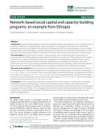
1. RSV alone induces airway obstruction (AO) and airway
hyperresponsiveness (AHR) in both C57BL/6 and BALB/c
mice
RSV infection alone, without allergic pre-sensitization
induced AO in both strains of mice as demonstrated by
significantly increased enhanced pause (Penh) values
compared with uninfected controls. Baseline Penh values
increased transiently on day 1 after RSV inoculation in
both strains, decreased by day 2, but continued to be sig-
nificantly greater than in controls. Airway obstruction
increased again and peaked on day 5, when C57BL/6 RSV-
infected mice showed significantly higher Penh values
than RSV-infected BALB/c mice (p < 0.001) (Figure 1). AO
decreased thereafter during the first two weeks after RSV
inoculation but remained significantly greater than the
respective controls in both strains, for 21 days in BALB/c
and 28 days in C57BL/6 mice (Figure 1 inset). RSV infec-
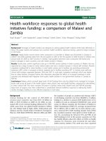
tion also induced AHR in both strains as evidenced by a
greater difference between pre- and post-methacholine
Penh values (delta Penh) compared with controls. Signif-
icantly increased AHR was persistently present for 42 days
post-inoculation in BALB/c mice, while C57BL/6 mice
showed significantly increased AHR for up to 28 days
post-inoculation (Figure 2).
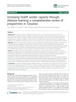
2. C57BL/6 and BALB/c mice demonstrate acute and
persistent inflammatory changes after RSV infection
RSV-inoculated C57BL/6 and BALB/c mice, compared
with their controls, showed greater histopathologic scores
(HPS) which peaked on day 5 after RSV inoculation (Fig-
ures 3, 4A–D). Although the acute inflammatory changes
observed in both strains gradually declined, RSV-infected
mice had significantly greater HPS than the sham-inocu-
lated controls for up to 77 days post-inoculation (Figures
3, 4E and 4F).
3. RSV infection induces similar cytokine production in the
respiratory tract of C57BL/6 and BALB/c mice
BAL concentrations of TNF-α, IFN-γ, MIG, RANTES, and
MIP-1α followed similar dynamics in both strains of mice
during the acute phase of the infection (Figure 5A–E).
Overall, there was a trend for greater BAL cytokine concen-
trations of IFN-γ, TNF-α, RANTES and MIP-1α in RSV-
infected BALB/c mice compared with C57BL/6 mice. (Fig-
ure 5A–E). No significant differences were observed in
BAL concentrations of IL-4 and IL-10 between controls
and infected mice of both strains at any time point evalu-
ated (data not shown).
4. RSV load dynamics
4a. RSV loads measured by plaque assay in BAL samples follow
similar dynamics in both C57BL/6 and BALB/c mice
On day 1 after RSV inoculation, RSV loads in BAL samples
from both C57BL/6 and BALB/c mice were significantly
greater than in controls by plaque assay (Figure 6). Com-
pared with day 1, plaque assay RSV loads peaked on days
3–5 after inoculation in both strains representing active
viral replication (p = 0.002 for day 1 vs days 3–5 in BALB/
c mice; ANOVA), and were significantly greater in BALB/c
than in C57BL/6 mice (Figure 7). BAL RSV loads declined
below the limit of detection by day 7 and remained unde-
tectable through day 77 post-inoculation.
4b. Real Time PCR (RLT-PCR) demonstrates RSV RNA after the virus
is no longer detectable by plaque assay
To further characterize the dynamics of RSV infection, we
used RLT-PCR in parallel with plaque assays, to measure
RSV loads in both BAL samples and lung homogenate
supernatants. These experiments were initially conducted
in BALB/c mice. RSV loads measured by RLT-PCR and
plaque assay in both BAL and lung supernatant samples
peaked on days 3 to 5 after inoculation. Similar to plaque
assay, RSV load by RLT-PCR also demonstrated a signifi-
cant increase in viral copies between day 1 and days 3–5,
likely demonstrating active replication (Figure 7). In con-
trast to RSV loads measured by plaque assay, which
became undetectable by day 7 after inoculation (Figure 7,
Virology Journal 2005, 2:46 />Page 3 of 14
(page number not for citation purposes)
dashed-line plots), RSV loads measured by RLT-PCR
remained positive for 42 days in BAL samples and
throughout 77 days in lung supernatants (Figure 7, solid
line plots).
Additional experiments in both mouse strains demon-
strated persistence of RSV RNA in lung supernatants for 77
days after inoculation (Figure 8) . Similar to the previous
findings using plaque assays, RSV loads were greater in
BALB/c than in C57BL/6 mice. Control mice of both
strains had undetectable RSV load by RLT-PCR.
5. Correlations among disease severity markers,
inflammatory indices, and viral load dynamics
Correlations were determined during both the acute
phase of the disease, day 5, and during the progression to
the chronic phase on days 14, 21 and 77 after inoculation.
During the acute phase in both mouse strains, airway
obstruction (AO) peaked on day 5 and strongly correlated
with histopathologic scores (HPS), BAL concentrations of
RANTES, IFN-γ, MIP-1α and MIG, and RSV loads meas-
ured by both plaque assay in BAL samples and RLT-PCR
Effect of RSV on airway obstruction (AO) in two mouse strainsFigure 1
Effect of RSV on airway obstruction (AO) in two mouse strains. BALB/c (᭝) and C57BL/6 (●) mice were inoculated
with sterile 10% EMEM (control) and were compared with RSV A2 infected BALB/c (ᮀ) and C57BL/6 (◆) mice to evaluate dif-
ferences in airway obstruction (AO), by measuring Penh via whole-body plethysmography. Penh values are presented as means
± SEM. Comparisons were made by t-test when data normally distributed, or by Mann-Whitney Rank sum test when data were
not normally distributed.
Days after RSV inoculation
02468101428425670
0.5
1.0
1.5
2.0
2.5
3.0
3.5
Control BALB/c AO
Control C57BL/6 AO
RSV BALB/c AO
RSV C57BL/6 AO
Penh
*
‡
*
‡
*
‡
#
*
‡
#
*
‡
*
‡
#
*
‡
*
‡
*
#
#
*
#
Days after RSV inoculation
14 21 28 35 42
Penh
0.5
0.6
0.7
0.8
0.9
1.0
* p<0.05 C57BL/6 RSV infected mice vs control
‡ p<0.05 BALB/c RSV infected mice vs control
# p<0.05 RSV BALB/c vs. RSV C57BL/6
n=10-30 mice per time point per group
Virology Journal 2005, 2:46 />Page 4 of 14
(page number not for citation purposes)
in lung supernatants (Additional file 1). In BALB/c mice,
AO also correlated with BAL TNF-α concentrations, RSV
loads measured by plaque assay in BAL samples signifi-
cantly correlated with those measured in lung superna-
tants by RLT-PCR in both C57BL/6 and BALB/c mice.
In addition, there were significant correlations between
airway hyperresponsiveness (AHR) and HPS on day 14 in
both BALB/c (r = 0.99, P = 0.0005, n = 4), and C57BL/6
mice (r = 0.85, P = 0.01, n = 7). AHR and HPS also corre-
lated significantly on day 77 in C57BL/6 mice (r = 0.95, P
= 0.012, n = 5).
In BALB/c mice alone, RSV loads measured by RLT-PCR
correlated with AHR on day 14 (r = 0.69, P = 0.01, n = 12).
RLT-PCR also correlated with AO on day 21 after RSV
inoculation (r = 0. 71, P = 0.01, n = 12).
Discussion
Intranasal inoculation of RSV induced acute and chronic
effects on the respiratory dynamics and inflammatory
response in C57BL/6 mice, similar to those previously
reported in BALB/c mice [7]. The acute manifestations of
RSV disease in C57BL/6 mice appeared similar but there
were several differences compared with BALB/c mice.
Airway Hyperresponsiveness (AHR) in BALB/c and C57BL/6 MiceFigure 2
Airway Hyperresponsiveness (AHR) in BALB/c and C57BL/6 Mice. Data presented as Delta Penh values which is the
difference between pre-and post methacholine Penh for each group of mice, in sham inoculated ( ) and RSV inoculated
( ) BALB/c mice, and sham inoculated ( ) and RSV inoculated ( ) C57BL/6 mice from days 14 to 77. Values represent
the mean SEM from 10–30 mice per group. Data shown are the result of four separate experiments. p < 0.05, comparison by
t-test when data normally distributed, or by Mann-Whitney Rank sum test when data were not normally distributed.
Days after RSV inoculation
14 21 28 35 42 49 56 63 70 77
DELTA Penh
0
1
2
3
4
Control BALB/c
Control C57BL/6
RSV BALB/c
RSV C57BL/6
* p<0.05 C57BL/6 RSV infected mice vs control
‡ p<0.05 BALB/c RSV infected mice vs control
# p<0.05 RSV BALB/c vs. RSV C57BL/6
n=10-30 mice per time point per group
*
‡
*
‡
*
‡
#
‡
‡
#
*
‡
Virology Journal 2005, 2:46 />Page 5 of 14
(page number not for citation purposes)
Acute airway obstruction was more severe and more
prolonged in C57BL/6 than in BALB/c mice. BALB/c mice,
however, demonstrated more severe histopathologic
changes, greater viral loads, and BAL concentrations of
most of the cytokines measured.
It is known that RSV infection causes variable degree of
disease severity in different mouse strains [8] but there
was no information on whether the different genetic back-
grounds influence the long term outcome of RSV infection
including chronic inflammation and persistent AO and
AHR. The varied responses of these two mouse strains to
different pathogens have been attributed in part to differ-
ences in their T helper (Th) lymphocyte response. Studies
in mice infected with Leishmania showed that a Th2
skewed response caused more severe disease in BALB/c
mice than in C57BL/6 mice that developed a Th1-domi-
nant response [9]. In some localized bacterial infections,
however, the severity of disease was milder in BALB/c
mice and was described as Th1 skewed [10]. Indistinct Th
responses were found in both mouse strains when
infected with certain viruses [11]. Hence, the differences
in the immune response might not only depend on the
genetic background of the host, but also on the site and
the specific pathogen involved in the infectious process.
Several groups have demonstrated AHR in C57BL/6 mice
Comparison of acute and long-term histopathologic scores after RSV inoculationFigure 3
Comparison of acute and long-term histopathologic scores after RSV inoculation. BALB/c (᭝) and C57BL/6 (●)
mice were inoculated with sterile 10% EMEM (control) and were compared with RSV A2 infected BALB/c (ᮀ) and C57BL/6
(◆) mice. Serial formalin fixed lung samples were obtained between day 0 (+2 hours) and day 77 after inoculation. HPS scores
are represented as means ± SEM. p < .05, by t-test when data normally distributed, or by Mann-Whitney Rank sum test when
data were not normally distributed.
Days after RSV inoculation
0 2 4 6 8101214 2835424956637077
HPS
0
2
4
6
8
10
12
14
Control BALB/c
Control C57BL/6
RSV BALB/c
RSV C57BL/6
*
‡
*
‡
*
‡
*
‡
*
‡
*
‡
‡
* p<0.05 C57BL/6 RSV infected mice vs control
‡ p<0.05 BALB/c RSV infected mice vs control
# p<0.05 RSV BALB/c vs. RSV C57BL/6
n=2-8 mice per time point per group
t-tests or Mann Whitney with Bonferroni correction
Virology Journal 2005, 2:46 />Page 6 of 14
(page number not for citation purposes)
during the acute stages of RSV infection. [22-24]. How-
ever, so far, our study is the first to shed light on the
dynamics of inflammatory infiltrates, airway dysfunction
(AO and AHR), and inflammatory cytokines during the
long-term phase of RSV-induced disease, up to 77 days
after primary infection in both C57BL/6 and BALB/c mice.
Researchers found that during RSV re-challenge following
sensitization with different RSV proteins, mice developed
a variable Th cytokine response [12-15] which was not
exclusively dependent on the strain haplotype [16]. Our
experiments, conducted in mice with primary RSV infec-
tion, demonstrated that IFN-γ, a Th1 cytokine, was
elevated after RSV infection in both strains. IFN-γ was
increased in the respiratory tract of children with acute
RSV infection [17,18] and correlated with disease severity
[19]. In mice, IFN-γ also correlates with the development
of RSV disease [20]. Our studies demonstrate that RSV
infection induces IFN-γ in both strains of mice and the
magnitude of the response is not consistent with the
traditional notion that BALB/c are Th-2 weighted and
C57BL/6 are Th1-weighted. BALB/c mice showed greater
IFN-γ response than C57BL/6 mice as previously shown
by others [21]. In our experiments IFN-γ demonstrated the
greatest correlation with disease severity (Additional file
1).
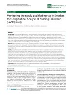
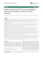
RSV induced histopathologyFigure 4
RSV induced histopathology. Lung specimens were harvested on days 5 and 77 from groups of C57BL/6 and BALB/c mice
inoculated with sterile medium (control) or RSV. Sections from control C57BL/6 and BALB/c mice above (4A and 4B, respec-
tively), show rare, scattered, small lymphocytic infiltrates on day 5 after inoculation with medium, similar to sections of control
mice harvested 77 days after (not shown). Acute and chronic inflammatory infiltrates, surrounding airways and vessels are
demonstrated in RSV-inoculated mice of both strains, on days 5 and 77 after inoculation (4C to F).
C57BL/6
BALB/c
Control Day 5
RSV Day 77
A.
B. D.
C. E.
F.
RSV Day 5
F.
E.
Virology Journal 2005, 2:46 />Page 7 of 14
(page number not for citation purposes)
Cytokine and chemokine concentrations in bronchoalveolar lavage (BAL) samples after RSV inoculationFigure 5
Cytokine and chemokine concentrations in bronchoalveolar lavage (BAL) samples after RSV inoculation. BAL
samples were obtained from BALB/c (᭝) and C57BL/6 (●) mice inoculated with sterile 10% EMEM (control), and RSV A2
infected BALB/c (ᮀ) and C57BL/6 (◆) mice, to measure concentrations of pro-inflammatory cytokines (A) IFN-γ and (B)TNF-
α; and the chemokines (C) RANTES, (D) MIP-1α, and (E) MIG. Values presented in means ± SEM pg/ml. p < .05, by t-test when
data were normally distributed, or by Mann-Whitney Rank sum test when data were not normally distributed.
Days after RSV inoculation
13579 1214
pg/mL
0
2000
4000
6000
8000
10000
12000
0
*
‡
#
Days after RSV inoculation
13579 1214
pg/mL
0
2000
4000
6000
8000
10000
12000
0
*
‡
#
Days after RSV inoculation
13579 1214
pg/mL
2000
4000
6000
8000
10000
0
#
‡
*
*
‡
#
*
‡
Days after RSV inoculation
13579 1214
pg/mL
2000
4000
6000
8000
10000
0
Days after RSV inoculation
13579 1214
pg/mL
2000
4000
6000
8000
10000
0
#
‡
*
*
‡
#
*
‡
Days after RSV inoculation
13579 12
pg/mL
0
1000
2000
3000
4000
#
*
‡
*
‡
Days after RSV inoculation
13579 12
pg/mL
0
1000
2000
3000
4000
#
*
‡
*
‡
Days after RSV inoculation
13579 1214
pg/mL
0
2000
4000
6000
8000
10000
12000
14000
0
*
‡
#
‡
Days after RSV inoculation
13579 1214
pg/mL
0
2000
4000
6000
8000
10000
12000
14000
0
*
‡
#
‡
Days after RSV inoculation
13579 1214
pg/mL
100
1000
10000
0
‡
#
*
‡
#
Days after RSV inoculation
13579 1214
pg/mL
100
1000
10000
0
Days after RSV inoculation
13579 1214
pg/mL
100
1000
10000
0
‡
#
*
‡
#
* p<0.05 C57BL/6 RSV infected mice vs control
‡ p<0.05 BALB/c RSV infected mice vs control
# p<0.05 RSV BALB/c vs. RSV C57BL/6
n=2-8 mice per time point per group
t-tests or Mann Whitney with Bonferroni correction
Control BALB/c
Control C57BL/6
RSV BALB/c
RSV C57BL/6
A. IFN-γ
γγ
γ
B. TNF-α
αα
α
C. RANTES D. MIP-1α
αα
α
E. MIG
Virology Journal 2005, 2:46 />Page 8 of 14
(page number not for citation purposes)
Like IFN-γ, the dynamics of TNF-α, RANTES, MIP-1α were
comparable in both mouse strains. TNF-α has been linked
to RSV disease severity [25]. In our model, BAL concentra-
tions of TNF-α peaked shortly after RSV infection and
correlated with markers of disease severity in BALB/c mice
(Additional file 1), and were significantly greater in BALB/
c than in C57BL/6 mice (Figure 5).
The CC chemokines, RANTES and MIP-1α, and the CXC
chemokine MIG were also elevated in mice of both strains
shortly after RSV inoculation. The biphasic production of
RANTES, MIP-1α and MIG after RSV infection, with the
second peak coinciding with the peak of viral replication
and histological inflammation, has been previously
described in BALB/c mice [7,26] but not in C57BL/6 mice.
Although MIP-1α has been detected in C57BL/6 mice
[27], no previous studies demonstrated the production of
RANTES in the respiratory tract of these mice after RSV
infection. These two chemokines correlate with acute dis-
ease severity both in humans and in mice. [19,28-30].
Increased production of MIG after RSV infection has been
documented in vitro [31] and by us in BALB/c mice[7].
Others have linked MIG to persistent airway inflamma-
tion due to continuous chemotaxis of mononuclear cells
[32]. In the present study we demonstrate significant cor-
relation between BAL MIG concentrations and AO, and
RSV loads in BAL samples measured by the plaque assay methodFigure 6
RSV loads in BAL samples measured by the plaque assay method. Groups of 4–16 BALB/c (᭝) and C57BL/6 (●)
mice per group per time point were inoculated intranasally with sterile 10% EMEM (control), and were compared with RSV A2
infected BALB/c (ᮀ) and C57BL/6 (◆) mice. Viral load was determined by HEp-2 plaque assay in BAL samples. Data are pre-
sented as mean ± SEM Log10 PFU/ml of BAL. p < .05, by t-test when data normally distributed, or by Mann-Whitney Rank sum
test when data were not normally distributed.
Days after RSV inoculation
1 3 5 9 12 21 42 77
2.0
2.5
3.0
3.5
4.0
Control BALB/c
Control C57BL/6
RSV BALB/c
RSV C57BL/6
Log
10
PFU/mL
#
‡
*
#
‡
*
‡
*
* p<0.05 C57BL/6 RSV infected mice vs C57BL/6 control
‡ p<0.05 BALB/c RSV infected mice vs BALB/c control
# p<0.05 RSV BALB/c vs. RSV C57BL/6
n=4-16 mice per time point per group
t-tests or Mann Whitney with Bonferroni correction
Virology Journal 2005, 2:46 />Page 9 of 14
(page number not for citation purposes)
RSV loads in RSV infected BALB/c mice BAL and lung supernatant samples measured by PCR vs. plaque assayFigure 7
RSV loads in RSV infected BALB/c mice BAL and lung supernatant samples measured by PCR vs. plaque assay.
RSV loads measured by PCR in BAL samples (Ќ) remain positive up to 42 days after inoculation, while viral loads measured by
plaque assay (ᮀ) become negative on day 7 post-inoculation (upper panel). Viral load measured in lung supernatants by the
plaque assay also become undetectable by day 7 after inoculation, whereas RSV loads measured by PCR in lung supernatants
remain detectable throughout all the time points evaluated (lower panel). All pair-wise multiple comparisons made by One-
Way ANOVA. † p < 0.01 between D1 and D5 and *p < 0.05 comparing D1 with D3 and D5.
Days after RSV inoculation
1 3 5 7 14 21 42 77
0
2
4
8
Log
10
PFU/mL
2
3
4
5
6
7
8
6
Log
10
copies/mL
Viral Load by PCR vs Cultures in BAL
13
5 7 14 21 42 77
0
1
2
3
4
5
6
2
3
4
5
6
Viral Load by PCR vs Cultures in Lung Homogenate Supernatants
Log
10
PFU/mL
Log
10
copies/mL
*
*
*
*
*
*
†
PCR viral load
Plaque assay viral load
PCR viral load
Plaque assay viral load
Virology Journal 2005, 2:46 />Page 10 of 14
(page number not for citation purposes)
viral load dynamics in both strains of mice. Taken
together, these results indicate that RSV induces severe
acute pulmonary disease in both strains of mice despite
different genetic backgrounds. Although there are quanti-
tative differences during the acute phase of the disease,
there were no significant distinctions in the pattern of
cytokine responses, lung inflammation or clinical mani-
festations of disease, and both strains developed long-
term airway disease defined by chronic inflammatory
infiltrates and abnormal AHR. These findings provide fur-
ther experimental support to the link between RSV and
chronic airway disease in humans. We believe that this is
the first description of RSV-induced long-term pulmonary
disease in C57BL/6 mice.
The exact mechanism by which different inflammatory
mediators participate in the pathogenesis of the acute
RSV-induced disease process is not completely under-
stood, and their potential role in determining the chronic
consequences of RSV infection, such as AO, AHR and
chronic inflammation is even less characterized. Studies
in murine models and in humans have not demonstrated
yet a direct relation between the acute inflammatory
response to RSV and its chronic consequences in the res-
Comparison of RSV loads measured by PCR in lung supernatants of BALB/c and C57BL/6 miceFigure 8
Comparison of RSV loads measured by PCR in lung supernatants of BALB/c and C57BL/6 mice. Groups of 2–12
BALB/c (᭝) and C57BL/6 (●) mice per group per time point were inoculated intranasally with sterile 10% EMEM (control), and
were compared with RSV A2 infected Balb/c (ᮀ) and C57Bl/6 (◆) mice. Viral load was determined by PCR to detect RSV N
gene. Data are presented as mean ± SEM Log10 PFU/ml of BAL. p < .05, by t-test when data normally distributed, or by Mann-
Whitney Rank sum test when data were not normally distributed.
Days after RSV inoculation
135 12 21 42 77
1
2
3
4
5
6
7
Control BALB/c
Control C57BL/6
RSV BALB/c
RSV C57BL/6
Log
10
copies/ml
#
‡
‡
#
‡
*
‡
*
‡
* p<0.05 C57BL/6 RSV infected mice vs control
‡ p<0.05 BALB/c RSV infected mice vs control
# p<0.05 RSV BALB/c vs. RSV C57BL/6
n=2-12 mice per time point per group
T-tests or Mann Whitney with Bonferroni correction
Virology Journal 2005, 2:46 />Page 11 of 14
(page number not for citation purposes)
piratory tract pathology. Our studies demonstrated
prolonged AO and persistent AHR in both mouse strains,
long after any of the cytokines, potentially responsible for
persistent inflammation that we measured, became unde-
tectable in BAL samples. It is possible that the use of more
sensitive methods, perhaps in specific compartments, will
contribute to clarify the role of these molecules in the
chronic respiratory function abnormalities induced by
RSV.
Until recently, it was suggested that initial RSV infection,
although of short duration, was sufficient to elicit an exag-
gerated inflammatory response which would evolve into
chronic inflammation and long-term airway abnormali-
ties. An alternative, but not contradictory explanation for
the development of chronic airway disease could be
related to long-term antigenic stimulation. In this hypoth-
esis, the presence of low level viral infection could repre-
sent a persistent stimulus responsible for the chronic
inflammatory changes and the lasting pulmonary func-
tion abnormalities. The application of the RLT-PCR assay
to this model demonstrated the persistence of RSV RNA in
the respiratory tract for 11 weeks after inoculation in both
strains of mice and was also associated with long-term
pulmonary disease.
RSV can persist and replicate in vitro [33-35]. The persist-
ence of RSV genomic RNA sequences and viral antigens
has been described in guinea pigs for several weeks after
acute primary infection [36-38]. Bovine RSV infection in
calves resulted in persistent detection of RSV N gene for
several weeks [39]. Miller et al. reported detection of G
protein RNA by RLT-PCR in lungs of BALB/c mice up to
day 12 after inoculation. [26].
It appears unlikely for a RNA virus to persist without at
least low-level replication in the host. This is supported by
the studies by Schwarze et al., who found RSV genomic
and messenger RNA in mouse lung homogenates for 100
days after inoculation, and were able to recover low titer
virus in culture after treatment with anti-CD4 and anti-
CD8 antibodies [40].
Clinical studies have demonstrated that adults with stable
COPD without acute exacerbation had RSV RNA detected
by PCR in the upper respiratory tract [41]. The persistence
of RSV RNA in children has not been extensively studied.
Post-mortem specimens of children who died of sudden
infant death syndrome showed positive results by PCR
targeting the RSV N gene in 27% of cases and in 18% of
the controls. Since some children died during summer the
authors suggested the possibility of viral persistence [42].
The persistence of RSV in the respiratory tract of children
and its potential contribution to the development and
maintenance of AHR is a critical question that needs to be
addressed.
In summary, we provide evidence that RSV-induced acute
and chronic airway disease is reproducible in two geneti-
cally distinct mouse strains. The long-term airway disease
coincided with persistent detection of RSV genome in the
respiratory tract. It is yet unclear whether the persistence
of RSV RNA plays a role in the development of chronic air-
way disease. Further studies are needed to characterize the
potential mechanisms that would allow viral persistence
and its possible contribution to the pathogenesis of RSV-
induced long-term pulmonary disease.
Materials and methods
Mice
Female, 7–8-week old pathogen-free C57BL/6 and BALB/
c mice (Charles River Laboratories, Wilmington, MA)
were maintained in filter top cages, and routinely moni-
tored for other pathogens [7]. After inoculation, all mice
were kept under the same conditions and were provided
identical care. Sentinel mice housed in the mouse storage
room are routinely used for health surveillance. Sentinel
mice had no detectable antibodies against mouse hepati-
tis virus, Sendai virus, pneumonia virus of mice, reo-3
virus, mouse encephalitis virus (GD-7), mouse rotavirus
(EDIM), minute virus of mice, and Mycoplasma pulmonis;
screening for pinworm and mites was also negative. This
study was approved by the Institutional Animal Care and
Use Committee at the University of Texas Southwestern
Medical Center at Dallas.
Virus
RSV A-2 strain was maintained in our laboratory as
described [7]. Results of plaque assays were reported in
Log
10
PFU/mL, with 1.7 Log
10
PFU/mL being the lowest
limit of detection.
Inoculation
Mice were anesthetized using inhaled methoxyfluorane
and intranasally inoculated with 10
7
PFU of RSV in 100 µl
of 10% Eagle's minimal essential medium (EMEM) [6].
Uninfected control animals were sham-inoculated with
100 µl of sterile 10% EMEM. Animals were allowed 30
seconds to aspirate the inoculum while held upright until
fully recovered from the anesthesia.
Plethysmography
Unrestrained, whole-body plethysmography (Buxco Elec-
tronics, Inc. Sharon, CT) was used to measure the
Enhanced Pause (Penh) to evaluate airway obstruction
(AO) and airway hyperresponsiveness (AHR), as previ-
ously described [7,43]. Briefly, mice were allowed to accli-
mate to the chamber, and then plethysmograph readings
were recorded to establish baseline Penh values to
Virology Journal 2005, 2:46 />Page 12 of 14
(page number not for citation purposes)
determine AO. Next the mice were exposed to aerosolized
methacholine (Sigma; 50 mg/ml) for 4 minutes; after
exposure, plethysmograph readings were recorded again
to determine AHR. Groups of infected and control mice
were always evaluated in parallel at all time points during
the entire study. Preliminary studies showed that normal
C57BL/6 mice have greater baseline Penh values than nor-
mal BALB/c mice; therefore, groups of infected and con-
trol mice of each strain were evaluated in parallel during
the entire study. Plethysmography was performed in the
groups of mice to be sacrificed for sample collection at
each time point, and in an additional group of mice that
were followed throughout each experiment. A total of 4
independent experiments including 10–30 mice per
group per time point were conducted.
Sample collection
Mice were anesthetized with an intraperitoneal injection
of 75 mg/kg of ketamine and 5 mg/kg of acepromazine
before euthanasia by exsanguination. On average, 4–12
mice were sacrificed per group per time point. Bronchoal-
veolar lavage (BAL) specimens were obtained as described
previously [7,43] to measure cytokine concentrations and
RSV loads. Histologic evaluation was performed in whole-
lung specimens fixed with a 10% buffered formalin solu-
tion. Whole lung samples were also collected from
another group of mice for determination of viral loads by
plaque assay and by real-time polymerase chain reaction
(RLT-PCR). Specimens were obtained on days 0 (within 2
hours of inoculation), 1, 3, 5, 8, 9, 12, 14, 21, 28, 42, and
77 post-inoculation.
Histopathology
Formalin fixed lungs were paraffin embedded, sectioned
and stained with hematoxylin and eosin. Histopathologic
score (HPS) was determined by a pathologist, unaware of
the infection status of the animals. Each section was
graded on the basis of a cumulative score from 5 catego-
ries: (1) peribronchiolar and bronchial infiltrates, (2)
bronchiolar and bronchial luminal exudates, (3) perivas-
cular infiltrates, (4) the number of monocytes, and (5)
parenchymal pneumonia. This HPS system assigns values
from 0 to 21. This scoring system has been previously val-
idated in RSV infection [7,43].
BAL cytokines
Concentrations of TNF-α, IFN-γ, IL-4, IL-10, MIG,
RANTES, and MIP-1α were measured in BAL specimens by
ELISA (R&D Systems, Minneapolis, Minn.) for up to 14
days after inoculation. The lower limit of detection for
each of these assays were: 120 pg/mL for TNF-α, 50 pg/mL
for IFN-γ, 40 pg/mL for IL4, 16 pg/mL for IL-10, 16 pg/ mL
for MIG, 200 pg/mL for RANTES and 60 pg/mL for MIP-
1α. For statistical analysis, samples with optical density
readings below the limit of the standard curve of the assay
were assigned a value half that of the detection level.
RSV loads by plaque assay and Real-time Polymerase
Chain Reaction (RLT-PCR)
Plaque assay tissue culture was used to measure viral loads
in BAL specimens and lung homogenate supernatants as
previously described [7]. Lung supernatant samples were
prepared by placing whole lung specimens in 1 mL of
10% EMEM, on ice. Lungs were homogenized using ster-
ile probes and a rotor tissue homogenizer (Omni Interna-
tional Inc., Marietta, GA). Lung supernatants were
separated by centrifugation at 2000 g for 10 minutes at
4°C and 100 µL were used immediately for determination
of viral loads by plaque assay; the remaining supernatant
was frozen at -80°C for viral load measurements by RLT-
PCR. In a subset of experiments, lung samples used for
RLT-PCR were placed in 1.5 mL of RNA stabilization rea-
gent (RNA later, Qiagen Inc., Valencia, CA) immediately
after sample collection. Lung supernatants were then
obtained by homogenization and centrifugation. The
RLT-PCR viral load values measured using this method
were similar to those obtained when no RNA later was
used; therefore, all results of lung homogenate superna-
tants are presented together. One sample per mouse was
evaluated as a single specimen. Quantitative RLT-PCR was
used targeting the conserved region of the RSV N-gene.
Forward (5'-AGA TCA ACT TCT GTC ATC CAG CAA) and
reverse (5'-TTC TGC ACA TCA TAA TTA GGA GTA TCA
AT) primers amplified an 85-bp region containing the 25-
mer FAM-labeled probe (5'-CAC CAT CCA ACG GAG
CAC AGG AGA T), as previously described [44]. RSV RNA
was extracted from 1 mL of lung supernatant samples
using ion-exchange mini-columns (Qiagen RNeasy Mini
Kit, Valencia, CA, USA) and cDNA was prepared by reverse
transcription using 2.5 uM random hexamers for 10 min
at 22°C, 30 min at 42°C and 5 min at 95°C. Real-time
PCR was performed using a Perkin-Elmer /Applied Biosys-
tems 7700 sequence detector (Foster City, CA) using 10 µl
cDNA/ in a total volume of 50 µl master mix with the fol-
lowing run conditions: 1 cycle for 2 min at 50°C and 10
min at 95°C each, followed by 50 cycles for 15 seconds at
95°C and 60 seconds at 60°C. RSV A known concentra-
tions were used to derive a standard curve. Standards and
negative controls were run together with each PCR assay.
The lower limit of detection of the assay was 10 viral
copies/mL.
Statistical methods
T-test or Mann-Whitney Rank Sum test were used accord-
ing to data distribution for comparisons between groups
and the Spearman Rank Order test was used for correla-
tions, as all the data taken together were not normally dis-
tributed, using the SigmaStat
®
(SPSS, Inc. Chicago,
Illinois) software package.
Virology Journal 2005, 2:46 />Page 13 of 14
(page number not for citation purposes)
Abreviations used
PCR polymerase chain reaction
TNF tumor necrosis factor
IL interleukin
IFN interferon
MIP macrophage inflammatory protein
RANTES regulated on activation, normal T-cell expressed
and secreted
MIG monokine induced by interferon-gamma
Competing interests
The author(s) declare that they have no competing
interests.
Authors' contributions
SCB study design, RSV infection, data analyses; AM BAL,
formalin fixation, plethysmography; AMG histopathol-
ogy and special staining; KDO real-time PCR; AMR and
MFA plethysmography and BAL; OR data interpretation;
HSJ project design, experimental analyses and
interpretation.
Additional material
Acknowledgements
S.C.B. is supported in part by a Pediatric Infectious Disease Society Fellow-
ship Award sponsored by GlaxoSmithKline Pharmaceuticals.
A.M. was supported in part by the Pediatric Fellowship Award in Viral Res-
piratory Infectious Diseases supported by MedImmune Inc., at the Pediatric
Academic Societies Annual Meeting.
H.S.J. is supported in part by grants from the Children's Clinical Research
Advisory Committee and the Children's Medical Center of Dallas Founda-
tion and American Lung Association.
Approved by the Institutional Animal Care and Use Committee at the Uni-
versity of Texas Southwestern Medical Center at Dallas.
References
1. Shay DK, Holman RC, Newman RD, Liu LL, Stout JW, Anderson LJ:
Bronchiolitis-associated hospitalizations among US children,
1980-1996. Jama 1999, 282(15):1440-1446.
2. Martinez FD, Wright AL, Taussig LM, Holberg CJ, Halonen M, Morgan
WJ: Asthma and wheezing in the first six years of life. The
Group Health Medical Associates. N Engl J Med 1995,
332(3):133-138.
3. Stein RT, Sherrill D, Morgan WJ, Holberg CJ, Halonen M, Taussig LM,
Wright AL, Martinez FD: Respiratory syncytial virus in early life
and risk of wheeze and allergy by age 13 years. Lancet 1999,
354(9178):541-545.
4. Sigurs N, Bjarnason R, Sigurbergsson F, Kjellman B: Respiratory
syncytial virus bronchiolitis in infancy is an important risk
factor for asthma and allergy at age 7. Am J Respir Crit Care Med
2000, 161(5):1501-1507.
5. Sigurs N, Gustafsson PM, Bjarnason R, Lundberg F, Schmidt S, Sigurb-
ergsson F, Kjellman B: Severe respiratory syncytial virus bron-
chiolitis in infancy and asthma and allergy at age 13. Am J
Respir Crit Care Med 2005, 171(2):137-141.
6. Karron RA, Singleton RJ, Bulkow L, Parkinson A, Kruse D, DeSmet I,
Indorf C, Petersen KM, Leombruno D, Hurlburt D, Santosham M,
Harrison LH: Severe respiratory syncytial virus disease in
Alaska native children. RSV Alaska Study Group. J Infect Dis
1999, 180(1):41-49.
7. Jafri HS, Chavez-Bueno S, Mejias A, Gomez AM, Rios AM, Nassi SS,
Yusuf M, Kapur P, Hardy RD, Hatfield J, Rogers BB, Krisher K, Ramilo
O: Respiratory syncytial virus induces pneumonia, cytokine
response, airway obstruction, and chronic inflammatory
infiltrates associated with long-term airway hyperrespon-
siveness in mice. J Infect Dis 2004, 189(10):1856-1865.
8. Byrd LG, Prince GA: Animal models of respiratory syncytial
virus infection. Clin Infect Dis 1997, 25(6):1363-1368.
9. Sacks D, Noben-Trauth N: The immunology of susceptibility
and resistance to Leishmania major in mice. Nat Rev Immunol
2002, 2(11):845-858.
10. Hazlett LD: Pathogenic mechanisms of P. aeruginosa kerati-
tis: a review of the role of T cells, Langerhans cells, PMN, and
cytokines. DNA Cell Biol 2002, 21(5-6):383-390.
11. Ramakrishna C, Ravi V, Desai A, Subbakrishna DK, Shankar SK, Chan-
dramuki A: T helper responses to Japanese encephalitis virus
infection are dependent on the route of inoculation and the
strain of mouse used. J Gen Virol 2003, 84(Pt 6):1559-1567.
12. Bangham CR, Cannon MJ, Karzon DT, Askonas BA: Cytotoxic T-
cell response to respiratory syncytial virus in mice. J Virol
1985, 56(1):55-59.
13. Alwan WH, Record FM, Openshaw PJ: Phenotypic and functional
characterization of T cell lines specific for individual respira-
tory syncytial virus proteins. J Immunol 1993,
150(12):5211-5218.
14. Hancock GE, Tebbey PW, Scheuer CA, Pryharski KS, Heers KM,
LaPierre NA: Immune responses to the nonglycosylated ecto-
domain of respiratory syncytial virus attachment glycopro-
tein mediate pulmonary eosinophilia in inbred strains of
mice with different MHC haplotypes. J Med Virol 2003,
70(2):301-308.
15. Hussell T, Georgiou A, Sparer TE, Matthews S, Pala P, Openshaw PJ:
Host genetic determinants of vaccine-induced eosinophilia
during respiratory syncytial virus infection. J Immunol 1998,
161(11):6215-6222.
16. Srikiatkhachorn A, Chang W, Braciale TJ: Induction of Th-1 and
Th-2 responses by respiratory syncytial virus attachment
glycoprotein is epitope and major histocompatibility com-
plex independent. J Virol 1999, 73(8):6590-6597.
17. Garofalo RP, Hintz KH, Hill V, Ogra PL, Welliver RCS: Production
of interferon gamma in respiratory syncytial virus infection
of humans is not associated with interleukins 12 and 18. J Med
Virol 2004, 73(2):289-294.
18. van Benten IJ, van Drunen CM, Koopman LP, KleinJan A, van Middelk-
oop BC, de Waal L, Osterhaus AD, Neijens HJ, Fokkens WJ: RSV-
induced bronchiolitis but not upper respiratory tract infec-
tion is accompanied by an increased nasal IL-18 response. J
Med Virol 2003, 71(2):290-297.
19. Garofalo RP, Patti J, Hintz KA, Hill V, Ogra PL, Welliver RC: Macro-
phage inflammatory protein-1alpha (not T helper type 2
Additional File 1
Significant Correlations on Day 5 after RSV Inoculation in C57BL/6
and BALB/c mice. r = correlation coefficient; p = p-value, n = number of
samples analyzed. Spearman's rank order analysis was used. Statistically
significant correlations (p < 0.05) listed in bold font. N/A, not applicable,
samples from different animals; * TNF-
α
concentrations undetectable on
day 5.
Click here for file
[ />422X-2-46-S1.xls]
Publish with BioMed Central and every
scientist can read your work free of charge
"BioMed Central will be the most significant development for
disseminating the results of biomedical research in our lifetime."
Sir Paul Nurse, Cancer Research UK
Your research papers will be:
available free of charge to the entire biomedical community
peer reviewed and published immediately upon acceptance
cited in PubMed and archived on PubMed Central
yours — you keep the copyright
Submit your manuscript here:
/>BioMedcentral
Virology Journal 2005, 2:46 />Page 14 of 14
(page number not for citation purposes)
cytokines) is associated with severe forms of respiratory syn-
cytial virus bronchiolitis. J Infect Dis 2001, 184(4):393-399.
20. van Schaik SM, Obot N, Enhorning G, Hintz K, Gross K, Hancock GE,
Stack AM, Welliver RC: Role of interferon gamma in the patho-
genesis of primary respiratory syncytial virus infection in
BALB/c mice. J Med Virol 2000, 62(2):257-266.
21. Johnson TR, Hong S, Van Kaer L, Koezuka Y, Graham BS: NK T cells
contribute to expansion of CD8(+) T cells and amplification
of antiviral immune responses to respiratory syncytial virus.
J Virol 2002, 76(9):4294-4303.
22. Tekkanat KK, Maassab H, Berlin AA, Lincoln PM, Evanoff HL, Kaplan
MH, Lukacs NW: Role of interleukin-12 and stat-4 in the regu-
lation of airway inflammation and hyperreactivity in respira-
tory syncytial virus infection. Am J Pathol 2001, 159(2):631-638.
23. Makela MJ, Kanehiro A, Dakhama A, Borish L, Joetham A, Tripp R,
Anderson L, Gelfand EW: The failure of interleukin-10-deficient
mice to develop airway hyperresponsiveness is overcome by
respiratory syncytial virus infection in allergen-sensitized/
challenged mice. Am J Respir Crit Care Med 2002, 165(6):824-831.
24. Schwarze J, Cieslewicz G, Hamelmann E, Joetham A, Shultz LD, Lam-
ers MC, Gelfand EW: IL-5 and eosinophils are essential for the
development of airway hyperresponsiveness following acute
respiratory syncytial virus infection. J Immunol 1999,
162(5):2997-3004.
25. Rutigliano JA, Graham BS: Prolonged production of TNF-alpha
exacerbates illness during respiratory syncytial virus
infection. J Immunol 2004, 173(5):3408-3417.
26. Miller AL, Bowlin TL, Lukacs NW: Respiratory syncytial virus-
induced chemokine production: linking viral replication to
chemokine production in vitro and in vivo. J Infect Dis 2004,
189(8):1419-1430.
27. Domachowske JB, Bonville CA, Gao JL, Murphy PM, Easton AJ,
Rosenberg HF: MIP-1alpha is produced but it does not control
pulmonary inflammation in response to respiratory syncytial
virus infection in mice. Cell Immunol 2000, 206(1):1-6.
28. Noah TL, Becker S: Chemokines in nasal secretions of normal
adults experimentally infected with respiratory syncytial
virus. Clin Immunol 2000, 97(1):43-49.
29. Noah TL, Ivins SS, Murphy P, Kazachkova I, Moats-Staats B, Hender-
son FW: Chemokines and inflammation in the nasal passages
of infants with respiratory syncytial virus bronchiolitis. Clin
Immunol 2002, 104(1):86-95.
30. Sheeran P, Jafri H, Carubelli C, Saavedra J, Johnson C, Krisher K,
Sanchez PJ, Ramilo O: Elevated cytokine concentrations in the
nasopharyngeal and tracheal secretions of children with res-
piratory syncytial virus disease. Pediatr Infect Dis J 1999,
18(2):115-122.
31. Bitko V, Garmon NE, Cao T, Estrada B, Oakes JE, Lausch RN, Barik
S: Activation of cytokines and NF-kappa B in corneal epithe-
lial cells infected by respiratory syncytial virus: potential rel-
evance in ocular inflammation and respiratory infection.
BMC Microbiol 2004, 4(1):28.
32. Kelsen SG, Aksoy MO, Yang Y, Shahabuddin S, Litvin J, Safadi F, Rog-
ers TJ: The Chemokine Receptor, CXCR3, and its Splice Var-
iants are Expressed in Human Airway Epithelial Cells. Am J
Physiol Lung Cell Mol Physiol 2004.
33. Pringle C R, Shirodaria PV, Cash P, Chiswell DJ, Malloy P: Initiation
and maintenance of persistent infection by respiratory syn-
cytial virus. J Virol 1978, 28(1):199-211.
34. Panuska JR, Cirino NM, Midulla F, Despot JE, McFadden ERJ, Huang
YT: Productive infection of isolated human alveolar macro-
phages by respiratory syncytial virus. J Clin Invest 1990,
86(1):113-119.
35. Guerrero-Plata A, Ortega E, Gomez B: Persistence of respiratory
syncytial virus in macrophages alters phagocytosis and pro-
inflammatory cytokine production. Viral Immunol 2001,
14(1):19-30.
36. Hegele RG, Robinson PJ, Gonzalez S, Hogg JC: Production of acute
bronchiolitis in guinea-pigs by human respiratory syncytial
virus. Eur Respir J 1993, 6(9):1324-1331.
37. Hegele RG, Hayashi S, Bramley AM, Hogg JC: Persistence of respi-
ratory syncytial virus genome and protein after acute bron-
chiolitis in guinea pigs. Chest 1994, 105(6):1848-1854.
38. Streckert HJ, Philippou S, Riedel F: Detection of respiratory syn-
cytial virus (RSV) antigen in the lungs of guinea pigs 6 weeks
after experimental infection and despite of the production of
neutralizing antibodies. Arch Virol 1996, 141(3-4):401-410.
39. Valarcher JF, Bourhy H, Lavenu A, Bourges-Abella N, Roth M,
Andreoletti O, Ave P, Schelcher F: Persistent infection of B lym-
phocytes by bovine respiratory syncytial virus. Virology 2001,
291(1):55-67.
40. Schwarze J, O'Donnell DR, Rohwedder A, Openshaw PJ: Latency
and persistence of respiratory syncytial virus despite T cell
immunity. Am J Respir Crit Care Med 2004, 169(7):801-805.
41. Rohde G, Wiethege A, Borg I, Kauth M, Bauer TT, Gillissen A, Bufe
A, Schultze-Werninghaus G: Respiratory viruses in exacerba-
tions of chronic obstructive pulmonary disease requiring
hospitalisation: a case-control study. Thorax 2003, 58(1):37-42.
42. Cubie HA, Duncan LA, Marshall LA, Smith NM: Detection of respi-
ratory syncytial virus nucleic acid in archival postmortem tis-
sue from infants. Pediatr Pathol Lab Med 1997, 17(6):927-938.
43. Mejias A, Chavez-Bueno S, Rios AM, Saavedra-Lozano J, Fonseca Aten
M, Hatfield J, Kapur P, Gomez AM, Jafri HS, Ramilo O: Anti-respira-
tory syncytial virus (RSV) neutralizing antibody decreases
lung inflammation, airway obstruction, and airway hyperre-
sponsiveness in a murine RSV model. Antimicrob Agents
Chemother 2004, 48(5):1811-1822.
44. van Woensel JB, Lutter R, Biezeveld MH, Dekker T, Nijhuis M, van
Aalderen WM, Kuijpers TW: Effect of dexamethasone on tra-
cheal viral load and interleukin-8 tracheal concentration in
children with respiratory syncytial virus infection. Pediatr
Infect Dis J 2003, 22(8):721-726.