Báo cáo sinh học: " Peptide inhibitors of dengue virus and West Nile virus infectivity" pot
Bạn đang xem bản rút gọn của tài liệu. Xem và tải ngay bản đầy đủ của tài liệu tại đây (2.1 MB, 10 trang )
BioMed Central
Page 1 of 10
(page number not for citation purposes)
Virology Journal
Open Access
Research
Peptide inhibitors of dengue virus and West Nile virus infectivity
Yancey M Hrobowski
1
, Robert F Garry
1,2
and Scott F Michael*
3
Address:
1
Department of Microbiology and Immunology, Tulane University Health Sciences Center, New Orleans, Louisiana 70112 USA,
2
Graduate Program in Cellular and Molecular Biology, Tulane University, New Orleans, LA 70112 USA and
3
Biotechnology Program, Florida Gulf
Coast University, Fort Myers, FL 33965 USA
Email: Yancey M Hrobowski - ; Robert F Garry - ; Scott F Michael* -
* Corresponding author
Abstract
Viral fusion proteins mediate cell entry by undergoing a series of conformational changes that result
in virion-target cell membrane fusion. Class I viral fusion proteins, such as those encoded by
influenza virus and human immunodeficiency virus (HIV), contain two prominent alpha helices.
Peptides that mimic portions of these alpha helices inhibit structural rearrangements of the fusion
proteins and prevent viral infection. The envelope glycoprotein (E) of flaviviruses, such as West
Nile virus (WNV) and dengue virus (DENV), are class II viral fusion proteins comprised
predominantly of beta sheets. We used a physio-chemical algorithm, the Wimley-White interfacial
hydrophobicity scale (WWIHS) [1] in combination with known structural data to identify potential
peptide inhibitors of WNV and DENV infectivity that target the viral E protein. Viral inhibition
assays confirm that several of these peptides specifically interfere with target virus entry with 50%
inhibitory concentration (IC50) in the 10 µM range. Inhibitory peptides similar in sequence to
domains with a significant WWIHS scores, including domain II (IIb), and the stem domain, were
detected. DN59, a peptide corresponding to the stem domain of DENV, inhibited infection by
DENV (>99% inhibition of plaque formation at a concentrations of <25 µM) and cross-inhibition of
WNV fusion/infectivity (>99% inhibition at <25 µM) was also demonstrated with DN59. However,
a potent WNV inhibitory peptide, WN83, which corresponds to WNV E domain IIb, did not inhibit
infectivity by DENV. Additional results suggest that these inhibitory peptides are noncytotoxic and
act in a sequence specific manner. The inhibitory peptides identified here can serve as lead
compounds for the development of peptide drugs for flavivirus infection.
Introduction
Enveloped viruses utilize membrane-bound fusion pro-
teins to mediate attachment and entry into specific target
host cells. During the virion assembly process, newly syn-
thesized envelope proteins are targeted to the endoplas-
mic reticulum and golgi apparatus where initial folding
and post-transcriptional processing occurs, including
multimerization, glycosylation, and proteolysis. This ini-
tial folding and processing is required to achieve a confor-
mation where the proteins are held in a metastable state
prior to virion release. Post virion release, the multimeric
envelope proteins are poised to undergo structural rear-
rangement leading to fusion of the virion and the new tar-
get cell lipid bilayer membranes. Depending on the virus
system, the rearrangement trigger can take the form of spe-
cific receptor binding, multiple receptor binding,
decreased pH following receptor mediated endocytosis, or
a combination of triggers.
Published: 01 June 2005
Virology Journal 2005, 2:49 doi:10.1186/1743-422X-2-49
Received: 20 May 2005
Accepted: 01 June 2005
This article is available from: />© 2005 Hrobowski et al; licensee BioMed Central Ltd.
This is an Open Access article distributed under the terms of the Creative Commons Attribution License ( />),
which permits unrestricted use, distribution, and reproduction in any medium, provided the original work is properly cited.
Virology Journal 2005, 2:49 />Page 2 of 10
(page number not for citation purposes)
The prototypic viral envelope fusion protein, the hemag-
glutinin of influenza virus, contains short alpha helical
domains in the trimeric virion configuration. In response
to receptor binding and decreased pH, the short helices
rearrange with adjoining sequences to produce a longer
helix, thus exposing an N-terminal fusion peptide that is
believed to interact directly with the target cell membrane.
This is followed by a hinge-like bending of the entire com-
plex to adjoin and fuse the two lipid membranes [2,3].
The structural rearrangements that result in extrusion of
the fusion peptide and subsequent collapse involve alter-
ations in packing between regions both within individual
fusion proteins as well as between monomeric subunits in
the trimeric structures. Several disparate viruses, including
arenaviruses, coronaviruses, filoviruses, orthomyxovi-
ruses, paramyxoviruses and retroviruses, encode similar
proteins that together are classified as class I fusion pro-
teins. These class I viral fusion proteins vary in length and
sequence, but are similar in overall structure [4,5].
Qureshi et al. (1990) demonstrated that a peptide from
one of the two extended helical domains of the HIV-1
transmembrane protein can block virion infectivity. Sub-
sequently, the FDA approved anti-HIV-1 drug Fuzeon™
(aka DP178, T-20, enfuvirtide) and other N- and C-helix
inhibitory peptides were developed [6,7]. These results
have greatly motivated the search for other HIV-1 inhibi-
tory peptides [8,9]. Additional peptide mimics of the
fusion proteins of other retroviruses, and of orthomyxovi-
ruses, paramyxoviruses, filoviruses, coronaviruses, and
herpesviruses have also been identified and shown to
inhibit viral entry [10-18]
The envelope fusion proteins of several virus types,
including the flaviviruses and alphaviruses, have a struc-
ture distinct from class I viral fusion proteins. The enve-
lope glycoprotein (E) of the flavivirus tick-borne
encephalitis virus (TBEV) consists of three domains: a
structurally central amino terminal domain (domain I), a
dimerization domain (domain II) and a carboxyl terminal
Ig-like domain (domain III), all containing predomi-
nantly beta sheet folds [19]. The primary sequence of E1,
the fusion protein of Semliki Forest virus, an alphavirus,
revealed a remarkable fit to the scaffold of TBEV E [20]
suggesting the existence of a second class of viral fusion
proteins. The dengue virus (DENV) E protein has also
been shown to have a class II structure [21]. Recent studies
of flavivirus virions and proteins by cryoelectron micros-
copy and crystal structure analysis have lead to a greatly
increased understanding of the function of these class II
viral envelope proteins, including the structural rearrange-
ments they undergo during maturation, triggering and
fusion [21-28].
The flaviviruses, which include DENV, West Nile virus
(WNV), yellow fever virus, Japanese encephalitis virus
(JEV), and TBEV, among others, are transmitted between
vertebrate hosts by insect vectors. The most serious mani-
festations of DENV infection are dengue hemorrhagic
fever (DHF) and dengue shock syndrome (DSS). There are
four serotypes of DENV (1–4), which together cause an
estimated 50 million human infections per year [29], and
each can cause DF, DHF or DSS. Because of the phenom-
enon of antibody-dependent enhancement (ADE), or
other immune phenomena, protection against one DENV
serotype increases the risk of DHF or DSS when the indi-
vidual is exposed to another serotype [30-32]. Cross-reac-
tive, but non-neutralizing antibodies can mediate entry of
DENV into macrophages, dendritic cells and other viral
target cells via Fc receptors, increasing virus titers and thus
pathology. Multivalent DENV vaccines have shown some
promise in humans [32-39] and in nonhuman primate
studies [40,41], but face several obstacles. Antiviral drugs,
which target each of the four serotypes of DENV without
enhancing pathogenesis of any serotype, are urgently
needed. The recent introduction of WNV in the United
States further highlights the public health challenges
posed by flaviviruses. No effective vaccine or antiviral
drug therapy is currently available against either DENV or
WNV.
Although there are many differences between the struc-
tures of class I and class II viral fusion proteins, we
hypothesized that they function through a similar mem-
brane fusion mechanism involving rearrangements of
domains, and that peptides mimicking portions of class II
viral fusion proteins would inhibit virion fusion and entry
steps thereby serving as lead compounds for the develop-
ment of antivirals. We used a physio-chemical algorithm,
the Wimley-White interfacial hydrophobicity scale [1] in
combination with known structural data to predict
regions of the DENV and WNV E proteins that may play
roles in protein-protein rearrangements or bilayer mem-
brane interactions during the entry and fusion process.
Several of these peptides specifically inhibit DENV or
WNV infection.
Results
Identification of Flavivirus inhibitory peptides
The domains that precede the transmembrane anchors of
most class I fusion proteins are not highly hydrophobic,
however, they usually contain a cluster of aromatic amino
acids and display a tendency to partition into bilayer
membranes, as revealed by analyses using the experimen-
tally-determined Wimley-White interfacial hydrophobic-
ity scale (WWIHS) [42]. Fuzeon's corresponding sequence
overlaps the aromatic pre-anchor domain of HIV-1 TM.
Synthetic peptides corresponding to other domains of
class I viral fusion proteins with significant WWIHS scores
Virology Journal 2005, 2:49 />Page 3 of 10
(page number not for citation purposes)
may also inhibit viral infectivity [43]. Previously, we sug-
gested that peptide drugs analogous to Fuzeon might be
developed for HCV and other members of the Flaviviridae
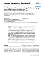
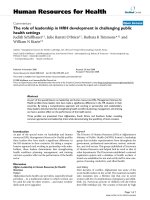
[44]. DENV E contains five domains with significant
WWIHS scores (Fig. 1, WWIHS sequences in black). These
include the fusion peptide domain, a portion of sub-
domain IIb, the pre-anchor stem region following domain
III, and the transmembrane domain. Sequences with high
WWIHS scores are similarly located in the X-ray structures
of WNV E and alphavirus (SFV and Sindbis virus – SINV)
E1, and potentially also in the putative class II fusion pro-
teins of hepatitis C virus (HCV), pestiviruses and bunyavi-
ruses [44,45]. Regions with high WWIHS scores are
predicted to play a role in protein-protein interactions
during structural rearrangements or protein-lipid interac-
tions during bilayer fusion, and we predicted that syn-
thetic peptides corresponding to these regions may have
the potential to inhibit flavivirus infectivity.
To test this hypothesis, an initial set of synthetic peptides
representing sequences of DENV E and WNV E with signif-
icant WWIHS scores was synthesized and screened for the
Diagramatic structure of DENV envelope protein showing inhibitory peptide regionsFigure 1
Diagramatic structure of DENV envelope protein showing inhibitory peptide regions. Grey lines: dicysteine link-
ages. Black stick figures: N-glycosylation sites. Regions with significant Wimley-White interfacial hydrophobicity scale scores
were predicted with MPeX (Boxed in left depiction; black in right depiction). Sequences of DENV peptides and the location of
WNV homologs are indicated.
Virology Journal 2005, 2:49 />Page 4 of 10
(page number not for citation purposes)
ability to inhibit plaque formation by these flaviviruses
(Table 1). Peptides corresponding to the transmembrane
domain were not tested because this region is not exposed
during the entry process. Initial assays for inhibitory activ-
ity were performed using the highest concentration of
each peptide that could be obtained in aqueous solution
with a maximum of 1% DMSO (between 29 and 128 µM).
Plaque reduction in which the inhibitor is removed after
virus adsorption is the most stringent test of an antiviral
agent. Prior to initiating these studies, we developed a
new immunoplaque assay for DENV and WNV. Approxi-
mately 200 focus forming units (FFU) of either WNV or
DENV were preincubated with each of the peptides and
used to infect monolayers of LLCKM-2 monkey kidney
epithelial cells. The number of resulting viral foci was
determined from three experiments and normalized to a
Table 1: Initial peptides synthesized and tested for inhibitory activity.
Peptide Sequence Location
a
Concentration (µM) % Inhibition +/- SD
DN80 MVDRGWGNHAGLFGKGSIV 386–400 49.9 17 +/- 10
DN57 AWLVHTQWFLDLPLPWLPGADTQGSNWI 485–503 30.6 7 +/- 4
DN81 AWLVHRQWFLDLPLPWLPG 485–512 42.6 25 +/- 8
DN59 MAILGDTAWDFGSLGGVFTSIGKALHQVFGAIY 692–724 29.0 93 +/- 2
WN82 VVDRGWGNGAGLFGKGSID 396–410 52.5 4 +/- 13
WN53 TFLVHREWFMDLNLPWSSAGSTVWR 500–518 98.7 56 +/- 5
WN83 TFLVHREWFMDLNLPWSSA 500–524 128.0 70 +/- 2
a
numbering from the beginning of the E polyprotein in either DENV or WNV.
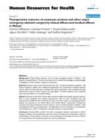
Plaque inhibition assayFigure 2
Plaque inhibition assay. (A) Preincubation of DENV with neutralizing antiserum reduces plaque number by 74%. (B) Prein-
cubation of DENV with a non-inhibitory peptide shows no reduction in plaques. (C) Preincubation of DENV with one of the
inhibitory peptides (DN59) shows a greater than 95% reduction in plaques.
Virology Journal 2005, 2:49 />Page 5 of 10
(page number not for citation purposes)
no-peptide control to calculate the percent inhibition.
Our screening of this initial set detected several peptides
that were able to inhibit infection by DENV or WNV
(Table 1, Fig. 2). Peptides similar in sequence to domains
with a significant WWIHS scores, including domain II
(IIb) (WN53 and WN83), and stem domain (DN59),
were found to have inhibitory activity.
Determination of 50% inhibitory concentrations
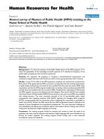
Dose-response curves were determined for the most
potent of the peptides WN53 and WN83 against WNV
and also for peptide DN59 against DENV (Fig. 3). The
WN53 peptide showed a maximum inhibitory activity
against WNV of 56.0 +/- 3.0% (mean +/- SD) at 99 µM.
The inhibitory activity decreased with decreasing concen-
tration with a 50% inhibitory concentration (IC50) at
roughly 10 µM. The WN83 peptide showed a maximum
inhibitory activity against WNV of 70.0 +/- 3.0% at 128
µM. The inhibitory activity decreased with decreasing con-
centration with an IC50 of roughly 10 µM. The DN59
peptide showed a maximum inhibitory activity against
DENV of 100.0 +/- 0.5% at 20 µM. The inhibitory activity
decreased with decreasing concentration with an IC50 of
at roughly 10 µM. DENV stem peptide 59 (DN59) and
WNV peptides 53 and 83 (WN53, WN83) reproducibly
inhibited infectivity at low µM concentrations.
Specificity of peptide inhibitory activity
The DN59 peptide matches a pre-anchor domain
sequence that is highly conserved among insect-transmit-
ted flaviviruses. DN59 inhibited infection by DENV
(>99% inhibition of plaque formation at a concentrations
of <25 µM). Cross-inhibition of WNV fusion/infectivity
(>99% inhibition at <25 µM) was also reproducibly dem-
onstrated with DN59 (Fig. 6). However, WNV inhibitory
peptide WN83 did not inhibit infectivity by DENV.
To determine if these peptides specifically inhibit infectiv-
ity of the viruses for which they were designed, the WN53,
WN83 and DN59 peptides were tested for inhibitory
effects against Sindbis virus (SINV), an alphavirus that
encodes a class II fusion protein. None of the peptides
showed a statistically significant effect against SINV infec-
tivity (Fig. 4). Peptides with the same amino acid compo-
sition as WN83 and DN59, but a scrambled sequence
(Scrambled WN83: VATWHLDWSREFPLFLMNS;
Scrambled DN59: YFIDTSGAIWGASHLTGVLFDFM-
GIQGGAVLAK) were synthesized and tested for the ability
to inhibit infection by WNV and DENV respectively. Nei-
ther scrambled peptide significantly inhibited infection
by these viruses (Fig. 5). These results provide evidence
that the action of these inhibitory peptides not due to gen-
eral inactivation of enveloped virions and is sequence
specific.
Dose-response curves for WN53, WN83 and DN59 peptidesFigure 3
Dose-response curves for WN53, WN83 and DN59
peptides. (A) Increasing concentrations of peptide WN53
produce a corresponding increase in WNV inhibition with an
IC50 in the 10µ range. (B) Increasing concentrations of pep-
tide WN83 produce a corresponding increase in WNV inhi-
bition with an IC50 in the 10 µM range. (C) Increasing
concentrations of peptide DN59 also produce a correspond-
ing increase in DNV inhibition with an IC50 in the 10 µM
range. All measurements were made in triplicate, with mean
+/- SD shown.
Virology Journal 2005, 2:49 />Page 6 of 10
(page number not for citation purposes)
Peptide toxicity
It is possible that inhibitory peptides induce cellular alter-
ations or toxicity that can block flavivirus entry or other
steps in the replication cycle. To address this possibility,
LLCMK-2 monkey kidney epithelial cell monolayers were
exposed to 100 mg/ml concentrations of WN53, WN83
and DN59 peptides for 24 hrs, and cell viability was
assayed with an MTT assay. No statistical difference was
observed between the viability of control cells versus cells
exposed to the peptides or DMSO (Fig. 7). This result sug-
gests that these inhibitory peptides are not blocking infec-
tivity via effects on host cell metabolism or viability.
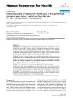
Non-synergistic activity of combined peptides
When added in combination, peptides that block entry at
different steps or that target different domains may pro-
duce greater inhibition of DENV-2 infectivity than either
peptide alone. Synergistic or antagonistic effects are also
possible, if a peptide that alters protein-protein interac-
tions allows greater or lesser access to E domains targeted
by another peptide. Since the WN53, WN83 and DN59
peptides all inhibited WNV entry, the possibility of antag-
onistic or synergistic function was examined by testing
WN53 and DN59 alone or in combination at three con-
centrations (5, 10 and 20 µM). At all three concentrations,
the peptide combination was more effective than WN53
alone, but less effective than DN59 alone. This indicates
that the activity of the WN53 peptide has an antagonistic
effect on the function of the DN59 peptide (Fig. 6).
Discussion
Synthetic peptides corresponding to sequences in DENV
and WNV E proteins were identified that inhibited infec-
tivity of these viral pathogens of major public health
importance. The inhibitory effects of these peptides were
dose dependent with IC50s in the range of 10 µM. Several
of the most potent of these peptides showed no inhibitory
activity against SINV, an alphavirus that possesses a class
II viral fusion protein with a similar overall structure as
flavivirus E. Scrambled peptides with the same amino acid
composition as the inhibitory peptides, but with a differ-
ent primary sequence, failed to inhibit DENV and WNV
infection. None of the DENV or WNV inhibitory peptides
induced gross cytopathic effects, killing of cultured cells or
showed evidence of in vitro cellular toxicity. These results
indicate that these inhibitory peptides function through a
sequence-specific mechanism and are not merely
cytotoxic.
Effect of DENV and WNV specific peptides against another virusFigure 4
Effect of DENV and WNV specific peptides against
another virus. Peptides DN59, WN83 and WN53 at 100
µg/ml concentrations were tested for inhibitory activity
against the alphavirus SINV in a similar plaque reduction
assay. Results are shown as the mean of three trials +/- SEM.
None of the peptides showed a statistically significant inhibi-
tory effect against SINV (ANOVA, p = 0.705, with Dunnett's
posthoc test).
Effect of scrambled peptide order on inhibitory functionFigure 5
Effect of scrambled peptide order on inhibitory func-
tion. Scrambled versions of peptides DN59 and WN83 at
100 µg/ml concentrations were tested for inhibitory effect
against DENV and WNV, respectively. Scrambled versions of
the peptides showed no inhibitory activity compared to the
original DN59 and WN83 peptides.
Virology Journal 2005, 2:49 />Page 7 of 10
(page number not for citation purposes)
Membrane fusion by both class I and II viral fusion pro-
teins is initiated by interaction of the fusion peptide with
the target cell membrane. In class I viral fusion proteins, a
subsequent rearrangement of a trimer of the proteins,
each with two α helices, to form a six-helix bundle brings
the viral and cell membranes into closer proximity. Inhib-
itors of viruses with class I fusion proteins, such as
Fuzeon™ that mimic a portion of one or the other of the
two α helices, interfere with a step proximal to six-helix
bundle formation possibly by forming an inactive
aggregate with the opposite helix. Recent studies indicate
that after insertion of the fusion peptide, class II viral
fusion proteins likewise undergo rearrangements. In this
case, intraprotein interactions may occur between the
stem domain and domains I, II and/or III [21-28,46].
According to this model, the viral and cellular membranes
are brought closer by interactions of the stem with other
portions of E, resulting in bilayer fusion. DN59, WN53
and WN83 peptides may interfere with the intramolecular
interactions between the stem and other portions of class
II viral fusion proteins, a possibility suggested previously
[23,24,46].
Two of the inhibitory peptides (WN53 and WN83) are
designed from overlapping regions of the E protein
domain I/II junction and are specifically inhibitory
against WNV. Recently, other investigators have hypothe-
sized that small molecule inhibitors to this domain I/II
junction region might be developed. Modis et al (2003;
2004) predicted that interactions near this region (the k-l
loop) that are involved in the rotational changes between
these domains might be blocked by small molecule inhib-
itors. However, our similar peptides designed from the
analogous region of the DENV E protein (D57 and D81)
failed to inhibit DENV infectivity.
The possibility that WN53, WN83 and DN59 interact with
some target cell surface component to exert their inhibi-
tory effects cannot be ruled out. However, the majority of
flavivirus neutralizing antibodies that appear to be
involved in receptor blocking bind to domain III, and sol-
uble domain III itself can block flavivirus entry, appar-
ently through competition for cellular receptors [47-51].
In contrast, the domains that correspond to WN53, WN83
and DN59 peptides, IIb and the pre-anchor stem, appear
to be involved in structural rearrangements during fusion,
rather than direct interactions with cellular receptors.
Interestingly, previous observations indicate that some
monoclonal antibodies block virion entry at a post attach-
ment step, indicating that they may interfere with
conformational changes necessary for fusion [52]. That
possibility that some antibodies can gain access to regions
important for conformational changes and block these
changes suggests that these inhibitory peptide regions
might be candidates for novel vaccine designs that either
Inhibitory effect of peptides WN53 and DN59 alone and in combinationFigure 6
Inhibitory effect of peptides WN53 and DN59 alone
and in combination. WN53 and DN59 peptides were
tested alone (■ DN59 alone, ● WN53 alone) or together
(◆) with WNV and inhibitory activity at three concentra-
tions was measured (mean of three trials +/- SD shown).
DN59 and WN53 together have an effect intermediate
between the two peptides alone.
Toxicity of inhibitory peptides in cell cultureFigure 7
Toxicity of inhibitory peptides in cell culture. MTT
assays for cell viability were performed after 24 hr incubation
of cells with 100 µg/ml of peptides WN83, WN53, and
DN59 (mean from three experiments +/- SEM). No statisti-
cally significant differences in cell viability were observed
(ANOVA p = 0.672, with Dunnett's posthoc test).
Virology Journal 2005, 2:49 />Page 8 of 10
(page number not for citation purposes)
utilize the inhibitory peptide regions directly as antigens,
or target the regions that interact with the inhibitory
peptides. Further studies are needed to define the exact
mechanism of inhibition of these DENV and WNV pep-
tides, and the specific nature or location of their interac-
tions with viral targets.
The DN59 peptide is inhibitory against DENV as well as
WNV. The corresponding pre-anchor region is highly con-
served between DENV and WNV as well as among other
flaviviruses (Table 2) and probably functions in a similar
manner during entry of all flaviviruses [53]. Thus, DN59
or similar peptides may act as broad-spectrum flavivirus
inhibitors. Other flaviviruses considered potential bioter-
rorism agents, including JEV, Kyasanur Forest disease
virus and TBEV, may also be inhibited by DN59, a DN59
derivative, or by an analogous peptide. Unlike proposed
DENV vaccines, which must be multivalent (ie. simulta-
neously effective against each of the four DENV serotypes
because of the phenomenon of antibody dependent
enhancement), peptide drugs targeting the highly
conserved stem conserved motifs in flavivirus E may dem-
onstrate cross-strain efficacy.
IC50s in the µM range have been considered promising
for class I viral fusion protein inhibitor development
[54,55]. Thus, the peptides identified here can serve as
lead compounds that may be developed as peptide drugs
against the four serotypes of DENV, WNV and potentially
other flaviviruses. We anticipate that such peptide inhibi-
tors may be as successful as the HIV-1 inhibitory peptide
Fuzeon™. Unlike persistant HIV infections, immune
responses against DENV and other flaviviruses are capable
of clearing the viruses in individuals that survive the
initial infection. By reducing the viral load during the ini-
tial stages of infection, it may be possible to extend the
window of time during which an immune response could
arise, and thus enable more individuals to control,
eliminate and survive infections by these agents. Evidence
for the ability to therapeutically intervene in flavivirus-
induced diseases has been demonstrated with the recent
observation that administration of neutralizing antibod-
ies against WNV can be curative, even after symptom ini-
tiation [56]. Development of resistant mutants will be a
concern, but should be a less problematic than in the case
of long-term treatment of persistent retroviral infections.
It is worth noting that the HIV inhibitor Fuzeon™, was ini-
tially identified using a predictive strategy without the
availability of structural data [6,7,57]. The fact that we
developed these peptides using a predictive algorithm val-
idates our approach as well as the accuracy of the flavivi-
rus E protein structural data. A similar approach may be
useful for the large number of other viruses with class II
envelope fusion proteins with or without known
structures.
Materials and methods
Design and synthesis of peptides
Sequences of DENV and WNV E with positive Wimley-
White interfacial hydrophobicity scale scores were deter-
mined using the program Membrane Protein eXplorer [1].
After consideration of the known secondary structures for
several subdomains of E, selected peptides were synthe-
sized by solid-phase conventional N-α-9-flurenylmethyl-
oxycarbonyl chemistry (Genemed Synthesis, San
Francisco, CA). Peptides were purified by reverse-phase
high performance liquid chromatography and confirmed
by amino acid analysis and electrospray mass
spectrometry. Peptide stock solutions were prepared in
Table 2: Alignment of preanchor domain sequences from representative flaviviruses.
Virus
a
Pre-anchor sequence
b
Location
c
DENV-1 AILGDTAWDFGSIGGVFTSVGKLIHQIFGTA 693–723
DENV-2 AILGDTAWDFGSLGGVFTSIGKALHQVFGAI 693–723
DENV-3 AILGDTAWDFGSVGGVLNSLGKMVHQIFGSA 691–721
DENV-4 AILGETAWDFGSVGGVLTSLGKAVHQVFGSV 692–722
WNV AVLGDTAWDFGSVGGVFTSVGKAVHQVFGGA 709–739
YFV AVMGDTAWDFSSAGGFFTSVGKGIHTVFGSA 695–725
SLEV AVLGDTAWDFGSIGGVFTSIGKALHQVFGGA 707–737
JEV AALGDTAWDFGSIGGVFNSIGKAVHQVFGGA 712–742
TBEV TVIGEHAWDFGSAGGFLSSIGKAVHTVLGGA 694–724
OHFV TVLGEHAWDFGSTGGFLSSIGKALHTVLGGA 694–724
KFV TVVGEHAWDFGSVGGMLSSVGKALHTAFGAA 695–725
POWV SVVGEHAWDFGSVGGVLSSVGKAIHTVLGGA 693–723
a
DENV: dengue virus; WNV: West Nile virus; YFV: yellow fever virus; SLEV: St. Louis encephalitis virus; JEV: Japanese encephalitis virus; TBEV: Tick-
borne encephalitis virus; OHFV: Omsk hemorrhagic fever virus; KFV: Kyasanur Forest virus; POWV: Powassan virus
b
* = identical or chemically similar amino acids in every sequence
c
numbering from the beginning of the E polyprotein.
Virology Journal 2005, 2:49 />Page 9 of 10
(page number not for citation purposes)
20% (v/v) dimethyl sulfoxide (DMSO): 80% (v/v) H
2
0,
and concentrations determined by absorbance of aro-
matic side chains at 280 nm. Scrambled peptides
sequences were obtained by drawing from a hat.
Viruses and Cells
DENV strain New Guinea-2 and WNV strain Egypt 101
were obtained from R. Tesh at the World Health Organi-
zation Arbovirus Reference Laboratory at the University of
Texas at Galveston. DENV and WNV were propagated in
the African green monkey kidney epithelial cell line,
LLCKM-2, a gift of K. Olsen at Colorado State University.
Sindbis virus (SINV) containing the enhanced green fluo-
rescent protein (EGFP) protein expression cassette was
obtained from K. Ryman at Louisiana State University at
Shreveport and was propagated in baby hamster kidney
cells. All cells were grown in Dulbecco's modified eagle
medium (DMEM) with 10% (v/v) fetal bovine serum
(FBS), 100 U/ml penicillin G and 100 mg/ml streptomy-
cin, at 37°C with 5% (v/v) CO
2
.
Viral plaque reduction assays
LLCKM-2 target cells were seeded at a density of 3 × 10
5
cells in each well of a 6-well plate 48 h prior to infection.
Approximately 200-focus forming units (FFU) of DENV,
WNV, or SINV/EGFP were incubated with or without pep-
tides in serum-free DMEM for 1 h at rt. Virus/peptide or
virus/control mixtures were allowed to infect confluent
LLCKM-2 monolayers for 1 h at 37°C, after which time
the medium was removed and the cells were washed once
with phosphate buffered saline (PBS) and overlaid with
fresh DMEM/10% (v/v) FBS containing 0.85% (w/v) Sea-
Plaque Agarose (Cambrex Bio Science, Rockland, ME).
Cells were then incubated at 37°C with 5% CO
2
for 1 day
(Sindbis virus), 3 days (WNV) or 6 days (DNV). Sindbis
virus infections were quantified by directly counting green
fluorescing foci. Cultures infected with DENV were fixed
with 10% formalin overnight at 4°C and permeablized
with 70% (v/v) ethanol prior to immunostaining and vis-
ualization using a human polyclonal anti-flavivirus anti-
body (a gift of V. Vorndam, CDC, San Juan) followed by
horseradish peroxidase (HRP) conjugated goat anti-
human immunoglobulin (Pierce, Rockford, IL) and AEC
chromogen substrate (Dako, Carpinteria, CA). WNV
plaques were similarly visualized using a mouse anti-
WNV antibody (Chemicon, Temecula, CA) and an HRP
conjugated goat anti-mouse antibody (Dako, Carpinteria,
CA).
Toxicity assay
Peptide cytotoxicity was measured using the TACS™ MTT
cell proliferation assay (R&D systems, Minneapolis, MN)
according to the manufacturer's instructions.
Acknowledgements
The authors would like to thank S. Isern, B. Sainz, W. Wimley and W. Gal-
laher for helpful discussions and technical assistance.
References
1. White SH, Snider C, Jaysinghe S, Kim J: Membrane Protein
Explorer version 2.2a. http://blancobiomoluciedu/mpex/ 2003.
2. Carr CM, Kim PS: A spring-loaded mechanism for the confor-
mational change of influenza hemagglutinin. Cell 1993,
73:823-832.
3. Wilson IA, Skehel JJ, Wiley DC: Structure of the haemagglutinin
membrane glycoprotein of influenza virus at 3 A resolution.
Nature 1981, 289:366-373.
4. Gallaher WR: Similar structural models of the transmem-
brane glycoproteins of Ebola and avian sarcoma viruses. Cell
1996, 85:1-2.
5. Gallaher WR, Ball JM, Garry RF, Griffin MC, Montelaro RC: A gen-
eral model for the transmembrane proteins of HIV and
other retroviruses. AIDS Res Hum Retroviruses 1989, 5:431-440.
6. Wild CT, Shugars DC, Greenwell TK, McDanal CB, Matthews TJ:
Peptides corresponding to a predictive alpha-helical domain
of human immunodeficiency virus type 1 gp41 are potent
inhibitors of virus infection. Proc Natl Acad Sci U S A 1994,
91:9770-9774.
7. Wild C, Greenwell T, Matthews T: A synthetic peptide from
HIV-1 gp41 is a potent inhibitor of virus-mediated cell-cell
fusion. AIDS Research & Human Retroviruses 1993, 9:1051-1053.
8. Pozniak A: HIV fusion inhibitors. J HIV Ther 2001, 6:91-94.
9. Sodroski JG: HIV-1 entry inhibitors in the side pocket. Cell
1999, 99:243-246.
10. Lambert DM, Barney S, Lambert AL, Guthrie K, Medinas R, Davis DE,
Bucy T, Erickson J, Merutka G, Petteway SRJ: Peptides from con-
served regions of paramyxovirus fusion (F) proteins are
potent inhibitors of viral fusion. Proc Natl Acad Sci U S A 1996,
93:2186-2191.
11. Young JK, Li D, Abramowitz MC, Morrison TG: Interaction of pep-
tides with sequences from the Newcastle disease virus fusion
protein heptad repeat regions. J Virol 1999, 73:5945-5956.
12. Watanabe S, Takada A, Watanabe T, Ito H, Kida H, Kawaoka Y:
Functional importance of the coiled-coil of the Ebola virus
glycoprotein. J Virol 2000, 74:10194-10201.
13. Bosch BJ, van der Zee R, de Haan CA, Rottier PJ: The coronavirus
spike protein is a class I virus fusion protein: structural and
functional characterization of the fusion core complex. J Virol
2003, 77:8801-8811.
14. Bultmann H, Brandt CR: Peptides containing membrane-trans-
iting motifs inhibit virus entry. J Biol Chem 2002,
277:36018-36023.
15. Markosyan RM, Bates P, Cohen FS, Melikyan GB: A study of low
pH-induced refolding of Env of avian sarcoma and leukosis
virus into a six-helix bundle. Biophys J 2004, 87:3291-3298.
16. Rapaport D, Ovadia M, Shai Y: A synthetic peptide correspond-
ing to a conserved heptad repeat domain is a potent inhibi-
tor of Sendai virus-cell fusion: an emerging similarity with
functional domains of other viruses. Embo J 1995,
14:5524-5531.
17. Okazaki K, Kida H: A synthetic peptide from a heptad repeat
region of herpesvirus glycoprotein B inhibits virus
replication. J Gen Virol 2004, 85:2131-2137.
18. Yao Q, Compans RW: Peptides corresponding to the heptad
repeat sequence of human parainfluenza virus fusion protein
are potent inhibitors of virus infection. Virology 1996,
223:103-112.
19. Rey FA, Heinz FX, Mandl C, Kunz C, Harrison SC: The envelope
glycoprotein from tick-borne encephalitis virus at 2 A
resolution. Nature 1995, 375:291-298.
20. Lescar J, Roussel A, Wien MW, Navaza J, Fuller SD, Wengler G, Rey
FA: The Fusion glycoprotein shell of Semliki Forest virus: an
icosahedral assembly primed for fusogenic activation at
endosomal pH. Cell 2001, 105:137-148.
21. Kuhn RJ, Zhang W, Rossmann MG, Pletnev SV, Corver J, Lenches E,
Jones CT, Mukhopadhyay S, Chipman PR, Strauss EG, Baker TS,
Strauss JH: Structure of dengue virus: implications for flavivi-
rus organization, maturation, and fusion. Cell 2002,
108:717-725.
Virology Journal 2005, 2:49 />Page 10 of 10
(page number not for citation purposes)
22. Zhang Y, Corver J, Chipman PR, Zhang W, Pletnev SV, Sedlak D,
Baker TS, Strauss JH, Kuhn RJ, Rossmann MG: Structures of imma-
ture flavivirus particles. Embo J 2003, 22:2604-2613.
23. Bressanelli S, Stiasny K, Allison SL, Stura EA, Duquerroy S, Lescar J,
Heinz FX, Rey FA: Structure of a flavivirus envelope glycopro-
tein in its low-pH-induced membrane fusion conformation.
Embo J 2004, 23:728-738.
24. Modis Y, Ogata S, Clements D, Harrison SC: Structure of the den-
gue virus envelope protein after membrane fusio. Nature
2004, 427:313-319.
25. Modis Y, Ogata S, Clements D, Harrison SC: A ligand-binding
pocket in the dengue virus envelope glycoprotein. Proc Natl
Acad Sci U S A 2003, 100:6986-6991.
26. Mukhopadhyay S, Kim BS, Chipman PR, Rossmann MG, Kuhn RJ:
Structure of West Nile virus. Science 2003, 302:248.
27. Mukhopadhyay S, Kuhn RJ, Rossmann MG: A structural perspec-
tive of the flavivirus life cycle. Nat Rev Microbiol 2005, 3:13-22.
28. Rey FA: Dengue virus envelope glycoprotein structure: new
insight into its interactions during viral entry. Proc Natl Acad
Sci U S A 2003, 100:6899-6901.
29. WHO: Dengue and dengue haemorrhagic fever. http://
www.who.int/mediacentre/factsheets/fs117/en/, ; 2002.
30. Hotta H, Sanchez LF, Takada H, Homma M, Kotani S: Enhancement
of dengue virus infection in cultured mouse macrophages by
lipophilic derivatives of muramyl peptides. Microbiol Immunol
1985, 29:533-541.
31. Halstead SB: Pathogenesis of dengue: challenges to molecular
biology. Science 1988, 239:476-481.
32. Guy B, Chanthavanich P, Gimenez S, Sirivichayakul C, Sabchareon A,
Begue S, Yoksan S, Luxemburger C, Lang J: Evaluation by flow
cytometry of antibody-dependent enhancement (ADE) of
dengue infection by sera from Thai children immunized with
a live-attenuated tetravalent dengue vaccine. Vaccine 2004,
22:3563-3574.
33. Sabchareon A, Lang J, Chanthavanich P, Yoksan S, Forrat R, Attanath
P, Sirivichayakul C, Pengsaa K, Pojjaroen-Anant C, Chokejindachai W,
Jagsudee A, Saluzzo JF, Bhamarapravati N: Safety and immuno-
genicity of tetravalent live-attenuated dengue vaccines in
Thai adult volunteers: role of serotype concentration, ratio,
and multiple doses. Am J Trop Med Hyg 2002, 66:264-272.
34. Edelman R, Wasserman SS, Bodison SA, Putnak RJ, Eckels KH, Tang
D, Kanesa-Thasan N, Vaughn DW, Innis BL, Sun W: Phase I trial of
16 formulations of a tetravalent live-attenuated dengue
vaccine. Am J Trop Med Hyg 2003, 69:48-60.
35. Kanesa-Thasan N, Sun W, Ludwig GV, Rossi C, Putnak JR, Mangiafico
JA, Innis BL, Edelman R: Atypical antibody responses in dengue
vaccine recipients. Am J Trop Med Hyg 2003, 69:32-38.
36. Kanesa-thasan N, Sun W, Kim-Ahn G, Van Albert S, Putnak JR, King
A, Raengsakulsrach B, Christ-Schmidt H, Gilson K, Zahradnik JM,
Vaughn DW, Innis BL, Saluzzo JF, Hoke CHJ: Safety and immuno-
genicity of attenuated dengue virus vaccines (Aventis Pas-
teur) in human volunteers. Vaccine 2001, 19:3179-3188.
37. Bhamarapravati N, Sutee Y: Live attenuated tetravalent dengue
vaccine. Vaccine 2000, 18 Suppl 2:44-47.
38. Barrett AD: Current status of flavivirus vaccines. Ann N Y Acad
Sci 2001, 951:262-271.
39. Sun W, Edelman R, Kanesa-Thasan N, Eckels KH, Putnak JR, King AD,
Houng HS, Tang D, Scherer JM, Hoke CHJ, Innis BL: Vaccination of
human volunteers with monovalent and tetravalent live-
attenuated dengue vaccine candidates. Am J Trop Med Hyg
2003, 69:24-31.
40. Guirakhoo F, Pugachev K, Zhang Z, Myers G, Levenbook I, Draper K,
Lang J, Ocran S, Mitchell F, Parsons M, Brown N, Brandler S, Fournier
C, Barrere B, Rizvi F, Travassos A, Nichols R, Trent D, Monath T:
Safety and efficacy of chimeric yellow Fever-dengue virus
tetravalent vaccine formulations in nonhuman primates. J
Virol 2004, 78:4761-4775.
41. Guirakhoo F, Pugachev K, Arroyo J, Miller C, Zhang ZX, Weltzin R,
Georgakopoulos K, Catalan J, Ocran S, Draper K, Monath TP:
Viremia and immunogenicity in nonhuman primates of a
tetravalent yellow fever-dengue chimeric vaccine: genetic
reconstructions, dose adjustment, and antibody responses
against wild-type dengue virus isolates. Virology 2002,
298:146-159.
42. Wimley WC, White SH: Experimentally determined hydropho-
bicity scale for proteins at membrane interfaces. Nat Struct
Biol 1996, 3:842-848.
43. Giannecchini S, Bonci F, Pistello M, Matteucci D, Sichi O, Rovero P,
Bendinelli M: The membrane-proximal tryptophan-rich region
in the transmembrane glycoprotein ectodomain of feline
immunodeficiency virus is important for cell entry. Virology
2004, 320:156-166.
44. Garry RF, Dash S: Proteomics computational analyses suggest
that hepatitis C virus E1 and pestivirus E2 envelope glyco-
proteins are truncated class II fusion proteins. Virology 2003,
307:255-265.
45. Garry CE, Garry RF: Proteomics computational analyses sug-
gest that the carboxyl terminal glycoproteins of Bunyavi-
ruses are class II viral fusion protein (beta-penetrenes). Theor
Biol Med Model 2004, 1:10.
46. Gibbons DL, Vaney MC, Roussel A, Vigouroux A, Reilly B, Lepault J,
Kielian M, Rey FA: Conformational change and protein-protein
interactions of the fusion protein of Semliki Forest virus.
Nature 2004, 427:320-325.
47. Beasley DW, Barrett AD: Identification of neutralizing epitopes
within structural domain III of the West Nile virus envelope
protein. J Virol 2002, 76:13097-13100.
48. Volk DE, Beasley DW, Kallick DA, Holbrook MR, Barrett AD, Goren-
stein DG: Solution structure and antibody binding studies of
the envelope protein domain III from the New York strain of
West Nile virus. J Biol Chem 2004, 279:38755-38761.
49. Chen Y, Maguire T, Marks RM: Demonstration of binding of den-
gue virus envelope protein to target cells. J Virol 1996,
70:8765-8772.
50. Bhardwaj S, Holbrook M, Shope RE, Barrett AD, Watowich SJ: Bio-
physical characterization and vector-specific antagonist
activity of domain III of the tick-borne flavivirus envelope
protein. J Virol 2001, 75:4002-4007.
51. Wu KP, Wu CW, Tsao YP, Kuo TW, Lou YC, Lin CW, Wu SC,
Cheng JW: Structural basis of a Flavivirus recognized by its
neutralizing antibody: Solution structure of the domain III of
the Japanese Encephalitis virus envelope protein. J Biol Chem
2003.
52. Hung SL, Lee PL, Chen HW, Chen LK, Kao CL, King CC: Analysis
of the steps involved in Dengue virus entry into host cells.
Virology 1999, 257:156-167.
53. Allison SL, Stiasny K, Stadler K, Mandl CW, Heinz FX: Mapping of
functional elements in the stem-anchor region of tick-borne
encephalitis virus envelope protein E. J Virol 1999,
73:5605-5612.
54. Liu S, Xiao G, Chen Y, He Y, Niu J, Escalante CR, Xiong H, Farmar J,
Debnath AK, Tien P, Jiang S: Interaction between heptad repeat
1 and 2 regions in spike protein of SARS-associated corona-
virus: implications for virus fusogenic mechanism and identi-
fication of fusion inhibitors. Lancet 2004, 363:938-947.
55. Sia SK, Carr PA, Cochran AG, Malashkevich VN, Kim PS: Short con-
strained peptides that inhibit HIV-1 entry. Proc Natl Acad Sci U
S A 2002, 99:14664-14669.
56. Oliphant T, Engle M, Nybakken GE, Doane C, Johnson S, Huang L,
Gorlatov S, Mehlhop E, Marri A, Chung KM, Ebel GD, Kramer LD,
Fremont DH, Diamond MS: Development of a humanized mon-
oclonal antibody with therapeutic potential against West
Nile virus. Nat Med 2005, 11:522-30.
57. Qureshi NM, Coy DH, Garry RF, LA H: Characterization of a
putative cellular receptor for HIV-1 transmembrane glyco-
protein using synthetic peptides. AIDS 1990, 4:553-558.