Báo cáo sinh học: " Human cytomegalovirus uracil DNA glycosylase associates with ppUL44 and accelerates the accumulation of viral DNA" pdf
Bạn đang xem bản rút gọn của tài liệu. Xem và tải ngay bản đầy đủ của tài liệu tại đây (1.53 MB, 14 trang )
BioMed Central
Page 1 of 14
(page number not for citation purposes)
Virology Journal
Open Access
Research
Human cytomegalovirus uracil DNA glycosylase associates with
ppUL44 and accelerates the accumulation of viral DNA
Mark N Prichard*
1
, Heather Lawlor
2
, Gregory M Duke
2
, Chengjun Mo
2
,
Zhaoti Wang
2
, Melissa Dixon
2
, George Kemble
2
and Earl R Kern
1
Address:
1
Department of Pediatrics, University of Alabama at Birmingham, Birmingham AL, USA and
2
Department of Research, MedImmune
Vaccines Inc., Mountain View, CA, USA
Email: Mark N Prichard* - ; Heather Lawlor - ;
Gregory M Duke - ; Chengjun Mo - ; Zhaoti Wang - ;
Melissa Dixon - ; George Kemble - ; Earl R Kern -
* Corresponding author
Abstract
Background: Human cytomegalovirus UL114 encodes a uracil-DNA glycosylase homolog that is
highly conserved in all characterized herpesviruses that infect mammals. Previous studies
demonstrated that the deletion of this nonessential gene delays significantly the onset of viral DNA
synthesis and results in a prolonged replication cycle. The gene product, pUL114, also appears to
be important in late phase DNA synthesis presumably by introducing single stranded breaks.
Results: A series of experiments was performed to formally assign the observed phenotype to
pUL114 and to characterize the function of the protein in viral replication. A cell line expressing
pUL114 complemented the observed phenotype of a UL114 deletion virus in trans, confirming that
the observed defects were the result of a deficiency in this gene product. Stocks of recombinant
viruses without elevated levels of uracil were produced in the complementing cells; however they
retained the phenotype of poor growth in normal fibroblasts suggesting that poor replication was
unrelated to uracil content of input genomes. Recombinant viruses expressing epitope tagged
versions of this gene demonstrated that pUL114 was expressed at early times and that it localized
to viral replication compartments. This protein also coprecipitated with the DNA polymerase
processivity factor, ppUL44 suggesting that these proteins associate in infected cells. This apparent
interaction did not appear to require other viral proteins since ppUL44 could recruit pUL114 to
the nucleus in uninfected cells. An analysis of DNA replication kinetics revealed that the initial rate
of DNA synthesis and the accumulation of progeny viral genomes were significantly reduced
compared to the parent virus.
Conclusion: These data suggest that pUL114 associates with ppUL44 and that it functions as part
of the viral DNA replication complex to increase the efficiency of both early and late phase viral
DNA synthesis.
Published: 15 July 2005
Virology Journal 2005, 2:55 doi:10.1186/1743-422X-2-55
Received: 18 May 2005
Accepted: 15 July 2005
This article is available from: />© 2005 Prichard et al; licensee BioMed Central Ltd.
This is an Open Access article distributed under the terms of the Creative Commons Attribution License ( />),
which permits unrestricted use, distribution, and reproduction in any medium, provided the original work is properly cited.
Virology Journal 2005, 2:55 />Page 2 of 14
(page number not for citation purposes)
Background
The enzymatic removal of uracil from DNA occurs in all
free-living organisms. Both the misincorporation of dUTP
by DNA polymerase and the spontaneous deamination of
cytosine are relatively frequent events and give rise to
uracil residues covalently linked to the genome, with the
latter resolving into A:T transition mutations in one of the
nascent strands [4,42]. Human herpesviruses, poxviruses
and retroviruses either encode or recruit uracil DNA glyc-
osylase (UNG) homologs, presumably to remove uracil
bases from genomic DNA [5]. A number of studies used
site directed mutagenesis to characterize the function of
this gene in the life cycle of these viruses and most have
described unexpected facets of the phenotype that involve
DNA (or RNA) replication [5]. Studies described here with
human cytomegalovirus (CMV) suggest that the UNG is
part of the replication complex and that it functions in the
replication of the viral genome.
Highly conserved mechanisms have evolved to minimize
the presence of uracil in genomic DNA, presumably to
prevent damage to the genome [30,44,46]. In humans, at
least five base excision repair enzymes are capable of
removing uracil bases incorporated in DNA. The human
UNG gene expresses distinct nuclear and mitochondrial
forms of this enzyme, designated UNG2 and UNG1,
respectively [18]. In addition, a thymine(uracil) DNA gly-
cosylase, a cyclin-like UNG, and a new gene SMUG1 have
all been shown to possess this activity [24,26,27]. The rel-
ative function of each of these molecules remains to be
characterized, but it appears that these molecules have
developed specialized roles in mammals. Recent studies
describing the phenotype of UNG knockout mice did not
identify a greatly increased spontaneous mutation rate, in
contrast to studies in both prokaryotes and sacharomyces
[18]. SMUG1 appears to be responsible for recognizing
and repairing uracil residues resulting from the spontane-
ous deamination of cytosine [26], whereas UNG2 colocal-
izes with replication foci in dividing cells and is thought
to remove uracil during the replication process [18]. An
ancillary role for this enzyme in mammalian DNA repli-
cation is also supported by the fact that UNG2 interacts
physically with both replication protein A [25], as well as
proliferating cell nuclear antigen (PCNA) which is a cen-
tral regulator of DNA synthesis [28]. Further, these inter-
actions suggest that UNG2 participates in the PCNA-
requiring 2–8 bp patch base excision repair pathway [39].
A number of virus families appear to recruit UNG2, or to
encode UNG2 homologs for use in the replication proc-
ess. In human immunodeficiency virus (HIV) type 1, the
vpr gene product interacts specifically with UNG2 [3]. The
Vpr from simian immunodeficiency virus also binds
UNG2 in a similar manner, however, it doesn't appear to
impact the phenotype of cell cycle arrest associated with
Vpr [38]. UNG2 is packaged inside retrovirus virions by an
integrase dependent mechanism [45], and physically
associates with integrase as well as reverse transcriptase in
the pre-integration complex [33]. Lysates from purified
virions demonstrated that UNG2 remained functional
and was capable of directing the repair of uracil from a
synthetic oligonucleotide template in conjunction with
reverse transcriptase in a manner that is independent of
apurinic/apyrimidinic endonuclease [33]. The function
that UNG2 serves in HIV replication is unclear. However,
the misincorporation of dUTP in a RT/RNAse H assay
does not appear to affect first strand DNA synthesis by RT,
but rather, it affects the specificity of cleavage by RNAse H
resulting in reduced second strand synthesis from the
RNA primers [17]. Poxviruses also encode a UNG2
homologs that perform an essential function in the repli-
cation of this virus [22,41,43] and are thought to act at the
level of DNA synthesis [8]. More recent studies confirmed
that D4R is essential for vaccinia DNA synthesis, and that
its essential function is unrelated to its ability to excise
uracil from DNA [7].
Herpesviruses all encode UNG homologs that do not
appear to be required for replication in cell culture
[23,31,36], although the deletion of the homolog in her-
pes simplex virus appears to reduce neuroinvasiveness in
animal models [35]. CMV is unique among these viruses
in that the deletion of this ORF results in a distinct pheno-
type characterized by a marked delay in the onset of DNA
synthesis despite the normal temporal expression of early
genes involved in this process [29,31]. The phenotype is
less apparent in rapidly dividing cells, suggesting that a
cellular gene might compensate at least to some degree
[6]. Another interesting aspect of the UNG
-
phenotype
occurs late in infection where the mutant virus fails to ini-
tiate robust DNA synthesis and concurrently fails to incor-
porate uracil in the genome, suggesting that the removal
of these moieties may be related to the switch to late phase
DNA synthesis [6]. It is unclear why this phenotype is
observed in CMV and not in other herpesviruses, but it
may be related to the distinct mechanisms that this virus
has evolved to replicate its genome that is independent of
origin binding proteins encoded by most other
herpesviruses.
To help understand how the UL114 gene product func-
tions in viral DNA synthesis, a complementing cell line
was constructed and recombinant viruses in which this
gene product was epitope tagged were used to characterize
its expression and localization in the context of a viral
infection. Herein, we demonstrate that pUL114 localizes
to the viral replication compartments and associates with
the accessory factor of the DNA polymerase (ppUL44,
ICP36), and that the absence of this molecule results in
Virology Journal 2005, 2:55 />Page 3 of 14
(page number not for citation purposes)
delayed onset of viral DNA synthesis as well as inefficient
replication of the viral genome.
Results
Restoration of UL114
Recombinant viruses with deletions in UL114 express
early gene products with normal kinetics, yet exhibit a
marked delay in the onset of DNA synthesis [6,31]. This
phenotype was assigned to UL114, since two independent
isolates of the recombinant virus exhibited the same phe-
notype. To formally ascribe the observed phenotype to
this locus, the lesion was repaired with an Eag I DNA frag-
ment (AD169 coordinates 162693–164080) that spans
the deletion in the mutant virus (Fig. 1). Plaques resistant
to high concentrations of xanthine were isolated and were
shown to have restored the deleted sequences as deter-
mined by Southern analysis (data not shown). Kinetics of
viral DNA synthesis were examined in HEL cells infected
with the parent virus, the mutant (RC2620) and the res-
cued virus (RQ2620) to determine if the restoration of the
UL114 locus reverted the phenotype of delayed DNA syn-
thesis. As observed previously, the mutant exhibited very
little DNA synthesis in the first three days of infection (Fig
2A). In contrast, the rescued virus appeared to synthesize
DNA with the same kinetics as the parent virus suggesting
that the defect was due to the engineered mutation rather
than to mutations elsewhere in the genome. These data
were confirmed in HEL cells in an experiment in which
single-step replication kinetics were examined. Delayed
viral replication was observed in the mutant virus,
whereas, no difference was observed between the wt virus
and the recombinant virus in which the UL114 lesion was
repaired (Fig. 2B). Thus, two facets of the described phe-
notype (DNA synthesis and replication kinetics) were
reverted upon restoration of this gene and we formally
assigned this phenotype to the engineered mutation. This
phenotype was also reproduced in Towne strain of CMV
when the UL114 open reading frame was disrupted.
Complementation of the UNG deficient mutant in trans
and the effect of uracil content on the phenotype
Previous work demonstrated that virion DNA from the
mutant virus contained modestly elevated levels of uracil
compared to the wt virus, which is a predicted phenotype
[31]. Thus, it is possible that the delay in DNA synthesis
simply reflects the time required to repair misincorpo-
rated uracil residues in the input viral genomes, and once
this is accomplished, DNA synthesis proceeds normally.
To test this hypothesis, a cell line that could complement
the mutant virus in trans was constructed by methods
described previously [32]. Virus stocks produced in the
complementing cell line (HL114) were determined to
possess normal levels of uracil, suggesting that the cell line
was able to compensate for the deficiencies in the deletion
mutant (data not shown). Thus, subsequent infection of
HEL cells with these complemented virus stocks should
reveal effects that are related to the genetic differences of
the viruses, rather than the physical characteristics of the
input genomes.
Complemented virus stocks were used to infect both HEL
cells and HL114 cells at an MOI of 5 PFU/cell and kinetics
of viral DNA synthesis were determined. In HEL cells, the
mutant virus failed to induce detectable DNA synthesis at
72 hpi, whereas cells infected with repaired virus synthe-
sized large quantities of viral DNA (Fig. 3). A similar result
was obtained when uncomplemented virus stocks were
used to infect these cells (data not shown). This suggested
that the defect in DNA synthesis was likely related to a
deficiency in pUL114 rather than the uracil content of the
input viral genomes. As a control, both viruses were used
to infect the complementing cells and both viruses pro-
duced similar quantities of DNA by 72, hpi, indicating
that pUL114 supplied in trans could complement the
observed defect in DNA synthesis. The complementation
did not appear to be complete however, and there does
appear to be a slight lag in DNA synthesis by the mutant
virus. These results were confirmed by titering progeny
virus at 96 hpi, when the mutant virus exhibits titers that
are more than ten-fold lower than the parent virus in pri-
mary fibroblasts. Infection of complementing cells pro-
duced indistinguishable titers of both the mutant and
restored viruses, while titers of the deletion virus were
reduced more than ten-fold in primary fibroblasts (data
not shown). Thus, the physical characteristic of the dele-
tion mutant's genome appear to be unrelated to the
observed phenotype and it appears more likely that the
observed defects are due to a deficiency in pUL114 during
the lytic replication cycle.
Construction of epitope tagged viruses
To investigate a potential role for pUL114 in viral DNA
replication, it was necessary to characterize the expression
and intracellular localization of this gene product during
the replication cycle. Site directed mutagenesis in very
large constructs is difficult to accomplish using standard
techniques, so a rapid method for epitope tagging viral
genes was developed. Homologous recombination in Sac-
charomyces cerevisae was conducted by methods similar to
those described earlier in yeast artificial chromosomes
[19]. A previous report described a method for recycling
the KanMX selectable marker in yeast, through the induc-
tion of CRE recombinase that resulted in the loxP depend-
ent excision of this marker. This construct was modified
such that a precise deletion of the marker would yield an
in frame 35 aa insertion including the ICP4 epitope tag.
Amplification of pkanMX-ICP4 allowed the insertion of
this epitope tag anywhere in the viral cosmid with primers
containing 40 bp 5' extensions to target the desired locus
in the DNA (Fig. 4). This technique was used to construct
Virology Journal 2005, 2:55 />Page 4 of 14
(page number not for citation purposes)
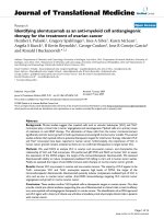
three cosmids in which UL114 was tagged at the amino
(UL114NTAG) and the carboxyl (UL114CTAG) termini,
as well as the precise replacement of UL114 with the 35 aa
ORF containing the epitope tag (UL114KO) (Fig. 1).
Resulting cosmids were used in a standard cotransfection
to generate three tagged recombinant viruses by methods
described previously [15].
Localization to replication compartments and association
with ppUL44
Previous work used immunofluorescence microscopy to
examine the nature and distribution of CMV replication
components at various times in the virus life cycle [29].
This work suggested that various members of the viral rep-
lication complex, including ppUL44, the DNA polymer-
ase processivity factor, localize into specific replication
compartments in patterns that are characteristic of a given
point in the replication cycle. In light of the putative role
of the UL114 gene product in viral DNA replication, sim-
ilar studies were undertaken, using the epitope-tagged
viruses described above to determine the location of
pUL114 in infected fibroblasts. HEL cells were infected
with the recombinant viruses and were examined by fluo-
rescence microscopy using anti-ICP4 and anti-UL44 mon-
oclonal antibodies. At 48 hpi, ppUL44 localized to the
nucleus in small foci in a pattern that was very similar to
that for pUL114 (Fig 5A–C). By 72 hpi, epitope tagged
pUL114 expressed from the CTAG virus partitioned to the
replication compartments within the nucleus as defined
by ppUL44 staining (Fig. 5D–F) and light punctate
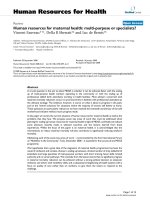
Recombinant virusesFigure 1
Recombinant viruses. The top line represents the CMV genome with the region surrounding UL114 expanded below. The
second line represents the structure of the region in the parent virus (AD169). The third line labeled "RC2620" depicts the 1.2
kb insertion containing the E. coli gpt gene (white arrow) that replaces most of the UL114 ORF. The final three lines represent
the same region in Towne and depict the placement of the 35 aa ICP4 epitope tags in the ORF. The entire ORF was also
deleted in Towne as a control and resulted in the same slow replication phenotype as was observed in the AD169 strain.
Virology Journal 2005, 2:55 />Page 5 of 14
(page number not for citation purposes)
cytoplasmic staining was also observed in some cells. The
recombinant UL114 NTAG virus did not exhibit the
strong nuclear localization observed with UL114 CTAG
and it is possible that fusing the ICP4 epitope to this part
of the molecule may have interfered with its normal local-
ization (data not shown).
The localization pattern exhibited by the tagged versions
of pUL114 suggested that it might be physically interact-
ing with the viral DNA replication machinery. We hypoth-
esized that pUL114 might interact with ppUL44
analogous to the UNG2 interaction with PCNA that
occurs the human DNA replication complex [28]. Extracts
of cells infected with the epitope tagged viruses and a wt
virus were immunoprecipitated with a monoclonal anti-
body to ppUL44. Precipitated proteins were separated on
denaturing polyacrylamide gels, transferred to nitrocellu-
lose and a monoclonal antibody specific for the ICP4
epitope was used to detect the tagged pUL114 molecules.
A protein with a predicted molecular weight of 32 kDa
was specifically detected from the recombinant virus in
which pUL114 was tagged at the carboxyl terminus (Fig.
6A). A very light band with the same migration rate was
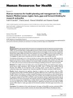
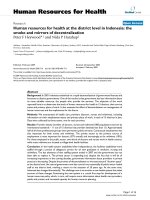
Repair of RC2620Figure 2
Repair of RC2620. (A) HEL cells were infected at an MOI
of 5 PFU/cell and total DNA was harvested at the indicated
times. The quantity of viral DNA for AD169 (black squares),
RC2620 (black circles), and RQ2620 (open circles) were
determined by dot blot hybridization as described in materi-
als and methods. (B) Titers of AD169 (black squares),
RC2620 (black circles), and RQ2620 (open circles) are
shown. The time point at 0 hpi represents the titer of the
input virus.
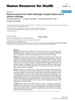
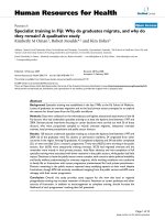
Kinetics of DNA synthesis and viral replication in comple-menting cellsFigure 3
Kinetics of DNA synthesis and viral replication in
complementing cells. Virus stocks of the parent virus and
the mutant virus were produced in the complementing cell
line (HL114) and used to infect either HEL cells or IHL114
cells at an MOI of 5 PFU/cell. Circular and square symbols
represent quantities of DNA from RC2620 and the repaired
virus respectively while solid and open symbols represent
DNA isolated from HEL cells and HL114 cells respectively.
The average of triplicate values are shown.
Virology Journal 2005, 2:55 />Page 6 of 14
(page number not for citation purposes)
detected from UL114 NTAG-infected cells upon long
exposure, consistent with its reduced localization to the
nucleus. No specific species were detected in extracts pre-
pared from the wt virus. The reverse experiment was per-
formed with pUL114-EGFP fusion proteins that were
precipitated with a monoclonal antibody specific for GFP
and the monoclonal antibody to ppUL44 was used to
detect the coprecipitated protein. This experiment con-
firmed the earlier result and demonstrated that it was also
possible to specifically coprecipitate ppUL44 with
pUL114 fusion proteins (Fig. 6B). Consistent with the
previous result, the coprecipitation appeared to be less
efficient for pUL114 labeled at the amino terminus.
To confirm these results, plasmids expressing ppUL44
(pMP62) and pUL114 with a carboxyl terminal EGFP tag
were transfected into monolayers of primary foreskin
fibroblast cells. In cells transfected with pMP62 alone,
ppUL44 localized exclusively to the nucleus and is shown
merged with DAPI image (Fig 7A), which was similar to
the localization observed in infected cells early in infec-
tion. Cells expressing either the full length pUL114-EGFP
fusion protein (pMP39), or the fusion protein in which aa
3–24 were deleted from pUL114 (pMP41) exhibited
punctate cytoplasmic fluorescence (Fig 7B, C). This local-
ization pattern was distinct from the nuclear staining
observed with the UL114 CTAG recombinant virus. How-
ever, when ppUL44 and full length pUL114 fusion pro-
teins were coexpressed in the same cell, pUL114 was
recruited to the nucleus with ppUL44 (Fig 7D–F), consist-
ent with its nuclear localization in the context of infected
cells. A small quantity of ppUL44 also appeared to local-
ize to a subset of the cytoplasmic punctae containing
pUL114. Deletion of aa 3–24 from the pUL114 fusion
protein eliminated its recruitment to the nucleus by
ppUL44, suggesting that this domain is required for the
interaction ppUL44 (Fig 7G–I). This interpretation of the
data is consistent with the impaired nuclear localization
observed with UL114 NTAG-infected cells, in which the
amino terminal domain of pUL114 was altered through
the addition of the ICP4 epitope tag (data not shown).
Also consistent with this result, is the inefficient coprecip-
itation of ppUL44 with pUL114 fusion proteins when the
tags were fused to the amino terminus (Fig. 6). These data
suggest that these proteins associate in a manner that is
dependent on aa 3–24 of pUL114, and independent of
other viral proteins or viral DNA. These experiments do
not, however, eliminate the possibility that they might
associate in an indirect manner through cellular proteins.
Characterizing the defect in DNA synthesis
The localization of pUL114 to replication compartments,
and its apparent association with ppUL44, which is
known to interact with the DNA polymerase [9] imply
that this molecule is part of the viral DNA replication
complex. This interpretation of the data is consistent with
the observed phenotype of delayed DNA synthesis in the
UL114 deletion virus [6,31], and is also consistent with
results reported for the human UNG2 that has been
shown to localize to replication complexes [28]. If this
assumption is correct and the viral UNG is an important
part of the replication complex, then the defect in viral
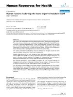
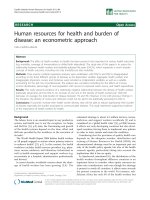
Rapid epitope tagging strategy in yeastFigure 4
Rapid epitope tagging strategy in yeast. The top line
represents the target ORF in the context of a large yeast
plasmid or YAC. Line 2 shows a PCR product containing the
epitope tagging cassette with 40 bp targeting sequences
homologous to the regions designated by the dashed lines.
Line 3 shows the site-specific integration of the cassette
resulting from homologous recombination in yeast. The final
line represents UL114 in the YAC with an in frame 35 aa
amino terminal insertion containing the ICP4 epitope and a
single loxP site. This strategy can be used to place the epitope
tag anywhere in the ORFs on the YAC by changing the tar-
geting sequences on the PCR primers.
Virology Journal 2005, 2:55 />Page 7 of 14
(page number not for citation purposes)
DNA synthesis should be apparent throughout the viral
DNA replication process. To characterize the affect of
pUL114 on DNA synthesis, triplicate monolayers of repli-
cating primary foreskin fibroblasts were infected with
either Towne, or an isogenic recombinant virus without
UL114 and the accumulation of viral DNA was quantified
with a TaqMan-based assay. Input copy number following
infection was determined at 2 hpi and yielded average val-
ues of 4.2 × 10
4
and 2.3 × 10
4
, for the wt and mutant
viruses respectively with standard deviations of <15% for
both values. During the course of infection, genome copy
number was determined in total DNA and the data were
normalized relative to the input copy number (Fig. 8).
During the first 18 h of infection, copy number of the wt
and deletion virus genomes decreased at the same rate
with a half-life of approximately 8 h (Fig 8B). This is con-
sistent with data presented earlier, which suggested that
increased uracil levels did not substantially affect genomic
integrity and were unlikely to be responsible for the
observed defects in DNA synthesis. This analysis also
revealed two features of the defect in DNA synthesis. First,
the accumulation rate of viral DNA synthesis was signifi-
cantly reduced in the recombinant virus with a deletion in
UL114 (Fig. 8A). A 7-fold increase in copy number was
attained in the parent virus at 25 hpi, but this same level
was not achieved in the mutant until 48 hpi. By this time,
the wt virus had attained a 300-fold amplification of the
input genome, which was not attained by the mutant even
after an additional 48 h of incubation. Exponential
growth rates were calculated from curves fitted to the
experimental data for both viruses. The wt rate (r) was
determined to be approximately 0.2 h
-1
, whereas the copy
number of the mutant expanded at a rate of about 0.1 h
-1
.
This decreased rate of DNA accumulation is consistent
Localization of pUL114 in infected HEL cellsFigure 5
Localization of pUL114 in infected HEL cells. Cells were infected with a recombinant virus with an epitope tag in the
carboxyl terminus of UL114. Monolayers were fixed and stained with an anti-ppUL44 monoclonal antibody (FITC) and an anti-
ICP4 mouse monoclonal antibody (Texas Red). Cells were fixed at 48 hpi and images of FITC, Texas Red, and a merged image
with DAPI are shown(A-C). Cells were fixed at 72 hpi and images of FITC, Texas Red, and a merged are shown (D-F). All
images were captured digitally and prepared in Adobe Photoshop.
Virology Journal 2005, 2:55 />Page 8 of 14
(page number not for citation purposes)
with the observed decrease in viral DNA described previ-
ously [31] and also with the data showing a defect in the
transition to late phase DNA synthesis reported recently
by Courcelle et al. [6]. A second defect in DNA synthesis
was also observed. The initial doubling of the wt genome
was detected at 21 hpi and the copy number increased
Coprecipitation of pUL114 and ppUL44Figure 6
Coprecipitation of pUL114 and ppUL44. (A) Primary
foreskin fibroblast cells were infected either with ICP4-
tagged recombinant viruses or Towne at an MOI of approxi-
mately 1 PFU/cell. Cells were lysed at 48 hpi, and extracts
were immunoprecipitated with a monoclonal antibody to
ppUL44 and separated on an SDS-PAGE gel. Proteins were
transferred to a membrane and a monoclonal antibody to the
ICP4 epitope was used to detect coprecipitated poteins in
the immunoblot. (B) EGFP.373 and C1-114.373 cells were
infected with AD169 at an MOI of 2 PFU/cell and harvested
at 24 hpi. Fusion proteins were precipitated with a mono-
clonal antibody to EGFP, separated on non-denaturing SDS
PAGE gels, transferred to nitrocellulose, and immmunoblot-
ting was performed with monoclonal antibody to ppUL44.
Arrows designate the specific bands.
Recruitment of pUL114 to the nucleus by ppUL44Figure 7
Recruitment of pUL114 to the nucleus by ppUL44.
Plasmids expressing ppUL44 or pUL114-EGFP fusion pro-
teins were transfected into primary fibroblast cells and visual-
ized by immunofluorescent staining. In the first row of
images, ppUL44 stained with Texas Red exhibited strong
nuclear localization as evidenced by the colocalization with
DAPI in the merged image (violet). The pUL114-EGFP fusion
protein and a similar protein containing a 25 aa amino termi-
nal deletion (green) both localized to the cytoplasm and are
shown merged with DAPI staining (blue). In the second row
of images, the coexpression of ppUL44 (Texas red), pUL114-
EGFP (green) and a merged image show that ppUL44 can
recruit pUL114 to the nucleus. In third row of images
ppUL44 (Texas red) and pUL114-EGFP containing a 25 aa
amino terminal deletion (green) did not colocalize to the
nucleus when co-expressed.
Virology Journal 2005, 2:55 />Page 9 of 14
(page number not for citation purposes)
exponentially to a 7-fold increase by 25 hpi (Fig. 8B). Dur-
ing this period of time, no increase in the copy number of
mutant virus genomes was observed. Thus, the initial
phase of DNA synthesis also appears to be compromised
in the absence of pUL114, despite the fact that early genes
are expressed at normal levels at this point in time
[29,31]. If viral DNA synthesis in the mutant had initiated
at the same time as the parent virus, the increased copy
number should have been easily detectable by 25 hpi,
even at the reduced rate of accumulation we report here.
Thus, either the initiation or the early theta-type DNA rep-
lication postulated for this family of viruses appears to be
compromised in absence of pUL114. These data suggest
that pUL114 acts during both the onset and the subse-
quent expansion phase of viral DNA synthesis and sug-
gests that this gene product functions as part of the viral
DNA replication machinery.
Discussion
Perhaps the simplest explanation of the observed pheno-
type associated with UL114 deletion viruses is that the
recombinant virus fails to remove uracil residues from its
genome and that these lesions decrease genome stability
and impede DNA synthesis. Two lines of evidence argue
against this interpretation of the data. First, input
genomes of the recombinant virus in infected cells
appeared to be as stable as the wt genomes in infected cells
and had similar initial half lives (Fig 8B). Second, the
complementing cell line reduced the uracil content of the
mutant genomes to levels indistinguishable from the
parent virus, yet the observed phenotype of these comple-
mented virus stocks in non-complementing cells was
unaffected. Thus, it appears that the viral UNG plays a
more direct role in the synthesis of viral DNA. However,
these data do not exclude the possibility that the removal
of uracil may be important late in infection. We suggest
that the HCMV UNG2 homolog functions as part of the
DNA replication machinery and that it significantly accel-
erates the synthesis of genomic DNA.
The parallels between this system and the recent results
for human UNG2 are striking. PCNA and ppUL44 are
thought to perform a similar function and associate with
human DNA polymerase δ and the HCMV DNA polymer-
ase, respectively. Despite the fact that these processivity
factors do not share significant aa sequence homology
and exhibit different 3-D structures [1], they retain inter-
actions with their respective DNA polymerases [21], as
well as an association with their respective UNG
homologs. The fact that the amino terminal domains of
both pUL114 and UNG2 are required to mediate these
interactions suggests that this might be a common feature
among all UNG2 homologs. This relationship is also con-
served in vaccinia virus where the viral UNG2 homolog
(D4R) was shown to physically associate with the A20R
Defects in DNA synthesis associated with pUL114Figure 8
Defects in DNA synthesis associated with pUL114.
Triplicate wells of HEL cells were infected at an MOI of 0.01
with Towne (black circles) or the isogenic deletion virus,
UL114 KO tag, (shaded squares). Total DNA was harvested
at the indicated times, and the genome copy number was
determined with a TaqMan assay using a standard curve of
virion DNA. Copy number was normalized to the quantity of
input genomes determined at 2 hpi with error bars repre-
senting the standard deviation of the triplicate samples. (A)
The log of the accumulated viral DNA copy number is shown
versus time post infection. The wt exponential rate of accu-
mulation (r) was determined to be approximately 0.2 h
-1
,
whereas the copy number of the mutant expanded only at a
rate of about 0.1 h
-1
. (B) Data for the first 24 h replotted on
a linear scale show the delayed onset of DNA synthesis dur-
ing the first duplication of the viral genome.
Virology Journal 2005, 2:55 />Page 10 of 14
(page number not for citation purposes)
DNA polymerase processivity factor [14]. In this system,
the viral UNG was shown to be essential for viral DNA
synthesis, and this requirement was unrelated to the abil-
ity of the molecule to excise uracil [7]. A potential role for
UNG in DNA replication was also noted in Epstein Barr
Virus where the UNG2 homolog (BKRF3) increased the
efficiency of replication of a transfected plasmid contain-
ing the origin of replication [10] and was absolutely
required when the core essential genes were supplied on a
set of cosmid clones [11]. Less analogous but equally
compelling, is the recruitment of UNG2 to the preintegra-
tion complex in HIV and its specific interaction with both
the integrase as well as the reverse transcriptase [33]. The
conserved relationship between UNG2 homologs and
DNA replication complexes in these diverse systems sug-
gests that it performs a conserved function in mammals. It
is unclear if this function is related to UNG enzymatic
activity, and it is likely that these molecules perform an
additional function replication that remains uncharacter-
ized. This view is supported by the fact that the UNG enzy-
matic activity can be eliminated without severely affecting
the replication of vaccinia virus, whereas larger mutations
are lethal [7]. A specialized role for UNG2 has also been
proposed in mammalian systems since UNG
-
/UNG
-
mice
are viable and do not exhibit the phenotype of highly ele-
vated mutation frequency that would be predicted by ear-
lier studies in prokaryotes and Sacharomyces. Information
garnered in future studies with HCMV will be particularly
helpful in shaping our understanding of the function of
UNG2 in the DNA replication foci of mammalian cells.
The unique phenotype associated with pUL114 in HCMV
infection and the fact that this simple system closely
resembles that in humans make it an attractive system to
probe the unique function of mammalian UNG2
homologs in DNA synthesis.
In HSV, the deletion of the UNG homolog (UL2) affects
the ability of the virus to replicate in mice, particularly the
CNS. The deletion of UL2 resulted in a 100,000-fold
reduction in the neuroinvasiveness and may represent a
potential attenuating mutation in candidate vaccines [34]
Previous studies with UNG deletion mutants in HSV were
not shown to affect replication in tissue culture, they rep-
licated to lower titers in vivo and were orders of magnitude
less neuroinvasive than control viruses [34]. To investigate
the possibility that the phenotype might be more pro-
nounced in vivo, we infected human fetal retinal tissue
implanted in a SCID-hu mouse [2,16]. In this model, a
deficiency in pUL114 resulted in a decreased infection
rate (P = 0.015) as well as significantly reduced titers in
infected animals (P = 0.0063). However, the observed
defects in vivo were not more pronounced that the repli-
cation defects in cell culture and were not similar to
results observed with HSV.
Conclusion
The work presented here suggests that pUL114 is part of
the DNA replication machinery and that it significantly
accelerates the synthesis of genomic DNA. This interpreta-
tion of the data is consistent with the early expression
kinetics and the nuclear localization exhibited by this
molecule in infected cells, which are both predicted char-
acteristics of an enzyme presumed to act in DNA repair.
Equally consistent is the observed intranuclear localiza-
tion to viral replication compartments at a time when
viral DNA synthesis is known to occur [29]. The fact that
pUL114 appears to associate with ppUL44 is intriguing,
because of the central role that ppUL44 plays in the
synthesis of viral DNA [9,20,21]. These data taken
together with the observed defects in the onset and expan-
sion of viral DNA synthesis suggest that it functions as
part of the DNA replication machinery.
We propose a model in which pUL114 functions as part
of the viral DNA polymerase complex and is required for
the efficient establishment and expansion of viral DNA
synthesis. Results presented here suggest that the perform-
ance of the DNA replication machinery is significantly
impaired without pUL114. The precise mechanism that
this molecule uses to affect DNA synthesis is unclear but
it may or may not be related to its ability to excise uracil
from DNA. The interaction with ppUL44 suggest that this
molecule might be close to the replication forks where it
might help destabilize double stranded DNA through a
scanning and pinching base flipping mechanism similar
to that described for the human homolog [12]. Additional
experiments in this system will be required to determine
the correlation between uracil excision activity and the
efficiency of viral DNA replication.
The evolving view of UNG function in the life cycle of
viruses increases its appeal as a target for antiviral chemo-
therapy, particularly in poxviruses where it is essential for
virus replication. This approach may also be valuable in
herpesviruses given its proximity to the replication com-
plex as well as its important role in vivo. It is certainly pos-
sible to obtain specific inhibitors of viral UNG molecules
based on their ability to block the enzyme's ability to
excise uracil, however at present, it is unclear that this
enzymatic activity is responsible for the interesting affects
observed both in vitro and in vivo. Rational drug strategies
should be possible, but their development is dependent
upon a better understanding of the biological functions of
this molecule in virus replication.
Methods
Plasmids
Construction of pON2619 and pON2620 were described
previously [31]. To construct a retroviral vector, a 1782 bp
EcoRI fragment (coordinates 163071 to 164853 AD169
Virology Journal 2005, 2:55 />Page 11 of 14
(page number not for citation purposes)
genome) containing the UNG open reading frame was
inserted into the MfeI site in pLXIN (Clontech, Palo Alto,
CA) to yield pON2159. EGFP fusion constructs were con-
structed by amplifying an 800 bp DNA fragment contain-
ing the UL114 open reading frame using the forward
primer 5'-GGA CTC AGA TCT ATG GCC CTC AAG CAG
TGG ATG-3' and the reverse primer 5'-GTC GAC TGC
AGA GAA TCT CCC ACA GAG TCG CCA GTC C-3'. The
resulting fragment was purified from an agarose gel and
cloned into the Bgl II and Pst I sites of pEGFPC1 and
pEGFPN3 (Clontech, Palo Alto, CA) to generate plasmids
pEGFPC1/UL114 and pEGFPN3/UL114. Plasmids
pMP39 and pMP41 were constructed by amplifying with
forward primers 5'-ATG GCC CTC AAG CAG TGG-3' and
5'-ATG GCC GCT CGC GTG TTT TGT CTG AGC-3' respec-
tively, with reverse primer 5'-TCA TCT GAG TCC GGA
CTT GTA CA-3' using pEGFPN3/UL114 as a template and
cloning into pcDNA3.1. The resulting plasmids were
sequenced and express proteins of the predicted molecu-
lar weight. The UL114 open reading frame in pMP41 con-
tains a deletion of aa 3 to 24. Primers 5'-CAC CAT GGA
TCG CAA GAC GCG C-3' and 5'-CTA GCC GCA CTT TTG
CTT CT-3' were used to amplify UL44 and the PCR prod-
uct was cloned into a eukaryotic expression vector to yield
pMP62. The plasmid pUG6 contains a recyclable genetic
marker for site directed mutagenesis in yeast [13]. This
cassette was amplified with the forward primer 5'-CAG
GTC GAC AAC CCT TAA TAT AAC TTC GTA TAA TGT ATG
CTA TAC GAA GTT ATT AGG TCT AGA GAT CTG TTT
AGC TTG C-3' and the reverse primer 5'-TCC TGG AGC
TCG ATC TCC TGC TGC ATC TGC TGC ATC ATC ATA
TTC ATC ACC TAA TAA CTT CGT ATA GCA TAC ATT ATA
CGA AGT TAT ATT AAGGGT TCT CG-3'. The resulting
product was TOPO-cloned into pcDNA3.1 (Invitrogen,
Carlsbad, CA) to yield pKan-ICP4 and used as a template
for subsequent amplifications. The Sma I – Sca I fragment
of pRS413, which contains ARS4, CEN6 and the HIS3
selectable marker, was ligated into the XmnI site of
pACYC184. A single EcoRI site in the resulting intermedi-
ate construct was converted to a unique PacI site by liga-
tion to EcoRI PacI adapters to produce pACYC ars cen. PacI
fragments from cosmids described previously [15] were
cloned in the PacI site for subsequent experiments.
Mutagenesis in Yeast
PCR products for site directed mutagenesis were generated
using the forward primer 5'-AGG TCG ACA ACC CTT AAT
ATA ACT-3' and reverse primer 5'-TCC TGG AGC TCG
ATC TCC TGC TGC AT-3' and were targeted by adding 40
bp of homologous sequence to the 5' end of each primer.
PCR products were cotransformed by a standard lithium
acetate protocol with target viral cosmids in Saccharomyces
cerevisiae strain CGY2570 carrying plasmid pSH47 [13]
which expresses CRE recombinase under control of the
GAL promoter. Recombinants were selected on yeast com-
plete medium plates containing 400 µg/ml G418 and the
selectable marker was excised through the galactose-
dependent expression CRE recombinase to yield 35 aa in
frame insertions containing the HSV ICP4 epitope tag.
Cells and virus
Primary human foreskin fibroblast (HFF) cells and
human embryonic lung (HEL) cells were grown in mon-
olayer cultures in Dulbecco's modified Eagle medium
(Gibco BRL, Gaithersberg, MD) supplemented with 100
units/ml penicillin G, 100 µg/ml streptomycin sulfate and
10% fetal bovine serum (FBS). Parental virus (AD169)
was obtained from the ATCC and virus stocks were
obtained and titered as described previously [40]. Cell
lines expressing EGFP fusion proteins were constructed by
transfecting 10 µg of linearized pEGFPC1/UL114,
pEGFPN3/UL114 or pN3EGFP inU373 cells with Lipofec-
tin (Gibco BRL, Gaithersberg, MD) according to the man-
ufacturers recommendations. Stably transfected C1-
114.373, N3-114.373 and EGFP.373 cells were selected
with 1 mg/ml G418, and resulting colonies were isolated
and frozen at passage 5. The construction and propaga-
tion of RC2620 as well as the production of high MOI
growth curves were described previously [31]. The con-
struction of RQ2620 was performed as described previ-
ously [31] and the resulting repaired virus was plaque
purified 3 times after it was shown to be free of the con-
taminating parent virus by Southern analysis. Epitope
tagged viruses were constructed by cotransfecting a set of
8 cosmids derived from the Towne strain of HCMV [15],
including one cosmid that was subjected to site directed
mutagenesis in yeast as described above. The epitope
tagged viruses replicated to high titers in HFF cells and do
not appear to be replication impaired.
Construction of HL114 cells
pON2159 was tranfected into PA317 cells to produce
defective retrovirus stocks that were subsequently used to
transduce the UL114 gene into low passage primary HEL
by methods described previously [32]. Transduced cells
were selected with 400 µg/ml G418 starting at 24 hpi. Sur-
viving cells were passaged in G418 and were used as a
mixed population.
DNA synthesis kinetics
Confluent monolayers of HFF cells in 6-well cluster dishes
were infected at an MOI of 5 PFU/cell with either the
AD169, RC2620, or RQ2620. Total DNA was extracted as
described previously [31], diluted and transferred to a
Hybond N+ membrane in a dot-blot manifold and
probed with a plasmid containing viral sequences
(89797-94860 in the AD169 genome). The resulting film
was captured digitally and quantified with Scan Analysis
(Biosoft, Cambridge, UK).
Virology Journal 2005, 2:55 />Page 12 of 14
(page number not for citation purposes)
Determination of genome copy number
Towne and an isogenic recombinant virus containing a
deletion in UL114 were used to infect dividing HFF cells
at an MOI of 0.02 PFU/cell in 12-well plates. Monolayers
were rinsed three times at 2 hpi and supplemented with
fresh media. At harvest, monolayers were rinsed twice
with fresh media, and frozen at -80°C in a final volume of
0.2 ml. Total DNA was extracted using a QIAamp DNA
blood minikit according to the manufacturers recommen-
dations (Qiagen, Valencia, CA). Copy number was deter-
mined using the ABI PRISM 7700 sequence detection
system and TaqMan Universal PCR Master Mix. Forward
primer (50 nM), 5'-CCG AGG TGG GTT ACT ACA ACG-
3', reverse primer (300 nM), 5'-GGA AGG GTA GAG GCT
GGC A-3', and fluorogenic probe (75 nM), 5' FAM-CCC
CGT GGC CGT GTT CGA CT-3' TAMRA were used in a 50
µl reaction volume with conditions as follows: 2 min at 50
?C, 10 min at 95 ?C, and 40 cycles of (15 sec at 95 ?C, 1
min at 60 ?C,). The DNA templates from triplicate wells
were analyzed in a volume of 5 µl per reaction. Copy
number was compared to a standard curve generated from
HCMV genomic DNA.
Immunofluorescence microscopy
Immunofluorescence staining was performed as previ-
ously described [37]. 8-well chamber slides of confluent
HFF cells (LF1043) were infected with recombinant
HCMV strains UL114 NTAG, UL114 CTAG, and Towne
(control) at an MOI of 0.5 PFU/cell. Slides were washed
once with PBS, fixed with 1% formalin for 15 min, and
washed again three times with PBS + 0.2% BSA. Perme-
ablization was performed in PBS with 0.2% Triton X-100
for 15 min, followed by one wash step with PBS + 0.2%
BSA and a blocking step in 5% normal horse serum (Vec-
tor Labs, Burlingame, CA) also for 15 min. Monolayers of
HFF cells on coverslips were transfected with Lipo-
fectamine 2000 according to the manufacturer's protocol
(Invitrogen). Transfected cells were fixed and permeabi-
lized by the same methods described above. Monoclonal
antibodies to ICP4 (Rumbaugh-Goodwin Institute, Plan-
tation, FL), or ppUL44 (gift from Bill Britt, University of
Alabama at Birmingham) were incubated with cells for 1
h at 37°C. Monolayers were washed and incubated with a
goat anti-mouse secondary antibody conjugated to FITC
(Southern Biotechnology Associates, Birmingham, AL).
For dual labeling experiments, monoclonal antibodies
were directly labeled with a Zenon Texas Red labeling kit
as per manufacturer's directions (Molecular Probes,
Eugene, OR). After three washes, Vectashield mounting
medium containing DAPI (Vector Labs, Burlingame, CA)
was added to each slide along with a glass coverslip. Cells
were examined with a Nikon TE2000 Microscope using a
40 × objective. Fluorescence images of stained cells were
captured with Hamamatsu ORCFA-100 digital camera
and recorded using Simple PCI software. All photographs
were prepared in Adobe Photoshop CS.
Immunoprecipitations
T-25 flasks or 6-well dishes of confluent HFF cells, C1-
114.373, N3-114.373, or EGFP.373 cells were infected
with AD169, Towne, UL114 CTAG, or UL114 NTAG at an
MOI of 5 PFU/cell. At 24 and 48 hpi, the monolayers were
washed with PBS and lysed on ice in 500 µl of lysis buffer
containing 50 mM HEPES, pH 7.5, 150 mM NaCl, 2.5
mM EGTA, 1 mM EDTA, 1% Triton X-100, 1 mM PMSF,
and 1 proteinase inhibitor tablet (Boehringer Man-
nheim). Cell lysates were preadsorbed for 1 h at 4°C with
30 µl of a 50% suspension of protein A-Sepharose in lysis
buffer. Proteins were precipitated with 30 µl of the protein
A-Sepharose suspension, and 1 µl of a monoclonal anti-
body to the EGFP domain (Clontech, Palo Alto, CA), the
ICP4 epitope tag, or ppUL44 (Rumbaugh-Goodwin Insti-
tute, Plantation, FL). The tubes were rocked overnight at
4°C. The protein A-Sepharose beads were washed twice in
lysis buffer and once in lysis buffer with the addition of
0.1% SDS, and 1% sodium deoxycholate (RIPA). Samples
were boiled 5 min in 80 mM Tris, pH 6.8, 2% SDS, 10%
sucrose, and 0.004% bromophenol blue and the proteins
were separated by SDS polyacrylamide electrophoresis
(48) and transfered to an Immobilon-P membrane (Mill-
ipore) at 200 mA for 1 h. Blots were blocked at room tem-
perature for 30 min with 5% (w/v) skim milk in 50 mM
Tris, pH7.5, 0.2 M NaCl, 0.01% Tween 20, and incubated
at room temperature for 1 h with a monoclonal antibody
specific for ppUL44, ICP4, or EGFP diluted 1:1000 in
washing buffer (1% (w/v) skim milk, 50 mM Tris, pH7.5,
0.2 M NaCl, 0.01% Tween 20). Blots were washed three
times with washing buffer, and incubated at room tem-
perature for 1 h with anti-Mouse IgG(H+L) HRP conju-
gate (New England Biolabs) diluted 1:2500 in washing
buffer. The unbound secondary antibody was removed in
three washes with 50 mM Tris, pH7.5, 0.2 M NaCl. The
membrane was subsequently developed with LumiGLO
or ECL according to the manufacturer's recommendations
and used to expose Kodak biomax film.
List of Abbreviations
HCMV human cytomegalovirus
HIV human immunodeficiency virus
dUTP deoxyuridine triphosphate
RT reverse transcriptase
HEL human embryonic lung
UNG uracil DNA glycosylase
Virology Journal 2005, 2:55 />Page 13 of 14
(page number not for citation purposes)
PCNA proliferating cell nuclear antigen
EGFP green fluorescent protein
SCID severe combined immunodeficiency
FITC fluoroscein isothiocyanate
MEM minimal essential medium
TR texas red
DNA deoxyribonucleic acid
Competing interests
Some of the authors of this publication are supported
financially by salary and shares of MedImmune Inc. The
authors declare that they have no other competing
interest.
Authors' contributions
MNP generated the recombinant viruses and cell lines
described here, performed the complementation experi-
ments and made the yeast mutagenesis constructs. HL per-
formed the immunoprecipitations. GMD and MD worked
on the yeast mutagenesis system and assisted in the pro-
duction of recombinant viruses. CM worked on the
immunofluorescence studies. ZW performed the TaqMan
analyses. GK and ERK provided critical intellectual input.
Acknowledgements
We thank Giesela Mosig, and Charmain Tan Courcelle for helpful discus-
sions and William J. Britt for his gift of monoclonal antibodies and critical
reading of the manuscript. This work was supported in part by the contract
NO1-AI-30049 from the National Institute for Allergy and Infectious
Diseases.
References
1. Brynolf K, Eliasson R, Reichard P: Formation of Okazaki frag-
ments in polyoma DNA synthesis caused by misincorpora-
tion of uracil. Cell 1978, 13(3):573-580.
2. Tye BK, Lehman IR: Excision repair of uracil incorporated in
DNA as a result of a defect in dUTPase. J Mol Biol 1977,
117(2):293-306.
3. Chen R, Wang H, Mansky LM: Roles of uracil-DNA glycosylase
and dUTPase in virus replication. J Gen Virol 2002, 83(Pt
10):2339-2345.
4. Percival KJ, Klein MB, Burgers PM: Molecular cloning and primary
structure of the uracil-DNA-glycosylase gene from Saccha-
romyces cerevisiae. J Biol Chem 1989, 264(5):2593-2598.
5. Varshney U, Hutcheon T, van de Sande JH: Sequence analysis,
expression, and conservation of Escherichia coli uracil DNA
glycosylase and its gene (ung). J Biol Chem 1988,
263(16):7776-7784.
6. Yang H, Chiang JH, Fitz-Gibbon S, Lebel M, Sartori AA, Jiricny J, Slup-
ska MM, Miller JH: Direct interaction between uracil-DNA gly-
cosylase and a proliferating cell nuclear antigen homolog in
the crenarchaeon Pyrobaculum aerophilum. J Biol Chem 2002,
277(25):22271-22278.
7. Krokan HE, Otterlei M, Nilsen H, Kavli B, Skorpen F, Andersen S,
Skjelbred C, Akbari M, Aas PA, Slupphaug G: Properties and func-
tions of human uracil-DNA glycosylase from the UNG gene.
Prog Nucleic Acid Res Mol Biol 2001, 68:365-386.
8. Muller SJ, Caradonna S: Isolation and characterization of a
human cDNA encoding uracil-DNA glycosylase. Biochim Bio-
phys Acta 1991, 1088(2):197-207.
9. Nilsen H, Haushalter KA, Robins P, Barnes DE, Verdine GL, Lindahl
T: Excision of deaminated cytosine from the vertebrate
genome: role of the SMUG1 uracil-DNA glycosylase. Embo J
2001, 20(15):4278-4286.
10. Nilsen H, Otterlei M, Haug T, Solum K, Nagelhus TA, Skorpen F,
Krokan HE: Nuclear and mitochondrial uracil-DNA glycosy-
lases are generated by alternative splicing and transcription
from different positions in the UNG gene. Nucleic Acids Res
1997, 25(4):750-755.
11. Nagelhus TA, Haug T, Singh KK, Keshav KF, Skorpen F, Otterlei M,
Bharati S, Lindmo T, Benichou S, Benarous R, Krokan HE: A
sequence in the N-terminal region of human uracil-DNA gly-
cosylase with homology to XPA interacts with the C-termi-
nal part of the 34-kDa subunit of replication protein A. J Biol
Chem 1997, 272(10):6561-6566.
12. Otterlei M, Warbrick E, Nagelhus TA, Haug T, Slupphaug G, Akbari
M, Aas PA, Steinsbekk K, Bakke O, Krokan HE: Post-replicative
base excision repair in replication foci. Embo J 1999,
18(13):3834-3844.
13. Simbulan-Rosenthal CM, Rosenthal DS, Hilz H, Hickey R, Malkas L,
Applegren N, Wu Y, Bers G, Smulson ME: The expression of
poly(ADP-ribose) polymerase during differentiation-linked
DNA replication reveals that it is a component of the multi-
protein DNA replication complex. Biochemistry 1996,
35(36):11622-11633.
14. Bouhamdan M, Benichou S, Rey F, Navarro JM, Agostini I, Spire B,
Camonis J, Slupphaug G, Vigne R, Benarous R, Sire J: Human immu-
nodeficiency virus type 1 Vpr protein binds to the uracil
DNA glycosylase DNA repair enzyme. J Virol 1996,
70(2):697-704.
15. Selig L, Benichou S, Rogel ME, Wu LI, Vodicka MA, Sire J, Benarous R,
Emerman M: Uracil DNA glycosylase specifically interacts with
Vpr of both human immunodeficiency virus type 1 and sim-
ian immunodeficiency virus of sooty mangabeys, but binding
does not correlate with cell cycle arrest. J Virol 1997,
71(6):4842-4846.
16. Willetts KE, Rey F, Agostini I, Navarro JM, Baudat Y, Vigne R, Sire J:
DNA repair enzyme uracil DNA glycosylase is specifically
incorporated into human immunodeficiency virus type 1
viral particles through a Vpr-independent mechanism. J Virol
1999, 73(2):1682-1688.
17. Priet S, Navarro JM, Gros N, Querat G, Sire J: Functional role of
HIV-1 virion-associated uracil DNA glycosylase 2 in the cor-
rection of G:U mispairs to G:C pairs. J Biol Chem 2003,
278(7):4566-4571.
18. Klarmann GJ, Chen X, North TW, Preston BD: Incorporation of
uracil into minus strand DNA affects the specificity of plus
strand synthesis initiation during lentiviral reverse
transcription. J Biol Chem 2003, 278(10):7902-7909.
19. Millns AK, Carpenter MS, DeLange AM: The vaccinia virus-
encoded uracil DNA glycosylase has an essential role in viral
DNA replication. Virology 1994, 198(2):504-513.
20. Stuart DT, Upton C, Higman MA, Niles EG, McFadden G: A poxvi-
rus-encoded uracil DNA glycosylase is essential for virus
viability. J Virol 1993, 67(5):2503-2512.
21. Upton C, Stuart DT, McFadden G: Identification of a poxvirus
gene encoding a uracil DNA glycosylase. Proc Natl Acad Sci U S
A 1993, 90(10):4518-4522.
22. Ellison KS, Peng W, McFadden G: Mutations in active-site resi-
dues of the uracil-DNA glycosylase encoded by vaccinia virus
are incompatible with virus viability. J Virol 1996,
70(11):7965-7973.
23. De Silva FS, Moss B: Vaccinia virus uracil DNA glycosylase has
an essential role in DNA synthesis that is independent of its
glycosylase activity: catalytic site mutations reduce viru-
lence but not virus replication in cultured cells. J Virol 2003,
77(1):159-166.
24. Mullaney J, Moss HW, McGeoch DJ: Gene UL2 of herpes simplex
virus type 1 encodes a uracil-DNA glycosylase. J Gen Virol 1989,
70 ( Pt 2):449-454.
25. Prichard MN, Duke GM, Mocarski ES: Human cytomegalovirus
uracil DNA glycosylase is required for the normal temporal
Publish with BioMed Central and every
scientist can read your work free of charge
"BioMed Central will be the most significant development for
disseminating the results of biomedical research in our lifetime."
Sir Paul Nurse, Cancer Research UK
Your research papers will be:
available free of charge to the entire biomedical community
peer reviewed and published immediately upon acceptance
cited in PubMed and archived on PubMed Central
yours — you keep the copyright
Submit your manuscript here:
/>BioMedcentral
Virology Journal 2005, 2:55 />Page 14 of 14
(page number not for citation purposes)
regulation of both DNA synthesis and viral replication. J Virol
1996, 70(5):3018-3025.
26. Reddy SM, Williams M, Cohen JI: Expression of a uracil DNA gly-
cosylase (UNG) inhibitor in mammalian cells: varicella-
zoster virus can replicate in vitro in the absence of detecta-
ble UNG activity. Virology 1998, 251(2):393-401.
27. Pyles RB, Thompson RL: Evidence that the herpes simplex virus
type 1 uracil DNA glycosylase is required for efficient viral
replication and latency in the murine nervous system. J Virol
1994, 68(8):4963-4972.
28. Penfold ME, Mocarski ES: Formation of cytomegalovirus DNA
replication compartments defined by localization of viral
proteins and DNA synthesis. Virology 1997, 239(1):46-61.
29. Courcelle CT, Courcelle J, Prichard MN, Mocarski ES: Requirement
for uracil-DNA glycosylase during the transition to late-
phase cytomegalovirus DNA replication. J Virol 2001,
75(16):7592-7601.
30. Prichard MN, Gao N, Jairath S, Mulamba G, Krosky P, Coen DM,
Parker BO, Pari GS: A recombinant human cytomegalovirus
with a large deletion in UL97 has a severe replication
deficiency. J Virol 1999, 73(7):5663-5670.
31. Larionov V, Kouprina N, Solomon G, Barrett JC, Resnick MA: Direct
isolation of human BRCA2 gene by transformation-associ-
ated recombination in yeast. Proc Natl Acad Sci U S A 1997,
94(14):7384-7387.
32. Kemble G, Duke G, Winter R, Spaete R: Defined large-scale alter-
ations of the human cytomegalovirus genome constructed
by cotransfection of overlapping cosmids. J Virol 1996,
70(3):2044-2048.
33. Ertl PF, Powell KL: Physical and functional interaction of
human cytomegalovirus DNA polymerase and its accessory
protein (ICP36) expressed in insect cells. J Virol 1992,
66(7):4126-4133.
34. Appleton BA, Loregian A, Filman DJ, Coen DM, Hogle JM: The
cytomegalovirus DNA polymerase subunit UL44 forms a C
clamp-shaped dimer. Mol Cell 2004, 15(2):233-244.
35. Loregian A, Appleton BA, Hogle JM, Coen DM: Specific residues in
the connector loop of the human cytomegalovirus DNA
polymerase accessory protein UL44 are crucial for interac-
tion with the UL54 catalytic subunit. J Virol 2004,
78(17):9084-9092.
36. Ishii K, Moss B: Role of vaccinia virus A20R protein in DNA
replication: construction and characterization of tempera-
ture-sensitive mutants. J Virol 2001, 75(4):1656-1663.
37. Fixman ED, Hayward GS, Hayward SD: Replication of Epstein-
Barr virus oriLyt: lack of a dedicated virally encoded origin-
binding protein and dependence on Zta in cotransfection
assays. J Virol 1995, 69(5):2998-3006.
38. Fixman ED, Hayward GS, Hayward SD: trans-acting requirements
for replication of Epstein-Barr virus ori-Lyt. J Virol 1992,
66(8):5030-5039.
39. Pyles RB, Sawtell NM, Thompson RL: Herpes simplex virus type
1 dUTPase mutants are attenuated for neurovirulence, neu-
roinvasiveness, and reactivation from latency. J Virol 1992,
66(11):6706-6713.
40. Bidanset DJ, Rybak RJ, Hartline CB, Kern ER: Replication of human
cytomegalovirus in severe combined immunodeficient mice
implanted with human retinal tissue. J Infect Dis 2001,
184(2):192-195.
41. Kern ER, Rybak RJ, Hartline CB, Bidanset DJ: Predictive efficacy of
SCID-hu mouse models for treatment of human cytomega-
lovirus infections. Antivir Chem Chemother 2001, 12 Suppl
1:149-156.
42. Loregian A, Appleton BA, Hogle JM, Coen DM: Residues of human
cytomegalovirus DNA polymerase catalytic subunit UL54
that are necessary and sufficient for interaction with the
accessory protein UL44. J Virol 2004, 78(1):158-167.
43. Fuxreiter M, Luo N, Jedlovszky P, Simon I, Osman R: Role of base
flipping in specific recognition of damaged DNA by repair
enzymes. J Mol Biol 2002, 323(5):823-834.
44. Guldener U, Heck S, Fielder T, Beinhauer J, Hegemann JH: A new
efficient gene disruption cassette for repeated use in budding
yeast. Nucleic Acids Res 1996, 24(13):2519-2524.
45. Spaete RR, Mocarski ES: Regulation of cytomegalovirus gene
expression: alpha and beta promoters are trans activated by
viral functions in permissive human fibroblasts. J Virol 1985,
56(1):135-143.
46. Sakakibara A, Furuse M, Saitou M, Ando-Akatsuka Y, Tsukita S: Pos-
sible involvement of phosphorylation of occludin in tight
junction formation. J Cell Biol 1997, 137(6):1393-1401.