Báo cáo sinh học: " G0/G1 arrest and apoptosis induced by SARS-CoV 3b protein in transfected cells" pot
Bạn đang xem bản rút gọn của tài liệu. Xem và tải ngay bản đầy đủ của tài liệu tại đây (537.11 KB, 5 trang )
BioMed Central
Page 1 of 5
(page number not for citation purposes)
Virology Journal
Open Access
Short report
G0/G1 arrest and apoptosis induced by SARS-CoV 3b protein in
transfected cells
Xiaoling Yuan, Yajun Shan, Zhenhu Zhao, Jiapei Chen and Yuwen Cong*
Address: Department of Pathophysiology, Beijing Institute of Radiation Medicine, Beijing, 100850, China
Email: Xiaoling Yuan - ; Yajun Shan - ; Zhenhu Zhao - ;
Jiapei Chen - ; Yuwen Cong* -
* Corresponding author
Abstract
Severe Acute Respiratory Syndrome coronavirus (SARS-CoV), cause of the life-threatening atypical
pneumonia, infects many organs, such as lung, liver and immune organ, and induces parenchyma
cells apoptosis and necrosis. The genome of SARS-CoV, not closely related to any of the previously
characterized coronavirus, encodes replicase and four major structural proteins and a number of
non-structural proteins. Published studies suggest that some non-structural proteins may play
important roles in the replication, virulence and pathogenesis of viruses. Among the potential
SARS-CoV non-structural proteins, 3b protein (ORF4) is predicted encoding 154 amino acids,
lacking significant similarities to any known proteins. Till now, there is no report about the function
of 3b protein. In this study, 3b gene was linked with the EGFP tag at the C- terminus. Through cell
cycle analysis, it was found that over-expression of 3b-EGFP protein in Vero, 293 and COS-7 cells
could induce cell cycle arrest at G0/G1 phase, and that especially in COS-7 cells, expression of 3b-
EGFP was able to induce the increase of sub-G1 phase from 24 h after transfection, which was most
obvious at 48 h. The apoptosis induction of 3b fusion protein in COS-7 cells was further confirmed
by double cell labeling with 7-AAD and Annexin V, the function of 3b protein inducing cell G0/G1
arrest and apoptosis may provide a new insight for further study on the mechanism of SARS
pathogenesis.
The outbreak of Severe Acute Respiratory Syndrome
(SARS) posed a great global threat. SARS is a system dis-
ease which impairs many organs, such as lung, liver and
immune organ. Respiratory distress and decreased
immune function are the main causes of SARS patient
death [1-3]. SARS was found to be caused by a novel coro-
navirus which was designated as SARS coronavirus (SARS-
CoV), and the genome of SARS-CoV contains 11 to 14
open reading frames (ORF) and 5 to 8 potential non-
structural proteins [4,5]. The virus non-structural pro-
teins, which vary widely among different coronavirus spe-
cies, are dispensable for virus replication. It has been
known that some non-structural proteins play important
roles in virulence and pathogenesis, such as X protein of
hepatitis B virus and ORF 8 protein of bovine herpes virus
1U(S) [6,7].
SARS-CoV 3b (ORF4) (ZJ01, AY297028) encodes a 154-
amino-acid protein, lacking significant similarities to any
previously known proteins [8]. With bioinformatics anal-
ysis, using the PSORT II server, it was shown that C- or N-
terminal signal peptide, coiled-coil regions and trans-
membrane region allocation were not detected, however,
two potential nuclear localization signals (NLS) were
Published: 17 August 2005
Virology Journal 2005, 2:66 doi:10.1186/1743-422X-2-66
Received: 30 April 2005
Accepted: 17 August 2005
This article is available from: />© 2005 Yuan et al; licensee BioMed Central Ltd.
This is an Open Access article distributed under the terms of the Creative Commons Attribution License ( />),
which permits unrestricted use, distribution, and reproduction in any medium, provided the original work is properly cited.
Virology Journal 2005, 2:66 />Page 2 of 5
(page number not for citation purposes)
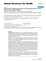
Induction of cell cycle arrest and cell apoptosis by 3b protein expressionFigure 1
Induction of cell cycle arrest and cell apoptosis by 3b protein expression. p3b/EGFP-N1 plasmid was transfected into
COS-7 cells, and the DNA contents of cells were measured by flow cytometry. EGFP expression positive and negative cells
were gated with forward scatter (on the left row). The middle and right rows were the assay of cell cycle in p3b/EGFP-N1 neg-
ative and positive cells. In p3b/EGFP-N1 positive cells, sub-G1 phase was changed from 11.80% to 53.50%, 48.36% at 24 h or 36
h, 48 h separately. The proportion was decreased to 23.34 and 24.85% at 60 and 72 h respectively. However, in EGFP negative
cells, the changes of sub-G1 phase were not obvious.
EGFP expression p3b/EGFP-N1 (-) p3b/EGFP-N1 (+)
R2
R3
40
10
4
10
3
70
Counts
Counts
10
GFP
2
10
1
10
0
24h
49.09%
48.16%
0
0
R2
R3
R2
R3
R2
R3
R2
R3
Figure 1
36h
48h
60h
72h
51.22%
44.54%
25.11%
72.10%
19.24%
78.40%
0 1000
DNA Content
10
4
10
3
10
2
10
1
10
0
GFP
01
DNA
000
Content
240
0
Counts
00001
DNA Content
40
Counts
0
250
Counts
0
00001
DNA Content
80
Counts
0
00001
DNA Content
70
Counts
0
01000
DNA Content
70
0
Counts
01000
DNA Content
10
4
10
3
10
2
10
1
10
0
GFP
01000
DNA Content
01
DNA
00000
Content
0
Content
01
DNA
01
DNA
0
Conten
Content
00
t
10
4
10
3
80
70
Counts
Counts
10
2
10
1
10
0
GFP
0
54.20%
42.52%
0
01
DNA
01
DNA
000
01
DNA
0
Conten
00
000
10
4
10
3
t
t
Conten
10
2
10
1
10
0
GFP
01
DNA
0
Conten
00
t
Virology Journal 2005, 2:66 />Page 3 of 5
(page number not for citation purposes)
predicted. The cellular localization of 3b protein by con-
focal microscopy was performed and the nucleolus local-
ization was confirmed. And the nucleolus localization
signal sequences of 3b protein may localize in the C-ter-
minal regions from 134 to 154 amino acids [9].
Nucleolus localization of SARS-CoV 3b protein suggested
that the expression of 3b protein may interfere with cell
cycle regulation in transfected cells. Flow cytometry was
performed on COS-7 cells transfected with pEGFP-N1
(Clontech) or p3b/EGFP-N1, which allowed examination
of two intra-culture populations with EGFP expression
indicative of transfected cells. PI staining revealed that a
similar pattern of phase distribution in EGFP positive and
negative cells transfected with pEGFP-N1 from 24 h to 72
h periods (data not shown). However, for p3b/EGFP-N1
transfected cells, the phase distribution of positive cells
was significantly different from that of negative cells (Fig-
ure 1). When compared with EGFP negative cells, the pop-
ulations of positive transfected cells displayed a
significant increase in G0/G1 phase, an obvious decrease
in S phase, and an emergence of sub-G1 phase compared
with negative cells at 24 h after transfection. At 36 h, the
increase in G0/G1 phase in positive cells was also signifi-
cant, but less than that at 24 h, while cells in S phase was
more decreased, and cells in sub-G1 phase continued
increase compared with negative ones. At 48 h, the
increase in G0/G1 phase and decrease in S phase in posi-
tive cells were not significant, but the percentage of sub-
G1 phase was significantly raised in positive cells (49.89%
vs. 5.26%). From 60 to 72 h post-transfection, the
increase in sub-G1 phase in positive cells was also signifi-
cant, but less than that at 48 h, while G0/G1 phases in
positive and negative cells were comparable (Figure 2).
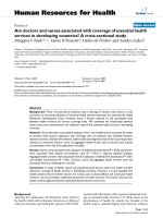
Histogram of cell cycle arrest and cell apoptosisFigure 2
Histogram of cell cycle arrest and cell apoptosis. Histogram showing the percentages of cells at various phases of cell
cycle. p3b/EGFP-N1 positive cells were showed with grey columns, and p3b/EGFP-N1 negative cells were showed with criss-
cross. Data were means of three independent experiment ± s.d. (bars).
70
60
50
40
30
20
10
0
Cells in each phase (%)
SubG1 G0/G1 S G2M SubG1 G0/G1 S G2M SubG1 G0/G1 S G2M SubG1 G0/G1 S G2M SubG1 G0/G1 S G2M
24h 36h 48h 60h 72h
positive cells
negative cell
s
Figure 2
Virology Journal 2005, 2:66 />Page 4 of 5
(page number not for citation purposes)
These data indicated that expression of 3b protein blocked
or delayed the progression of cells from G0/G1 phase into
S phase, and induced cells toward apoptosis.
Next, using similar method, we investigated the effects of
p3b/EGFP-N1 on cell cycles in 293 and Vero cells. In these
cells, the p3b/EGFP-N1 positive cells were arrested at G0/
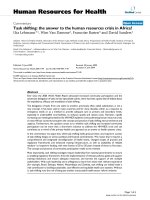
Apoptosis assay of COS-7 cells transfected with p3b/EGFP-N1Figure 3
Apoptosis assay of COS-7 cells transfected with p3b/EGFP-N1. COS-7 cells were transfected with pEGFP-N1 and
p3b/EGFP-N1 respectively. At 48 h after transfection, cells were collected and resuspended in binding buffer containing
Annexin V-PE and 7-AAD, and then processed for flow cytometry analysis. On the left row, EGFP positive cells were gated.
The middle and right rows were the results of EGFP negative and positive cells analyzed with Annexin V-PE and 7-AAD stain-
ing. In each box, the upper left corner included damaged cells, the lower left corner included viable cells, which were negative
for 7-AAD and Annexin V-PE binding, the upper right corner included necrotic or late apoptotic cells, which were positive for
Annexin V-PE staining and for 7-AAD uptake, while the lower right corner included apoptotic cells, which were Annexin V-PE
positive but impermeable to 7-AAD. In p3b/EGFP-N1 transfected cells (a), the percentage of apoptosis cells in EGFP positive
cells increased significantly, compared with negative ones, while there were no changes between positive and negative cells
transfected with pEGFP-N1 (b). One of three experiments with similar results was shown.
a
10
4
10
3
10
4
10
3
EGFP expression p3b/EGFP-N1 (-) p3b/EGFP-N1 (+)
b
EGFP expression EGFP (-) EGFP (+)
Figure 3
48h
48h
31.57%
0.49% 2.39%
7.94%
0.63% 7.43%
32.17%
32.40%
0.75% 1.73%
1.60%
0.48% 0.29%
1.24%
0 1000
DNA Content
10
4
10
3
10
2
1
GFP
0
1
10
0
110
4
Annexin V PE
10
4
10
3
10
2
10
1
10
0
210
4
Annexin V PE
10
4
10
3
1
1
7-AAD
0
2
0
1
10
0
7-AAD
0 1000
DNA Content
1
GFP
0
2
10
1
10
0
010
4
10
4
10
3
1
7-AAD
0
2
10
1
10
0
1
7-AAD
0
2
10
1
10
0
4
010
Annexin V PE
Annexin V PE
Publish with BioMed Central and every
scientist can read your work free of charge
"BioMed Central will be the most significant development for
disseminating the results of biomedical research in our lifetime."
Sir Paul Nurse, Cancer Research UK
Your research papers will be:
available free of charge to the entire biomedical community
peer reviewed and published immediately upon acceptance
cited in PubMed and archived on PubMed Central
yours — you keep the copyright
Submit your manuscript here:
/>BioMedcentral
Virology Journal 2005, 2:66 />Page 5 of 5
(page number not for citation purposes)
G1 phase compared with negative ones after transfection
(55.25% vs. 37.17% in 293 cells and 82.27% vs. 56.00%
in Vero cells at 48 h), but the percentages of sub-G1 phase
in positive and negative cells were all too low and compa-
rable. These data indicated that the role of 3b protein in
inducing cell cycle G0/G1 phase arrest was a conserved
character, but the apoptosis induction of 3b protein might
be a cell type specific.
In order to further confirm the function of 3b protein in
inducing cell apoptosis, a more definitive study using
double cell labeling with Annexin V-PE and 7-AAD was
performed. COS-7 cells were transiently transfected with
pEGFP-N1 and p3b/EGFP-N1 separately. At 48 h after
transfection, apoptosis analysis was carried out. As shown
in figure 3, the different populations in EGFP positive and
negative cells were measured by flow cytometry. It was
revealed that the percentages in apoptosis and necrosis
were both lower and comparable in the positive and neg-
ative pEGFP-N1 transfected cells (Figure 3b). However,
for p3b/EGFP-N1 transfected cells, the apoptosis cells in
the positive cells increased over 4-fold compared with that
in the negative ones (Figure 3a). These data further dem-
onstrated that over-expression of 3b protein could induce
cell apoptosis.
Published data showed that massive necrosis was found
in lung, spleen and lymph nodes in SARS patients. As
compared with normal tissues, apoptosis cells increased
significantly in the spleen, liver, lung, and lymph nodes of
SARS patients. The apoptosis cells were further demon-
strated to be pneumocytes, lymphocytes, and monocytes
[10,11]. Taken together, the data we presented here, as
well as the apoptosis and necrosis data of SARS patients,
suggest that 3b is an apoptosis-related gene in SARS
genome, which induce cell or tissue specific apoptosis in
transfected cells.
Competing interests
The author(s) declare that there are no competing
interests.
Authors' contributions
SY and ZZ conducted all the experiments. YX wrote the
maunscript and coordinated the research efforts. CY con-
ceived the study and edited the paper. CJ revised the
article.
Acknowledgements
We kindly thank Prof. Wei Kang for reading of the manuscript and Dr. Liu
Hong-yan, Dr. Li Su-yan, Yao Zhen-yu, Wu Jie and Li Jian-yong for construc-
tion of some plasmids.
References
1. Nicholls JM, Poon LL, Lee KC, Ng WF, Lai ST, Leung CY, Chu CM,
Hui PK, Mak KL, Lim W, Yan KW, Chan KH, Tsang NC, Guan Y, Yuen
KY, Peiris JS: Lung pathology of fatal severe acute respiratory
syndrome. Lancet 2003, 361:1773-1778.
2. Ding Y, Wang H, Shen H, Li Z, Geng J, Han H, Cai J, Li X, Kang W,
Weng D, Lu Y, Wu D, He L, Yao K: The clinical pathology of
severe acute respiratory syndrome (SARS): a report from
China. J Pathol 2003, 200:282-289.
3. Lang Z, Zhang L, Zhang S, Meng X, Li J, Song C, Sun L, Zhou Y: Path-
ological study on severe acute respiratory syndrome. Chin
Med J (Engl) 2003, 116:976-980.
4. Rota PA, Oberste MS, Monroe SS, Nix WA, Campagnoli R, Icenogle
JP, Penaranda S, Bankamp B, Maher K, Chen MH, et al.: Characteri-
zation of a novel coronavirus associated with severe acute
respiratory syndrome. Science 2003, 300:1394-1399.
5. Marra MA, Jones SJ, Astell CR, Holt RA, Brooks-Wilson A, Butterfield
YS, Khattra J, Asano JK, Barber SA, Chan SY, et al.: The Genome
sequence of the SARS-associated coronavirus. Science 2003,
300:1399-1404.
6. Oh JC, Jeong DL, Kim IK, Oh SH: Activation of calcium signaling
by hepatitis B virus-X protein in liver cells. Exp Mol Med 2003,
35:301-309.
7. Nakamichi K, Matsumoto Y, Otsuka H: Bovine herpesvirus 1 U(S)
ORF8 protein induces apoptosis in infected cells and facili-
tates virus egress. Virology 2002, 304:24-32.
8. Drosten C, Gunther S, Preiser W, van der Werf S, Brodt HR, Becker
S, Rabenau H, Panning M, Kolesnikova L, Fouchier RA, et al.: Identi-
fication of a novel coronavirus in patients with severe acute
respiratory syndrome. N Engl J Med 2003, 348:1967-1976.
9. Yuan X, Yao Z, Shan Y, Chen B, Yang Z, Wu J, Zhao Z, Chen J, Cong
Y: Nucleolar localization of non-structural protein 3b, a pro-
tein specifically encoded by the severe acute respiratory syn-
drome coronavirus. Virus Res 2005 in press.
10. Chau TN, Lee KC, Yao H, Tsang TY, Chow TC, Yeung YC, Choi KW,
Tso YK, Lau T, Lai ST, Lai CL: SARS-associated viral hepatitis
caused by a novel coronavirus: report of three cases. Hepatol-
ogy 2004, 39:302-310.
11. Zhang QL, Ding YQ, He L, Wang W, Zhang JH, Wang HJ, Cai JJ, Geng
J, Lu YD, Luo YL: Detection of cell apoptosis in the pathological
tissues of patients with SARS and its significance. Di Yi Jun Yi
Da Xue Xue Bao 2003, 23:770-773.