Báo cáo sinh học: " Evidence that spontaneous reactivation of herpes virus does not occur in mice" pdf
Bạn đang xem bản rút gọn của tài liệu. Xem và tải ngay bản đầy đủ của tài liệu tại đây (882.61 KB, 12 trang )
BioMed Central
Page 1 of 12
(page number not for citation purposes)
Virology Journal
Open Access
Research
Evidence that spontaneous reactivation of herpes virus does not
occur in mice
Bryan M Gebhardt*
1
and William P Halford
2
Address:
1
LSU Eye Center, Louisiana State University Health Sciences Center, New Orleans, LA 70112 USA and
2
Department of Veterinary
Microbiology, Montana State University, Bozeman, MT 59718 USA
Email: Bryan M Gebhardt* - ; William P Halford -
* Corresponding author
Abstract
Background: Some species, including humans and rabbits, exhibit periodic viral reactivation and
shed infectious virus at the infected end organ. Mice may be an exception, because spontaneous
shedding of infectious virus rarely, if ever, occurs. However, spontaneous molecular reactivation,
i.e., the expression of a few viral genes and the synthesis of the viral glycoproteins coded for by
these genes, has been reported. This finding has prompted the assumption that molecular
reactivation is an indicator of reactivation and the production of infectious virus. The goal of this
study was to differentiate between viral gene expression during latency and the episodic production
of infectious virus in mice.
Results: Viral reactivation and infection were not seen in herpes simplex virus type 1 (HSV-1)
latent ganglion graft recipient BALB/c scid or immunocompetent BALB/c mice, which survived the
65-day observation period with no evidence of viral infection although the immunocompetent mice
developed cellular and humoral immunity to HSV-1. In contrast, BALB/c scid recipients of ganglia
containing reactivating virus invariably developed a local and, subsequently, systemic viral infection
and died within 14 days. Immunocompetent BALB/c mice that received ganglion grafts containing
reactivating virus survived the infection and became immune to the virus. Trigeminal ganglia
removed from scid and immunocompetent recipient graft sites 5, 14, and 28 days after
transplantation contained latent virus and viable neurons.
Conclusion: The results suggest that, within the limits of detection of the experiments,
spontaneous episodic production of immunogenic viral antigens but not of infectious virus occurs
in mouse neural ganglia during latency.
Background
The infectious cycle of herpes simplex virus type 1 (HSV-
1) in experimental animals is similar to that which occurs
in humans, but there may be a significant difference as
well. HSV-1 readily infects epithelial surfaces of most
mammalian species, replicates in these cells, enters the
nervous system, and achieves a latent state in neurons in
the peripheral nervous system. A notable species differ-
ence is that the virus undergoes spontaneous, episodic
reactivation with or without evidence of recurrent disease
in humans and rabbits, whereas mice either do not
undergo spontaneous reactivation or undergo spontane-
ous reactivation at such a low frequency that it is difficult
to document [1].
Published: 18 August 2005
Virology Journal 2005, 2:67 doi:10.1186/1743-422X-2-67
Received: 17 June 2005
Accepted: 18 August 2005
This article is available from: />© 2005 Gebhardt and Halford; licensee BioMed Central Ltd.
This is an Open Access article distributed under the terms of the Creative Commons Attribution License ( />),
which permits unrestricted use, distribution, and reproduction in any medium, provided the original work is properly cited.
Virology Journal 2005, 2:67 />Page 2 of 12
(page number not for citation purposes)
Testing an end organ such as the eye or the site of viral
latency, the sensory ganglia, for infectious virus during
latency in mice fails to yield virus [2-5]. However, evi-
dence of viral gene expression in the trigeminal ganglia of
mice during latency has been reported [6,7]. In addition
to the expression of the latency-associated transcript
(LAT), the expression of other viral genes and their prod-
ucts has been found in a small number of ganglion cells.
Feldman et al. [8] described "abundant" expression of
viral genes and proteins and noted viral DNA synthesis in
occasional neurons. This process was termed "spontaneous
molecular reactivation"; no evidence of infectious virus was
reported in this study [8].
Stevens and Cook [3] transplanted ganglia from latent
mice into mice that were actively immunized with irradi-
ated virus or passively immunized with anti-HSV anti-
body and concluded that antiviral antibody helped
maintain viral latency. Tenser et al. [4] reported that viral
reactivation occurred in ganglion transplants after ex vivo
explantation. The occurrence of secondary latency was
proposed as a consequence of viral reactivation and infec-
tion of "secondary" neurons in the grafts; however, infec-
tious virus was not found in ganglion homogenates [4].
The current study was designed to differentiate between
viral gene expression and the production of infectious
virus in latent mouse ganglia in vivo. The experimental sys-
tem was designed to assess for the production of small
numbers of infectious viral particles which would lead to
morbidity and, ultimately, mortality in the host mice. In
the results reported here, molecular reactivation (i.e.,
expression of HSV-1 genes and production of glycopro-
teins during latency) did not proceed to the production of
detectable infectious virus in immune-deficient mice. The
results suggest that viral reactivation does not occur spon-
taneously and episodically in the mouse trigeminal gan-
glion in vivo.
Results
Absence of infectious virus in the trigeminal ganglion
during latency
Infectious virus was present on the ocular surface and in
both trigeminal ganglia of a group of five BALB/c mice
sacrificed 5 days after topical ocular infection (Table 1).
On days 10, 20, 30, 50, 70, and 100 after infection, both
the ocular surface and the trigeminal ganglion homoge-
nates of latently infected mice failed to yield infectious
virus as evidenced by cytopathic effect on Vero cells (Table
1).
Sensitivity of the ganglion assay
Assay of trigeminal ganglion homogenates for infectious
virus immediately after microinjection of a known
number of PFU of virus revealed that the limit of sensitiv-
ity for the in vitro assay was between 50 and 100 PFU per
ganglion (Table 2). All ganglia injected with 100 PFU
yielded plaques and 6 of 10 ganglia injected with 50 PFU
yielded plaques (Table 2). Injection of smaller numbers of
PFU did not reproducibly yield plaques (Table 2).
Ten out of 10 of the BALB/c scid mice receiving ganglia
injected with 100 PFU and 9 of 10 mice receiving ganglia
injected with 50 PFU died from complications of viral
pathogenesis within 12 days of receiving ganglion trans-
plants (Table 3). Five of 10 BALB/c scid mice receiving
ganglion grafts containing 10 PFU and 1 of 10 mice receiv-
ing grafts containing 5 PFU died within 14 days of receiv-
ing the grafts (Table 3). None of the 10 animals receiving
the ganglion grafts containing 1 PFU gave evidence of viral
infection and viral pathogenesis over a 35-day observa-
tion period.
Outcome of ganglion transplantation
Acute protocol
Results from three replicate experiments revealed that the
BALB/c scid recipients of ganglion transplants from
acutely infected BALB/c donors transplanted 3 days after
infection (N = 19) all died, with a mean time to death of
14 days and a standard deviation of ± 2 days (Fig. 1). In
contrast, all of the immunocompetent BALB/c recipients
of acutely infected ganglia (N = 15) survived (Fig. 1).
The cause of death in the BALB/c scid mice was not exten-
sively examined in this study. As the animals became pro-
gressively moribund, it was evident that they were
experiencing a neurological disease resembling encephali-
tis. In randomly chosen animals, virus was isolated from
the ear graft site at the time that the animal died. Confir-
mation that virus was replicating at the transplant site is
provided below.
Table 1: Analysis of infectious virus in the eye and trigeminal
ganglion during establishment of latency
Location Days after infection
a
5 1020305070100
Eye 5/5
b
0/5 0/5 0/5 0/5 0/5 0/5
Trigeminal ganglia 10/10 0/10 0/10 0/10 0/10 0/10 0/10
a
Infected mice (N = 5) were killed and eye swabs (both combined) and
trigeminal ganglion homogenates (separately) tested for infectious
virus.
b
The numbers indicate number of eye swabs and trigeminal ganglion
homogenates containing infectious virus/number of each tested at
each time.
Virology Journal 2005, 2:67 />Page 3 of 12
(page number not for citation purposes)
Latent protocol
In a series of three experiments involving a total of 100
recipients (70 BALB/c scid, 30 immunocompetent BALB/
c), only 1 of the 70 BALB/c scid recipients of a latent gan-
glion transplant died (28 days after grafting). Virus was
not isolated from the graft site or the brain of this animal.
As shown in Figure 2, the remainder of the BALB/c scid
recipients survived without evidence of viral infection up
to 65 days after grafting. All of the 30 immunocompetent
BALB/c recipients of latent ganglion transplants survived
for the duration of the experiment (Fig. 2). Mice in both
of these groups were bled at 21, 45, and 65 days after graft-
ing to test their sera for anti-HSV-1 antibodies, as
described below.
Reactivation protocol
Ganglia from BALB/c mice latent for HSV-1 were passaged
in tissue culture for 3 days and then transplanted into
BALB/c scid recipients (N = 24) and immunocompetent
BALB/c recipients (N = 16). All of the BALB/c scid recipi-
ents developed viral infections and viral-mediated neuro-
logic disease and died, with a mean time to death of 14
days (Fig. 3). Analyses of the transplanted tissue and the
local graft site supported the conclusion that the cause of
death was the reactivated virus. In contrast, all of the
immunocompetent BALB/c recipients of reactivated gan-
Table 2: Detection of virus injected into the trigeminal ganglion by plaque assay
a
Number of PFU injected/ganglion Number of ganglia containing infectious
virus/number of ganglia tested
Mean PFU ± standard deviation
100 10/10 28 ± 7
50 6/10 7 ± 4
10 1/10 1
50/100
10/100
a
Intact trigeminal ganglia were injected with the amounts of virus indicated and homogenized. The homogenate was tested for infectious virus by
plaque assay.
Table 3: Detection of virus injected into the trigeminal ganglion by transplantation into scid mice
a
Number of PFU injected/ganglion Number of mice dead/number of mice grafted
100 10/10
50 9/10
10 5/10
5 1/10
1 0/10
a
Trigeminal ganglia injected with the amount of virus indicated were transplanted to BALB/c scid mice and the animals observed for viral
pathogenesis and death over a 35-day period.
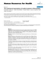
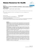
Acute Protocol: Kaplan Meier analysis of the fate of ganglion transplant recipientsFigure 1
Acute Protocol: Kaplan Meier analysis of the fate of
ganglion transplant recipients. BALB/c scid mice (N =
19) and immunocompetent BALB/c mice (N = 15) were
observed daily for evidence of infection and death. The day
of death was recorded as the number of days after grafting.
BALB/c scid recipients all died by day 16, whereas all of the
immunocompetent BALB/c recipients survived throughout
the entire observation period (P < 0.0001).
Virology Journal 2005, 2:67 />Page 4 of 12
(page number not for citation purposes)
glia survived without evidence of disease (Fig. 3) and ulti-
mately became immune to the virus.
Serum anti-HSV-1 antibody responses in ganglion
recipients
None of the BALB/c scid recipients of latently infected
ganglia developed a serum IgG antibody response as
measured by enzyme-linked immunosorbent assay
(ELISA). Randomly chosen BALB/c scid mice tested at 21,
45, and 65 days after transplantation showed no evidence
of having developed a humoral immune response to the
virus (Fig. 4a). The BALB/c mice that received latent gan-
glion transplants did not exhibit an anti-HSV-1 antibody
response on day 21 after transplantation, but had serum
antibody on days 45 and 65 after grafting (Fig. 4a). The
immunocompetent BALB/c recipients of acutely infected
ganglia or of ganglia containing reactivating virus devel-
oped serum antibody IgG responses by 21 days after infec-
tion, which were present also at 45 and 65 days after
transplantation (Fig. 4b).
Cell-mediated immunity in ganglion transplant recipients
BALB/c scid mice from the Latent Protocol that survived
the 65-day observation period were tested by footpad
swelling assay. None of the animals tested gave evidence
of a delayed-type hypersensitivity response to viral anti-
gens (Fig. 5). In contrast, all of the immunocompetent
BALB/c recipients of acutely infected ganglia and recipi-
ents of ganglia undergoing viral reactivation exhibited
delayed-type hypersensitivity responses on day 65 after
ganglion transplantation (Fig. 5). Four of seven immuno-
competent BALB/c recipients of latent ganglia also had
positive delayed-type hypersensitivity responses on day
65 after transplantation (Fig. 5).
Viability of the ganglion transplants
Latent ganglia that had been transplanted into BALB/c
scid and immunocompetent BALB/c recipients were
removed from the graft recipients on days 5, 14, or 28
after transplantation and placed into tissue culture. Seven-
teen of 18 latent ganglia removed from BALB/c scid ani-
mals underwent reactivation in vitro (Table 4), typically
within 3 to 6 days after explantation. All of the latent
ganglia recovered from immunocompetent BALB/c recip-
ients underwent reactivation in tissue culture between
days 6 and 10 after explantation.
The histology of ganglion transplants was examined on
days 5, 28, and 65 after transplantation. Although the
architecture of transplanted ganglia was somewhat altered
compared with that of freshly isolated ganglia (Fig. 6a),
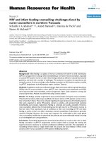
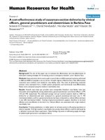
Latent Protocol: Kaplan Meier analysis of the fate of BALB/c scid (N = 70) and immunocompetent BALB/c (N = 30) gan-glion recipientsFigure 2
Latent Protocol: Kaplan Meier analysis of the fate of
BALB/c scid (N = 70) and immunocompetent BALB/
c (N = 30) ganglion recipients. Mice were observed daily
for evidence of infection and death. The day of death was
recorded as the number of days after grafting. One of the
BALB/c scid mice died on day28, but virus was not found in
the graft site or brain of this animal. There was no significant
difference between the survival of the BALB/c scid and
immunocompetent BALB/c mice (P > 0.05).
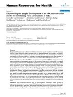
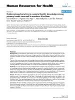
Reactivation Protocol: Kaplan Meier analysis of the fate of BALB/c scid (N = 24) and immunocompetent BALB/c (N = 16) recipients of ganglion graftsFigure 3
Reactivation Protocol: Kaplan Meier analysis of the
fate of BALB/c scid (N = 24) and immunocompetent
BALB/c (N = 16) recipients of ganglion grafts. Mice
were observed daily for infection and death. The day of death
was recorded as the number of days after grafting. All of the
BALB/c scid recipients were dead by day 18, whereas all of
the immunocompetent BALB/c recipients survived through-
out the entire observation period (P < 0.0001).
Virology Journal 2005, 2:67 />Page 5 of 12
(page number not for citation purposes)
numerous large cells with the morphology of neurons
were seen in latent ganglia transplanted to BALB/c scid
(Fig. 6b) and immunocompetent BALB/c (Fig. 6c)
recipients.
Table 5 presents the results of the vital dye staining of cells
isolated from ganglion transplants. Viable small (non-
neuronal) and large (neurons) cells were found in roughly
the same ratios on days 5, 14, 28, and 65 after transplan-
tation. These ratios were similar to the ratios of small and
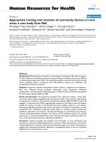
Serum anti-HSV-1 antibody responsesFigure 4
Serum anti-HSV-1 antibody responses. (a) Serum anti-
HSV-1 antibody responses of BALB/c scid (N = 8) and immu-
nocompetent BALB/c (N = 8) mice in the Latent Protocol.
The mice were grafted with ganglia containing latent virus
and randomly chosen mice were bled on days 21, 45, and 65
after transplantation. The corrected optical density readings
indicate that the BALB/c scid mice did not produce IgG anti-
body, whereas the immunocompetent BALB/c mice all had
serum IgG anti-HSV-1 antibody on days 45 and 65 after
transplantation. (b) Serum antibody responses of BALB/c
mice receiving acutely infected ganglia (N = 4) or ganglia con-
taining reactivating virus (N = 4). The optical density readings
indicate that the immunocompetent mice in the Acute Pro-
tocol and the Reactivation Protocol developed serum anti-
HSV-1 antibody titers by day 21 after grafting and that this
antibody continued to be present on days 45 and 65 after
transplantation. At each of the three time points, symbols
representing one mouse each are spread out to avoid con-
cealment by overlap.
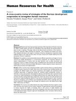
Footpad swelling responses to measure cell-mediated immunityFigure 5
Footpad swelling responses to measure cell-medi-
ated immunity. Footpad swelling responses in the BALB/c
scid mice in the Latent Protocol (N = 9) and immunocompe-
tent BALB/c recipients in the Latent (N = 7), Acute (N = 7),
and Reactivation (N = 6) Protocols are shown. Included in
the data sets are the footpad swelling responses of
immune(N = 6) and nonimmune (N = 6) immunocompetent
BALB/c mice. The BALB/c scid recipients of latent ganglia
failed to develop a cell-mediated immune response, whereas
four of the seven BALB/c wild-type recipients of latent gan-
glia showed a delayed-type hypersensitivity response. All of
the immunocompetent BALB/c recipients of ganglia in the
Acute and Reactivation Protocols exhibited cellular immune
reactivity. Comparison of the footpad swelling response of
the BALB/c immune mice, immunocompetent BALB/c recipi-
ents in the Reactivation Protocol and the Acute Protocol,
and immunocompetent BALB/c recipients in the Latent Pro-
tocol with the BALB/c scid recipients in the Latent Protocol
revealed that the response in the immunocompetent mice in
each group was significantly greater than the response in the
BALB/c scid mice (P < 0.001). Values are means ± SD.
Virology Journal 2005, 2:67 />Page 6 of 12
(page number not for citation purposes)
large cells obtained from freshly isolated ganglia (Table
5).
Confirmation that viral reactivation and viral glycopro-
tein synthesis was occurring in ganglia transplanted dur-
ing acute infection and in ganglia transplanted following
reactivation was obtained by performing immunohisto-
chemical staining for HSV-1 antigens in tissue sections.
Staining of viral antigens was seen in the ganglion trans-
plants and adjacent ear cells in BALB/c scid recipients of
acutely infected (Fig. 7a) and reactivating (Fig. 7b) gan-
glia, but not in recipients of latent ganglion transplants
(Fig. 7c).
Discussion
The results of these experiments indicate that little, if any,
infectious virus is produced during latency in mice. It has
been proposed that some component of the immune sys-
tem is necessary to induce HSV-1 into latency and prevent
viral reactivation [9-15]. Sawtell [16] reported that
immune cells in mouse ganglia do not inhibit viral reacti-
vation. Thus, the role of antiviral immunity in the estab-
lishment and maintenance of latency is still being
debated.
The immunocompetent BALB/c mice in the acute
, latent,
and reactivation
protocols developed cellular and
humoral immunity, indicating that there was an adequate
amount of viral antigen produced in all three circum-
stances to sensitize the recipients. The finding that T cell-
mediated and humoral responses developed and were
sustained for 65 days suggests that viral antigen expres-
sion during latency has a role in this process. BALB/c scid
mice lack an acquired immune system but have an intact
innate immune system, including cells such as macro-
phages and natural killer (NK) cells and antiviral
cytokines such as the types 1 and 2 interferons. NK cells
alone cannot protect scid mice from HSV-1 infection and
pathogenesis [17]. However, protection against viral-
mediated death can be provided by T lymphocytes [18-
21]. There is ample evidence to indicate that the
interferons modulate the level of viral replication and
spread, although this response is not known to protect
scid mice from HSV-1-mediated death [22-24]. However,
in the absence of an acquired immune system and, in par-
ticular, T lymphocytes, the virus evades the interferon
response, enters the nervous system, and replicates in vital
cells causing a fatal encephalitis.
The findings of the current study appear to imply that
mouse neural tissue containing latent HSV-1 (e.g., the
trigeminal ganglion) does not support periodic episodic
viral reactivation. Although spontaneous molecular viral
reactivation has been reported [8], the results presented
here suggest that this molecular reactivation does not pro-
ceed to the production of detectable infectious virus. It
may be that there are nonimmunological cellular or
molecular factors that prevent spontaneous viral reactiva-
tion in mice.
These findings in vivo are particularly important since it is
known that explanted mouse neural tissues latent for
HSV-1 demonstrate viral reactivation in culture. This sug-
gests that explantation itself or factors in tissue culture
that we do not understand may suppress or destroy the in
vivo factors that maintain viral latency.
A number of reports describe the induction of viral reacti-
vation from latency in mice using a variety of stimuli such
as immunosuppressive drugs, UV irradiation, and thermal
stress [25-29]. These induction protocols yield a variable
frequency of viral reactivation. There are no reports
confirming spontaneous episodic shedding of virus at
epithelial surfaces of mice, including the eye and
genitalia, although it has been reported recently that
infectious virus is present in the trigeminal ganglion up to
240 days after infection [16].
The possibility that the surgical trauma of ganglion trans-
plantation or the site in which the transplant was placed
(the ear) prevents viral reactivation from occurring and/or
prevents infectious virus from leaving this site to infect the
animal's nervous system must be considered. However, in
the Acute and Reactivation Protocol mice, it was found
Table 4: Recovery (reactivation) of virus in explanted ganglion grafts
a
Source of explants Day of explantation relative to day of grafting
51428
BALB/c scid Latent Protocol 5/5
b
6/6 6/7
Immunocompetent BALB/c Latent Protocol 4/4 7/7 5/5
a
Ganglion grafts were placed into culture on the day indicated and the culture medium tested for infectious virus on days 1, 3, 5, 7, 10, 14, and 21
of incubation.
b
Number of ganglia from which virus reactivated/number of ganglia tested.
Virology Journal 2005, 2:67 />Page 7 of 12
(page number not for citation purposes)
that ganglia containing infectious virus, either in the acute
stage of infection or following reactivation, placed into
the ear pocket resulted in spread of the virus from this site
to the nervous system of BALB/c scid mice, resulting in
encephalitis and death. Additionally, injection of 10 viral
particles into ganglion grafts resulted in viral infection
and death of 50% of BALB/c scid mice, demonstrating the
sensitivity of this in vivo system and confirming that the
subcutaneous ear site is not a sequestered site that pre-
vents the escape of infectious virus.
Conclusion
It is concluded that measurable infectious virus is not pro-
duced under the conditions of these experiments. Thus,
the technical approach used here appears to be a valid and
sensitive measure of the presence of infectious virus. The
Histology of ganglion graftsFigure 6
Histology of ganglion grafts. (a) Hematoxylin and eosin (H & E) stained section of a freshly isolated trigeminal ganglion.
The large neuron cell bodies (arrows) interspersed among a field of nerve fibers present the typical histologic appearance of
the trigeminal ganglion. (original magnification 400×) (b) H & E stained section of a latent ganglion graft removed from a BALB/
c scid mouse 45 days after grafting. In this section, neuron cell bodies (arrows) with typical morphology can be seen. (original
magnification 400×) (c) H & E stained section of alatent ganglion graft removed from an immunocompetent BALB/c recipient
45 days after transplantation. Clusters of neuron cell bodies (arrows) with typical morphology can be seen. (original magnifica-
tion 400×)
Virology Journal 2005, 2:67 />Page 8 of 12
(page number not for citation purposes)
results of this study reveal that molecular reactivation, i.e.,
expression of HSV-1 genes and production of glycopro-
teins during latency, occurs in mice and extends this
observation to establish that molecular reactivation does
not necessarily lead to the production of infectious viral
particles. The approach used in this investigation opens
up new vistas for studying herpesvirus latency and
reactivation.
Methods
Mice
Female BALB/cJ and BALB/c scidJ mice at 5 weeks of age
(The Jackson Laboratory, Bar Harbor, ME) were used.
Confirmation that the BALB/c scid mice were immune
deficient was achieved by performing flow cytometric
analysis of spleen cells for CD3
+
T cells and membrane
immunoglobulin-positive cells. No evidence of the pres-
ence of T or B lymphocytes in the BALB/c scid mice sacri-
ficed throughout the course of this study was obtained
(data not shown). Animals studies were approved by the
Louisiana State University Health Sciences Center Institu-
tional Animal Care and Use Committee (IACUC). All ani-
mals were provided with food and water ad libidum and
were cared for according to the NIH Guidelines on the
Care and Use of Animals in Research.
Virus
The McKrae strain of HSV-1, a strain which is known to
spontaneously reactivate in rabbits, was propagated in
and titered on Vero cells (American Type Culture Collec-
tion, Manassas, VA). At the time of infection, the virus
stock was thawed and diluted so as to deliver 1 × 10
5
PFU
in 4 µl of culture medium. BALB/c mice to be infected
were anesthetized, their corneas were lightly scratched in
a cross-hatched pattern, and 4 µl of the viral suspension
was placed on the surface of each eye. In order to ensure
survival, infected animals each received 0.1 ml of pooled
human serum (Chemicon International, Temecula, CA)
intraperitoneally at the time of infection. At 3 and 5 days
after infection, the ocular surface of the animals was
swabbed and tested for the presence of infectious virus by
the viral plaque assay. Animals not giving evidence of
infection were excluded from the study.
Analysis of the trigeminal ganglion for infectious virus
during latency
Thirty-five BALB/c strain mice were infected with the McK-
rae strain of HSV-1 by the topical ocular route as described
above. Five days after infection, the eyes of all animals
were swabbed for the determination of infectious virus.
Also on day 5, five animals were killed and their trigemi-
nal ganglia were separately homogenized in 0.5 ml of
tissue culture medium. The ganglion homogenates were
tested for infectious virus on Vero cell monolayers in 24-
well tissue culture plates. In this experiment, no attempt
was made to quantitate infectious virus, but only to note
the presence or absence of infectious virus in the ganglia.
Eye swabs and trigeminal ganglion homogenates were
similarly tested from five additional animals killed at each
of the following time points: 10, 20, 30, 50, 70, and 100
days after infection.
Determination of the sensitivity of the assay for infectious
virus in the ganglion
Groups of 10 uninfected BALB/c mice were sacrificed and
their trigeminal ganglia removed intact and placed into
tissue culture medium. The 20 ganglia from each group of
mice were positioned under a stereoscopic microscope
and each ganglion injected with a 5 µl volume of culture
medium containing 100, 50, 10, 5, or 1 PFU of the
McKrae strain of HSV-1. Ten ganglia from each group were
immediately homogenized in 0.5 ml of culture medium,
the homogenate clarified by centrifugation at 8000 × g in
a microcentrifuge, and the supernatant plated on Vero
cells in 24-well plates. Following viral attachment, the
supernatant was removed and a 0.5% methylcellulose
overlay was placed in each well. The plates were incubated
for 2 days and plaques counted. The remaining 10 ganglia
Table 5: Viability of cells in ganglion grafts
a
Source of ganglia Day of cell viability determination relative to day of grafting
5142865
Small Large Small Large Small Large Small Large
BALB/c scid Latent Protocol 328/416
b
(79) 54/72 (75) 477/519 (92) 38/49 (78) 622/705 (88) 88/97 (91) 219/279 (78) 41/54 (76)
Immuno-competent BALB/c
Latent Protocol
789/885 (89) 66/81 (81) 413/500 (83) 75/82 (91) 917/998 (92) 31/48 (65) 261/308 (85) 87/101 (86)
Freshly isolated ganglia 419/524 (80) 88/110 (80) 523/567 (92) 101/123 (82) 816/911 (90) 23/38 (61) 377/408 (92) 99/122 (81)
a
Ganglion grafts or freshly isolated ganglia were dissociated and stained with vital DNA dye and trypan blue. The total numbers of small cells (10–50
µm) and large cells (larger than 50 µm), as well as the number of live cells in each size category, were determined by microscopic examination.
b
Number of viable cells of each size/total number of cells of each size counted (%).
Virology Journal 2005, 2:67 />Page 9 of 12
(page number not for citation purposes)
were transplanted into subcutaneous ear pockets in BALB/
c scid mouse recipients as described below. The mice were
observed daily for signs of viral pathogenesis and death.
Ganglion transplantation
Recipient mice were anesthetized with a mixture of keta-
mine and xylazine and positioned under a stereoscopic
dissecting microscope such that the entire ear pinna could
be seen at 10× magnification. The tip of the ear was gently
grasped with sterile forceps and an incision made in the
dorsal skin surface with a sterile lamellar blade (Wilson
Ophthalmic, Mustang, OK). The lamellar blade was gen-
tly eased below the surface of the epithelium with a side-
to-side and insertion-retraction motion, creating a pocket
Immunohistochemical staining for viral antigensFigure 7
Immunohistochemical staining for viral antigens. (a) Immunohistochemical staining of a tissue section through the ear
graft site of a BALB/c scid mouse containing a ganglion from an acutely infected donor 7 days after grafting. In this immunoper-
oxidase-stained section, cells expressing HSV-1 antigens can be seen in the ganglion graft (arrows). (original magnification,
400×) (b) Immunohistochemical staining for HSV-1 antigens in the ganglion graft in a BALB/c scid mouse from the Reactivation
Protocol. Cells expressing HSV-1 antigen are seen in both the graft and the surrounding ear cells at the graft site (arrows).
(original magnification, 400×) (c) Immunohistochemical staining of a latent ganglion graft in a BALB/c scid recipient at 28 days
after grafting. No evidence of cells expressing HSV-1antigens was seen in these tissue sections. (original magnification, 400×)
Virology Journal 2005, 2:67 />Page 10 of 12
(page number not for citation purposes)
approximately 3 mm wide and 7 mm deep. Individual
ganglia were gently inserted into the ear pockets so as to
allow the open end of the ear pocket to close over the graft
and self-seal, thus enclosing the ganglion in the pocket
and avoiding the need for sutures. One application of
neomycin and polymixin sulfate ointment externally was
adequate to prevent bacterial infection. The recipient ani-
mals were returned to their cages for recovery from the
anesthetic and the external condition of the graft site was
observed daily to ensure the success of the transplant.
Three ganglion transplantation protocols were performed:
1) Acute Protocol:
BALB/c mice and BALB/c scid mice
received trigeminal ganglion grafts from BALB/c donors
that had been infected 5 days previously with McKrae
strain HSV-1. Recipient mice were observed for evidence
of viral infection, development of tissue pathology at the
site of the transplant, signs of morbidity, and death. At the
time of sacrifice or the time of death, serum was collected
for testing for antibodies to HSV-1.
2) Latent Protocol:
Trigeminal ganglia from BALB/c mice
that had been infected 35 days previously with McKrae
strain HSV-1 were transplanted into BALB/c and BALB/c
scid mice as described above. The recipient mice were
observed as described above for evidence of viral infec-
tion, viral-induced tissue pathology at the transplant site,
signs of morbidity, and death. In replicates of this
experiment, recipient mice were anesthetized on days 5,
14, or 28 after transplantation and the ganglion trans-
plants removed from the skin pockets and placed in tissue
culture to test for the presence of latent, reactivatable her-
pesvirus in the transplanted ganglion. For in vitro
incubation, ganglia from latent BALB/c mice and
explanted ganglion transplants were incubated in separate
wells of 12-well culture plates containing Dulbecco's
modified Eagle's medium (DMEM) supplemented with
10% fetal bovine serum (FBS) and an antibiotic/antimy-
cotic mixture (GIBCO, Carlsbad, CA). The culture
medium in each well was assayed for infectious virus on
Vero cell monolayers at 1, 3, 5, 7, 10, 14, and 21 days of
incubation.
3) Reactivation Protocol:
Trigeminal ganglia latent with
the McKrae strain of HSV-1 obtained from BALB/c mice
were incubated in tissue culture for 3 days, and then
transplanted into BALB/c and BALB/c scid recipients. The
mice were observed for evidence of viral infection, virus-
induced tissue pathology at the transplant site, signs of
morbidity, and death. The recipients were tested for the
development of antiviral antibody by ELISA.
ELISA for serum antibody
Serum was collected from the BALB/c and BALB/c scid
animals at the time of sacrifice. ELISA plates were coated
with a cell culture lysate from Vero cells infected 18 hours
previously with McKrae strain HSV-1. This lysate contains
a mixture of HSV-1 antigens and has been used in previ-
ous studies [30,31]. Equal numbers of ELISA plate wells
were coated with a cell culture lysate of uninfected Vero
cells. The plate wells were washed three times with Tris
buffered saline (TBS) and 1:50 dilutions of each serum
were tested in quadruplicate for reactivity with the
infected and uninfected cell lysates. Binding of serum
anti-HSV antibody was detected with a secondary rabbit
anti-mouse IgG antibody coupled to horseradish peroxi-
dase (Jackson ImmunoResearch Laboratories, Inc., West
Grove, PA). Following washing, the plate wells were incu-
bated in tetramethylbenzidine substrate
(DakoCytomation, Inc., Carpinteria, CA) for 15 minutes
at room temperature and the reaction stopped with 1 M
sulfuric acid. The optical densities were read at 450 nm in
a plate reader and the optical density values for each
serum sample tested on the infected cell lysate were cor-
rected by subtraction of the optical density obtained with
the serum on the uninfected cell lysate.
Footpad swelling assay for delayed type hypersensitivity
The same infected cell lysate used to coat the ELISA plates
was treated with UV light for 10 minutes to inactivate
infectious virus. Mice were anesthetized with a mixture of
ketamine and xylazine. The left hind footpads were
injected with 10 µl of the treated, infected lysate and the
right hind footpad received 10 µl of the uninfected cell
lysate. At 24 hours, the footpad swelling response was
measured using a spring-loaded micrometer gauge
(Starett, Inc., Athol, MA). Four measurements were made
of each right and left footpad. Delayed-type
hypersensitivity reactions were calculated as follows: spe-
cific footpad swelling = (24 hr measurement of left foot-
pad - 0 hr measurement of left footpad) - (24 hr
measurement of right footpad - 0 hr measurement of right
footpad) × 10
3
mm. In each experiment, in addition to the
test animals, mice not immune to HSV-1 and mice immu-
nized with UV-inactivated virus were used as negative and
positive controls.
Histology and immunohistochemical staining
Animals selected at random were sacrificed and the por-
tion of the ear containing the ganglion transplant site was
frozen and sectioned in a cryotome. The sections (10 µm)
were placed on microscope slides and fixed in cold ace-
tone for 5 minutes. Representative sections were stained
with hematoxylin and eosin for histopathologic examina-
tion. Additional sections were stained for cells expressing
HSV-1 antigens. A direct staining method employing a
polyclonal, horseradish peroxidase-conjugated rabbit
Virology Journal 2005, 2:67 />Page 11 of 12
(page number not for citation purposes)
anti-HSV-1 antibody (DakoCytomation) was used. The
antibody was diluted 1:200 in TBS containing 1% bovine
serum albumin. Following incubation for 1 hour, the
slides were washed in three washes of TBS for 5 minutes
each and then incubated in the substrate consisting of
diaminobenzidine and hydrogen peroxide (Pierce Bio-
technology, Inc., Rockford, IL). Color development was
stopped after 5 minutes and the sections were counter-
stained with methyl green. Control slides were incubated
in an irrelevant peroxidase-labeled rabbit antibody fol-
lowed by the substrate.
Cell viability in ganglion transplants
Transplanted ganglia were removed from ear pockets at 5,
14, 28, and 65 days after transplantation. Each ganglion
was teased apart with forceps in 2 ml of calcium/magne-
sium-free Hank's balanced salt solution (GIBCO) con-
taining 10 U DNase, 0.1 mg/ml dispase, 0.1 mg/ml
collagenase, and 0.1 mg/ml trypsin. The tissue fragments
were incubated with gentle stirring for 15 minutes at
37°C. The cells and tissue clumps were gently triturated
and washed two times in DMEM/10% FBS. The cells were
resuspended in 2 ml DMEM/10% FBS containing 1 µg/ml
of Hoechst 33342 vital DNA stain (H342, Calbiochem, La
Jolla, CA) for 15 minutes at 37°C. The cells were washed
twice in DMEM/FBS and resuspended in 2 ml of medium.
Living and dead cells were differentiated with the addition
of 0.1% trypan blue, which quenches the H342 fluores-
cence of dead cells [32]. The cells were placed onto clean
microscope slides, coverslipped, and immediately exam-
ined on a Nikon E600 fluorescence microscope with a UV-
2E/C (330–380 excitation; 435–485 barrier) filter. The
cells were categorized as either small (10–50 µm in diam-
eter, non-neurons) or larger than 50 µm in diameter (neu-
rons). For each suspension, 10 microscopic fields at 100×
magnification were examined, and the total number of
cells, the numbers of small and large cells, and the num-
bers of fluorescing (i.e., not quenched, viable) cells in
each of the two size ranges were determined.
Statistical analysis
Analysis of numerical data and statistical analyses were
performed with Microsoft Excel (Redmond, WA), Modstat
(Modern Microcomputers, Mechanicsville, VA), and CoS-
tat (Cohort Software, Monterey, CA). Fisher's exact test
was used to compare the differences in survival frequen-
cies between groups of mice. P values less than 0.01 were
considered significant.
Competing interests
The author(s) declare that they have no competing
interests.
Authors' contributions
BMG carried out the ganglion transplantation experi-
ments and immunoassays. WPH carried out the viral
infections and plaque assays. BMG and WPH conceived of
the study. BMG wrote the manuscript.
Acknowledgements
This work was supported in part by U.S. Public Health Service grants
R01EY002672 (BMG), R01AI054104 (WPH), and P30EY002377 (LSU Eye
Center Core grant) from the National Institutes of Health, Bethesda, Mar-
yland, an unrestricted challenge grant (LSU Eye Center) from Research to
Prevent Blindness, Inc., New York, New York, and a National Science
Foundation EPSCoR grant (EPS0346458) to Montana State University
(WPH).
References
1. Mester JC, Rouse BT: The mouse model and understanding
immunity to herpes simplex virus. Rev Infect Dis 1991,
13:S935-S945.
2. Hill TJ, Harbour DA, Blyth WA: Isolation of herpes simplex virus
from the skin of clinically normal mice during latent
infection. J Gen Virol 1980, 47:205-207.
3. Stevens JG, Cook ML: Maintenance of latent herpetic infection:
an apparent role for anti-viral IgG. J Immunol 1974,
113:1685-1693.
4. Tenser RB, Edris WA, Gaydos A, Hay KA: Secondary herpes sim-
plex virus latent infection in transplanted ganglia. J Virol 1994,
68:7212-7220.
5. Willey DE, Trousdale MD, Nesburn AB: Reactivation of murine
latent HSV infection by epinephrine iontophoresis. Invest
Ophthalmol Vis Sci 1984, 25:945-950.
6. Deatly AM, Spivack JG, Lavi E, Fraser NW: RNA from an immedi-
ate early region of the type 1 herpes simplex virus genome is
present in the trigeminal ganglia of latently infected mice.
Proc Natl Acad Sci USA 1987, 84:3204-3208.
7. Stevens JG, Wagner EK, Devi-Rao GB, Cook ML, Feldman LT: RNA
complementary to a herpesvirus alpha gene mRNA is prom-
inent in latently infected neurons. Science 1987, 235:1056-1059.
8. Feldman LT, Ellison AR, Voytek CC, Yang L, Krause P, Margolis TP:
Spontaneous molecular reactivation of herpes simplex virus
type 1 latency in mice. Proc Natl Acad Sci USA 2002, 99:978-983.
9. Halford WP, Gebhardt BM, Carr DJJ: Persistent cytokine expres-
sion in trigeminal ganglion latently infected with herpes sim-
plex virus type 1. J Immunol 1996, 157:3542-3549.
10. Keadle TL, Laycock KA, Miller JK, Hook KK, Fenoglio ED, Francotte
M, Slaoui M, Stuart PM, Pepose JS: Efficacy of a recombinant glyc-
oprotein D subunit vaccine on the development of primary
and recurrent ocular infection with herpes simplex virus
type 1 in mice. J Infect Dis 1997, 176:331-338.
11. Liu T, Khanna KM, Carriere BN, Hendricks RL: Gamma interferon
can prevent herpes simplex virus type 1 reactivation from
latency in sensory neurons. J Virol 2001, 75:11178-11184.
12. Liu T, Khanna KM, Chen X, Fink DJ, Hendricks RL: CD8
+
T cells can
block herpes simplex virus type 1 (HSV-1) reactivation from
latency in sensory neurons. J Exp Med 2000, 191:1459-1466.
13. Shimeld C, Easty DL, Hill TJ: Reactivation of herpes simplex
virus type 1 in the mouse trigeminal ganglion: an in vivo study
of virus antigen and cytokines. J Virol 1999, 73:1767-1773.
14. Shimeld C, Whiteland JL, Nicholls SM, Grinfeld E, Easty DL, Gao H,
Hill TJ: Immune cell infiltration and persistence in the mouse
trigeminal ganglion after infection of the cornea with herpes
simplex virus type 1. J Neuroimmunol 1995, 61:7-16.
15. Shimeld C, Whiteland JL, Williams NA, Easty DL, Hill TJ: Reactiva-
tion of herpes simplex virus type 1 in the mouse trigeminal
ganglion: an in vivo study of virus antigen and immune cell
infiltration. J Gen Virol 1996, 77:2583-2590.
16. Sawtell NM: Quantitative analysis of herpes simplex virus
reactivation in vivo demonstrates that reactivation in the
nervous system is not inhibited at early times
postinoculation. J Virol 2003, 77:4127-4138.
Publish with BioMed Central and every
scientist can read your work free of charge
"BioMed Central will be the most significant development for
disseminating the results of biomedical research in our lifetime."
Sir Paul Nurse, Cancer Research UK
Your research papers will be:
available free of charge to the entire biomedical community
peer reviewed and published immediately upon acceptance
cited in PubMed and archived on PubMed Central
yours — you keep the copyright
Submit your manuscript here:
/>BioMedcentral
Virology Journal 2005, 2:67 />Page 12 of 12
(page number not for citation purposes)
17. Halford WP, Maender JL, Gebhardt BM: Re-evaluating the role of
natural killer cells in innate resistance to herpes simplex
virus type 1. Virol J 2005, 2:56.
18. Minagawa H, Sakuma S, Mohri S, Mori R, Watanabe T: Herpes sim-
plex virus type 1 infection in mice with severe combined
immunodeficiency (SCID). Arch Virol 1988, 103:73-82.
19. Bouley DM, Kanangat S, Rouse BT: The role of the innate
immune system in the reconstituted SCID mouse model of
herpetic stromal keratitis. Clin Immunol Immunopathol 1996,
80:23-30.
20. Minagawa H, Yanagi Y: Latent herpes simplex virus-1 infection
in SCID mice transferred with immune CD4+ T cells: a new
model for latency. Arch Virol 2000, 145:2259-2272.
21. Irie H, Aita K, Koyama AH, Fukuda A, Yoshida T, Shiga J: The role
of donor CD4
+
T cells in the reconstitution of oral immunity
by herpes simplex virus type 1 in severe combined immuno-
deficiency mice. J Infect Dis 2002, 185:409-416.
22. Geiger K, Howes EL, Sarvetnick N: Ectopic expression of gamma
interferon in the eye protects transgenic mice from intraoc-
ular herpes simplex virus type 1 infections. J Virol 1994,
68:5556-5567.
23. Geiger KD, Nash TC, Sawyer S, Krahl T, Patstone G, Reed JC, Kra-
jewski S, Dalton D, Buchmeier MJ, Sarvetnick N: Interferon-
gamma protects against herpes simplex virus type 1-medi-
ated neuronal death. Virology 1997, 238:189-197.
24. Mikloska Z, Cunningham AL: Alpha and gamma interferons
inhibit herpes simplex virus type 1 infection and spread in
epidermal cells after axonal transmission. J Virol 2001,
75:11821-11826.
25. Blatt AN, Laycock KA, Brady RH, Traynor P, Krogstad DJ, Pepose JS:
Prophylactic acyclovir effectively reduces herpes simplex
virus type 1 reactivation after exposure of latently infected
mice to ultraviolet B. Invest Ophthalmol Vis Sci 1993,
34:3459-3465.
26. Cook SD, Paveloff MJ, Doucet JJ, Cottingham AJ, Sedarati F, Hill JM:
Ocular herpes simplex virus reactivation in mice latently
infected with latency-associated transcript mutants. Invest
Ophthalmol Vis Sci 1991, 32:1558-1561.
27. Gebhardt BM, Varnell ED, Hill JM, Kaufman HE: Animal models of
ocular herpes simplex virus infection (rabbits, primates,
mice). In Handbook of Animal Models of Infection: Experimental Models
in Antimicrobial Chemotherapy Edited by: Zak O, Sande MA. San Diego,
CA: Academic Press; 1999:919-926.
28. Sawtell NM, Thompson RL: Rapid in vivo reactivation of herpes
simplex virus in latently infected murine ganglionic neurons
after transient hyperthermia. J Virol 1992, 66:2150-2156.
29. Shimeld C, Hill TJ, Blyth WA, Easty DL: Reactivation of latent
infection and induction of recurrent herpetic eye disease in
mice. J Gen Virol 1990, 71:397-404.
30. Gebhardt BM, Hill JM: T lymphocytes in the trigeminal ganglia
of rabbits during corneal HSV infection. Invest Ophthalmol Vis Sci
1988, 29:1683-1691.
31. Gebhardt BM: Evidence for antigenic cross-reactivity between
herpesvirus and the acetylcholine receptor. J Neuroimmunol
2000, 105:145-153.
32. Reynolds CP, Black AT, Woody JN: Sensitive method for detect-
ing viable cells seeded into bone marrow. Cancer Res 1986,
46:5878-5881.