Báo cáo sinh học: " Involvement of PKR and RNase L in translational control and induction of apoptosis after Hepatitis C polyprotein expression from a Vaccinia virus recombinant" doc
Bạn đang xem bản rút gọn của tài liệu. Xem và tải ngay bản đầy đủ của tài liệu tại đây (1.27 MB, 19 trang )
BioMed Central
Page 1 of 19
(page number not for citation purposes)
Virology Journal
Open Access
Research
Involvement of PKR and RNase L in translational control and
induction of apoptosis after Hepatitis C polyprotein expression
from a Vaccinia virus recombinant
Carmen E Gómez, Andrée Marie Vandermeeren, María Angel García,
Elena Domingo-Gil and Mariano Esteban*
Address: Department of Molecular and Cellular Biology, Centro Nacional de Biotecnología, CSIC, Campus Universidad Autónoma, 28049
Madrid, Spain
Email: Carmen E Gómez - ; Andrée Marie Vandermeeren - ;
María Angel García - ; Elena Domingo-Gil - ; Mariano Esteban* -
* Corresponding author
Abstract
Background: Hepatitis C virus (HCV) infection is of growing concern in public health with around
350 million chronically infected individuals worldwide. Although the IFN-α/rivabirin is the only
approved therapy with 10–30% clinical efficacy, the protective molecular mechanism involved
during the treatment is still unknown. To analyze the effect of HCV polyprotein expression on the
antiviral response of the host, we developed a novel vaccinia virus (VV)-based delivery system
(VT7-HCV7.9) where structural and nonstructural (except part of NS5B) proteins of HCV ORF
from genotype 1b are efficiently expressed and produced, and timely regulated in mammalian cell
lines.
Results: Regulated transcript production and viral polypeptide processing was demonstrated in
various cell lines infected with the recombinant VT7-HCV7.9, indicating that the cellular and viral
proteolytic machineries are functional within these cells. The inducible expression of the HCV
polyprotein by VV inhibits the synthesis of both host and viral proteins over the time and also
induces apoptosis in HeLa and HepG2-infected cells. These effects occur accompanying with the
phosphorylation of the translation initiation factor eIF-2α. In cells co-infected with VT7-HCV7.9
and a recombinant VV expressing the dominant negative eIF-2α-S51A mutant in the presence of
the inductor isopropyl-thiogalactoside (IPTG), protein synthesis is rescued. The IFN-inducible
protein kinase PKR is responsible for the translational block, as demonstrated with PKR-/- and
PKR+/+ cell lines. However, apoptosis induced by VT7-HCV7.9 is mediated by the RNase L
pathway, in a PKR-independent manner.
Conclusion: These findings demonstrate the antiviral relevance of the proteins induced by
interferon, PKR and RNase L during expression from a VV recombinant of the HCV polyprotein in
human cell lines. HCV polyprotein expression caused a severe cytopathological effect in human
cells as a result of inhibition of protein synthesis and apoptosis induction, triggered by the activation
of the IFN-induced enzymes PKR and RNase L systems. Thus, the virus-cell system described here
highlights the relevance of the IFN system as a protective mechanism against HCV infection.
Published: 12 September 2005
Virology Journal 2005, 2:81 doi:10.1186/1743-422X-2-81
Received: 28 July 2005
Accepted: 12 September 2005
This article is available from: />© 2005 Gómez et al; licensee BioMed Central Ltd.
This is an Open Access article distributed under the terms of the Creative Commons Attribution License ( />),
which permits unrestricted use, distribution, and reproduction in any medium, provided the original work is properly cited.
Virology Journal 2005, 2:81 />Page 2 of 19
(page number not for citation purposes)
Background
The Hepatitis C virus (HCV) was identified as the causa-
tive agent for the majority of posttransfusion and sporadic
non-A, and non-B hepatitis cases [1,2]. The World health
organization (WHO) estimates that more than 3% of the
world's population is infected with the virus. HCV
belongs to the genus of Hepacivirus and is a member of
the Flaviviridae family, along with Pestivirus and Flavivirus
[3]. The HCV genome is a positively charged single
stranded RNA molecule that includes two untranslated
regions at the 5' and 3' ends, and a large open reading
frame (ORF) encoding a 3010–3030 amino acid polypro-
tein that is co- and posttranslationally cleaved by cellular
and viral proteases to produce mature structural (Core,
E1, E2 and p7) and nonstructural (NS2, NS3, NS4A,
NS4B, NS5A and NS5B) proteins [4,5]. One striking char-
acteristic of HCV is its strong propensity to persist in the
infected host, which often leads to severe liver damage,
ranging from chronic hepatitis to liver cirrhosis and even
hepatocellular carcinoma [6].
The IFN-α monotherapy became the mainstay for treat-
ment of HCV infection until recently, when IFN-α/ribavi-
rin, and pegylated IFN-α/ribavirin combination therapies
became available [7]. The IFN-based regimens are still the
only approved therapies for HCV [8]. Although the bene-
ficial effect has been documented by numerous studies [9-
11], only 10–40% of patients respond to treatment. The
molecular mechanisms involved in protection during IFN
therapy are not fully understood. Due to the clinical rele-
vance of HCV infection and the differential responses of
patients to IFN therapy, it is essential to investigate the
molecular mechanisms involved in the sensitivity and
resistance patterns of HCV infection in an appropriate
model system.
In order to establish a robust in vitro infection model sys-
tem for HCV, a variety of different approaches, mainly
those based on infection with human patient sera of pri-
mary human liver cells or diverse cell lines of hepatic or
lymphoid origin, have been explored [12,13]. Nonethe-
less, so far the success of these attempts has been limited
due to the extremely low HCV replication levels that pre-
vent detailed studies. The development of subgenomic
HCV replicons that generates high-level replication of
HCV RNAs in cell culture, has overcome this hurdle
[14,15]. In spite of an efficient expression of the structural
proteins and high levels of replication, it has not been
possible to generate viral particles in cell cultures. Moreo-
ver, important information on the potential effect of the
structural proteins on the host cell could not be obtained.
An alternative approach has been viral delivery systems. In
such systems, cells are transfected with a plasmid contain-
ing a cDNA clone under the control of a T7 promoter, and
then infected with a virus that expresses T7 RNA polymer-
ase. Although this approach has been met with some
degree of success [16-18], it is limited by the efficiency
with which the plasmid can be transfected into hosts cells.
In the case of hepatocyte derived cell lines, the transfec-
tion efficiency is often rather low. This inefficiency could
be overcome in certain cases, by using recombinant fowl-
pox viruses to deliver HCV minigenomes under the con-
trol of a T7 promoter into cells co-infected with an
adenovirus expressing T7 RNA polymerase [19]. Although
this system improved the efficiency of delivery, it was not
possible to control HCV gene expression. Recently, a virus
production system has been developed which is based on
the transfection of the human hepatoma cell line Huh-7
with a genomic HCV RNA replicon derived from an indi-
vidual with fulminant hepatitis [20]. The limited virus
yields and virus spread of this cell culture system has been
improved using a particular permissive cell line derived
from Huh-7 designated Huh-7.5.1 [21]. This provides a
significant advance in order to understand the biology of
HCV infection in culture systems.
To characterize the antiviral response of the host during
expression of the HCV polyprotein, we developed a novel
poxvirus-based delivery system (VT7-HCV7.9), that is
inducible and able to express structural and nonstructural
(except part of NS5B) proteins of HCV ORF from geno-
type 1b in hepatic and non-hepatic mammalian cell lines.
In this virus-cell system, we observed that HCV polypro-
tein expression controls cellular translation through eIF-
2α-S51 phosphorylation, with involvement of the IFN-
inducible double-stranded RNA-dependent protein
kinase PKR. Moreover, in VT7-HCV7.9 infected cells, we
found that HCV polyprotein expression brings about an
apoptotic response through the activation of the RNase L
pathway.
Results
Generation of a vaccinia virus recombinant expressing the
near full-length HCV genome under regulation (VT7-
HCV7.9)
In order to study the effect of HCV gene expression on
host cellular mechanisms, we developed a novel system
based on a poxvirus vector that when induced, expresses
the structural and nonstructural (except part of NS5B)
proteins of HCV ORF from genotype 1b. Briefly, BSC40
cells infected with the recombinant VT7lacOI virus, that
inducibly expresses the T7 RNA polymerase, were trans-
fected with the plasmid transfer vector pVOTE.1-HCV7.9.
This transfer vector directs the insertion of the HCV DNA
fragment into the viral hemagglutinin (HA) locus under
the transcriptional control of the T7 promoter, to generate
the recombinant VT7-HCV7.9 (Figure 1A). Upon induc-
tion with IPTG, the T7 RNA polymerase is expressed
which in turn, allows the transcription of HCV genes in
VT7-HCV7.9 infected cells.
Virology Journal 2005, 2:81 />Page 3 of 19
(page number not for citation purposes)
To confirm expression of HCV proteins from the VV
recombinant, we infected BSC40 cells with VT7-HCV7.9
and employed metabolic labelling, immunoblot and
immunofluorescence microscopic analyses. Continuous
metabolic labelling of BSC40 cells infected with VT7-
HCV7.9 in the presence of IPTG, revealed by SDS-PAGE
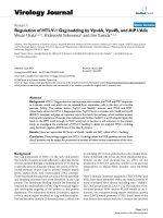
Construction and characterization of the recombinant VT7-HCV7.9 virusFigure 1
Construction and characterization of the recombinant VT7-HCV7.9 virus. A: Generation of recombinant VT7-
HCV7.9. A 7.9 Kb DNA fragment containing the structural (C, E1, E2 and p7) and nonstructural (NS3, NS4A, NS4B, NS5A and
the amino terminal region of NS5B) proteins of HCV from genotype 1b was cloned into a unique EcoRI restriction site of
pVOTE.1 to make the plasmid transfer vector pVOTE.1-HCV7.9. BSC40 cells infected with the recombinant VT7lacOI (VT7),
were transfected with the plasmid pVOTE.1-HCV7.9 as described in Materials and Methods to generate the recombinant VT7-
HCV7.9. B: Expression of HCV inhibits protein synthesis in mammalian cells. Monolayers of BSC40 cells were infected at 5
PFU/cell with either the parental VT7 or the recombinant VT7-HCV7.9 viruses in the presence (+) or absence (-) of the induc-
tor IPTG. Uninfected (U) and infected cells were metabolically labelled with
35
S-Met-Cys Promix (100 µCi/mL) from 4 to 24
h.p.i. as described in Materials and Methods. Approximately 100 µg of total cell protein extracted from uninfected (U) and
infected cells, was fractionated by SDS-PAGE followed by autoradiography. (*) represents new additional polypeptides corre-
sponding to the HCV proteins. C: Inducible expression of HCV proteins by recombinant VT7-HCV7.9 virus. BSC40 cells were
infected as described above. Total cell protein lysates from uninfected (U) and infected cells at 24 h.p.i. were analysed by West-
ern blot using a human anti-HCV antibody from an infected patient. The protein band migration of Core, E2, NS4B and NS5A,
as determined with specific antibodies, is indicated.
A.
HA
R
gpt
P
7.5
P
T7
SLO
EMC
HA
L
TT
HCV
7.9
P
11
P
7.5
T7gene
lacI
LacO
VT7-HCV
7.9
P
7.5
Homologous
recombination
MCS
HA
R
HA
L
gpt
P
7.5
P
T7
SLO
EMC
TT
pVOTE.1
P
11
T7gene
lacI
LacO
VT7lacOI
pcDNA-hcv1b
p7
NS2
CE2E1 NS3
NS4
AB
NS5A
NS5B
EcoRI EcoRI
/EcoRI
/EcoRI/CIP
HA
R
gpt
P
7.5
P
T7
SLO
EMC
HA
L
TT
HCV
7.9
pVOTE.1-HCV
7.9
C.B.
122
83
51
35
28
20
kDa U
VT7-HCV
7.9
IPTG
VT7
IPTG
+-+-
122
VT7-HCV
7.9
IPTG
VT7
IPTG
83
51
35
28
20
kDa
+-+-
U
E2
NS5A
NS4B
Core
Virology Journal 2005, 2:81 />Page 4 of 19
(page number not for citation purposes)
the synthesis of polypeptides not present in the absence of
IPTG (Figure 1B, see new proteins denoted with asteriks).
Significantly, in the presence of IPTG, overall protein syn-
thesis was reduced in VT7-HCV7.9 infected cells when
compared to protein synthesis in the absence of the induc-
tor. This translational inhibitory effect was specific, since
protein synthesis was not affected in cells infected with
VT7, with or without IPTG (Figure 1B). The synthesis of
HCV proteins in VT7-HCV7.9 infected cells was also doc-
umented by Western blot analysis, using sera from an
HCV-infected patient. As shown in Figure 1C, HCV pro-
teins of the expected size, for structural and nonstructural
polypeptides, were detected only in VT7-HCV7.9 infected
cells upon induction with IPTG. The size of specific HCV
proteins was confirmed following reactivity with antibod-
ies against Core, E2, NS4B and NS5A (not shown). A het-
erogeneous pattern of HCV-specific proteins was
observed, perhaps as a result of different stages of proteo-
lytic processing of the polyprotein. Confocal microscopy
using sera from an infected patient revealed that the HCV
proteins expressed in VT7-HCV7.9 infected cells upon
induction with IPTG, formed large cytoplasmic aggregates
and produced severe disruption of the golgi apparatus, a
phenomenon not observed in cells infected in the absence
of IPTG (Figure 2). The HCV proteins Core, E2, NS4B and
NS5A were individually detected intracellularly with spe-
cific antibodies in VT7-HCV7.9 infected HeLa cells upon
induction with IPTG (not shown).
The results of Figures 1, 2 reveal that the HCV ORF
included in the recombinant VT7-HCV7.9 is efficiently
transcribed during infection in the presence of IPTG, gen-
erating a viral polyprotein that is processed into mature
structural and nonstructural HCV proteins, triggering dis-
ruption of the golgi apparatus.
Expression of HCV polyprotein from VV inhibits the
production of vaccinia virus
To determine the impact of HCV gene expression on the
replication of the recombinant VT7-HCV7.9 virus, we
studied the production of infectious VV at 12, 24 and 48
h.p.i, in the presence or absence of the inductor IPTG. As
demonstrated in Figure 3 by virus plaque formation and
virus titration curves, the production of infectious VV was
significantly reduced (over 2 logs) during HCV gene
expression. These results reveal that expression of HCV
impairs VV replication.
Expression of HCV polyprotein from VV inhibits cellular
and viral protein synthesis through eIF-2
α
phosphorylation
Next, we determined the nature of the translational block
in cells infected with VT7-HCV7.9 in the presence of IPTG.
As a control, we included a recombinant VT7-VP3 induci-
bly expressing the IBDV capsid protein VP3. This virus was
constructed similarly to VT7-HCV7.9, and expresses an
mRNA encoding VP3 ORF from the vaccinia virus genome
via T7 polymerase. Cells infected with VT7-HCV7.9, in the
presence or absence of IPTG, were metabolically labelled
for 30 min with
35
S-Met-Cys Promix at 4, 8, 12 and 16
h.p.i., whole cell lysates fractionated by SDS-PAGE and
the protein pattern examined by autoradiography. As
shown in Figure 4, a clear reduction in cellular and viral
protein synthesis was observed after 4 h.p.i in cells
infected with the recombinant VT7-HCV7.9 virus in the
presence of IPTG, in contrast with cells infected in the
absence of the inductor, or in cells inducibly expressing
the VP3 protein (Figure 4A). The protein levels were quan-
tified by densitometry of the bands and are represented in
Figure 4B. A strong decrease in protein synthesis becomes
apparent by 8 h.p.i.
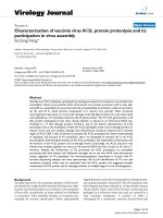
Cellular localization of HCV proteins by immunofluores-cence microscopyFigure 2
Cellular localization of HCV proteins by immunofluo-
rescence microscopy. Subconfluent HeLa cells were
infected at 5 PFU/cell with the recombinant VT7-HCV7.9 in
the presence (+) or absence (-) of the inductor IPTG. At 16
h.p.i, cells were doubly labelled with polyclonal antibody anti-
Gigantine to detect the Golgi complex (red) and a 1/200 dilu-
tion of serum from an HCV-infected patient (green) followed
by the appropriate fluorescent secondary antibody and
ToPro reagent.
VT7-HCV
7.9
-IPTG
VT7-HCV
7.9
+IPTG
Human α
αα
α-HCV
UNINFECTED
Virology Journal 2005, 2:81 />Page 5 of 19
(page number not for citation purposes)
Phosphorylation of the α subunit of the eukaryotic trans-
lation initiation factor 2 (eIF-2) on serine 51 leads to the
downregulation of translation initiation through a well-
characterized mechanism involving inhibition of eIF-2B
activity [22]. As such, we determined whether HCV poly-
protein expression altered this initiation step. Thus, the
levels of phospho-eIF-2α-S51 in VT7-HCV7.9 infected
cells, in the presence or absence of IPTG, were determined
by immunoblot analysis. The results obtained showed
that expression of HCV is related to levels of eIF-2α-S51
phosphorylation over time, relative to non-induced VT7-
HCV7.9 infected cells (Figure 4C). Similar levels of phos-
phorylation have been shown to cause growth inhibitory
effects in yeast, as well as in mammalian cells [23]. The
levels of phospho-eIF-2α-S51 in VT7-VP3 infected cells in
the presence of IPTG at the assayed times, were similar to
the levels obtained in uninduced VT7-HCV7.9 infected
cultures (Figure 4C), and represent the values usually
found in VV-infected cells. A shorter time-course analysis
of the extent of inhibition of protein synthesis and of eIF-
2α-S51 phosphorylation indicates that such effects are
clearly observed by 6 h.p.i in VT7-HCV7.9 infected cul-
tures in the presence of IPTG (not shown).
To further assess the role of eIF-2α phosphorylation on
the translational arrest, we examined whether expression
of the dominant negative non-phosphorylated mutant
Ser51-Ala (eIF-2α-S51A) was capable of rescuing the
translation inhibitory effects of HCV gene expression. To
this end, different combinations of recombinant viruses,
VT7-HCV7.9, VT7 and VV-eIF2αNP (inducibly expressing
the eIF-2α-S51A mutant), were assayed in the presence or
absence of IPTG. The metabolic labelling of infected cells
revealed that expression of eIF2α-S51A mutant in cells co-
infected with VT7-HCV7.9 in the presence of IPTG, res-
cues the translational block caused after HCV polyprotein
expression (Figure 5A: compare lanes 3, 4 and 6 with lanes
1 and 2). In the absence of IPTG, protein synthesis levels
were not affected (Figure 5B).
The above findings demonstrate that the translational
block induced after HCV polyprotein expression from VV
involves eIF-2α phosphorylation.
HCV polyprotein expression from VV in the hepatic cell
line HepG2 inhibits cellular and viral protein synthesis
The HCV is a hepatotropic virus, thus we set out to study
the effects of HCV gene expression in a hepatoblast cell
line. HepG2 cells were infected with VT7 or VT7-HCV7.9
in the presence or absence of IPTG, metabolically labelled
with
35
S-Met-Cys Promix from 4 to 24 h.p.i, cell extracts
fractionated by SDS-PAGE, and the protein pattern visual-
ized upon autoradiography analysis. As shown in Figure
6A, cells infected with the recombinant VT7-HCV7.9 virus
in the presence of IPTG demonstrated the synthesis of
new additional polypeptides corresponding to HCV pro-
teins (confirmed by Western blot, not shown), with a
marked reduction in protein synthesis, in comparison
with cells infected in the absence of the inductor, or in
those cells inducibly expressing the T7 RNA polymerase
(VT7). Expression of HCV results in decreased levels of VV
proteins, as shown by a Western blot using anti-VV anti-
bodies (Figure 6B) and increased phosphorylation levels
of eIF-2α-S51 (Figure 6C). These results indicate that HCV
Expression of HCV polyprotein inhibits the production of infectious VVFigure 3
Expression of HCV polyprotein inhibits the produc-
tion of infectious VV. BSC40 cells were infected at 5 PFU/
cell with the recombinant VT7-HCV7.9 in the presence or
absence of IPTG. After the indicated times postinfection the
cells were collected, centrifuged and resuspended in 300 µL
of DMEM. After three freeze-thawing cycles, followed by
sonication, the cell extracts were titrated in BSC40 cells. The
experiment was performed two times in duplicate. Means
and standard deviations are shown.
12h 24h 48h
IPTG+
5.7 x 10
5
IPTG-
1.1 x 10
8
1.6 x 10
6
7.0 x 10
5
1.0 x 10
8
6.6 x 10
8
12h 24h 48h
+IPTG
-IPTG
VT7-HCV
7.9
IPTG+
IPTG-
12h 24h 48h
10
5
10
6
10
7
10
8
10
9
Time
Log (titer)
Virology Journal 2005, 2:81 />Page 6 of 19
(page number not for citation purposes)
Time-course analysis of cellular and viral protein synthesis in cells expressing HCV polyproteinFigure 4
Time-course analysis of cellular and viral protein synthesis in cells expressing HCV polyprotein. A: BSC40 cells
infected with the recombinant VT7-HCV7.9 virus in the presence (+) or absence (-) of IPTG were metabolically labelled with
[
35
S] Met-Cys Promix (50 µCi/mL) at the indicated times (h.p.i) and analysed by SDS-PAGE (12%) and autoradiography. For
comparative purposes, we included a similar inducible recombinant virus but expressing the IBDV mature structural capsid
protein VP3 (VT7-VP3). B: Inhibition of VV proteins after expression of HCV. The levels of VV proteins were quantitated from
autoradiograms using a BioRad GS700 image densitometer and computer software as suggested by the manufacturer. C:
Immunoblot analysis of phospho-eIF-2α-S51 protein levels during the time-course of VT7-HCV7.9 infection. The number
appearing in each lane represents the ratio of phospho-eIF-2α-S51 levels in infected cells compared to levels in uninfected cells.
C.
Fold (x)
eIF2α
αα
α-P
1.3 4.5
5.9
5.0 1.7 2.5 2.5 2.1 1.8 2.5 2.5 2.7
+IPTG
u481216481216
4 8 12 16
-IPTG +IPTG
VT7-VP3
kDa
122
83
51
35
28
20
A.
VT7-HCV
7
.
9
VP3
B.
0
50
100
150
200
250
8h 12h 16h
VT7-HCV7.9
+
IPTG
VT7-HCV7.9
-
IPTG
VT7-VP3
+
IPTG
VV antigens
Time
OD Arbitrary units
Virology Journal 2005, 2:81 />Page 7 of 19
(page number not for citation purposes)
polyprotein expression from VV inhibits cellular and viral
protein synthesis in hepatoblast cells, which correlates
with eIF-2α-S51 phosphorylation.
Phosphorylation of eIF-2
α
and translational inhibition
induced by HCV polyprotein expression from VV is
mediated by PKR
Inhibition of translation through phosphorylation of eIF-
2α, is a major stress-responsive checkpoint employed by
at least four cellular kinases: PKR, PERK, GCN2, and HRI
[24-27]. In particular of these four kinases, PKR has been
shown to be the key regulator of cell defence against viral
infections, and mediates the antiviral and antiprolifera-
tive effects of interferon (IFN) [28]. Activated PKR phos-
phorylates the α subunit of eIF-2 on serine 51, thus
halting initiation of translation of both cellular and viral
proteins that eventually leads to inhibition of viral repli-
cation [24].
In order to determine if PKR was the kinase responsible
for eIF-2α phosphorylation following expression of HCV
from VV, we infected PKR knockout cells (PKR-/-) and
PKR WT cells (PKR+/+) with VT7 or VT7-HCV7.9
recombinant viruses in the presence of IPTG. As shown in
Figure 7A, higher eIF-2α phosphorylation levels were
observed in PKR+/+ than in PKR-/- cells after VT7-HCV7.9
infection. The total levels of eIF-2α and β-actin proteins
were similar for both cell lines, in uninfected, as well as in
Expression of the dominant negative eIF-2α-S51A mutant by VV-eIF2αNP rescues the translation inhibition induced by HCV polyproteinFigure 5
Expression of the dominant negative eIF-2α-S51A mutant by VV-eIF2αNP rescues the translation inhibition
induced by HCV polyprotein. BSC40 cells grown in 12-well plates were infected at a total of 9 PFU/cell with the viruses
indicated in the presence or absence of IPTG (1.5 mM). At 18 h.p.i. the cells were metabolically labeled with [
35
S] Met-Cys
Promix (50 µCi/mL) for 30 min. and analysed by SDS-PAGE (12%) and autoradiography.
123456
VT7-HCV7.9 6 PFU 3 PFU 3 PFU 3 PFU 6 PFU
VV-eIF2αNP 3PFU 6PFU 6PFU 3PFU
VT7 3 PFU 6 PFU 3 PFU 3 PFU
- -
-
U
122
83
51
35
28
20
kDa
7
1 2 345 612345 6
+IPTG -IPTG
Panel A Panel B
Virology Journal 2005, 2:81 />Page 8 of 19
(page number not for citation purposes)
VT7 or VT7-HCV7.9 infected cells. To corroborate whether
eIF-2α phosphorylation halts translation of cellular and
viral proteins, PKR-/- and PKR+/+ cells were infected with
VT7-HCV7.9 in the presence or absence of IPTG, metabol-
ically labelled, cell extracts fractionated by SDS-PAGE and
proteins pattern visualized employing autoradiography.
Only those PKR+/+ VT7-HCV7.9 infected cells in the pres-
ence of IPTG, showed a significant reduction of cellular
and viral protein synthesis (Figure 7B). As expected, the
expression of PKR by VV-PKR when used as a positive
control, suppressed protein synthesis in both cell lines.
Those data indicates that such cells are responsive to exog-
enous PKR delivered by VV.
These findings reveal that PKR is the kinase responsible
for eIF-2α phosphorylation as well as for the translational
block following HCV polyprotein expression from VV in
infected cells.
HCV polyprotein expression from VV induces apoptosis in
HeLa and HepG2 cells, an effect that is caspase-
dependent
It has been reported that expression in hepatic cells of all
structural and nonstructural proteins from HCV cDNA
[29] or from full-length RNA [30], can lead to apoptotic
cell death, which may be an important event in the
pathogenesis of chronic HCV infection in humans. To
Expression of HCV polyprotein from VV inhibits cellular and viral protein synthesis in the hepatic cell line HepG2Figure 6
Expression of HCV polyprotein from VV inhibits cellular and viral protein synthesis in the hepatic cell line
HepG2. A: Monolayers of HepG2 cells were infected (5 PFU/cell) with either VT7 or VT7-HCV7.9 recombinant viruses, in
the presence (+) or absence (-) of the inductor IPTG. Uninfected (U) and infected cells were metabolically labelled with [
35
S]
Met-Cys Promix (100 µCi/mL) from 4 to 24 h.p.i and treated as described under Materials and Methods. Approximately 100 µg
of total cell protein extracted from uninfected and infected cells was fractionated by SDS-PAGE followed by autoradiography.
(*) represents new additional polypeptides corresponding to the HCV proteins. B: Immunoblot analysis of total cell protein
lysates prepared from uninfected and infected cells at 24 h.p.i. The blot was probed with a rabbit polyclonal anti-serum raised
against live VV. C: The blot was stripped and probed again with a polyclonal antibody that recognized phospho-eIF-2α-S51
protein.
B
122
83
51
35
28
20
kDa
7
eIF2a-P
C
U
VT7-HCV
7.9
VT7
IPTG IPTG
-+ - +
U
VT7-HCV
7.9
VT7
IPTG IPTG
-+-+
A
122
83
51
35
28
20
kDa
*
*
*
*
Virology Journal 2005, 2:81 />Page 9 of 19
(page number not for citation purposes)
investigate whether apoptosis occurs in our virus-cell sys-
tem, HeLa and HepG2 cells were infected with the recom-
binant VT7-HCV7.9 or coinfected with the recombinant
VV-Bcl2 (that inducibly expresses the anti-apoptotic Bcl-2
polypeptide) in the presence or absence of IPTG. The
levels of apoptosis were determined at 24 h.p.i (for HeLa
cells) or at 48 h.p.i (for HepG2 cells), using an ELISA-
based assay that detects the amount of cytoplasmic his-
PKR mediates phosphorylation of eIF-2α and inhibition of translation caused by the expression of HCV polyproteinFigure 7
PKR mediates phosphorylation of eIF-2α and inhibition of translation caused by the expression of HCV poly-
protein. A: Immunoblot analysis of total cell protein lysates prepared from PKR knockout (PKR-/-) and PKR WT (PKR+/+)
cells infected with the parental (VT7) or the recombinant VT7-HCV7.9 viruses in the presence (+) of IPTG for 24 h. The blot
was first probed with a polyclonal antibody that recognized phospho-eIF-2α-S51 protein, stripped twice, and reprobed with a
polyclonal antibody that recognizes total eIF-2α protein and a monoclonal antibody against β-actin. B: Wild type and PKR-/-
cell lines infected with VT7-HCV7.9 in the presence (+) or absence (-) of IPTG were metabolically labelled with
35
S-Met-Cys
Promix (50 µCi/mL) at 16 h.p.i, fractionated by SDS-PAGE and analysed by autoradiography. The recombinant VV-PKR virus
was used as a control. U: uninfected cells.
A.
eIF2α
αα
α
eIF2α
αα
α-S51-P
β
ββ
β-actin
PKR+/+
U
VT7
VT7-
HCV
7.9
PKR-/-
U
VT7
VT7-
HCV
7.9
PKR+/+
U
VT7-HCV
7.9
VV
PKR
-
++IPTG
B.
U
VT7-HCV
7.9
VV
PKR
-
++ IPTG
PKR-/-
Virology Journal 2005, 2:81 />Page 10 of 19
(page number not for citation purposes)
tone-associated DNA fragments. As shown in Figure 8
(panels A and B), expression of HCV by VT7-HCV7.9 in
the presence of IPTG, induces apoptosis to levels similar
to those obtained in induced VV-PKR-infected cells, used
as a positive control. These apoptosis levels were two fold
higher than those found in uninduced VT7-HCV7.9
infected cells. Co-expression from VV of HCV and of Bcl-
2 in HeLa and HepG2 cells infected in the presence of
IPTG, generates a two-fold reduction in apoptosis levels.
A higher reduction in apoptosis was obtained by the Z-
VAD-FMK general caspase inhibitor. These results
revealed that HCV polyprotein expression from VV
induced an apoptotic response, an effect mediated by
caspases.
Apoptosis induced by HCV polyprotein expression from VV
is mediated by RNase L in a PKR-independent manner
In addition to PKR, the antiviral effects of IFN are executed
through the functions of various proteins, including 2'5
oligoadenylate synthetase (2'-5AS), RNase L and Mx [31-
34]. The 2'-5AS/RNase L and PKR pathways respond to
dsRNA produced during the course of viral infections, to
trigger an antiviral response in cells through RNA degrada-
tion and inhibition of protein synthesis. In contrast, Mx
proteins obstruct the replicative cycles of particular nega-
tive strand RNA viruses by interfering with the intracellu-
lar movement and functions of viral proteins [28].
Once it was verified that PKR was the kinase responsible
for eIF-2α phosphorylation and for the translational
block following expression of HCV from VV, we assayed
the activity of RNase L under the same conditions. HeLa
cells were infected with VT7 or VT7-HCV7.9 recombinants
in the presence or absence of IPTG for 24 h. Total RNA was
fractionated in 1% agarose-formaldehyde gel and stained
with ethidium bromide. As shown in Figure 9A, cells
infected with VT7-HCV7.9 in the presence of IPTG exhib-
ited ribosomal RNA degradation. This effect is mediated
by RNase L since a similar pattern of rRNA cleavage
products is observed by the co-expression of RNase L and
2-5AS delivered by the recombinant VVs, used as a posi-
tive control. In cells infected with either VT7 or VT7-
HCV7.9 in the absence of IPTG, ribosomal RNAs were
intact. The results of Figure 9A reveal that expression of
HCV from VV induces the activation of RNase L.
One interesting parallel between the PKR and 2-5A system
is that both pathways contribute to apoptosis [35,36]. In
order to compare the role of these pathways in the apop-
totic response induced by HCV, we used PKR and RNase L
knockout cells. PKR+/+ and PKR-/- as well as RL+/+ and
RL-/- cells were infected with VT7 or VT7-HCV7.9 recom-
binants in the presence of IPTG, and the apoptotic levels
were determined by ELISA at 24 h.p.i. As seen in Figure 9,
expression of HCV by VT7-HCV7.9 induces apoptosis in
PKR+/+ (Figure 9B) and RL+/+ cells (Figure 9C). The lev-
els of apoptosis were similar to those obtained after the
expression of PKR from VV-PKR, used as positive control.
The levels of apoptosis induced by VT7-HCV7.9 after
addition of IPTG, were significantly decreased in RL-/-
infected cells (Figure 9C), while in PKR-/- cells, such levels
remained similar to those in PKR+/+ cells (Figure 9B).
These findings indicate that expression of HCV by VT7-
HCV7.9 triggers apoptosis through RNase L, in a PKR-
independent pathway.
Finally, we analysed cellular and viral protein synthesis in
RNase L knockout cells expressing HCV. Consequently,
RL+/+ and RL-/- cells were infected with VT7-HCV7.9 in
the presence or absence of IPTG, metabolically labelled,
cell extracts fractionated by SDS-PAGE and the pattern of
proteins visualized using autoradiography. As shown in
Figure 10, the expression of HCV provokes a similar
reduction of cellular and viral protein synthesis in RL-/-
and RL+/+ infected cells upon induction with IPTG (Fig-
ure 10A). This translational block correlates with
increased levels of phosphorylation eIF-2α-S51 (Figure
10B) through PKR which is active in both cell lines. This
result corroborates that apoptosis induced by HCV
through RNase L is independent of the inhibition of pro-
tein synthesis caused by PKR.
Discussion
Understanding the molecular mechanisms by which IFN-
based therapies decreases HCV viral load, reduces the
number of viral quasispecies, improves liver function, and
reduces liver fibrosis in 15–30% of patients, is a priority
in HCV research. Consequently, both viral and host fac-
tors have been implicated during the effective clinical
response or resistance phenomenon of patients to IFN
treatment [37]. Different in vitro model systems have been
developed to study the role of HCV polyprotein on host
cell responses [12-21]. The implication of IFN-induced
genes and their action in the antiviral response of the host
to HCV expression is not yet fully understood.
To further characterize the antiviral response of the host
during expression of HCV polyprotein, we developed a
novel virus-cell system based on a poxvirus vector, that
inducibly expresses the structural and nonstructural
(except part of NS5B) proteins of HCV ORF from geno-
type 1b. The generated recombinant VT7-HCV7.9 virus
contains the HCV DNA coding region inserted within the
VV HA locus, under the transcriptional control of a T7
promoter, and expresses the T7 RNA polymerase upon
induction with IPTG (see Figure 1A). Current systems rely-
ing on viral delivery of T7 RNA polymerase are restricted
by the efficiency with which HCV cDNAs can be
transfected into cells, which in the case of hepatocyte and
hepatocyte-derived cell lines, is often low [16-18]. The
Virology Journal 2005, 2:81 />Page 11 of 19
(page number not for citation purposes)
Expression of HCV polyprotein from VV induces apoptosis in HeLa and HepG2 cells that is caspase-dependentFigure 8
Expression of HCV polyprotein from VV induces apoptosis in HeLa and HepG2 cells that is caspase-dependent.
A: HeLa cells were infected at 5 PFU/cell with the recombinant VT7-HCV7.9 individually or in combination (2.5 PFU of each
virus/cell) with the recombinant VV-Bcl2 (inducibly expressing the anti-apoptotic Bcl-2 polypeptide) or with a general caspase
inhibitor, Z-VAD-FMK (Calbiochem) at 50 µM, in the presence (+) or absence (-) of IPTG. The apoptotic levels were deter-
mined at 24 h.p.i by ELISA. B: HepG2 cells were infected at 10 PFU/cell with the recombinant VT7-HCV7.9 individually or in
combination (5 PFU of each virus/cell) with the recombinant VV-Bcl2 or with a general caspase inhibitor, Z-VAD-FMK (Calbio-
chem) at 50 µM, in the presence (+) or absence (-) of IPTG. The apoptotic levels were determined at 48 h.p.i by ELISA. VV-
PKR infected cells in the presence (+) of IPTG were used as positive controls.
B.
HepG2
VT7-HCV
7.9
+IPTG -IPTG
VT7-HCV
7.9
+VV-Bcl2
+IPTG -IPTG
VV-PKR
+IPTG
MOCK
0
0.5
1
1.5
OD
405nm
VT7-HCV
+ZVAD
+IPTG
A.
HeLa
VT7-HCV
7.9
+IPTG -IPTG
VT7-HCV
7.9
+VV-Bcl2
+IPTG -IPTG
VV-PKR
+IPTG
MOCK
0
0.5
1
1.5
2
OD
405nm
VT7-HCV
+ZVAD
+IPTG
Virology Journal 2005, 2:81 />Page 12 of 19
(page number not for citation purposes)
Expression of HCV polyprotein from VV induces ribosomal RNA degradation mediated by RNaseL and triggers apoptosis through RNase L independently of PKRFigure 9
Expression of HCV polyprotein from VV induces ribosomal RNA degradation mediated by RNaseL and trig-
gers apoptosis through RNase L independently of PKR. A: Monolayers of HeLa cells were either uninfected (U), single-
infected with VT7 (5 PFU/cell), single-infected with VT7-HCV7.9 (5 PFU/cell) in the presence (+) or absence (-) of IPTG, or tri-
ple-infected with VV-RL + VT7 + VV-25AS (2 PFU of each virus/cell) (C+). Infections proceeded for 24 hours. 2 µg of total
RNA was fractionated in 1% agarose-formaldehyde gel and stained with ethidium bromide. Abundant ribosomal RNAs 28S and
18S are indicated. B and C: PKR knockout (PKR-/-) and PKR WT cells (PKR+/+) (panel B), as well as RNase L knockout (RL-/
-) and RNase L WT cells (RL+/+) (panel C), were infected at 5 PFU/cell with the recombinant VT7-HCV7.9 virus, in the pres-
ence (+) or absence (-) of the inductor IPTG. The apoptotic levels in cell extracts were determined at 24 h.p.i. by ELISA. The
recombinant VV-PKR virus was used as a control. U: Uninfected cells.
0
0.1
0.2
0.3
0.4
0.5
-IPTG +IPTG
VT7-HCV
7.9
+IPTG
VV-PKR
OD
405 nm
PKR+/+
PKR-/-
0
0.1
0.2
0.3
0.4
0.5
0.6
0.7
0.8
-IPTG +IPTG
VT7-HCV
7.9
+IPTG
VV-PKR
OD
405 nm
RL+/+
RL-/-
B.
A.
U
28S r R NA
18S r R NA
Ribosomal RNA
cleavage products
V
T
7
V
T
7
-
H
C
V
7
.
9
++
-
C
+
I
P
T
G
C.
Virology Journal 2005, 2:81 />Page 13 of 19
(page number not for citation purposes)
Expression of HCV polyprotein from VV inhibits cellular and viral protein synthesis in RL+/+ and in RL-/- infected cellsFigure 10
Expression of HCV polyprotein from VV inhibits cellular and viral protein synthesis in RL+/+ and in RL-/-
infected cells. A: RL+/+ and RL-/- cells infected with VT7-HCV7.9 in the presence (+) or absence (-) of IPTG were metabol-
ically labelled with
35
S-Met-Cys Promix (50 µCi/mL) at 8 h.p.i, fractionated by SDS-PAGE and analysed by autoradiography. U:
uninfected cells. B: Immunoblot analysis of total cell protein lysates prepared from RL+/+ and RL-/- cells infected with VT7-
HCV7.9 in the presence (+) or absence (-) of IPTG for 8 h. The blot was first probed with a polyclonal antibody that recog-
nized phospho-eIF-2α-S51 protein, stripped and reprobed with a polyclonal antibody that recognizes total eIF-2α protein.
A.
U
VT7-HCV
7.9
+
IPTG
-
U
VT7-HCV
7.9
+
IPTG
-
RL-/-RL+/+
B.
RL+/+
U
RL-/-
U
VT7-HCV
7.9
-
+
VT7-HCV
7.9
-
+
eIF2α
αα
α
eIF2α
αα
α-S51-P
IPTG
Virology Journal 2005, 2:81 />Page 14 of 19
(page number not for citation purposes)
poxvirus-based system described here permits both the
regulated production of the HCV transcripts into cells and
the efficient delivery of the HCV genome into a wide vari-
ety of primary and continuous cell lines.
In this study, we demonstrate that upon induction with
IPTG, HCV proteins are efficiently produced in VT7-
HCV7.9 infected cells of various origins. This observation
indicates that the DNA fragment of HCV ORF included in
the VV genome, is efficiently transcribed and translated
into a viral polyprotein precursor that is correctly
processed into mature structural and nonstructural HCV
proteins, as confirmed with specific antibodies to individ-
ual HCV proteins. Significantly, inducible expression of
HCV polyprotein in VT7-HCV7.9 infected cells caused a
considerable reduction in the production of infectious VV,
as well as striking inhibition in total protein synthesis,
both viral and cellular. The translational block was
observed by 6 h.p.i when all of the HCV proteins were
produced. The inhibition of protein synthesis by HCV was
highly specific and could not be solely attributed to the
induction of HCV RNA transcript since cells infected with
VT7-VP3 that expressed the IBDV ORF VP3 mRNA, did
not trigger translational inhibition. Furthermore, the HCV
ORF included in the VT7-HCV7.9 recombinant virus lacks
the 5' UTR, bearing the HCV IRES, and the 3' UTR, both
implicated in HCV replication and liver injury [38]. The
inhibition of protein synthesis that we have observed in
induced VT7-HCV7.9 infected HeLa and HepG2 cells was
associated with a significant increase in the phospho-eIF-
2α-S51 levels, suggesting that HCV expression might
control the cellular translation through eIF-2α-S51 phos-
phorylation. This translational control was confirmed
with a dominant negative non-phosphorylated (NP)
mutant Ser51-Ala (eIF-2α-S51A). Expression of the eIF-
2α-S51A mutant in cells co-infected with VV-eIF-2α-NP
and VT7-HCV7.9 in the presence of IPTG, rescued the
translational block induced by HCV (Figure 6). Moreover,
we showed that phosphorylation of eIF-2α-S51 was car-
ried out by the cellular kinase PKR, as revealed in knock-
out PKR-/- cells (Figure 9). The role of PKR and eIF-2α-S51
phosphorylation in HCV infection has been widely stud-
ied due to the relevance of this kinase in the cellular anti-
viral response. As has been previously reported [23,39-
41], PKR mediated phosphorylation of eIF-2α-S51 results
in inhibition of translation and a blockade of viral protein
synthesis, which in turn, inhibits virus replication. For this
reason, viruses employ a variety of strategies to inhibit
PKR activation and function. Several groups have
described the role of certain HCV proteins in cellular
translation. HCV NS4A and NS4B proteins mediate trans-
lational inhibition and, perhaps, increased degradation of
certain cellular proteins [42,43]. In contrast, NS5A and E2
proteins are reported to enhance translation by inhibiting
PKR functions [44,45]. Therefore, it seems that during the
course of HCV infection, there is a balance between inhi-
bition and enhancement of host cell translation depend-
ing on the degree of activation/inhibition of the PKR
pathway. Most of these studies have relied on systems that
express HCV proteins individually. Nontheless, since all
HCV proteins are potentially produced in vivo during virus
infection of hepatocytes, it is important to use a full-
length genome rather than individual HCV proteins to
study the molecular mechanisms involved in virus-host
cell interactions and in HCV pathogenesis. In our viral
delivery system, the overall expression of structural and
nonstructural HCV proteins by recombinant VT7-HCV7.9
virus did not reverse the action of PKR, since host cell
translation was inhibited through phosphorylation of eIF-
2α-S51 by the kinase. An incapability to prevent PKR acti-
vation by HCV polyprotein expression was reported by
François and co-workers when they analysed the response
to IFN of the human cell line UHCV-11 engineered to
inducibly express the entire HCV genotype 1a polyprotein
[46]. Although we could not exclude the possibility that a
certain level of inhibition of PKR by NS5A or E2 occurs at
a much localized level, the resistance to IFN exhibited by
some HCV genotypes as a result of viral protein expres-
sion, cannot be explained solely by inhibition of the neg-
ative control of PKR translation. It is possible that during
the course of HCV infection, NS5A plays a role in inhibit-
ing PKR locally at the site of HCV protein synthesis. NS5A
may, however, participate in the blockade of IFN's antivi-
ral action through another mechanism, such as the
reported interaction with the Ras-associated Grb-2 protein
[47]. These results confirm the necessity to re-evaluate all
types of interactions between any particular HCV protein
and its cellular partner(s) in the context of expression of
all of the HCV proteins. Consequently, as shown here by
confocal microscopy (Figure 2), the HCV proteins are
localized within aggregates in the cell cytoplasm which
might influence their interaction with PKR, a protein
found surrounding the nucleus, in microsomes and in the
nucleolus [24,48].
Several in vitro studies reveal that synthesis of HCV struc-
tural proteins or the full-length genome have a direct
cytotoxic effect or activate an apoptotic response in
osteosarcoma, hepatoma and B cell lines [29,30,49-51].
Furthermore, the alteration of ER membranes [52] and
the activation of signalling pathways characteristic of an
ER-stress condition, have been found to be associated
with the expression of HCV proteins [53-55]. Although
these data suggest that HCV may alter intracellular events
with possible consequences on liver pathogenesis, the
complex mechanism and the role of the viral proteins
implicated are currently unknown. As we have shown in
this work, expression of most of the HCV genome from
VV induces a cell death phenomenon by apoptosis that
should contribute to liver pathogenesis. Apoptosis
Virology Journal 2005, 2:81 />Page 15 of 19
(page number not for citation purposes)
induced by HCV polyprotein expression was prevented by
Bcl-2 and by a general caspase inhibitor (Z-VAD-FMK)
indicating a caspase-dependent death process. Even
though PKR is the main kinase responsible for eIF-2α
phosphorylation and for translation inhibition induced
by the expression of HCV in VT7-HCV7.9 infected cells, it
does not appear to be involved in apoptosis within this
system, as revealed from studies performed in PKR+/+ and
PKR-/- knockout cells. The extent of apoptosis induction
by HCV expression was the same in PKR+/+ and in PKR-/
- cells (Figure 9B), suggesting that other pathways may be
involved. PKR induces apoptosis in response to activation
by different stimuli, such as the accumulation of dsRNA as
a by-product during virus replication [36], or when PKR is
overexpressed in cells [56]. Several authors, however, have
reported that PKR can also be activated through the
binding of heparin and other polyanions [57,58], or by
the cellular activator protein PACT/RAX [59,60]. The
events that mediate induction of apoptosis by PKR have
been widely studied and both PKR-induced translational
block by phosphorylation of eIF-2α, and NF-kB activa-
tion, have been shown to be activated during apoptosis
[61]. Since PKR has a number of potential substrates and
signalling targets, it is likely that the phosphorylation of
eIF-2α by PKR in response to HCV expression is not suffi-
cient to mediate the pro-apoptotic effects of this kinase.
In this study, we also demonstrate the activation of endog-
enous RNase L and its role in the apoptosis induced by
HCV expression (Figure 9A,C). Although it is widely
accepted that the IFN-induced proteins PKR and RNase L
require the expression of dsRNA for their activation
(either directly in the case of PKR or indirectly via 2'-5'-
OAS in the case of RNase L), there are several reports that
documented the effect of HCV proteins on PKR and 2'-5'-
OAS activation. The NS5A and E2 proteins can suppress
the PKR pathway [44,45], whereas the Core protein can
transcriptionally activate the 2'-5'-OAS gene through an
IRES present within IFN-inducible gene promoter [62].
Like PKR, the 2-5AS/RNase L system can control virus
growth by inducing apoptosis in response to viral infec-
tion [35,36]. Overexpression of RNase L or activation of
the endogenous enzyme induces apoptosis by a mito-
chondrial-caspase dependent pathway that is suppressed
by Bcl-2 [63-65]. Similarly, apoptosis induced by HCV
polyprotein expression was inhibited by Bcl-2 (Figure 8).
Although the apoptotic levels induced by HCV proteins
remain invariable in PKR+/+, PKR-/-, and RL+/+ cells, the
levels are significantly decreased in RL-/- cells, indicating
that inducible expression of HCV proteins by VT7-
HCV7.9 triggers apoptosis through RNase L in a PKR-
independent pathway. Under physiologic conditions,
RNase L activity is tightly regulated by 2'-phosphodieste-
rase and RNase L inhibitor [66,67] such that only a lim-
ited activation of RNase L occurs. The mechanism of the
regulation of RNase L inhibitor is unknown, but the
reduction of its expression seems to be advantageous for
host defence together with the enhanced 2-5 OAS activity.
Yu and co-workers [68] described that hepatic overexpres-
sion of PKR mRNA, and reduced expression of an RNase
L inhibitor mRNA, are parameters that seem to contribute
to an anti-HCV response. In agreement with our results, it
has been reported that the absence of RNase L has an anti-
apoptotic effect in multiple cell types treated with a variety
of different agents [69]. The effects that have been
observed in this study upon HCV polyprotein expression
from VV are likely to have biological significance during
HCV infection as there is ample evidence that VV recom-
binants can be used to study the function of multiple
genes and that the assigned function mimics the effects
described in non-viral systems [70].
Conclusion
We have developed an efficient viral delivery system
expressing the polyprotein of HCV in numerous mamma-
lian cell lines in a faithfully, efficient and time regulated
manner, allowing us to analyze the host response to HCV
proteins. We demonstrate that two components of the
interferon (IFN) system, protein kinase PKR and RNase L,
are activated during HCV polyprotein expression and are
responsible for translational control and induction of
apoptosis. These two pathways are likely to limit the rep-
lication capacity of HCV. Thus, the virus-cell system
described here highlights the relevance of the IFN system
as a protective mechanism against HCV infection.
Methods
Cells and viruses
Cells were maintained in a humidified air 5% CO
2
atmos-
phere at 37°C. African green monkey kidney cells
(BSC40) and human cells (HeLa) were grown in Dul-
becco's modified Eagle's medium (DMEM) supplemented
with 10% newborn calf serum (NCS). Human HepG2
hepatocellular carcinoma cells (ATCC HB-8065) were
maintained in DMEM supplemented with penicillin (0.6
µg/mL); streptomycin (60 µg/mL); glutamine (2 mM); N-
2-hydroxyethylpiperazine-N'-2-ethanosulfonic acid
(HEPES) buffer, pH 7.4 (20 mM) and 10% fetal calf serum
(FCS). Mouse 3T3-like fibroblasts derived either from
homozygous PKR knockout mice (PKR-/-) or PKR wild
type mice (PKR+/+) [71] were obtained from C. Weiss-
mann (University of Zurich, Switzerland) and grown in
DMEM supplemented with 10% FCS. Wild type mouse
embryo fibroblasts (MEFs) derived from C57BL6 mice
(RL+/+) and fibroblast lacking the RNase L gene (RL-/-
MEFs) derived from mice with the RNase L gene disrupted
[35], were propagated in DMEM supplemented with 10%
FCS, and were a gift from R. Silverman (Cleveland Clinic,
USA)
Virology Journal 2005, 2:81 />Page 16 of 19
(page number not for citation purposes)
The recombinant vaccinia virus (VV) that is inducible and
expresses the T7 RNA polymerase (VT7lacOI) was previ-
ously described [72]. Virus VT7-VP3 expressing the IBDV
mature structural capsid protein VP3 [73] was kindly pro-
vided by J.F. Rodríguez (CNB, Spain). VVeIF-2α NP was
generated through homologous recombination in TK
-
143B cells, as previously reported [74]. The recombinant
VV-PKR TK
-
expressing IPTG-inducible PKR was generated
by homologous recombination of their respective pPR35-
derived plasmid with the WR strain of VV in BSC40 cells,
as previously described [56]. VV recombinant expressing
Bcl-2 protein (VV-Bcl2) was generated as previously
reported [75]. The recombinant vaccinia viruses VV-RL
and VV-2-5AS were obtained after introduction of
plasmid pTM-RL and pSC-2-5AS respectively into the TK
region of wild-type vaccinia virus (WR) DNA by homolo-
gous recombination as described [76]. All VV
recombinants were grown in BSC40 cells and purified by
banding on sucrose gradients [77].
Generation of the recombinant vaccinia VT7-HCV7.9 virus
A 7.9 Kb DNA fragment containing the structural (C, E1,
E2 and p7) and nonstructural (NS2, NS3, NS4A, NS4B,
NS5A and the amino terminal region of NS5B) proteins of
HCV ORF from genotype 1b was excised with EcoRI from
the original full-length HCV genome containing plasmid
pcDNA-hcv1b (kindly provided by Ilkka Julkunen from
National Public Health Institute, Finland). This DNA frag-
ment was cloned into the VV insertion/expression vector
pVOTE.1 [72] previously digested with EcoRI and dephos-
phorylated by incubation with alkaline phosphatase, Calf
Intestinal (CIP) as described in Figure 1A. The resulting
plasmid, pVOTE.1-HCV7.9 directs the insertion of HCV
genes into the HA locus of the VT7lacOI genome under
the transcriptional control of the T7 promoter. BSC40
cells were infected with the recombinant vaccinia virus
VT7lacOI at a multiplicity of 0.05 PFU/cell, and then
transfected with 10 µg of plasmid DNA pVOTE.1-HCV7.9
using lipofectamine reagent according to manufacturer's
instructions (Invitrogen). The selection and amplification
of the recombinant VT7-HCV7.9 virus was carried out as
previously described [78]. The purity of the recombinant
virus was confirmed by PCR analysis. The plasmid
pVOTE.1 as well as the VV recombinant VT7lacOI, were
kindly provided by Bernard Moss (NIH, USA).
Metabolic labelling of proteins
Different cell lines grown in 12 well plates were infected
at an infection multiplicity of 5 PFU/cell with the viruses
indicated, and maintained either in the presence or
absence of the inductor isopropyl-β-D-thiogalactoside
(IPTG) (1.5 mM final concentration). For continuous
metabolic labelling of proteins, the cells were rinsed three
times with Met-Cys-free DMEM at 4 h post-infection (p.i)
and incubated with 100 µCi of [
35
S] Met-Cys Promix
(Amersham) per mL in a mixture of Met-Cys-free DMEM
and complete DMEM (9:1) for 16–20 h. After three
washes with phosphate buffered saline (PBS) cells were
resuspended in Laemmli buffer and analysed by sodium-
dodecyl sulfate-polyacrylamide gel electrophoresis (SDS-
PAGE) followed by autoradiography. For discontinuous
metabolic labelling of proteins, the cells were rinsed three
times and incubated with Met-Cys-free DMEM 30 min-
utes prior to labelling. After incubation, the medium was
removed and 50 µCi of [
35
S] Met-Cys Promix per mL in
Met-Cys-free DMEM was added for an additional 30 min-
utes. The cells were washed with PBS and treated as
described above.
Immunoblotting
The HCV-antibody positive human sera used in this study
was kindly provided by Dr Rafael Fernández from the
Ramón and Cajal Hospital (Spain). The rabbit polyclonal
anti-serum against live vaccinia virus was previously
described [79]. The rabbit polyclonal anti eIF2α [PS
51
]
phosphospecific antibody was supplied by BIOSOURCE.
The monoclonal antibody against β-actin was supplied by
SIGMA. Rabbit polyclonal anti-eIF2α antibody was sup-
plied by Santa Cruz, CA.
For immunoblot analyses, total cell extracts were boiled in
Laemmli sample buffer, and proteins were fractionated by
12% SDS-PAGE. After electrophoresis, proteins were
transferred to nitrocellulose membranes using a semi-dry
blotting apparatus (Gelman Sciences). Filters were mixed
with antisera in PBS containing non-fat dry milk at 5%
(BLOTTO), incubated overnight at 4°C, washed three
times with PBS, and further incubated with secondary
antibody coupled to horseradish peroxidase in BLOTTO.
After the PBS wash, the immunocomplexes were detected
by enhanced chemiluminescense Western blotting rea-
gents (ECL) (Amersham).
Immunofluorescence
Specific antibody for Golgi apparatus (anti-Gigantine)
was kindly provided by Manfred Renz from the Institute
of Immunology and Genetics Karlsruhe (Germany).
HeLa cells cultured on coverslips were infected at 5 PFU/
cell with VT7-HCV7.9 in the presence or absence of IPTG
(1.5 mM final concentration). At 16 h.p.i, cells were
washed with PBS, fixed with 4% paraformaldehyde and
permeabilized with 2% Triton X-100 in PBS (room tem-
perature, 5 min). Cells were incubated with a human anti-
body recognizing HCV proteins together with anti-
Gigantine antibody. Coverslips were then extensively
washed with PBS, and incubated in darkness for 1 h at
37°C, with secondary antibody conjugated with green
fluorochrome Cy2 (Jackson Immunoresearch) and with
the DNA staining reagent ToPro (Molecular Probes).
Virology Journal 2005, 2:81 />Page 17 of 19
(page number not for citation purposes)
Images were obtained by using Bio-Rad Radiance 2100
confocal laser microscope, were collected by using Laser-
sharp 2000 software and were processed in LaserPix.
Measurement of the extent of apoptosis
The cell death detection enzyme-linked immunosorbent
assay (ELISA) kit (Roche) was used according to manufac-
turer's instructions. This assay is based on the quantitative
sandwich enzyme immunoassay principle, and uses
mouse monoclonal antibodies directed against DNA and
histones to estimate the amount of cytoplasmic histone-
associated DNA fragments.
Total RNA isolation
Total RNA from uninfected or infected cells was isolated
using Ultraspect-II resin purification system (Biotecx).
RNA was denatured and analyzed in 1% formaldehyde
agarose gels and stained using ethidium bromide as previ-
ously described [76].
Competing interests
The author(s) declare that they have no competing
interests.
Authors' contributions
CEG has generated the vaccinia virus recombinant VT7-
HCV7.9 and has analyzed protein expression in culture
cells. AMV has performed confocal microscopy and
defined apoptosis in infected cells. MAG has performed
PKR and RNase L assays with KO cells. EDG has per-
formed rRNA cleavage assays. ME conceived the study, has
supervised the work, and provided the tools necessary for
the performance of the research.
Acknowledgements
This investigation was supported by research grants BIO2000-0340-P4,
BMC2002-03246 and Fundación Marcelino Botin from Spain and
QLK22002-00954 from the European Union to ME. CEG was supported by
a fellowship from Carolina Foundation and MAG from the Ministry of Sci-
ence and Technology of Spain. We thank the expert technical assistance of
Victoria Jiménez. We also thank JF Rodríguez, R. Bablanian and P. Martinez
for critically reviewing the manuscript.
References
1. Choo QL, Kuo G, Weiner AJ, Overby LR, Bradley DW, Houghton M:
Isolation of a cDNA clone derived from a blood-borne non-
A, non-B viral hepatitis genome. Science 1989, 244:359-362.
2. Kuo G, Choo QL, Alter HJ, Gitnick GL, Redeker AG, Purcell RH,
Miyamura T, Dienstag JL, Alter MJ, Stevens CE, et al.: An assay for
circulating antibodies to a major etiologic virus of human
non-A, non-B hepatitis. Science 1989, 244:362-364.
3. Robertson B, Myers G, Howard C, Brettin T, Bukh J, Gaschen B,
Gojobori T, Maertens G, Mizokami M, Nainan O, Netesov S, Nishioka
K, Shin i T, Simmonds P, Smith D, Stuyver L, Weiner A: Classifica-
tion, nomenclature, and database development for hepatitis
C virus (HCV) and related viruses: proposals for standardiza-
tion. International Committee on Virus Taxonomy. Arch Virol
1998, 143:2493-2503.
4. Bartenschlager R: The NS3/4A proteinase of the hepatitis C
virus: unravelling structure and function of an unusual
enzyme and a prime target for antiviral therapy. J Viral Hepat
1999, 6:165-181.
5. Reed KE, Rice CM: Overview of hepatitis C virus genome
structure, polyprotein processing, and protein properties.
Curr Top Microbiol Immunol 2000, 242:55-84.
6. Hoofnagle JH: Hepatitis C: the clinical spectrum of disease.
Hepatology 1997, 26:15S-20S.
7. Fabrizi F, Poordad FF, Martin P: Hepatitis C infection and the
patient with end-stage renal disease. Hepatology 2002, 36:3-10.
8. Hoofnagle JH: Management of hepatitis C: current and future
perspectives. J Hepatol 1999, 31 Suppl 1:264-268.
9. Thomas HC, Torok ME, Forton DM, Taylor-Robinson SD: Possible
mechanisms of action and reasons for failure of antiviral
therapy in chronic hepatitis C. J Hepatol 1999, 31 Suppl
1:152-159.
10. Poynard T, Moussalli J, Ratziu V, Regimbeau C, Opolon P: Effect of
interferon therapy on the natural history of hepatitis C virus-
related cirrhosis and hepatocellular carcinoma. Clin Liver Dis
1999, 3:869-881.
11. Manns MP, McHutchison JG, Gordon SC, Rustgi VK, Shiffman M, Rein-
dollar R, Goodman ZD, Koury K, Ling M, Albrecht JK: Peginter-
feron alfa-2b plus ribavirin compared with interferon alfa-2b
plus ribavirin for initial treatment of chronic hepatitis C: a
randomised trial. Lancet 2001, 358:958-965.
12. Bartenschlager R, Lohmann V: Replication of hepatitis C virus. J
Gen Virol 2000, 81:1631-1648.
13. Kato N, Shimotohno K: Systems to culture hepatitis C virus.
Curr Top Microbiol Immunol 2000, 242:261-278.
14. Lohmann V, Korner F, Koch J, Herian U, Theilmann L, Bartenschlager
R: Replication of subgenomic hepatitis C virus RNAs in a
hepatoma cell line. Science 1999, 285:110-113.
15. Ikeda M, Yi M, Li K, Lemon SM: Selectable subgenomic and
genome-length dicistronic RNAs derived from an infectious
molecular clone of the HCV-N strain of hepatitis C virus rep-
licate efficiently in cultured Huh7 cells. J Virol 2002,
76:2997-3006.
16. Chung RT, He W, Saquib A, Contreras AM, Xavier RJ, Chawla A,
Wang TC, Schmidt EV: Hepatitis C virus replication is directly
inhibited by IFN-alpha in a full-length binary expression
system. Proc Natl Acad Sci U S A 2001, 98:9847-9852.
17. Mizuno M, Yamada G, Tanaka T, Shimotohno K, Takatani M, Tsuji T:
Virion-like structures in HeLa G cells transfected with the
full-length sequence of the hepatitis C virus genome. Gastro-
enterology 1995, 109:1933-1940.
18. Myung J, Khalap N, Kalkeri G, Garry R, Dash S: Inducible model to
study negative strand RNA synthesis and assembly of hepa-
titis C virus from a full-length cDNA clone. J Virol Methods
2001, 94:55-67.
19. Yap CC, Ishii K, Aizaki H, Tani H, Aoki Y, Ueda Y, Matsuura Y, Miya-
mura T: Expression of target genes by coinfection with repli-
cation-deficient viral vectors. J Gen Virol 1998, 79 ( Pt
8):1879-1888.
20. Wakita T, Pietschmann T, Kato T, Date T, Miyamoto M, Zhao Z,
Murthy K, Habermann A, Krausslich HG, Mizokami M, Bartenschlager
R, Liang TJ: Production of infectious hepatitis C virus in tissue
culture from a cloned viral genome. Nat Med 2005, 11:791-796.
21. Zhong J, Gastaminza P, Cheng G, Kapadia S, Kato T, Burton DR, Wie-
land SF, Uprichard SL, Wakita T, Chisari FV: Robust hepatitis C
virus infection in vitro. Proc Natl Acad Sci U S A 2005,
102:9294-9299.
22. Rowlands AG, Panniers R, Henshaw EC: The catalytic mechanism
of guanine nucleotide exchange factor action and competi-
tive inhibition by phosphorylated eukaryotic initiation factor
2. J Biol Chem 1988, 263:5526-5533.
23. Clemens MJ: Protein kinases that phosphorylate eIF2a and
eIF2b and their role in eukaryotic cell translational control.
In Translational control Edited by: Hershey JWMMBSN. New York,
Cold Spring Harbor Laboratory Press; 1996:139-172.
24. Kaufman R: The double-stranded RNA-activated protein
kinase PKR. In Translational control of gene expression Edited by:
Sonenberg NHJWMMB. New York, Cold Spring Harbor Laboratory
Press; 2000:503-527.
25. Chen JJ: Heme-regulated eIF2a kinase. In Translational control of
gene expression Edited by: Sonenberg NHJWMMB. New York, Cold
Spring Harbor Laboratory Press; 2000:529-546.
Virology Journal 2005, 2:81 />Page 18 of 19
(page number not for citation purposes)
26. Hinnebusch AG: Mechanism and regulation of initiator
methionyl-tRNA binding to ribosomes. In Translational control of
gene expression Edited by: Sonenberg NHJWMMB. New York, Cold
Spring Harbor Laboratory Press; 2000:185-244.
27. Ron DHHP: PERK and translational control by stress in the
endoplasmic reticulum. In Translational control of gene expression
Edited by: Sonenberg NHJWMMB. New York, Cold Spring Harbor
Laboratory Press; 2000:547-560.
28. Stark GR, Kerr IM, Williams BR, Silverman RH, Schreiber RD: How
cells respond to interferons. Annu Rev Biochem 1998, 67:227-264.
29. Kalkeri G, Khalap N, Akhter S, Garry RF, Fermin CD, Dash S: Hep-
atitis C viral proteins affect cell viability and membrane
permeability. Exp Mol Pathol 2001, 71:194-208.
30. Kalkeri G, Khalap N, Garry RF, Fermin CD, Dash S: Hepatitis C
virus protein expression induces apoptosis in HepG2 cells.
Virology 2001, 282:26-37.
31. Staeheli P, Pavlovic J: Inhibition of vesicular stomatitis virus
mRNA synthesis by human MxA protein. J Virol 1991,
65:4498-4501.
32. Sen GC, Ransohoff RM: Interferon-induced antiviral actions and
their regulation. Adv Virus Res 1993, 42:57-102.
33. Hassel BA, Zhou A, Sotomayor C, Maran A, Silverman RH: A domi-
nant negative mutant of 2-5A-dependent RNase suppresses
antiproliferative and antiviral effects of interferon. Embo J
1993, 12:3297-3304.
34. Li XL, Blackford JA, Hassel BA: RNase L mediates the antiviral
effect of interferon through a selective reduction in viral
RNA during encephalomyocarditis virus infection. J Virol
1998, 72:2752-2759.
35. Zhou A, Paranjape J, Brown TL, Nie H, Naik S, Dong B, Chang A,
Trapp B, Fairchild R, Colmenares C, Silverman RH: Interferon
action and apoptosis are defective in mice devoid of 2',5'-oli-
goadenylate-dependent RNase L. Embo J 1997, 16:6355-6363.
36. Der SD, Yang YL, Weissmann C, Williams BR: A double-stranded
RNA-activated protein kinase-dependent pathway mediat-
ing stress-induced apoptosis. Proc Natl Acad Sci U S A 1997,
94:3279-3283.
37. Hu KQ, Vierling JM, Redeker AG: Viral, host and interferon-
related factors modulating the effect of interferon therapy
for hepatitis C virus infection. J Viral Hepat 2001, 8:1-18.
38. Chang M, Marquardt AP, Wood BL, Williams O, Cotler SJ, Taylor SL,
Carithers RLJ, Gretch DR: In situ distribution of hepatitis C
virus replicative-intermediate RNA in hepatic tissue and its
correlation with liver disease. J Virol 2000, 74:944-955.
39. Hershey JW: Translational control in mammalian cells. Annu
Rev Biochem 1991, 60:717-755.
40. Merrick WCHJW: The pathway and mechanism of eukaryotic
protein syntesis. In Translational control Edited by: Hershey
JWMMBSN. New York, Cold Spring Harbor Laboratory Press;
1996:31-70.
41. Korth MJ, Katze MG: Evading the interferon response: hepatitis
C virus and the interferon-induced protein kinase, PKR. Curr
Top Microbiol Immunol 2000, 242:197-224.
42. Florese RH, Nagano-Fujii M, Iwanaga Y, Hidajat R, Hotta H: Inhibi-
tion of protein synthesis by the nonstructural proteins NS4A
and NS4B of hepatitis C virus. Virus Res 2002, 90:119-131.
43. Kato J, Kato N, Yoshida H, Ono-Nita SK, Shiratori Y, Omata M: Hep-
atitis C virus NS4A and NS4B proteins suppress translation
in vivo. J Med Virol 2002, 66:187-199.
44. Gale MJJ, Korth MJ, Tang NM, Tan SL, Hopkins DA, Dever TE, Polyak
SJ, Gretch DR, Katze MG: Evidence that hepatitis C virus resist-
ance to interferon is mediated through repression of the
PKR protein kinase by the nonstructural 5A protein. Virology
1997, 230:217-227.
45. Taylor DR, Shi ST, Romano PR, Barber GN, Lai MM: Inhibition of
the interferon-inducible protein kinase PKR by HCV E2
protein. Science 1999, 285:107-110.
46. Francois C, Duverlie G, Rebouillat D, Khorsi H, Castelain S, Blum HE,
Gatignol A, Wychowski C, Moradpour D, Meurs EF: Expression of
hepatitis C virus proteins interferes with the antiviral action
of interferon independently of PKR-mediated control of pro-
tein synthesis. J Virol 2000, 74:5587-5596.
47. Tan SL, Nakao H, He Y, Vijaysri S, Neddermann P, Jacobs BL, Mayer
BJ, Katze MG: NS5A, a nonstructural protein of hepatitis C
virus, binds growth factor receptor-bound protein 2 adaptor
protein in a Src homology 3 domain/ligand-dependent man-
ner and perturbs mitogenic signaling. Proc Natl Acad Sci U S A
1999, 96:5533-5538.
48. Besse S, Rebouillat D, Marie I, Puvion-Dutilleul F, Hovanessian AG:
Ultrastructural localization of interferon-inducible double-
stranded RNA-activated enzymes in human cells. Exp Cell Res
1998, 239:379-392.
49. Moradpour D, Kary P, Rice CM, Blum HE: Continuous human cell
lines inducibly expressing hepatitis C virus structural and
nonstructural proteins. Hepatology 1998, 28:192-201.
50. Moradpour D, Wakita T, Wands JR, Blum HE: Tightly regulated
expression of the entire hepatitis C virus structural region in
continuous human cell lines. Biochem Biophys Res Commun 1998,
246:920-924.
51. Sung VM, Shimodaira S, Doughty AL, Picchio GR, Can H, Yen TS,
Lindsay KL, Levine AM, Lai MM: Establishment of B-cell lym-
phoma cell lines persistently infected with hepatitis C virus
in vivo and in vitro: the apoptotic effects of virus infection. J
Virol 2003, 77:2134-2146.
52. Egger D, Wolk B, Gosert R, Bianchi L, Blum HE, Moradpour D, Bienz
K: Expression of hepatitis C virus proteins induces distinct
membrane alterations including a candidate viral replication
complex. J Virol 2002, 76:5974-5984.
53. Tardif KD, Mori K, Siddiqui A: Hepatitis C virus subgenomic rep-
licons induce endoplasmic reticulum stress activating an
intracellular signaling pathway. J Virol 2002, 76:7453-7459.
54. Pavio N, Romano PR, Graczyk TM, Feinstone SM, Taylor DR: Pro-
tein synthesis and endoplasmic reticulum stress can be mod-
ulated by the hepatitis C virus envelope protein E2 through
the eukaryotic initiation factor 2alpha kinase PERK. J Virol
2003, 77:3578-3585.
55. Liberman E, Fong YL, Selby MJ, Choo QL, Cousens L, Houghton M,
Yen TS: Activation of the grp78 and grp94 promoters by hep-
atitis C virus E2 envelope protein. J Virol 1999, 73:3718-3722.
56. Lee SB, Esteban M: The interferon-induced double-stranded
RNA-activated protein kinase induces apoptosis. Virology
1994, 199:491-496.
57. George CX, Thomis DC, McCormack SJ, Svahn CM, Samuel CE:
Characterization of the heparin-mediated activation of PKR,
the interferon-inducible RNA-dependent protein kinase.
Virology 1996, 221:180-188.
58. Hovanessian AG, Galabru J: The double-stranded RNA-depend-
ent protein kinase is also activated by heparin. Eur J Biochem
1987, 167:467-473.
59. Ito T, Yang M, May WS: RAX, a cellular activator for double-
stranded RNA-dependent protein kinase during stress
signaling. J Biol Chem 1999, 274:15427-15432.
60. Patel RC, Sen GC: PACT, a protein activator of the interferon-
induced protein kinase, PKR. Embo J 1998, 17:4379-4390.
61. Gil J, Alcami J, Esteban M: Induction of apoptosis by double-
stranded-RNA-dependent protein kinase (PKR) involves the
alpha subunit of eukaryotic translation initiation factor 2 and
NF-kappaB. Mol Cell Biol 1999, 19:4653-4663.
62. Naganuma A, Nozaki A, Tanaka T, Sugiyama K, Takagi H, Mori M, Shi-
motohno K, Kato N: Activation of the interferon-inducible 2'-
5'-oligoadenylate synthetase gene by hepatitis C virus core
protein. J Virol 2000, 74:8744-8750.
63. Castelli JC, Hassel BA, Wood KA, Li XL, Amemiya K, Dalakas MC,
Torrence PF, Youle RJ: A study of the interferon antiviral mech-
anism: apoptosis activation by the 2-5A system. J Exp Med
1997, 186:967-972.
64. Diaz-Guerra M, Rivas C, Esteban M: Activation of the IFN-induc-
ible enzyme RNase L causes apoptosis of animal cells. Virology
1997, 236:354-363.
65. Rusch L, Zhou A, Silverman RH: Caspase-dependent apoptosis
by 2',5'-oligoadenylate activation of RNase L is enhanced by
IFN-beta. J Interferon Cytokine Res 2000, 20:1091-1100.
66. Benoit De Coignac A, Bisbal C, Lebleu B, Salehzada T: cDNA clon-
ing and expression analysis of the murine ribonuclease L
inhibitor. Gene 1998, 209:149-156.
67. Martinand C, Salehzada T, Silhol M, Lebleu B, Bisbal C: The RNase
L inhibitor (RLI) is induced by double-stranded RNA. J Inter-
feron Cytokine Res 1998, 18:1031-1038.
68. Yu SH, Nagayama K, Enomoto N, Izumi N, Marumo F, Sato C: Intra-
hepatic mRNA expression of interferon-inducible antiviral
genes in liver diseases: dsRNA-dependent protein kinase
Publish with BioMed Central and every
scientist can read your work free of charge
"BioMed Central will be the most significant development for
disseminating the results of biomedical research in our lifetime."
Sir Paul Nurse, Cancer Research UK
Your research papers will be:
available free of charge to the entire biomedical community
peer reviewed and published immediately upon acceptance
cited in PubMed and archived on PubMed Central
yours — you keep the copyright
Submit your manuscript here:
/>BioMedcentral
Virology Journal 2005, 2:81 />Page 19 of 19
(page number not for citation purposes)
overexpression and RNase L inhibitor suppression in chronic
hepatitis C. Hepatology 2000, 32:1089-1095.
69. Houge G, Robaye B, Eikhom TS, Golstein J, Mellgren G, Gjertsen BT,
Lanotte M, Doskeland SO: Fine mapping of 28S rRNA sites spe-
cifically cleaved in cells undergoing apoptosis. Mol Cell Biol
1995, 15:2051-2062.
70. Gil J, Esteban M: Vaccinia virus recombinants as a model sys-
tem to analyze interferon-induced pathways. J Interferon
Cytokine Res 2004, 24:637-646.
71. Yang YL, Reis LF, Pavlovic J, Aguzzi A, Schafer R, Kumar A, Williams
BR, Aguet M, Weissmann C: Deficient signaling in mice devoid
of double-stranded RNA-dependent protein kinase. Embo J
1995, 14:6095-6106.
72. Ward GA, Stover CK, Moss B, Fuerst TR: Stringent chemical and
thermal regulation of recombinant gene expression by vac-
cinia virus vectors in mammalian cells. Proc Natl Acad Sci U S A
1995, 92:6773-6777.
73. Fernandez-Arias A, Martinez S, Rodriguez JF: The major antigenic
protein of infectious bursal disease virus, VP2, is an apoptotic
inducer. J Virol 1997, 71:8014-8018.
74. Lee SB, Esteban M: The interferon-induced double-stranded
RNA-activated human p68 protein kinase inhibits the repli-
cation of vaccinia virus. Virology 1993, 193:1037-1041.
75. Lee SB, Rodriguez D, Rodriguez JR, Esteban M: The apoptosis path-
way triggered by the interferon-induced protein kinase PKR
requires the third basic domain, initiates upstream of Bcl-2,
and involves ICE-like proteases. Virology 1997, 231:81-88.
76. Diaz-Guerra M, Rivas C, Esteban M: Inducible expression of the
2-5A synthetase/RNase L system results in inhibition of vac-
cinia virus replication. Virology 1997, 227:220-228.
77. Esteban M: Defective vaccinia virus particles in interferon-
treated infected cells. Virology 1984, 133:220-227.
78. Earl PLMB: Generation of recombinant vaccinia viruses. In Cur-
rent protocols in Molecular Biology Volume 2. Edited by: Ausubel
FMBRKREMDSJLSJSK. New York, John Wiley and Sons;
1993:16.17.1-16.18.10.
79. Demkowicz WE, Maa JS, Esteban M: Identification and character-
ization of vaccinia virus genes encoding proteins that are
highly antigenic in animals and are immunodominant in vac-
cinated humans. J Virol 1992, 66:386-398.